Review

Selective Nerve Root Block in Treatment of Lumbar Radiculopathy: A Narrative Review

by
Jacqueline Chu Ruo Yang
1,†,
Shi Ting Chiu
2,†,
Jacob Yoong-Leong Oh
3 and
Arun-Kumar Kaliya-Perumal
1,*
1
Lee Kong Chian School of Medicine, Nanyang Technological University, Singapore 308232, Singapore
2
Yong Loo Lin School of Medicine, National University of Singapore, Singapore 117597, Singapore
3
Division of Spine, Department of Orthopaedic Surgery, Tan Tock Seng Hospital, Singapore 308433, Singapore
*
Author to whom correspondence should be addressed.
These authors contributed equally to this work.
Surgeries 2022, 3(3), 259-270; https://doi.org/10.3390/surgeries3030028
Submission received: 22 August 2022 / Revised: 1 September 2022 / Accepted: 8 September 2022 / Published: 14 September 2022

Abstract

:
Selective Nerve Root Block (SNRB) is a precise local injection technique that can be utilised to target a particular inflamed nerve root causing lumbar radiculopathy for both diagnostic and therapeutic purposes. Usually, for SNRB to be therapeutic, a combination of a local anaesthetic agent and a steroid is injected under imaging guidance, whereas for diagnostic purposes, just the local anaesthetic agent is injected. While the ideal treatment strategy is to relieve the nerve root from its compressing pathology, local injection of steroids targeted at the affected nerve root can also be attempted to reduce inflammation and thus achieve pain relief. Although the general principle for administering an SNRB remains largely the same across the field, there are differences in techniques depending on the region and level of the spine that is targeted. Moreover, drug combinations utilised by clinicians vary based on preference. The proven benefits of SNRBs largely outweigh their risks, and the procedure is deemed safe and well tolerated in a majority of patients. In this narrative, we explore the existing literature and seek to provide a comprehensive understanding of SNRB as a treatment for lumbar radiculopathy, its indications, techniques, outcomes, and complications.

1. Introduction

1.1. Low Back Pain and Lumbar Radiculopathy

Low back pain is one of the most common complaints faced in medical practice [1], with an alarming increase in incidence and prevalence over the past 20 years [2]. Various studies have assessed the risk factors leading to back pain, including genome-wide association studies, with the primary aim of understanding the biology behind this problem and thus progress towards identifying novel therapeutic strategies [3,4,5,6,7,8]. Looking at the pain generators, degeneration involving the bony elements of the spine or the intervertebral discs plays a major role [9,10]. Such degeneration could also lead to compression of nerve roots. Here, the cause of compression could be a herniating disc, thickened ligamentum flavum, hypertrophied facet or neural foraminal stenosis secondary to disc height loss. In these conditions, patients often present with lumbar radiculopathy where there is shooting pain down their legs along the course of an affected nerve. Associated symptoms include tingling and numbness, and in severe cases, motor weakness [11].

1.2. Diagnosing Lumbar Radiculopathy

Lumbar radiculopathy can be diagnosed clinically by doing various sciatic stretch tests. The most commonly used is the straight leg raising test (SLRT) where the patient is positioned supine and the clinician passively lifts the patient’s affected leg, while the knee is fully extended. Doing so generates tensile stresses at the sciatic nerve and the lumbosacral nerve roots, and a positive test is when radicular pain is reproduced between 30 to 70 degrees of hip flexion [12]. In addition, once the SLRT turns out to be positive, the leg can be lowered just below the pain threshold and the foot can be passively dorsiflexed. If this manoeuvre causes a similar pain as that of the SLRT, then Bragard’s sign is said to be positive [13]. Another sensitive diagnostic test is the slump test where the patient, being seated with hands behind the back is asked to slump forward, followed by flexion of the neck to achieve chin on chest, followed by extension of affected side knee and then dorsiflexion of the ipsilateral ankle [12,14]. This progressive series of manoeuvres generate increasing tension at the sciatic nerve roots and the test is considered positive when radicular pain is reproduced at any step of the procedure. The slump test when combined with the Dejerine triad, which includes performing a Valsalva manoeuvre, coughing, and sneezing was shown to have high diagnostic validity [13]. Ultimately, Magnetic Resonance (MR) imaging is the gold standard for identifying the exact pathology that is affecting the nerve root [15]. Electrodiagnostic testing using sensory nerve action potentials and compound muscle action potentials can also be performed to differentiate other neurologic conditions that may present similar to lumbar radiculopathy. Typically, such assessment is indicated in patients who present with sensory or motor loss without any correlation to MR imaging findings [16,17].

1.3. Management Strategies

Multiple reports provide evidence for complete resolution of lumbar radiculopathy symptoms caused by various pathologies with conservative management, analgesics, rest and physiotherapy [18,19,20,21,22]. Hence, first-line management is predominantly conservative unless otherwise indicated. Usually, a trial of oral non-steroidal anti-inflammatory drugs (NSAIDs) and in severe cases corticosteroids along with non-pharmacological interventions, such as rest and traction physiotherapy are attempted [1,23], whereas surgery at the first instance is reserved for patients with red flag signs, such as neurological deficits or loss of bladder and bowel function [24]. However, there is always a dilemma regarding when conservative treatment should be abandoned in favour of other interventions [19,24]. Reports suggest that if symptoms worsen or persist for more than six weeks despite conservative management or if there is neurological deterioration, invasive procedures may be considered [25,26,27]. While surgery to relieve the compression on the nerve root is the most ideal option, local injection of steroids targeted at the affected nerve root or epidural space of the affected level can also be attempted to reduce inflammation and thus achieve pain relief [28]. In all cases, patients need to be clearly explained the pros and cons of SNRB in comparison to other surgical options available and a concordant decision needs to be made.
Targeting the epidural space for such injections can be via interlaminar, transforaminal or caudal approaches. The interlaminar approach is the midline approach where the needle is advanced between the laminae of two adjacent vertebrae towards the epidural space [29]. The transforaminal approach is where the needle is inserted far lateral to the midline on the affected side and advanced towards the intervertebral foramen of the affected disc level. This approach is similar to targeting the affected nerve root for a selective nerve root block (SNRB) and is performed under imaging guidance [30]. The caudal approach is through the sacral hiatus where the needle is advanced into the sacral canal through the sacrococcygeal ligament and into the epidural space [31,32]. Moreover, in circumstances where a patient is reluctant to go for surgery despite being indicated, a steroid injection can provide temporary pain relief before deciding on the next line of management [33,34]. Even though SNRBs have become increasingly popular, there is still discourse over many aspects of their administration, such as the medications used and the method of administration. Here, we seek to provide a comprehensive understanding of SNRB and its existing literature, including its indications, methods, outcomes and complications.

2. Selective Nerve Root Block

Selective Nerve Root Block (SNRB) is a precise local injection procedure where a particular inflamed nerve root causing lumbar radiculopathy can be targeted both for diagnostic and therapeutic purposes [35,36,37,38]. Usually, for SNRB to be therapeutic, a combination of a local anaesthetic and a steroid is injected around the affected nerve root under imaging guidance, whereas for diagnostic purposes, it is just the local anaesthetic that is injected. Immediate relief of pain indicates that the targeted nerve root is the cause of pain; besides, no relief of pain is also an important indicator that the pain is originating from a different level or nerve root [39]. It is for this reason that SNRB is considered by various authors a useful diagnostic tool [35,36,40]. In addition, owing to its therapeutic efficiency whenever a steroid and local anaesthetic combo is injected, many pain physicians, interventional radiologists and spine surgeons have adopted this procedure in their routine practice for therapeutic purposes.

2.1. Indications for Therapeutic SNRB

Since therapeutic SNRB works well for reducing pain caused by inflammation of a particular nerve root, it is advised after a trial of failed conservative management for unilateral lumbar radiculopathy where only a single nerve root is affected [30]. However, it can also be used for bilateral or ipsilateral multilevel pathology as in most cases of spondylosis [41], but it should be noted that injecting steroids at multiple levels or in higher volumes may lead to complications.

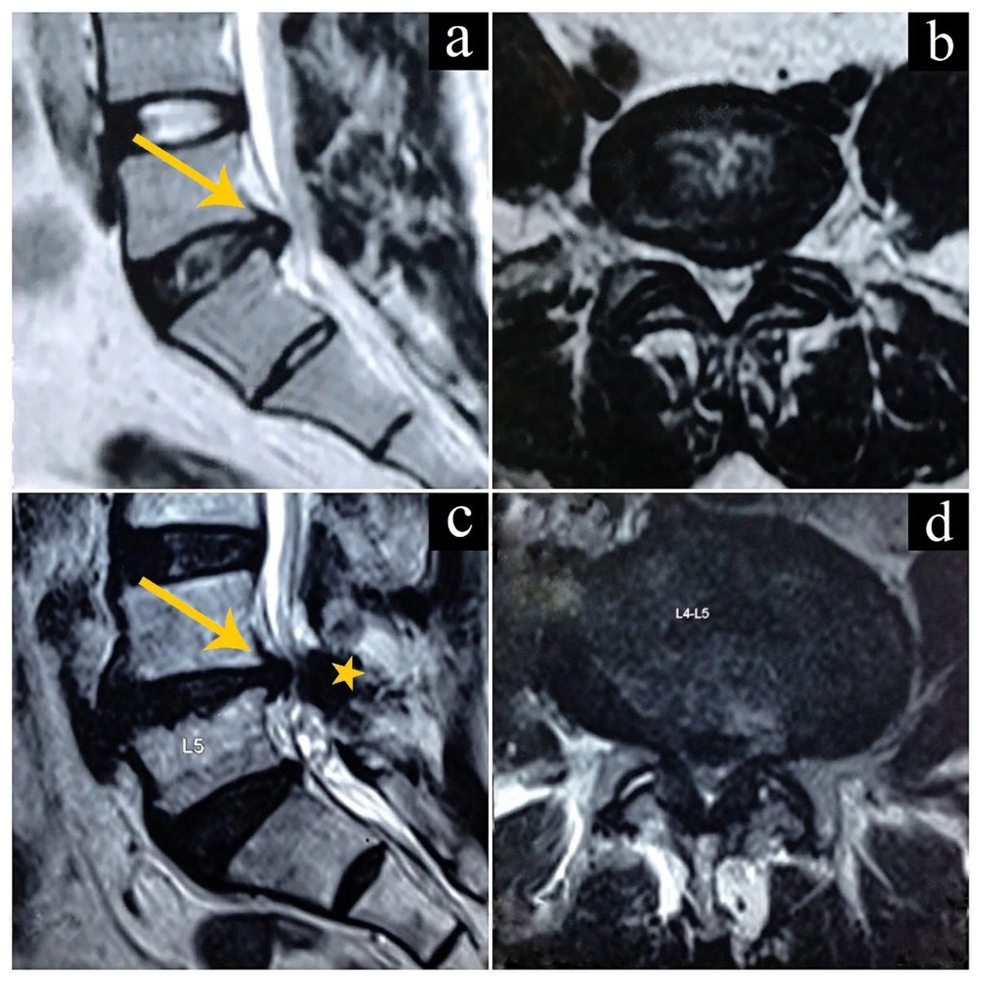
2.2. Intervertebral Disc Herniations

The most common pathology causing nerve root inflammation leading to lumbar radiculopathy is intervertebral disc herniation where the nucleus pulposus gets displaced from its normal location (Figure 1a,b). This can happen acutely due to an injury or more chronically when the intervertebral disc gets degenerated and desiccated as part of the natural ageing process [42]. There are multiple nomenclature systems to describe disc herniations, with many existing classification methods. Broadly, disc herniations can be categorised based on the anatomical location of the herniation, which can be defined as central, paracentral, foraminal or far lateral [43]. It can also be described as protrusion, extrusion, or sequestration, depending on the morphology of the displaced disc material [44]. A more elaborate system based on the morphology of the herniation is the Michigan State University (MSU) classification system [45]. Here, grading is based on the size and location of disc herniation as visualised on a T2 axial cut MR image at the level of maximal disc herniation [45]. Meanwhile, Pfirrmann’s grading also utilises a similar T2 axial cut MR image at the level of maximal disc herniation but grades the amount of nerve root compromise caused by the herniated disc into four categories demonstrating a high correlation with surgical findings [46]. While it is theoretically possible to try out therapeutic SNRBs for any type of disc herniations described in these classification systems causing radiculopathy, it is often not used for severe cases for the reason that those with severe disc herniations get no relief except for temporary postprocedural pain relief [33]. However, not many studies assess and describe outcomes following SNRBs based on these elaborate classification systems; hence, a structured evidence-based guideline is lacking.

2.3. Spondylosis

Spondylosis is a general term that is given for a wide range of age-related degenerative wear and tear that affects all the components of the spine including the bony elements of the vertebra, intervertebral discs, ligamentum flavum and facet joints [47]. Some of these conditions could result in foraminal narrowing leading to nerve root compromise and result in radiculopathy. Firstly, the most common form of spondylosis is intervertebral disc degeneration, which can cause significant disc height loss and stiffness [48,49]. When this happens, the neural foraminal height also decreases, which can potentially cause exiting nerve root compromise, leading to radiculopathy. It should also be noted that structural changes from such degenerative discs increase the risk for intervertebral disc herniation.
Secondly, the ligamentum flavum, bridging the upper and lower lamina of every spinal level, maintains tension when in motion and also in the resting state [50]. It gets thicker and stiffer with age secondary to cumulative mechanical stress [51], and also gets buckled inside the spinal canal as the disc height decreases due to degeneration [50]. While an intervertebral disc herniation can compress the nerve root from the front, ligamentum flavum hypertrophy can cause a similar compression from the back leading to radiculopathy [52]. In some cases of spondylosis, the nerve root can be sandwiched between a herniating disc from the front and a thickened and buckled ligamentum flavum from the back (Figure 1c,d), causing severe symptoms even if the compression caused by the herniating disc is minimal.
Similarly, the facet joints, which are paired synovial joints that play important roles in load transmission and stability maintenance during spinal movements, can also be the cause of nerve root compromise [53]. The neural foramen is bound posteriorly by the facet joint, formed by the superior and inferior articular processes of two adjacent vertebrae [54]. Whenever there is spondylosis due to ageing or abnormal mechanics of the body, inflammation and hypertrophy of the facet joint capsule can occur. Besides hypertrophy, there can also be formation of osteophytes or spurs that further enlarge the facet joints [55]. Additionally, osteoarthritis of the joints can lead to the formation of synovial cysts [56]. In all such cases, there is a possibility for the degenerated and enlarged facet joint to cause compression of the nerve root leading to radiculopathy.
Whatever the indication as described above, if MR imaging is clearly suggestive of the causative lesion in the foraminal-extraforaminal zone compressing the nerve root, which can be correlated to the radiculopathy, then that particular nerve root can be targeted with an SNRB to achieve pain relief [57]. However, the severity of the lesion will be the deciding factor as to whether the SNRB will work well as a therapeutic intervention [33]. Here, it should be noted that the SNRB only reduces the inflammation, but the mechanical compression causing the inflammation will prevail and hence, in most cases, SNRB may not be the definite therapeutic solution.

3. Procedure for SNRB

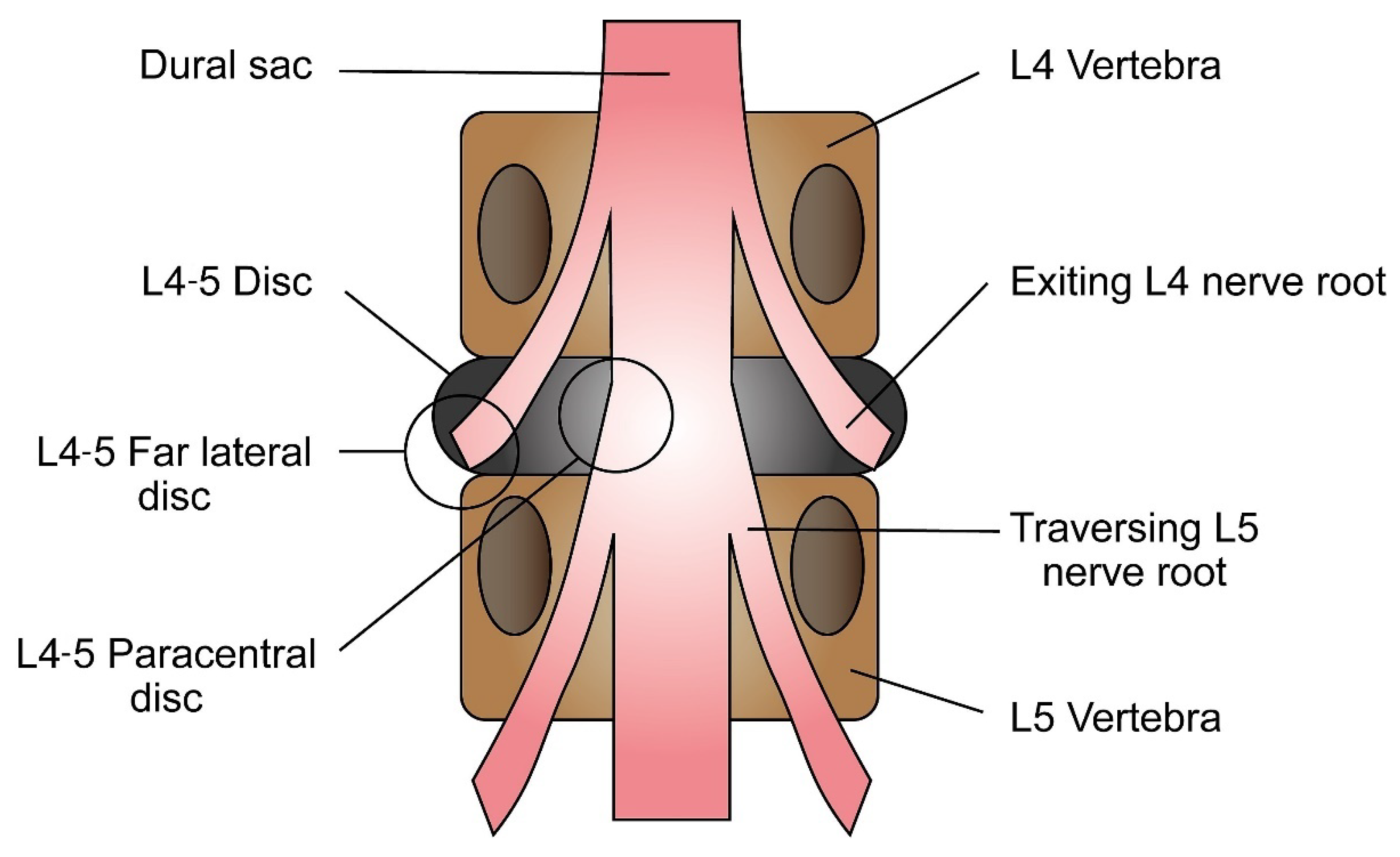
3.1. Identifying the Affected Nerve Root

In the case of disc herniation, the nerve root affected by disc herniation depends on both the level and the location of the herniation. In paracentral or posterolateral herniations, the traversing nerve root is affected. On the other hand, far lateral herniations would affect the exiting nerve root. Here, it should be noted that, unlike the exiting nerve root, the traversing nerve root exits one level below the level of the compression. For example, a paracentral/posterolateral disc herniation at L4-5 would affect the L5 nerve root, which is the traversing nerve root. A far lateral disc herniation at the same level would affect the exiting L4 nerve root instead (Figure 2) [58].
The affected nerve root needs to be targeted at the point where it exits the neural foramen. Hence, needle placement is the most important step of the procedure. Based on expertise, different approaches can be used; however, the “oblique Scottie dog” approach is practised widely due to its high success rates. Here, “Scottie dog” represents the appearance of the bony vertebra in an oblique view X-ray image taken during the procedure where the needle tip is placed below the neck of the “Scottie dog”. When a similar needle placement is achieved without the need for an oblique view X-ray image, the procedure can be termed the anteroposterior (AP) approach [59]. It should be noted that in both approaches, the needle tip is aimed for the so-called “safe zone” or “safe triangle”, while the needle track is more or less the same, the only main difference here is the X-ray view. This zone is an inverted right-angled triangle with the pedicle as its base, lateral vertebral border as the side, which is at a right angle to the base and the exiting nerve root forming the hypotenuse (Figure 3).

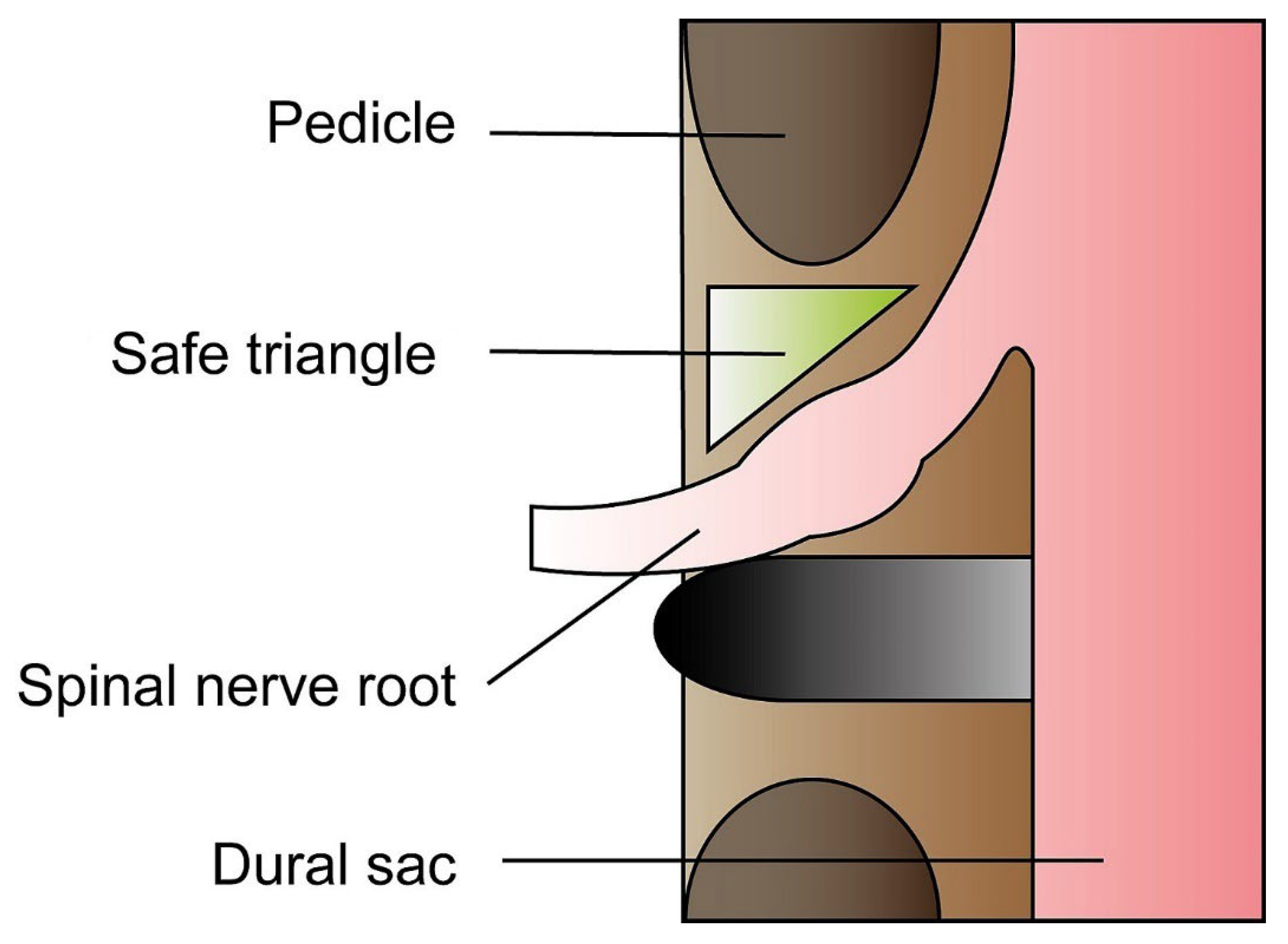
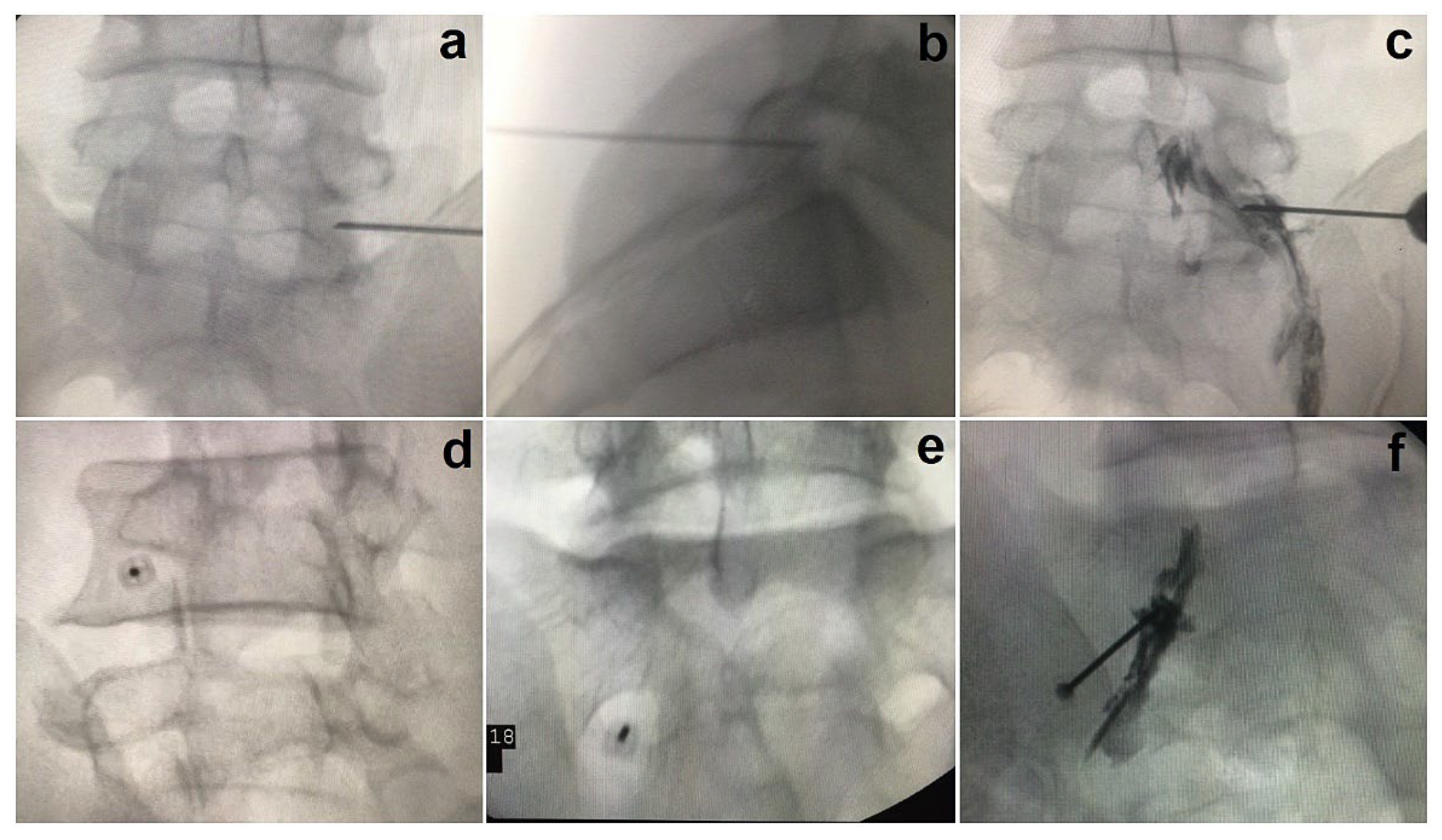
3.2. The Anteroposterior Approach

This approach requires the patient to lie down prone on a radiolucent operating table. After preparation and draping of the patient, the C-arm machine is brought in and tilted cephalocaudally (compensating for lordosis) to get a true AP image with the affected disc level endplates parallel to each other. The vertebra corresponding to the target nerve root level is identified on the AP view. The entry point is marked a few centimetres lateral to the lateral border of the pedicle on the affected side which is followed by local anaesthetic infiltration of the skin. An 18-gauge needle is directed diagonally to a point just below and lateral to the pedicle on the affected side which corresponds to the lateral side of the “safe zone” [33,59]. The drawback here is that the nerve root at this zone is completely covered by the pars intrarticularis. Hence, a bony resistance might be felt when advancing the needle [33]. In such circumstances, the needle can be walked over the bone laterally to an ideal point where the bony resistance disappears. In order to prevent X-ray exposure to the administrator’s hand, the needle is held using a long sponge holder or any other instrument that can hold it without interfering with the needle’s position on the C-arm image. Advancing the needle, a bit further, would help its tip enter the neural foramen at the safe triangle (Figure 4a). This needs to be confirmed with a lateral view X-ray image (Figure 4b). Once a satisfactory placement is achieved in both AP and lateral view images, Iohexol dye is injected to confirm placement (Figure 4c), followed by a combination of a steroid and local anaesthetic.

3.3. The Oblique Scottie Dog Approach

This approach requires similar patient positioning on a radiolucent table. The C-arm is positioned for a true AP view, as previously described. Maintaining the cephalocaudal tilt, the C-arm is positioned to take an oblique view X-ray image of the affected level. Here, the vertebra corresponding to the nerve root that has to be targeted (for example, the L5 vertebra in cases where the L5 nerve root is to be targeted) needs to be visualised as a “Scottie dog” in the oblique view image [57,59]. Once satisfactory C-arm positioning is obtained, the site of injection (corresponding to the neck of the “Scottie dog”) is marked, and the skin is infiltrated with a local anaesthetic agent. Then, an 18-gauge spinal needle is inserted and advanced to a point just below the neck of the “Scottie dog”.
Throughout the advancement of the needle, it is maintained in an “end on” position along with the direction of the X-ray beam so that the needle appears as a single point in the C-arm image (Figure 4d) [59]. In a subsequent lateral view X-ray image, the needle tip position is confirmed to be at the level of the neural foramen. Once a satisfactory placement of the needle is obtained in both oblique and lateral views, the C-arm is re-positioned for an AP view image and a radiopaque dye (Iohexol) is injected through the needle without disturbing its placement. The accuracy and the success rates of the oblique Scottie dog approach are said to be high, and appropriate spread of the dye along the targeted nerve is visualised in most cases [59]. Following this, a combination of a steroid and local anaesthetic is injected. While the local anaesthetic gives immediate temporary pain relief, the steroid acts to reduce inflammation of the affected nerve root and helps with prolonged pain relief.

3.4. SNRB Targeting S1

Targeting the S1 nerve root is completely different from the rest of the lumbar nerve roots [60]. Here, while the patient is lying down prone on the radiolucent table, the S1 foramen needs to be visualised in the C-arm image; the C-arm needs to be tilted cephalocaudally until the S1 foramen, both the dorsal and ventral aspects, appear overlapped. This is required for the needle to approach the dorsal S1 foramen without encountering any bony structures. Once the S1 foramen is clearly visualized, the site is marked on the surface and local anaesthesia infiltration is given. Then, a spinal needle is advanced up to the dorsal S1 foramen in line with the beam of the X-ray. Once the needle tip is at the required position as confirmed by a lateral view C-arm image (Figure 4e), Iohexol dye is injected, and it should spread along the spinal nerve and subsequently flow into the epidural space medial to the S1 pedicle (Figure 4f).

3.5. Ultrasonogram (USG) Guided SNRB

USG-guided SNRB for lumbar levels is recently gaining popularity due to the avoidance of excessive radiation to the patient as only confirmatory X-ray images are required during the procedure [61]. The technique could be quite demanding for first users of ultrasound as there might be difficulty in visualizing the final needle tip due to shadowing of the foraminal area with bony structures in the ultrasound image [62]. Currently, two approaches have been described: The axial approach, where the ultrasound transducer is placed perpendicular to the long axis of the body and the parasagittal approach, where the ultrasound transducer is placed parallel to the long axis of the body with the needle orientation being in-plane for both approaches [62]. The probe used is generally a curvilinear probe, which best suits the visualization of deep structures [63]. Probably due to the difficulty in visualizing the needle tip in an axial scan, authors have used different final ultrasound images showing the various bony elements of the vertebra during the placement of the needle; however, studies describing the parasagittal scan are consistent in identifying the plane between adjacent transverse processes [62]. Nevertheless, a final X-ray is required to confirm the level, placement, and spread of dye [64]. Even though the current evidence is not adequate to propose USG as an alternative to the use of X-rays, further randomized trials comparing both techniques hold the key to determining if this could be true.

4. The Pharmacological Formulae

Various authors have reported the use of different steroids, both particulate (triamcinolone acetonide, methylprednisolone acetate and betamethasone acetate) and nonparticulate (betamethasone sodium phosphate and dexamethasone sodium phosphate) in combination with local anaesthetic agents, such as lidocaine or bupivacaine [65,66,67,68]. Even though nonparticulate corticosteroids are preferred for cervical epidural steroid injections, authors utilise both particulate and non-particulate preparations when it comes to lumbar selective nerve root blocks without serious neurological complications [69]. The reported benefits of particulate corticosteroids with regard to treatment efficacy and duration of relief may outweigh their risk, especially at the lumbar levels and in those who do not respond to nonparticulate soluble preparations [67,70]. However, there could be adverse effects due to the preservatives and drug vehicles used in the different formulations of corticosteroids and hence they are always to be used with caution. In addition, it should be noted that local anaesthetic agents could cause central nervous system disruption, or cardiotoxicity, if there is any unwanted intravascular or intrathecal injection in large doses [71]. However, during routine selective nerve root blocks, the concentrations used and exposure durations are unlikely to cause such toxicity [69].

5. Outcomes Following SNRB

Multiple reports have shown varied therapeutic efficiencies for SNRBs [33,34,57,72]. This is because outcomes following SNRB depend on various factors, especially the severity of nerve root compromise, selection of patients and the pharmacological formulation used. In addition, due to the heterogeneity among studies, reported data including appropriate dosage, number of procedures required and adverse effects vary. Only well-designed, large, randomized studies can provide a clear consensus regarding these aspects. However, the current literature does provide evidence for both short-term and long-term relief of radicular pain following SNRBs [73]. A systematic review by Roberts et al. showed that SNRBs are not only superior to placebo but also to interlaminar epidural steroid injections and caudal epidural steroid injections in treating radicular pain [74]. Another review by Bhatia et al. showed that while there was an analgesic benefit at 3 months, there was no impact on the incidence of surgery among those who took SNRBs [75]. Hence, it is often portrayed as more of an intermediate treatment modality that offers temporary pain relief for a few months without altering the long-term prognosis, especially in those with a severe pathology compressing the nerve root [33].

6. Complications of SNRB

Some of the large studies, as that of Manchikanti et al. [76], Karaman et al. [77] and McGrath et al. [78], which assessed 1310, 1305 and 4104 injections, respectively, have reported mostly transient minor complications, such as intravascular penetration, bleeding, local hematoma, bruising, vasovagal reaction, nerve root irritation, facet joint or disc entry, facial flushing, impotency, increased pain and numbness, injection site pain, flushing headache and weakness [79]. McGrath et al. also concluded that transforaminal injections result in fewer minor complications, as mentioned above, than interlaminar injections. However, reports do exist of major complications, which are extremely rare, such as paraplegia, epidural abscess, epidural hematoma, and dural puncture [80,81,82,83,84,85,86,87]. Based on this data, it can be understood that SNRB is a well-tolerated management strategy for lumbar radiculopathy; even though minor side effects seem to happen more frequently, major complications are rare and hence the procedure can be considered safe, especially in expert hands and when due safety precautions are taken [77].

7. Conclusions

Selective nerve root block is both a useful diagnostic tool and a therapeutic procedure that has been growing in popularity in the clinical field. It can be effective in treating lumbar radiculopathy caused by a wide variety of conditions. The procedure itself can be approached in many ways, as deemed appropriate by respective clinicians. This can include different methods of visualisation of the spine with placement of the needle and different drug combinations. As with all procedures, SNRB comes with its own set of possible complications. However, major complications are extremely rare, and the benefits largely outweigh the risks. In future, SNRBs may become more popular with further advancements, such as better standardisation, and optimisation of its process to ensure maximum periods of effectiveness.

Author Contributions

Conceptualization, J.C.R.Y., S.T.C., J.Y.-L.O. and A.-K.K.-P.; methodology, J.C.R.Y., S.T.C. and A.-K.K.-P.; resources, J.C.R.Y. and S.T.C.; data curation, J.C.R.Y., S.T.C. and A.-K.K.-P.; writing—original draft preparation, J.C.R.Y., S.T.C. and A.-K.K.-P.; writing—review and editing, J.Y.-L.O. and A.-K.K.-P.; supervision, J.Y.-L.O. and A.-K.K.-P.; project administration, J.Y.-L.O. and A.-K.K.-P. All authors have read and agreed to the published version of the manuscript.

Funding

This research received no external funding.

Institutional Review Board Statement

Not applicable.

Informed Consent Statement

Written informed consent was obtained from patients whose X-ray and MRI images are shown here.

Data Availability Statement

Not applicable.

Acknowledgments

The authors sincerely thank Esther Ivorra-Molla for help with preparing the figures.

Conflicts of Interest

The authors declare no conflict of interest.

References

  1. Alexander, C.E.; Varacallo, M. Lumbosacral radiculopathy. In StatPearls; StatPearls Publishing: Treasure Island, FL, USA, 2021. [Google Scholar]
  2. Mattiuzzi, C.; Lippi, G.; Bovo, C. Current epidemiology of low back pain. J. Hosp. Manag. Health Policy 2020, 4, 15. [Google Scholar] [CrossRef]
  3. Suri, P.; Palmer, M.R.; Tsepilov, Y.A.; Freidin, M.B.; Boer, C.G.; Yau, M.S.; Evans, D.S.; Gelemanovic, A.; Bartz, T.M.; Nethander, M.; et al. Genome-wide meta-analysis of 158,000 individuals of European ancestry identifies three loci associated with chronic back pain. PLoS Genet. 2018, 14, e1007601. [Google Scholar] [CrossRef] [PubMed]
  4. Suri, P.; Stanaway, I.B.; Zhang, Y.; Freidin, M.B.; Tsepilov, Y.A.; Carrell, D.S.; Williams, F.M.K.; Aulchenko, Y.S.; Hakonarson, H.; Namjou, B.; et al. Genome-wide association studies of low back pain and lumbar spinal disorders using electronic health record data identify a locus associated with lumbar spinal stenosis. Pain 2021, 162, 2263–2272. [Google Scholar] [CrossRef] [PubMed]
  5. Zafar, F.; Qasim, Y.F.; Farooq, M.U.; Shamael, I.; Khan, I.U.; Khan, D.H. The Frequency of Different Risk Factors for Lower Back Pain in a Tertiary Care Hospital. Cureus 2018, 10, e3183. [Google Scholar] [CrossRef]
  6. Wong, A.Y.L.; Karppinen, J.; Samartzis, D. Low back pain in older adults: Risk factors, management options and future directions. Scoliosis Spinal Disord. 2017, 12, 14. [Google Scholar] [CrossRef]
  7. Alhowimel, A.S.; Alodaibi, F.; Alshehri, M.M.; Alqahtani, B.A.; Alotaibi, M.; Alenazi, A.M. Prevalence and Risk Factors Associated with Low Back Pain in the Saudi Adult Community: A Cross-Sectional Study. Int. J. Environ. Res. Public Health 2021, 18. [Google Scholar] [CrossRef]
  8. Mukasa, D.; Sung, J. A prediction model of low back pain risk: A population based cohort study in Korea. Korean J. Pain 2020, 33, 153–165. [Google Scholar] [CrossRef]
  9. Allegri, M.; Montella, S.; Salici, F.; Valente, A.; Marchesini, M.; Compagnone, C.; Baciarello, M.; Manferdini, M.E.; Fanelli, G. Mechanisms of low back pain: A guide for diagnosis and therapy. F1000Research 2016, 5, F1000 Faculty Rev-1530. [Google Scholar] [CrossRef]
  10. Zheng, C.J.; Chen, J. Disc degeneration implies low back pain. Theor. Biol. Med. Model 2015, 12, 24. [Google Scholar] [CrossRef]
  11. Dydyk, A.M.; Khan, M.Z.; Singh, P. Radicular back pain. In StatPearls; StatPearls Publishing: Treasure Island, FL, USA, 2021. [Google Scholar]
  12. Majlesi, J.; Togay, H.; Unalan, H.; Toprak, S. The sensitivity and specificity of the Slump and the Straight Leg Raising tests in patients with lumbar disc herniation. J. Clin. Rheumatol. 2008, 14, 87–91. [Google Scholar] [CrossRef]
  13. Gonzalez Espinosa de Los Monteros, F.J.; Gonzalez-Medina, G.; Ardila, E.M.G.; Mansilla, J.R.; Exposito, J.P.; Ruiz, P.O. Use of Neurodynamic or Orthopedic Tension Tests for the Diagnosis of Lumbar and Lumbosacral Radiculopathies: Study of the Diagnostic Validity. Int. J. Environ. Res. Public Health 2020, 17, 7046. [Google Scholar] [CrossRef] [PubMed]
  14. Urban, L.M.; MacNeil, B.J. Diagnostic Accuracy of the Slump Test for Identifying Neuropathic Pain in the Lower Limb. J. Orthop. Sports Phys. Ther. 2015, 45, 596–603. [Google Scholar] [CrossRef] [PubMed]
  15. Rao, D.; Scuderi, G.; Scuderi, C.; Grewal, R.; Sandhu, S.J. The Use of Imaging in Management of Patients with Low Back Pain. J. Clin. Imaging Sci. 2018, 8, 30. [Google Scholar] [CrossRef] [PubMed]
  16. Reza Soltani, Z.; Sajadi, S.; Tavana, B. A comparison of magnetic resonance imaging with electrodiagnostic findings in the evaluation of clinical radiculopathy: A cross-sectional study. Eur. Spine J. 2014, 23, 916–921. [Google Scholar] [CrossRef] [PubMed]
  17. Tamarkin, R.G.; Isaacson, A.C. Electrodiagnostic evaluation of lumbosacral radiculopathy. In StatPearls; StatPearls Publishing: Treasure Island, FL, USA, 2022. [Google Scholar]
  18. Chiu, C.C.; Chuang, T.Y.; Chang, K.H.; Wu, C.H.; Lin, P.W.; Hsu, W.Y. The probability of spontaneous regression of lumbar herniated disc: A systematic review. Clin. Rehabil. 2015, 29, 184–195. [Google Scholar] [CrossRef] [PubMed]
  19. Gugliotta, M.; da Costa, B.R.; Dabis, E.; Theiler, R.; Juni, P.; Reichenbach, S.; Landolt, H.; Hasler, P. Surgical versus conservative treatment for lumbar disc herniation: A prospective cohort study. BMJ Open 2016, 6, e012938. [Google Scholar] [CrossRef]
  20. Schoenfeld, A.J.; Weiner, B.K. Treatment of lumbar disc herniation: Evidence-based practice. Int. J. Gen. Med. 2010, 3, 209–214. [Google Scholar] [CrossRef]
  21. Hahne, A.J.; Ford, J.J.; McMeeken, J.M. Conservative management of lumbar disc herniation with associated radiculopathy: A systematic review. Spine 2010, 35, E488–E504. [Google Scholar] [CrossRef]
  22. Hakan, T.; Gurcan, S. Spontaneous Regression of Herniated Lumbar Disc with New Disc Protrusion in the Adjacent Level. Case Rep. Orthop. 2016, 2016, 1538072. [Google Scholar] [CrossRef]
  23. Zhang, X.; Zhang, Z.; Wen, J.; Lu, J.; Sun, Y.; Sang, D. The effectiveness of therapeutic strategies for patients with radiculopathy: A network meta-analysis. Mol. Pain 2018, 14, 1744806918768972. [Google Scholar] [CrossRef]
  24. Sabnis, A.B.; Diwan, A.D. The timing of surgery in lumbar disc prolapse: A systematic review. Indian J. Orthop. 2014, 48, 127–135. [Google Scholar] [CrossRef]
  25. Gregory, D.S.; Seto, C.K.; Wortley, G.C.; Shugart, C.M. Acute lumbar disk pain: Navigating evaluation and treatment choices. Am. Fam. Physician 2008, 78, 835–842. [Google Scholar] [PubMed]
  26. Yoon, W.W.; Koch, J. Herniated discs: When is surgery necessary? EFORT Open Rev. 2021, 6, 526–530. [Google Scholar] [CrossRef] [PubMed]
  27. Lorio, M.; Kim, C.; Araghi, A.; Inzana, J.; Yue, J.J. International Society for the Advancement of Spine Surgery Policy 2019-Surgical Treatment of Lumbar Disc Herniation with Radiculopathy. Int. J. Spine Surg. 2020, 14, 1–17. [Google Scholar] [CrossRef] [PubMed]
  28. Abram, S.E. Treatment of lumbosacral radiculopathy with epidural steroids. Anesthesiology 1999, 91, 1937–1941. [Google Scholar] [CrossRef]
  29. Hakim, B.R.; Munakomi, S. Interlaminar epidural injection. In StatPearls; StatPearls Publishing: Treasure Island, FL, USA, 2021. [Google Scholar]
  30. Viswanathan, V.K.; Kanna, R.M.; Farhadi, H.F. Role of transforaminal epidural injections or selective nerve root blocks in the management of lumbar radicular syndrome—A narrative, evidence-based review. J. Clin. Orthop. Trauma 2020, 11, 802–809. [Google Scholar] [CrossRef]
  31. Singh, S.; Kumar, S.; Chahal, G.; Verma, R. Selective nerve root blocks vs. caudal epidural injection for single level prolapsed lumbar intervertebral disc—A prospective randomized study. J. Clin. Orthop. Trauma 2017, 8, 142–147. [Google Scholar] [CrossRef]
  32. Murakibhavi, V.G.; Khemka, A.G. Caudal epidural steroid injection: A randomized controlled trial. Evid. Based Spine Care J. 2011, 2, 19–26. [Google Scholar] [CrossRef]
  33. Arun-Kumar, K.; Jayaprasad, S.; Senthil, K.; Lohith, H.; Jayaprakash, K.V. The Outcomes of Selective Nerve Root Block for Disc Induced Lumbar Radiculopathy. Malays. Orthop. J. 2015, 9, 17–22. [Google Scholar] [CrossRef]
  34. Dhakal, G.R.; Hamal, P.K.; Dhungana, S.; Kawaguchi, Y. Clinical Efficacy of Selective Nerve Root Block in Lumbar Radiculopathy due to Disc Prolapse. J. Nepal Health Res. Counc. 2019, 17, 242–246. [Google Scholar] [CrossRef]
  35. Yeom, J.S.; Lee, J.W.; Park, K.W.; Chang, B.S.; Lee, C.K.; Buchowski, J.M.; Riew, K.D. Value of diagnostic lumbar selective nerve root block: A prospective controlled study. AJNR Am. J. Neuroradiol. 2008, 29, 1017–1023. [Google Scholar] [CrossRef]
  36. Huston, C.W.; Slipman, C.W. Diagnostic selective nerve root blocks: Indications and usefulness. Phys. Med. Rehabil. Clin. N. Am. 2002, 13, 545–565. [Google Scholar] [CrossRef]
  37. Kanaan, T.; Abusaleh, R.; Abuasbeh, J.; Al Jammal, M.; Al-Haded, S.; Al-Rafaiah, S.; Kanaan, A.; Alnaimat, F.; Khreesha, L.; Al Hadidi, F.; et al. The Efficacy of Therapeutic Selective Nerve Block in Treating Lumbar Radiculopathy and Avoiding Surgery. J. Pain Res. 2020, 13, 2971–2978. [Google Scholar] [CrossRef] [PubMed]
  38. Narozny, M.; Zanetti, M.; Boos, N. Therapeutic efficacy of selective nerve root blocks in the treatment of lumbar radicular leg pain. Swiss Med. Wkly. 2001, 131, 75–80. [Google Scholar]
  39. Beynon, R.; Elwenspoek, M.M.C.; Sheppard, A.; Higgins, J.N.; Kolias, A.G.; Laing, R.J.; Whiting, P.; Hollingworth, W. The utility of diagnostic selective nerve root blocks in the management of patients with lumbar radiculopathy: A systematic review. BMJ Open 2019, 9, e025790. [Google Scholar] [CrossRef] [PubMed]
  40. Slipman, C.W.; Issac, Z. The role of diagnostic selective nerve root blocks in the management of spinal pain. Pain Physician 2001, 4, 214–226. [Google Scholar] [CrossRef] [PubMed]
  41. Bartynski, W.S.; Kang, M.D.; Rothfus, W.E. Adjacent double-nerve root contributions in unilateral lumbar radiculopathy. AJNR Am. J. Neuroradiol. 2010, 31, 327–333. [Google Scholar] [CrossRef] [PubMed]
  42. Sharrak, S.; Al Khalili, Y. StatPearls; StatPearls Publishing: Treasure Island, FL, USA, 2021. [Google Scholar]
  43. Fardon, D.F.; Williams, A.L.; Dohring, E.J.; Murtagh, F.R.; Gabriel Rothman, S.L.; Sze, G.K. Lumbar disc nomenclature: Version 2.0: Recommendations of the combined task forces of the North American Spine Society, the American Society of Spine Radiology and the American Society of Neuroradiology. Spine J. 2014, 14, 2525–2545. [Google Scholar] [CrossRef]
  44. Fardon, D.F.; Milette, P.C. Nomenclature and classification of lumbar disc pathology. Recommendations of the Combined task Forces of the North American Spine Society, American Society of Spine Radiology, and American Society of Neuroradiology. Spine 2001, 26, E93–E113. [Google Scholar] [CrossRef]
  45. Mysliwiec, L.W.; Cholewicki, J.; Winkelpleck, M.D.; Eis, G.P. MSU classification for herniated lumbar discs on MRI: Toward developing objective criteria for surgical selection. Eur. Spine J. 2010, 19, 1087–1093. [Google Scholar] [CrossRef]
  46. Pfirrmann, C.W.; Dora, C.; Schmid, M.R.; Zanetti, M.; Hodler, J.; Boos, N. MR image-based grading of lumbar nerve root compromise due to disk herniation: Reliability study with surgical correlation. Radiology 2004, 230, 583–588. [Google Scholar] [CrossRef] [PubMed]
  47. Middleton, K.; Fish, D.E. Lumbar spondylosis: Clinical presentation and treatment approaches. Curr. Rev. Musculoskelet. Med. 2009, 2, 94–104. [Google Scholar] [CrossRef] [PubMed]
  48. Rustenburg, C.M.E.; Emanuel, K.S.; Peeters, M.; Lems, W.F.; Vergroesen, P.A.; Smit, T.H. Osteoarthritis and intervertebral disc degeneration: Quite different, quite similar. JOR Spine 2018, 1, e1033. [Google Scholar] [CrossRef] [PubMed]
  49. Rider, S.M.; Mizuno, S.; Kang, J.D. Molecular Mechanisms of Intervertebral Disc Degeneration. Spine Surg. Relat. Res. 2019, 3, 1–11. [Google Scholar] [CrossRef] [PubMed]
  50. Mihara, A.; Nishida, N.; Jiang, F.; Ohgi, J.; Imajo, Y.; Suzuki, H.; Funaba, M.; Yamagata, H.; Chen, X.; Sakai, T. Tensile Test of Human Lumbar Ligamentum Flavum: Age-Related Changes of Stiffness. Appl. Sci. 2021, 11, 3337. [Google Scholar] [CrossRef]
  51. Sairyo, K.; Biyani, A.; Goel, V.; Leaman, D.; Booth, R., Jr.; Thomas, J.; Gehling, D.; Vishnubhotla, L.; Long, R.; Ebraheim, N. Pathomechanism of ligamentum flavum hypertrophy: A multidisciplinary investigation based on clinical, biomechanical, histologic, and biologic assessments. Spine 2005, 30, 2649–2656. [Google Scholar] [CrossRef]
  52. Kolte, V.S.; Khambatta, S.; Ambiye, M.V. Thickness of the ligamentum flavum: Correlation with age and its asymmetry-an magnetic resonance imaging study. Asian Spine J. 2015, 9, 245–253. [Google Scholar] [CrossRef]
  53. Inoue, N.; Orias, A.A.E.; Segami, K. Biomechanics of the Lumbar Facet Joint. Spine Surg. Relat. Res. 2020, 4, 1–7. [Google Scholar] [CrossRef]
  54. Gilchrist, R.V.; Slipman, C.W.; Bhagia, S.M. Anatomy of the intervertebral foramen. Pain Physician 2002, 5, 372–378. [Google Scholar] [CrossRef]
  55. Jaumard, N.V.; Welch, W.C.; Winkelstein, B.A. Spinal facet joint biomechanics and mechanotransduction in normal, injury and degenerative conditions. J. Biomech. Eng. 2011, 133, 071010. [Google Scholar] [CrossRef]
  56. Bureau, N.J.; Kaplan, P.A.; Dussault, R.G. Lumbar facet joint synovial cyst: Percutaneous treatment with steroid injections and distention--clinical and imaging follow-up in 12 patients. Radiology 2001, 221, 179–185. [Google Scholar] [CrossRef] [PubMed]
  57. Kanna, R.M.; Shetty, A.P.; Rajasekaran, S. Predictors of Successful Outcomes of Selective Nerve Root Blocks for Acute Lumbar Disc Herniation. Glob. Spine J. 2019, 9, 473–479. [Google Scholar] [CrossRef] [PubMed]
  58. Amin, R.M.; Andrade, N.S.; Neuman, B.J. Lumbar Disc Herniation. Curr. Rev. Musculoskelet. Med. 2017, 10, 507–516. [Google Scholar] [CrossRef] [PubMed]
  59. Kaliya-Perumal, A.K.; Yeh, Y.C.; Luo, C.A.; Joey-Tan, K.Y. Assessment of Anteroposterior Subpedicular Approach and Oblique Scotty Dog Subpedicular Approach for Selective Nerve Root Block. Clin. Orthop. Surg. 2017, 9, 71–76. [Google Scholar] [CrossRef]
  60. Mansfeld, E.E. Sacral Transforaminal Epidural Injection (Selective Nerve Root Block), Interventional Pain ed.; Stogicza, A.R., Mansano, A.M., Trescot, A.M., Staats, P.S., Eds.; Springer: Cham, Switzerland, 2020. [Google Scholar]
  61. Emami, S.A.; Sanatkar, M.; Espahbodi, E.; Pestehei, S.K. Ultrasound and nerve stimulator guidance lumbar transforaminal epidural block for the treatment of patients with lumbosacral radicular pain. Sci. Rep. 2022, 12, 5954. [Google Scholar] [CrossRef]
  62. Soni, P.; Punj, J. Ultrasound-Guided Lumbar Transforaminal Epidural Injection: A Narrative Review. Asian Spine J. 2021, 15, 261–270. [Google Scholar] [CrossRef]
  63. Korbe, S.; Udoji, E.N.; Ness, T.J.; Udoji, M.A. Ultrasound-guided interventional procedures for chronic pain management. Pain Manag. 2015, 5, 465–482. [Google Scholar] [CrossRef] [PubMed]
  64. Sahu, D.K.; Sharma, A.; Kothari, K.; Wani, P.; Patel, C.; Parampill, R. Ultrasound-guided fluoroscopic-verified lumbar transforaminal epidural injection: A clinical evaluation of technique. Indian J. Pain 2016, 30, 158–161. [Google Scholar] [CrossRef]
  65. Chatterjee, N.; Roy, C.; Das, S.; Al Ajmi, W.; Al Sharji, N.S.; Al Mandhari, A. Comparative Efficacy of Methylprednisolone Acetate and Dexamethasone Disodium Phosphate in Lumbosacral Transforaminal Epidural Steroid Injections. Turk. J. Anaesthesiol. Reanim. 2019, 47, 414–419. [Google Scholar] [CrossRef]
  66. Guyot, J.P. Lumbar Selective Nerve Root Block: Comparative Study Using Two Pharmacological Formulae. Global Spine J. 2018, 8, 374–377. [Google Scholar] [CrossRef]
  67. Jonayed, S.A.; Kamruzzaman, M.; Saha, M.K.; Alam, S.; Akter, S. The Role of Selective Nerve Root Block in the Treatment of Lumbar Radicular Leg Pain. Mymensingh Med. J. 2016, 25, 141–147. [Google Scholar] [PubMed]
  68. McCormick, Z.; Kennedy, D.J.; Garvan, C.; Rivers, E.; Temme, K.; Margolis, S.; Zander, E.; Rohr, A.; Smith, M.C.; Plastaras, C. Comparison of Pain Score Reduction Using Triamcinolone vs. Betamethasone in Transforaminal Epidural Steroid Injections for Lumbosacral Radicular Pain. Am. J. Phys. Med. Rehabil. 2015, 94, 1058–1064. [Google Scholar] [CrossRef] [PubMed]
  69. MacMahon, P.J.; Huang, A.J.; Palmer, W.E. Spine Injectables: What Is the Safest Cocktail? AJR Am. J. Roentgenol. 2016, 207, 526–533. [Google Scholar] [CrossRef]
  70. Bensler, S.; Sutter, R.; Pfirrmann, C.W.A.; Peterson, C.K. Particulate versus non-particulate corticosteroids for transforaminal nerve root blocks: Comparison of outcomes in 494 patients with lumbar radiculopathy. Eur. Radiol. 2018, 28, 946–952. [Google Scholar] [CrossRef]
  71. MacMahon, P.J.; Eustace, S.J.; Kavanagh, E.C. Injectable corticosteroid and local anesthetic preparations: A review for radiologists. Radiology 2009, 252, 647–661. [Google Scholar] [CrossRef]
  72. Lee, J.W.; Kim, S.H.; Lee, I.S.; Choi, J.A.; Choi, J.Y.; Hong, S.H.; Kang, H.S. Therapeutic effect and outcome predictors of sciatica treated using transforaminal epidural steroid injection. AJR Am. J. Roentgenol. 2006, 187, 1427–1431. [Google Scholar] [CrossRef]
  73. Benny, B.; Azari, P. The efficacy of lumbosacral transforaminal epidural steroid injections: A comprehensive literature review. J. Back Musculoskelet. Rehabil. 2011, 24, 67–76. [Google Scholar] [CrossRef] [PubMed]
  74. Roberts, S.T.; Willick, S.E.; Rho, M.E.; Rittenberg, J.D. Efficacy of lumbosacral transforaminal epidural steroid injections: A systematic review. PM R 2009, 1, 657–668. [Google Scholar] [CrossRef]
  75. Bhatia, A.; Flamer, D.; Shah, P.S.; Cohen, S.P. Transforaminal Epidural Steroid Injections for Treating Lumbosacral Radicular Pain from Herniated Intervertebral Discs: A Systematic Review and Meta-Analysis. Anesth. Analg. 2016, 122, 857–870. [Google Scholar] [CrossRef] [PubMed]
  76. Manchikanti, L.; Malla, Y.; Wargo, B.W.; Cash, K.A.; Pampati, V.; Fellows, B. A prospective evaluation of complications of 10,000 fluoroscopically directed epidural injections. Pain Physician 2012, 15, 131–140. [Google Scholar] [CrossRef]
  77. Karaman, H.; Kavak, G.O.; Tufek, A.; Yldrm, Z.B. The complications of transforaminal lumbar epidural steroid injections. Spine 2011, 36, E819–E824. [Google Scholar] [CrossRef] [PubMed] [Green Version]
  78. McGrath, J.M.; Schaefer, M.P.; Malkamaki, D.M. Incidence and characteristics of complications from epidural steroid injections. Pain Med. 2011, 12, 726–731. [Google Scholar] [CrossRef] [PubMed]
  79. Chang, A.; Ng, A.T. Complications Associated with Lumbar Transforaminal Epidural Steroid Injections. Curr. Pain Headache Rep. 2020, 24, 67. [Google Scholar] [CrossRef]
  80. Lyders, E.M.; Morris, P.P. A case of spinal cord infarction following lumbar transforaminal epidural steroid injection: MR imaging and angiographic findings. AJNR Am. J. Neuroradiol. 2009, 30, 1691–1693. [Google Scholar] [CrossRef] [PubMed]
  81. Kabbara, A.; Rosenberg, S.K.; Untal, C. Methicillin-resistant Staphylococcus aureus epidural abscess after transforaminal epidural steroid injection. Pain Physician 2004, 7, 269–272. [Google Scholar] [CrossRef]
  82. Houten, J.K.; Errico, T.J. Paraplegia after lumbosacral nerve root block: Report of three cases. Spine J. 2002, 2, 70–75. [Google Scholar] [CrossRef]
  83. Huntoon, M.A.; Martin, D.P. Paralysis after transforaminal epidural injection and previous spinal surgery. Reg. Anesth. Pain Med. 2004, 29, 494–495. [Google Scholar] [CrossRef]
  84. Kennedy, D.J.; Dreyfuss, P.; Aprill, C.N.; Bogduk, N. Paraplegia following image-guided transforaminal lumbar spine epidural steroid injection: Two case reports. Pain Med. 2009, 10, 1389–1394. [Google Scholar] [CrossRef]
  85. Kim, S.I.; Lee, D.H.; Kim, S.H.; Cho, Y.H. Spinal epidural hematoma occurring at a distance from the transforaminal epidural injection site: A case report. Medicine 2019, 98, e16654. [Google Scholar] [CrossRef]
  86. Gungor, S.; Aiyer, R. Epidural hematoma development contralateral to dura after lumbar transforaminal epidural steroid injection. Pain Manag. 2017, 7, 367–375. [Google Scholar] [CrossRef]
  87. Goodman, B.S.; Bayazitoglu, M.; Mallempati, S.; Noble, B.R.; Geffen, J.F. Dural puncture and subdural injection: A complication of lumbar transforaminal epidural injections. Pain Physician 2007, 10, 697–705. [Google Scholar] [CrossRef] [PubMed]
Figure 1. MRI images showing intervertebral disc herniations. (a) Sagittal view showing L5-S1 disc herniation (arrow), (b) corresponding axial view, (c) Sagittal view showing front and back compression due to herniating L4-L5 disc (arrow) and a thick buckled ligamentum flavum (star), (d) corresponding axial view showing circumferential compression.
Surgeries 03 00028 g001
Figure 2. Representation of L4-L5 disc level showing how the anatomy of the disc herniation would affect the exiting or traversing nerve roots.
Surgeries 03 00028 g002
Figure 3. Representation of safe triangle and its boundaries.
Surgeries 03 00028 g003
Figure 4. C-arm images during the SNRB procedure. (a) Antero-posterior (AP) view of needle placement during the AP approach, (b) Lateral view showing needle tip positioned in the foramen, (c) AP view following injection of Iohexol dye showing appropriate spread along the L5 nerve root, (d) Oblique view showing needle placement below the neck of the Scottie dog during the Scottie dog approach, (e) AP view of needle placement in the S1 foramen, (f) Spread of dye along the S1 nerve root.
Surgeries 03 00028 g004
Publisher’s Note: MDPI stays neutral with regard to jurisdictional claims in published maps and institutional affiliations.

