Technical Note

A Novel Comprehensive Classification for Non-Prosthetic Peri-Implant Fractures

Department of General Surgery and Medical Surgical Specialties, Section of Orthopaedics and Traumatology, A.O.U.P. Policlinico Rodolico-San Marco, University of Catania, 95123 Catania, Italy
*
Author to whom correspondence should be addressed.
Surgeries 2023, 4(4), 530-543; https://doi.org/10.3390/surgeries4040052
Submission received: 18 September 2023 / Revised: 13 October 2023 / Accepted: 16 October 2023 / Published: 17 October 2023

Abstract

:
Non-prosthetic peri-implant fractures (NPPIFs) are often reported mixed with periprosthetic fractures (PPFs), but they are different entities. Due to the increase in the age of the world’s population and to the intensification of surgeries for fractures, nowadays, peri-implant fractures are a very frequent entity in clinical practice, with an increasing trend expected in the future. A clear exclusive classification of NPPIFs is not reported in the literature. The aim of this study is to provide a valid comprehensive classification for all the NPPIFs. X-rays of all the peri-implant cases treated in our unit in a 3-year period were retrospectively collected. Five orthopedic surgeons reviewed 30 X-rays of NPPIFs, providing a code according to the classification proposed. After a 3-month interval, they reviewed the same X-rays. Eighteen femoral, eight humeral, and four forearm peri-implant fractures were collected and showed to the raters. Inter- and intra-observer reliability was calculated using a k-statistic, showing a moderate agreement between observers (κ = 0.73) and a substantial agreement between the observations of the same viewer (κ = 0.82). The literature lacks a comprehensive classification for peri-implant fractures that considers all the bones and all the types of implants. The proposed classification is meant to be an instrument for orthopedic surgeons to categorize these types of fractures and seems to be simple, easy to comprehend, and reproducible. This new classification can provide the orthopedic surgeon a reliable method to clearly catalogue different fractures according to the site and the implants; the physicians can use it, through a code, in clinical practice to describe an NPPIF without the need of images. Further studies may be necessary to confirm the validity and eventually to improve the suggested classification.

1. Introduction

Fractures around orthopedic implants, different from the prosthesis, are often seen in clinical practice, and a clear distinction with periprosthetic fractures (PPFs) is not always considered [1]. This specific category of lesions is named “non-prosthetic peri-implant fractures” (NPPIFs) [2] and is underreported [3], often described as mixed with PPFs that, differently, are fractures that occur in the proximity of an orthopedic prosthesis and are a well-known and studied entity [4]. Diversely, NPPFIs can be defined as fractures of bones with pre-existing non-prosthetic implants [2] (plate and screws, only screws, an intramedullary nail, or K-wires) and are considered ‘modern’ fractures because of their increasing incidence in the last years.
In fact, considering that the world’s population is ageing and considering how frequent osteoporosis and arthritis are right now (and how frequent they are surgically treated with prostheses and implants), seeing periprosthetic and peri-implant fractures is a regular occurrence that is expanding [5].
Other than epidemiological reasons, many risk factors need to be considered in periprosthetic and peri-implant fractures: age of the patients, osteoporosis, trauma, gender (it is more frequent in female patients), osteolysis, aseptic loosening, previous surgical techniques, and implants utilized [6,7,8,9].
Even if it is an increasingly frequent entity, in the literature and in clinical practice, there is not a comprehensive recognized classification system for NPPFIs. Valid tools helpful for the diagnosis and for a reliable evaluation of these fractures are not reported. Choice of treatment is often difficult because of altered anatomy, the presence of the previous implanted hardware, and phenomena such as stress shielding, osteopenia, and fracture remodeling [10]. To help the surgeons in the diagnosis and in the decision-making process, classifications play an important role.
Indeed, classifications help orthopedic surgeons to define a type of injury and sometimes indicate a potential treatment and prognosis. Furthermore, classifications play a key role in the collecting of epidemiological and clinical data, allowing uniform comparison and documentation of pathologies. Even if is often hard to define the validity of a classification, the reliability, using the kappa value between observations, is a dependable parameter. Many classification systems commonly used in orthopedics have not proved good reliability [11,12,13,14,15,16], and no classifications have shown a perfect agreement between observers. For this reason, new classifications are always needed and can provide a helpful contribution to surgeons to fully understand and categorize the fractures.
The best NPPIF classification was proposed by Chan et al. [2] in 2018 with a collection of 60 patients. However, the authors only considered two types of hardware (nails and plates and screws), and even if they collected NPPIFs of different bones (femur, forearm, humerus, tibia, and clavicle), their proposed novel classification was only limited to the femur. Some commonly used classifications only consider one district and are mainly about periprosthetic and not about peri-implant fractures, such as the Vancouver classification, largely used for hip periprosthetic fractures [11].
A single comprehensive classification that considers all the skeletal bones and all the different types of implants is not reported [12,13]. For the plain fractures (without any implant), many classifications exist, but the AO foundation provided the most complete and most used worldwide classification [14]. The AO classification considers some important aspects. First, it examines the bone and gives a number (from 1 to 15). Then, it evaluates if the fracture involves the diaphysis or the epiphysis (the location of the fracture). Next step is to distinguish the type of fracture. Other groups and subgroups are also given using letters or numbers. In a similar way, the Unified Classification System for Periprosthetic Fractures (UCPF) [15] provided a comprehensive classification for PPFs, considering the joint (using Roman numerals), the bone (using numbers), and the type of fracture (using letters). These classifications regard all the skeletal bones. The literature lacks similar classifications for NPPIFs, and the few classifications reported are only about some specific districts [2,3].
So far, a comprehensive classification (regarding all the skeletal bones and all different implants) for NPPIFs that is also easy to understand and reliable is not reported in the literature. Our study aims to provide a new valid comprehensive classification for all the NPPIFs.

2. Materials and Methods

The aim of our classification is to provide a reliable code to all the types of NPPIFs for all the bones of the human body of orthopedic competence, providing an idea of the pre-existing implant and the location of the fractures.
All the peri-implant cases treated in the Orthopaedics and Traumatology Unit, A.O.U. Policlinico, San Marco, University of Catania, Catania, Italy, between 1 January 2020, and 31 December 2022, were collected. Exclusion criteria for the study were PPF, more than two implants, and more than two fractures in the same bone. During the search, 7 interimplant fractures and 102 periprosthetic fractures were found. After excluding all the periprosthetic and interimplant fractures, 30 X-rays were considered eligible for the study.
Five orthopedic surgeons rated 30 X-rays of peri-implant fractures, providing a code according to the proposed classification. The X-rays of these 30 patients were then deidentified and were reviewed by 5 fellowship-trained orthopedic surgeons. No additional information, other than age and gender, was provided to reviewers. The observers were able to classify each X-ray in around 10 to 30 s, and it took less than 20 min to review all the X-rays. None of these surgeons were authors of the study. The raters had an average experience of 7 years as orthopedic surgeons (rate 5–12) working in a trauma center. All of them were confident with AO classification and the Vancouver classification [11].
The given classification was fully explained to the observers, offering them the instruction reported in Appendix A.
The described classification is characterized by 4 main aspects: bone, type of hardware, hardware length, and level of the fracture.
The different bones are identified by a number similar to AO classification [17] (Figure 1, Table 1): “1” identifies the humerus, “2” the forearm, “3” is related to the femur, “4” considers the Tibia, “5” can refer to the spine, “6” is about the pelvis, “7” characterizes the hand and consists of eight under-categories (lunate “71”, scaphoid “72”, capitate “73”, hamate “74”, trapezium “75”, other carpal bones “76”, metacarpals “77”, and phalanges “78”), “8” characterizes the foot and consists of eight under-categories (talus “81”, calcaneus “82”, navicular “83”, cuboid “84”, cuneiforms “85”, metatarsals “87”, phalanges “88”), “14” identifies the scapula, and “15” refers to the clavicle.
The hardware can be identified by a letter of the Greek alphabet (Figure 2, Table 2): “α” for plate and screws, “β” for screws, “γ” for a nail, “δ” for K-wires, and “ε” for external fixator.
The length of the hardware can be distinguished using the Roman numbers (Figure 3, Table 3): “I” when the hardware is in the proximal third of the corresponding bone, “II” when the hardware is in the middle third of the corresponding bone, “III” when the hardware is in the distal third of the corresponding bone, “IV” when the hardware is in the proximal two-thirds of the bone, “V” when the hardware is in the distal two-thirds of the bone, and “VI” when the hardware lies along the entire length of the bone.
The site of the fracture is described compared to the hardware and can be described by a capital letter, along the lines of the Vancouver classification [18] (Figure 4, Table 4 and Table 5): “A” when the fracture is proximal to the hardware, “B” when the fracture is corresponds with the hardware (“B1” if the fracture is in the proximal part of the hardware, “B2” if it is in the middle part of the hardware, and “B3” if the fracture is in the distal part of the hardware), and “C” if the fracture is distal to the hardware.
According to the proposed classification, to correctly classify a peri-implant fracture, the observer must provide 4 numbers or letters as reported in Table 5 and in Figure 5.
Only 2 plain X-rays for each case were shown to the observers. No CT scans were exhibited. The only information provided to the observers was the age and sex of the patient. No additional information was provided to reviewers about patients’ treatments or outcomes. After a 3-month interval, each surgeon performed the evaluation of the same X-rays and again provided a code to calculate the intra-observer reliability. The orders in which they reviewed the patients were randomly scrambled. Data were analyzed using k value as a measurement of inter- and intra-observer agreement.
The average agreement between and within each observer was calculated. The reliability was calculated using k statistics. It was interpreted as none (0–0.20), minimal (0.21–0.39), weak (0.4–0.59), moderate (0.6–0.79), strong (0.80–0.90), or almost perfect (>0.90) reliability.
All statistical tests were performed using SPSS 28 statistical software. Significance was set at p < 0.05.

3. Results

A total of 30 X-rays were evaluated by 5 observers. A new evaluation was conducted 3 months later to calculate intra-observer reliability. Eighteen femoral, eight humeral and four forearm peri-implant fractures were collected and showed to five orthopedic surgeons. Each surgeon rated the same 30 X-rays at the beginning and then after 3 months in random order. A total of 300 observations were conducted. An inter-rater reliability analysis was performed between the dependent samples of the five observers. The codes provided by each rater were compared to the ones given by the other observers, with a total of 10 possible combinations (rater 1—rater 2, rater 1—rater 3, rater 1—rater 4, rater 1—rater 5, rater 2—rater 3, rater 2—rater 4, rater 2—rater 5, rater 3—rater 4, rater 3—rater 5, rater 4—rater 5) that were then analyzed using a k statistics. The Fleiss kappa was calculated as a measure of the agreement between more than two dependent categorical samples.
The inter-rater reliability of the projected classification was considered as none (0–0.20), minimal (0.21–0.39), weak (0.4–0.59), moderate (0.6–0.79), strong (0.80–0.90), or almost perfect (>0.90) reliability.
The Fleiss kappa showed that there was a substantial agreement between observers with κ = 0.73 (range: 0.66 to 0.81) (Table 6). Inter-rater reliability showed substantial agreement. The form of distribution was quite symmetrical. The confidence level selected was 95%.
The inter-observer reliability between observers is reported in Table 7.
A comparison between observations made by the same observer at two time points (at least 3 months apart) was conducted. The k-statistics were analyzed. In the same way, the agreement was considered as none (0–0.20), minimal (0.21–0.39), weak (0.4–0.59), moderate (0.6–0.79), strong (0.80–0.90), or almost perfect (>0.90) reliability.
The Cohens kappa showed a substantial intra-observer agreement. The average intra-observer reliability was κ = 0.82 (range: 0.67 to 0.96), indicating strong reliability (Table 8). The form of distribution was quite symmetrical. The confidence level selected was 95%.
The intra-observer reliability of each observer is reported in Table 9.

4. Discussion

Our results show that when classifying peri-implant fractures with this new classification from plain X-rays, there is good intra- and inter-observer agreement.
Many authors have reported different studies about periprosthetic fractures [19]. However, just a few studies specifically regard NPPIFs. The UCS broadly classified periprosthetic fractures, but to our knowledge, a similar comprehensive classification for peri-implant fractures is not yet described. Indeed, some authors reported interesting classifications about peri-implant fractures but only for a precise bone [20].
Egol et al. [21] proposed an interesting classification model mainly focused on the implant (plate and screws or intramedullary nail) and where the fracture was (proximal or distal to the implant, at the tip or within). However, they did not provide information about the bone.
On the other hand, some authors focused on some specific bones, mainly the femur [12]. Chan et al. [2] centered on the proximal femur developing a categorization according to the type of initial fracture, the implant used, and the location of the fracture.
Muller et al. [22] stated a higher risk of peri-implant fractures for femoral fractures treated with nails than for those treated with screws. In contrast, Kruse et al. [23] reported a higher incidence of fractures in previously implanted sliding-hip devices than in cephalomedullary nails.
One of the biggest studies about femoral peri-implant fractures was conducted by Videla-Cés et al. [13] in 2019. The authors described a new classification of peri-implant femoral fractures after collecting 143 cases in a multicenter study (12 centers).
Recently, Bidolegui [20] proposed a very useful classification for peri-implant femoral fractures with an algorithm of possible treatments and suggestions for removal (partial or total) of the previous hardware.
Prieto et al. [10] stated that NPPIFs are an under-reported entity, above all about upper limbs, and indicated that only two cases of both bone NPPIFs of the forearm are described in the literature.
So far, our study does not provide suggestions on the treatment. Currently, the main goal of the study is just to provide a comprehensive classification that can be an instrument for orthopedic surgeons to categorize, distinguish, and talk about peri-implant fractures, even without seeing X-rays. Furthermore, the choice of treatment for NPPIFs is very complex and variable; it needs to be decided according to the patients’ characteristics and not only to the imaging studies. Additionally, the surgeons’ preference must be considered. In any event, the two main options to treat an NPPIF are the following: remove the previous implant and undertake a new and longer osteosynthesis, or fix the fracture with new hardware.
Our classification can provide all the necessary information about an NPPIF just using a code. The big innovation of this classification is to provide not only information about the fracture (bone and location) but also about the implant (type of hardware and length). Many important classifications (like the UCPF used for PPF, for example) do not provide information about the type of prosthesis previously implanted. Differently, our classification gives all the useful information about the implant.
The utility of this comprehensive classification is the same as the other classifications used for plain fractures (without implants) such as the AO classification [14]. Simply by using a code, orthopedic surgeons can clearly understand the type of fracture without seeing the X-ray.
In the future, our classification can be improved, providing advice on possible surgical solutions for peri-implant fractures.
Liporace et al. [24] described the main principles, strategies, and surgeries for complex clinical scenarios characterized by inter-prosthetic and inter- or peri-implant fractures. Similarly, Bonnomet et al. [25] conducted a fascinating study about femoral fractures between prosthesis and implants. This scenario is very complex and can be treated with a prosthesis revision or with a new fixation. Considering that our classification is preliminary, we preferred to exclude fractures with more than one implant.
Other authors considered different bones than the femur: Stramazzo et al. [26] reviewed all cases of peri-implant radius fracture reported in the literature and described a new classification for this type of fracture.
The most comprehensive study conducted about peri-implant fractures was directed by Chan et al. [2]. The authors collected 60 NPPIFs in 53 patients; 38 fractures involved the femur, 12 the forearm, 5 the humerus, 3 the tibia and/or fibula, and 1 the clavicle. Similarly, we collected 18 femoral, 8 humeral, and 4 forearm peri-implant fractures. No cases of tibia/fibula or clavicle were reported in our sample.
As already mentioned, the most-used comprehensive classification for plain fractures is the AO classification, while for periprosthetic fractures, it is the UCPF (that still does not give complete information about the prosthesis implanted). However, many classifications are reported for a specific district.
Proximal humerus fractures (PHF) were initially classified by Neer in 1970 [27], but lately, other classifications have been reported other than AO, like the Hertel classification [28]. Even if all those classifications are well known and largely reported in the literature, many studies have shown a low inter- and intra-observer reliability [29,30,31,32]. The same happens for other bones. If we consider the tibial plateau for example, the most known classification is the Schatzker classification [33], but some studies have shown moderate or substantial reliability [34]. Similarly, our study did not show perfect reliability but a moderate one, which is often sufficient in the literature to classify a fracture.
As mentioned before, our classification considers four main categories: the bone, the implant, the length of the hardware, and the site of the fracture (compared to the hardware). Agreement between observers and observations about the first two categories was 100%. In fact, all the authors were always agreeing about the interested bone and about the type of hardware. On the other hand, the main disagreement was about the length of the hardware and the site of the fractures (Figure 6). Above all for femoral fractures, some authors maintained that the hardware was in the proximal third of the femur (I), while others stated it was in the proximal two-thirds of the bone (IV). Similarly, some reviewers thought the fracture was distal to the nail (C), while for others, it was in proximity of the distal part of the nail (B3). Overall, our study showed good reliability between observers and observations. The intra-rater kappa values did not show any difference with rater experience (defined as years of practice as orthopedic surgeons), suggesting that the classification is easy to understand both for youngest and oldest surgeons.
The main disagreement was limited to selected cases (and was mostly about the length of the hardware and the location of the fracture).
It was not always clear if the fracture was at the tip of the hardware or distal to it. In fact, the main disagreement was between types C and B3 (or B1), as happens for the Vancouver classification as well.
Furthermore, the reliability of the proposed classification was calculated only on the X-ray evaluation; however, it can potentially be analyzed using a CT scan. A tridimensional exam may allow the achievement of a better agreement between observers and observations.
All the raters worked together in the same department and had an average experience of 7 years as orthopedic surgeons (rate: 5 to 12). This low variation in the experience level of the graders and their being part of the same team can also explain the agreement between observers. Probably, a significant variation of years of practice and observers taken from different contexts may have led to a lower reliability.
There are some limitations in our study. First, only 30 cases are presented. We also did not consider an association of two or more previous surgeries (either fixation or replacement) because only one previous implant can be easily classified. Equally, more bones with previous fixations (a forearm with two plates, for example) or multiple fractures in the same bone can be hardly classified. Another limitation of the study concerns bones different from long bones, spine “5”, pelvis “6”, and scapula “14”, considering their shape and characteristics (either of the bone and the used implants) may be more difficult to classify, but NPPIFs at these levels are very rare.
As stated before, this new proposed classification may need to be improved to provide useful information on the treatment choice. However, the simplicity and comprehensiveness make it a potentially valuable instrument to fully understand and categorize all the peri-implant fractures.

5. Conclusions

Due to the increase in the age of the world’s population and to the intensification of surgeries for fractures, nowadays, peri-implant fractures are very frequent entities in clinical practice. However, most of the classifications reported in the literature are about periprosthetic fractures. The classifications for NPPIFs reported in the literature are limited to some anatomical districts, and a single classification that considers all the bones, as happens for plain fractures and for PPFs, is not reported.
The proposed classification considers four main items: bone, type of implant, hardware length, and site of the fracture. It seems to be simple, easy to comprehend, reproducible, and extremely comprehensive. Further studies may be necessary to confirm the validity and eventually to improve the suggested classification.
With the rising use of osteosynthesis and an aging population, the number of NPPIFs will increase ulteriorly and will be even more an important and frequent challenge. For this reason, surgeons should be aware of the knowledge about these types of fractures.
A national NPPIF database that records patient and implant characteristics can be useful to additionally study this frequent entity.

Author Contributions

Conceptualization, L.L. and C.d.C.; methodology, G.T.; software, A.C.; validation, M.S., A.C. and G.T.; formal analysis, V.P.; investigation, G.T.; resources, L.L.; data curation, A.C. and M.S.; writing—original draft preparation, L.L., G.T., C.d.C., M.S. and A.C.; writing—review and editing, L.L, G.T., M.S., A.C. and C.d.C.; visualization, G.T.; supervision, V.P and L.C.; project administration, V.P. and L.C. All authors have read and agreed to the published version of the manuscript.

Funding

This research received no external funding.

Institutional Review Board Statement

Not applicable.

Informed Consent Statement

Not applicable.

Data Availability Statement

The data used to support the findings of this study are available from the corresponding author upon request.

Conflicts of Interest

The authors declare no conflict of interest.

Appendix A

To use the proposed classification, the questions to answer are, in order, the following:
  • Question about the BONE—In which bone is the fracture? Number 1–15;
  • Question about the HARDWARE—What is the hardware and what is its length? A–ε and I–VI;
  • Question about the FRACTURE—Where is the fracture (compared to the hardware)? A–C.
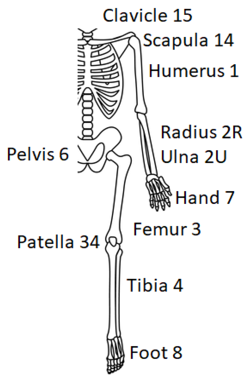
Classification:
  • BONE
The different bones of the human body can be distinguished in a similar way to the AO classification (Table 1) and are identified by a number (Figure 1).
  • Number 1 identifies the humerus.
  • Number 2 recognizes the forearm, and it can also be distinguished in 2R or 2U if only one bone of the forearm (radius or ulna) is considered.
  • Number 3 is related to the femur, while 34 is for the patella.
  • Number 4 considers the tibia (and 44, the malleoli).
  • Number 5 can refer to the spine (however, this bone can be considered a little bit different).
  • Number 6 is about the pelvis (same as number 5—spine).
  • Number 7 characterizes the hand and consists of eight under-categories (lunate 71, scaphoid 72, capitate 73, hamate 74, trapezium 75, other carpal bones 76, metacarpals 77, phalanges 78).
  • Number 8 characterizes the foot and consists of eight under-categories (talus 81, calcaneus 82, navicular 83, cuboid 84, cuneiforms 85, metatarsals 87, phalanges 88).
  • Number 14 identifies the scapula.
  • Number 15 refers to the clavicle.
Spine (5), pelvis (6), and scapula (14), considering their shapes and characteristics (either of the bone and the used implants), may be more difficult to classify, but NPPIFs at these levels are very rare.
2.
HARDWARE
The existing hardware can be identified by a letter of the Greek alphabet (Figure 2, Table 2).
  • α: plate and screws;
  • β: screws;
  • γ: nail;
  • δ: K-wires;
  • ε: external fixator.
3.
LENGTH OF THE HARDWARE
The other important factor to know is where in the bone is located the hardware and its length, compared to the bone in which it was implanted. This aspect can be distinguished using Roman numbers as follows (Figure 3, Table 3).
  • I: the hardware is in the proximal third of the corresponding bone;
  • II: the hardware is in the middle third of the corresponding bone;
  • III: the hardware is in the distal third of the corresponding bone;
  • IV: the hardware is in the distal two-thirds of the bone;
  • V: the hardware is in the proximal two-thirds of the bone;
  • VI: the hardware lies along the entire length of the bone.
4.
LEVEL OF FRACTURE
The site of the fracture is described compared to the hardware and can be described by a capital letter as follows (Figure 4, Table 4):
  • A: the fracture is proximal to the hardware.
  • B: the fracture is in correspondence of the hardware.
    B1: the fracture is in the proximal part of the hardware.
    B2: the fracture is in the middle part of the hardware.
    B3: the fracture is in the distal part of the hardware.
  • C: the fracture is distal to the hardware.

References

  1. Seo, J.H.; Lee, B.-S.; Kim, J.-M.; Kim, J.J.; Kim, J.W. Outcomes of Dual Plating for Unstable Distal Femoral Fractures: A Subgroup Comparison between Periprosthetic and Non-Periprosthetic Fractures. Int. Orthop. 2022, 46, 2685–2692. [Google Scholar] [CrossRef] [PubMed]
  2. Chan, L.W.M.; Gardner, A.W.; Wong, M.K.; Chua, K.; Kwek, E.B.K. Non-Prosthetic Peri-Implant Fractures: Classification, Management and Outcomes. Arch. Orthop. Trauma Surg. 2018, 138, 791–802. [Google Scholar] [CrossRef] [PubMed]
  3. Barrera-Ochoa, S.; Nuñez, J.H.; Mir, X. Peri-Implant Radial and Ulnar Shaft Fractures after Volar Locking Plate Fixation of the Distal Radius. J. Hand Surg. Eur. Vol. 2018, 43, 209–210. [Google Scholar] [CrossRef]
  4. Al-Jabri, T.; Ridha, M.; McCulloch, R.A.; Jayadev, C.; Kayani, B.; Giannoudis, P.V. Periprosthetic Distal Femur Fractures around Total Knee Replacements: A Comprehensive Review. Injury 2023, 54, 1030–1038. [Google Scholar] [CrossRef]
  5. Lehmann, W.; Rupprecht, M.; Nuechtern, J.; Melzner, D.; Sellenschloh, K.; Kolb, J.; Fensky, F.; Hoffmann, M.; Püschel, K.; Morlock, M.; et al. What Is the Risk of Stress Risers for Interprosthetic Fractures of the Femur? A Biomechanical Analysis. Int. Orthop. (SICOT) 2012, 36, 2441–2446. [Google Scholar] [CrossRef]
  6. Ammarullah, M.I.; Afif, I.Y.; Maula, M.I.; Winarni, T.I.; Tauviqirrahman, M.; Akbar, I.; Basri, H.; van der Heide, E.; Jamari, J. Tresca Stress Simulation of Metal-on-Metal Total Hip Arthroplasty during Normal Walking Activity. Materials 2021, 14, 7554. [Google Scholar] [CrossRef]
  7. Ammarullah, M.I.; Hartono, R.; Supriyono, T.; Santoso, G.; Sugiharto, S.; Permana, M.S. Polycrystalline Diamond as a Potential Material for the Hard-on-Hard Bearing of Total Hip Prosthesis: Von Mises Stress Analysis. Biomedicines 2023, 11, 951. [Google Scholar] [CrossRef]
  8. Ammarullah, M.; Santoso, G.; Sugiharto, S.; Supriyono, T.; Wibowo, D.; Kurdi, O.; Tauviqirrahman, M.; Jamari, J. Minimizing Risk of Failure from Ceramic-on-Ceramic Total Hip Prosthesis by Selecting Ceramic Materials Based on Tresca Stress. Sustainability 2022, 14, 13413. [Google Scholar] [CrossRef]
  9. Franklin, J.; Malchau, H. Risk Factors for Periprosthetic Femoral Fracture. Injury 2007, 38, 655–660. [Google Scholar] [CrossRef]
  10. Prieto Meré, J.A.; Barrera-Ochoa, S.; Liburd-Hernández, D.; Presas, J.P. Non-Prosthetic Peri-Implant Fracture of Both Forearm Bones. J. Surg. Case Rep. 2023, 2023, rjad300. [Google Scholar] [CrossRef] [PubMed]
  11. Brady, O.H.; Garbuz, D.S.; Masri, B.A.; Duncan, C.P. Classification of the hip. Orthop. Clin. N. Am. 1999, 30, 215–220. [Google Scholar] [CrossRef] [PubMed]
  12. Norris, R.; Bhattacharjee, D.; Parker, M.J. Occurrence of Secondary Fracture around Intramedullary Nails Used for Trochanteric Hip Fractures: A Systematic Review of 13,568 Patients. Injury 2012, 43, 706–711. [Google Scholar] [CrossRef] [PubMed]
  13. Videla-Cés, M.; Sales-Pérez, J.-M.; Sánchez-Navés, R.; Romero-Pijoan, E.; Videla, S. Proposal for the Classification of Peri-Implant Femoral Fractures: Retrospective Cohort Study. Injury 2019, 50, 758–763. [Google Scholar] [CrossRef] [PubMed]
  14. Müller, M.E.; Koch, P.; Nazarian, S.; Schatzker, J. The Comprehensive Classification of Fractures of Long Bones; Springer: Berlin/Heidelberg, Germany, 1990; ISBN 978-3-540-18165-1. [Google Scholar]
  15. Unified Classification System for Periprosthetic Fractures (UCPF). J. Orthop. Trauma 2018, 32, S141–S144. [CrossRef] [PubMed]
  16. Garbuz, D.S.; Masri, B.A.; Esdaile, J.; Duncan, C.P. Classification Systems in Orthopaedics. J. Am. Acad. Orthop. Surg. 2002, 10, 290–297. [Google Scholar] [CrossRef] [PubMed]
  17. Marsh, J.L.; Slongo, T.F.; Agel, J.; Broderick, J.S.; Creevey, W.; DeCoster, T.A.; Prokuski, L.; Sirkin, M.S.; Ziran, B.; Henley, B.; et al. Fracture and Dislocation Classification Compendium—2007: Orthopaedic Trauma Association Classification, Database and Outcomes Committee. J. Orthop. Trauma 2007, 21, S1–S6. [Google Scholar] [CrossRef]
  18. Masri, B.A.; Meek, R.M.D.; Duncan, C.P. Periprosthetic Fractures Evaluation and Treatment. Clin. Orthop. Relat. Res. 2004, 420, 80–95. [Google Scholar] [CrossRef] [PubMed]
  19. Giannoudis, P.V.; Kanakaris, N.K.; Tsiridis, E. Principles of Internal Fixation and Selection of Implants for Periprosthetic Femoral Fractures. Injury 2007, 38, 669–687. [Google Scholar] [CrossRef] [PubMed]
  20. Bidolegui, F.; Pereira, S.; Munera, M.A.; Garabano, G.; Pesciallo, C.A.; Pires, R.E.; Giordano, V. Peri-Implant Femoral Fractures: Challenges, Outcomes and Proposal of a Treatment Algorithm. Chin. J. Traumatol. 2022, 26, 211–216. [Google Scholar] [CrossRef]
  21. Egol, K.A.; Carlock, K.D.; Kelly, E.A.; Seetharam, A.; Mullis, B.H.; Marcantonio, A.J.; Bramlett, K.J.; Nchako, C.M.; Watson, J.T.; Cannada, L.K.; et al. Previous Implant Fractures: A New Descriptive Classification System. J. Orthop. Trauma 2019, 33, 423–427. [Google Scholar] [CrossRef]
  22. Müller, F.; Galler, M.; Zellner, M.; Bäuml, C.; Marzouk, A.; Füchtmeier, B. Peri-Implant Femoral Fractures: The Risk Is More than Three Times Higher within PFN Compared with DHS. Injury 2016, 47, 2189–2194. [Google Scholar] [CrossRef] [PubMed]
  23. Kruse, M.; Mohammed, J.; Sayed-Noor, A.; Wolf, O.; Holmgren, G.; Nordström, R.; Crnalic, S.; Sköldenberg, O.; Mukka, S. Peri-Implant Femoral Fractures in Hip Fracture Patients Treated with Osteosynthesis: A Retrospective Cohort Study of 1965 Patients. Eur. J. Trauma Emerg. Surg. 2022, 48, 293–298. [Google Scholar] [CrossRef] [PubMed]
  24. Liporace, F.A.; Yoon, R.S.; Collinge, C.A. Interprosthetic and Peri-Implant Fractures: Principles of Operative Fixation and Future Directions. J. Orthop. Trauma 2017, 31, 287–292. [Google Scholar] [CrossRef] [PubMed]
  25. Bonnomet, F.; Favreau, H.; Bonnevialle, P.; Adam, P.; Ehlinger, M. Interimplant Femoral Fractures. Orthop. Traumatol. Surg. Res. 2022, 108, 103117. [Google Scholar] [CrossRef] [PubMed]
  26. Stramazzo, L.; Rovere, G.; Cioffi, A.; Vigni, G.E.; Galvano, N.; D’Arienzo, A.; Letizia Mauro, G.; Camarda, L.; D’Arienzo, M. Peri-Implant Distal Radius Fracture: Proposal of a New Classification. J. Clin. Med. 2022, 11, 2628. [Google Scholar] [CrossRef]
  27. Neer, C.S. Displaced Proximal Humeral Fractures. I. Classification and Evaluation. J. Bone Jt. Surg. Am. 1970, 52, 1077–1089. [Google Scholar] [CrossRef]
  28. Sukthankar, A.V.; Leonello, D.T.; Hertel, R.W.; Ding, G.S.; Sandow, M.J. A Comprehensive Classification of Proximal Humeral Fractures: HGLS System. J. Shoulder Elb. Surg. 2013, 22, e1–e6. [Google Scholar] [CrossRef]
  29. Chelli, M.; Gasbarro, G.; Lavoué, V.; Gauci, M.-O.; Raynier, J.-L.; Trojani, C.; Boileau, P. The Reliability of the Neer Classification for Proximal Humerus Fractures: A Survey of Orthopedic Shoulder Surgeons. JSES Int. 2022, 6, 331–337. [Google Scholar] [CrossRef]
  30. Gumina, S.; Giannicola, G.; Albino, P.; Passaretti, D.; Cinotti, G.; Postacchini, F. Comparison between Two Classifications of Humeral Head Fractures: Neer and AO-ASIF. Acta Orthop. Belg. 2011, 77, 751–757. [Google Scholar] [PubMed]
  31. Marongiu, G.; Leinardi, L.; Congia, S.; Frigau, L.; Mola, F.; Capone, A. Reliability and Reproducibility of the New AO/OTA 2018 Classification System for Proximal Humeral Fractures: A Comparison of Three Different Classification Systems. J. Orthop. Traumatol. 2020, 21, 4. [Google Scholar] [CrossRef]
  32. Stoddart, M.; Pearce, O.; Smith, J.; McCann, P.; Sheridan, B.; Al-Hourani, K. Proximal Humerus Fractures: Reliability of Neer Versus AO Classification on Plain Radiographs and Computed Tomography. Cureus 2020, 12, e8520. [Google Scholar] [CrossRef] [PubMed]
  33. Schatzker, J.; McBroom, R.; Bruce, D. The Tibial Plateau Fracture. The Toronto Experience 1968–1975. Clin. Orthop. Relat. Res. 1979, 138, 94–104. [Google Scholar]
  34. Harish, S.; Roberts, C.; Blundell, C.; Walton, N.P. AO or Schatzker? How Reliable Is Classification of Tibial Plateau Fractures? Arch. Orthop. Trauma Surg. 2003, 123, 396–398. [Google Scholar] [CrossRef] [PubMed]
Figure 1. Interested bone and their corresponding numbers.
Surgeries 04 00052 g001
Figure 2. Types of hardware and their corresponding Greek letters.
Surgeries 04 00052 g002
Figure 3. Examples of where the hardware and its length compared to the bone where it was implanted is located.
Surgeries 04 00052 g003
Figure 4. Level of the fracture compared to the hardware.
Surgeries 04 00052 g004
Figure 5. Some examples of peri-implant fractures and their corresponding codes. (a) The X-ray shows a humerus (“1”) previously treated with plate and screws (α); the hardware is placed in the proximal third of the humerus (I), and the fracture is in correspondence with the distal part of the hardware (B3). The referring code is 1αIB3. (b) The X-ray shows a femur (“3”) previously treated with screws (β); the hardware is placed in the proximal third of the femur (I), and the fracture is distal to the hardware (C). The referring code is 3βIC. (c) The X-ray shows a radius (“2R”) previously treated with plate and screws (α); the hardware is placed between the middle third and distal third of the radius (V), and the fracture is in correspondence with the proximal aspect of the hardware (B1). The referring code is 2RαVB1.
Surgeries 04 00052 g005
Figure 6. An example of disagreement between the raters. This is the case with the highest disagreement. In fact, the five reviewers, during the two observations, classified the fracture as 3gammaIVB3, 3gammaIC, or 3gammaIVC. Considering the length of the hardware, some reviewers maintained that the nail is in the proximal third of the femur (I), while others stated it was in the proximal two-thirds of the bone (IV). Similarly, some reviewers thought the fracture was distal to the nail (C), while for others, it was in proximity of the distal part of the nail (B3).
Surgeries 04 00052 g006
Table 1. The bones and their corresponding numbers.
BONECorresponding Number
Humerus1
Forearm2R 2U
Femur3
Patella34
Tibia4
Spine5
Pelvis6
Hand7
Foot8
Scapula14
Clavicle15
Table 2. Types of hardware and their corresponding Greek letters.
HARDWARECorresponding Letter of the Greek Alphabet
Plate and screwsAlfa (α)
ScrewsBeta (β)
NailGamma (γ)
K wiresDelta (δ)
External fixatorEpsilon (ε)
Table 3. Classification of hardware length compared to the bone.
HARDWARE LENGTHCorresponding Roman Number
Proximal thirdI
Middle thirdII
Distal thirdIII
Proximal two-thirds of the boneIV
Distal two-thirds of the boneV
The entire length of the boneVI
Table 4. Level of the fracture compared to the hardware and the corresponding capital letter.
Level of Fracture Compared to the HardwareCorresponding
Capital
Letter
ProximalA
In correspondence
● in the proximal partB1
● in the middle part
● in the distal part
B2
B3
DistalC
Table 5. Summary of the main items considered for the classification and their corresponding numbers or letters.
BoneHardwareHardware LengthSite of the Fracture
1–15α–εI–VIA–C
Table 6. Inter-observer reliability.
Fleiss KappaStandard ErrorLower 95% CIUpper 95% CIp
0.730.040.660.81<0.001
Table 7. Inter-observer reliability between raters.
Inter-Rater Reliability
Rater 1Rater 2k = 0.73
Rater 1Rater 3k = 0.81
Rater 1Rater 4k = 0.69
Rater 1Rater 5k = 0.76
Rater 2Rater 3k = 0.77
Rater 2Rater 4k = 0.66
Rater 2Rater 5k = 0.73
Rater 3Rater 4k = 0.71
Rater 3Rater 5k = 0.66
Rater 4Rater 5k = 0.74
Averagek = 0.73
Table 8. Results of the average intra-observer reliability.
Cohens KappaStandard ErrorLower 95% CIUpper 95% CIp
0.820.070.670.96<0.001
Table 9. Intra-observer reliability of all raters.
Intra-Rater Reliability (Weighted Kappa)
Rater 1k = 0.79
Rater 2k = 0.84
Rater 3k = 0.96
Rater 4k = 0.67
Rater 5k = 0.82
Averagek = 0.82
Disclaimer/Publisher’s Note: The statements, opinions and data contained in all publications are solely those of the individual author(s) and contributor(s) and not of MDPI and/or the editor(s). MDPI and/or the editor(s) disclaim responsibility for any injury to people or property resulting from any ideas, methods, instructions or products referred to in the content.

