Article

Comparative Analysis of Open Transforaminal Lumbar Interbody Fusion and Wiltse Transforaminal Lumbar Interbody Fusion Approaches for Treating Single-Level Lumbar Spondylolisthesis: A Single-Center Retrospective Study

by
Renat Nurmukhametov
1,
Medet Dosanov
1,
Abakirov Medetbek
1,
Manuel De Jesus Encarnacion Ramirez
2,
Vishal Chavda
3,4,
Gennady Chmutin
2 and
Nicola Montemurro
5,*
1
Department of Spine Surgery, Central Clinical Hospital of the Russian Academy of Sciences, 121359 Moscow, Russia
2
Department of Neurosurgery, Russian People’s Friendship University, 121359 Moscow, Russia
3
Department of Pathology, Stanford of School of Medicine, Stanford University Medical Centre, Stanford, Palo Alto, CA 94304, USA
4
Department of Medicine, Multispeciality, Trauma and ICCU Center, Sardar Hospital, Ahmedabad 50700, Gujarat, India
5
Department of Neurosurgery, Azienda Ospedaliero Universitaria Pisana (AOUP), University of Pisa, 56100 Pisa, Italy
*
Author to whom correspondence should be addressed.
Surgeries 2023, 4(4), 623-634; https://doi.org/10.3390/surgeries4040060
Submission received: 12 October 2023 / Revised: 21 November 2023 / Accepted: 23 November 2023 / Published: 28 November 2023

Abstract

:
Background: The aim of this study is to compare the surgical outcomes of two different surgical approaches, open transforaminal lumbar interbody fusion (TLIF) and Wiltse TLIF, in the treatment of single-level lumbar spondylolisthesis and also to provide the advantages and disadvantages of each approach. Methods: This retrospective study included 600 patients with single-level lumbar spondylolisthesis who underwent fusion surgery at a single academic institution between January 2018 and December 2022. Patients were divided into two groups: traditional open TLIF (group A; 300 patients) and the Wiltse TLIF approach (group B; 300 patients). Preoperative diagnostic tests were performed on all patients. Results: The fluoroscopy time for the Wiltse TLIF group was longer, whereas the mean blood loss for the Wiltse TLIF approach was less. Both techniques resulted in significant improvements in pain relief and functional disability, with no significant difference between the two groups in terms of their pre- or post-operative (Oswestry Disability Index) ODI scores. The Wiltse TLIF technique resulted in significantly shorter hospital stays and had a lower rate of complications compared with the open TLIF technique. Conclusion: The Wiltse TLIF approach showed advantages in shorter surgical times, reduced blood loss, and shorter hospital stays, whereas the traditional open TLIF approach exhibited shorter fluoroscopy times.

1. Introduction

Spondylolisthesis (spondylos = vertebrae; listhesis = slippage) is defined as the forward slippage of one vertebra on another [1]. Of the five subtypes, degenerative and isthmic spondylolisthesis are the most common in adults [2]. Both can lead to compression and instability, which result in radicular and lower back pain [3]. Surgical fusion has emerged as a critical intervention in managing lumbar spondylolisthesis, aiming to stabilize the affected area and alleviate chronic pain. This surgical procedure involves the fusion of two or more vertebrae, often with the use of bone grafts and instrumentation, to promote spinal stability and reduce the discomfort experienced by patients. By restoring proper alignment and limiting the motion of the affected vertebrae, surgical fusion contributes to enhanced spinal function and improved quality of life for individuals grappling with lumbar spondylolisthesis. It is a vital treatment option that can offer relief and long-term benefits to those affected by this condition [4].
The transforaminal lumbar interbody fusion (TLIF) technique was first reported by Harms and Rolinger [5] in 1982. Thereafter, this technique has been increasingly used in a variety of lumbar diseases, including degenerative lumbar disc diseases, spondylolisthesis, degenerative scoliosis, and spinal instability [6]. Although TLIF is an effective procedure, extensive stripping of the paravertebral muscles and prolonged retraction are required for adequate exposure of the surgical field [7]. Iatrogenic muscle damage can lead to atrophy of the paraspinal muscles and chronic postoperative lower back pain [8,9].
Minimizing soft tissue and muscle damage is one of the main benefits of minimally invasive surgery (MIS) TLIF compared with traditional open TLIF surgery [10,11]. The use of retractors and the pressure they exert on the paraspinal muscles can cause muscle injury and negatively affect trunk muscle strength [12,13]. In addition to the physiological benefits, MIS TLIF has been shown to result in less intraoperative blood loss, lower postoperative pain, and faster recovery times compared with open TLIF [12,13]. The advent of advanced visualization tools, such as the microscope and exoscope, has been revolutionary in the field of spinal surgery, especially for procedures like TLIF [14,15,16]. Beyond visualization, its ergonomic design, which allows surgeons to view a screen rather than peer through eyepieces, mitigates physical strain, enabling longer periods of focused surgery. Moreover, its ability to broadcast a live view to a monitor means that the entire surgical team can share the same high-resolution perspective, fostering collaborative surgery and enhanced teaching scenarios [17]. The question of what constitutes a MIS TLIF in terms of the extent of surgical exposure has been a topic of considerable discussion within the medical community for the past decade. Since the inception of the MIS TLIF technique, surgeons and medical researchers have sought to outline the specific parameters that would differentiate it from its more traditional counterpart.
The aim of this study is to compare the outcomes of the open traditional TLIF and Wiltse TLIF techniques in the treatment of single-level lumbar spondylolisthesis, utilizing a retrospective analysis of 600 patients. This study aims to assess the efficacy, safety, and post-operative clinical outcomes of both techniques, with particular emphasis on muscle and soft tissue preservation, operative blood loss, postoperative pain, recovery times, and reoperation rates.

2. Materials and Methods

This was a retrospective observational cohort study conducted at the Central Clinical Hospital of the Russian Academy of Sciences in Moscow, Russia. The inclusion criteria were as follows: (1) patients who were diagnosed with single-level lumbar spondylolisthesis, presenting with symptoms of unilateral or bilateral sciatica and lumbosacral pain, typical in degenerative lumbar spine disease; (2) symptoms have not been relieved after 3 months or more of conservative treatment; (3) patients that require single-segment fusion surgery; and (4) age between 18 and 80 years. The exclusion criteria were as follows: (1) lumbosacral pain and lumbar degeneration caused by other reasons (tumors and infections); (2) patients that require fusion surgery of more than 2 segments; (3) Meyerding degree III and IV lumbar spondylolisthesis; (4) a history of previous lumbar spine surgery; and (5) trauma, active infection, and malignancy. We enrolled a total of 600 patients who were treated at the Central Clinical Hospital of the Russian Academy of Sciences (Moscow, Russia) between January 2018 and December 2022. According to different surgical treatments, they were divided into a traditional open TLIF group (group A; 300 patients) and a Wiltse TLIF approach group (group B; 300 patients). Before surgery, all patients provided consent to participate in this study and performed preoperative lumbar spine X-rays, CT or/and MRI scans, and lumbosacral dynamic position X-rays. There were no significant differences between the two groups in age and sex. This study was conducted in accordance with the Declaration of Helsinki and approved by the Local Ethics Committee (Feb 20/23).

2.1. Conservative Treatment Prior to Surgery

Before considering surgical intervention, patients enrolled in this study underwent a minimum of three months of conservative treatment. This period was essential to evaluate the effectiveness of non-surgical management in alleviating symptoms of lumbar spondylolisthesis. The conservative treatment approaches included physical therapy, pain management, activity modification, bracing, weight management, chiropractic care, and epidural steroid injections.
Regarding physical therapy, patients were provided with customized exercise programs focusing on strengthening abdominal and back muscles, improving flexibility, and enhancing spinal stability. These exercises were designed to improve core strength and reduce stress on the lumbar spine. Both over-the-counter and prescription pain relievers were utilized to manage pain and inflammation. The choice of medication was tailored based on individual patient needs and pain severity.
Patients were counseled to avoid activities that could exacerbate their symptoms. Guidance was given toward engaging in lower-impact exercises and lifestyle modifications to reduce strain on the lumbar spine. Some patients were recommended to use back braces to provide support, restrict movement, and consequently reduce pain and discomfort. Patients with excess body weight were advised on weight loss strategies to decrease stress on the lower back (weight management), which can be a contributing factor to symptom severity, whereas a subset of patients opted for chiropractic adjustments or manipulations, seeking relief from pain and discomfort associated with lumbar spondylolisthesis. For patients with significant inflammation and pain, epidural steroid injections were administered. These injections aimed to reduce inflammation in the epidural space, providing temporary pain relief.
It is important to note that the decision to proceed with surgical options such as TLIF was considered only after the persistence or worsening of symptoms despite these conservative measures. The decision was based on the severity of symptoms, the degree of spinal instability, and the overall health and preferences of the patient. This integration provides a clear and comprehensive overview of the conservative treatments undertaken prior to surgical intervention, which is crucial for understanding the patient journey and the rationale behind opting for surgery.

2.2. Indications for Selection of Open TLIF vs. Wiltse TLIF Approach

This study is retrospective. The decision to use either the open TLIF or the Wiltse TLIF approach for treating single-level lumbar spondylolisthesis was based on a comprehensive assessment of various factors, and among these factors were patient-specific anatomy, the extent of pathology, patients’ general health, surgeons’ expertise, and preference.
Regarding patient-specific anatomy, open TLIF was preferred for those patients with complex spinal anatomies or higher-grade spondylolisthesis, requiring broader spinal access, whereas the Wiltse TLIF approach was used in cases with less severe anatomical deformities, allowing less invasive access. Similarly, open TLIF was recommended for patients needing extensive decompression due to severe spinal stenosis or large disc herniations, whereas Wiltse TLIF was chosen for patients with milder spinal canal narrowing that needed minimal decompression. Finally, the surgeon’s experience and comfort with each technique influenced the decision, prioritizing the best outcome for the patient.
These individualized indications ensured that the surgical approach for each patient was tailored to optimize clinical outcomes and meet specific patient needs. This meticulous selection process was aimed at providing the most effective treatment for lumbar spondylolisthesis while minimizing potential risks and complications.

2.3. Surgical Technique in Open TLIF

Following the central longitudinal incision in the lumbar region corresponding to the level identified on lateral and anteroposterior view radiographs, the initial step involves a meticulous subperiosteal dissection. This procedure is undertaken to minimize muscular damage, thereby enhancing the clarity of exposure to the vertebral structures. Once the anatomy is adequately exposed and confirmed using intraoperative X-rays, the next stage involves the insertion of pedicle screws (as depicted in Figure 1). These pedicle screws are placed with the utmost care to ensure optimal stability. Following the secure placement of the pedicle screws, rods are contoured and interconnected across these screws. This step provides immediate spinal stability, which is crucial for the success of the procedure. Subsequently, to alleviate neural pressure and create space for the fusion process, a portion of the lamina is carefully removed via a laminectomy. On the symptomatic side, additional decompression is achieved through the removal of the facet joint, a procedure known as facetectomy.
The surgical process continues with a meticulous discectomy, where the damaged or problematic disc is carefully removed. To restore the integrity of the spinal column and promote fusion, a specially designed TLIF cage is inserted into the now vacant disc space. This cage is typically filled with local autologous bone grafts, which aid in the fusion process. This step is critical for ensuring long-term stability and the resolution of symptoms, as the fusion of adjacent vertebrae is essential for the overall success of the TLIF procedure. The combination of precise surgical techniques, careful instrumentation, and the incorporation of bone grafts sets the stage for a successful open TLIF procedure, ultimately providing relief to patients suffering from spinal issues.

2.4. Bilateral Wiltse TLIF Approach

The Wiltse TLIF approach utilizes a paramedian incision positioned 2 to 3 cm lateral to the spinal midline, strategically avoiding the dense central lumbar musculature. This incision, typically spanning 2 to 2.5 cm longitudinally, creates a surgical corridor between the multifidus and longissimus muscles, which allows for better access to the target area. Following the incision, the interbody preparation is a critical step in the procedure. There are two primary methodologies for interbody preparation in the Wiltse TLIF approach. The first method involves the use of “Serial Dilators & Tubular Retractors”. In this technique, progressively sized dilators are inserted sequentially to expand the surgical pathway, creating the necessary space for the procedure (as shown in Figure 2). The second method is the “Pedicle Screw-Based Retractor Method”. This method integrates pedicle screws as anchoring points, around which a retractor is securely fastened. This approach ensures both stability and accessibility during the procedure. With the chosen retraction system in place, an ipsilateral complete facetectomy is performed, further enhancing access to the target area.
The Wiltse TLIF approach is valued for its ability to minimize disruption of the central lumbar musculature while providing effective access to the spine for interbody fusion procedures. This technique combines precise incision placement, innovative retraction methods, and meticulous surgical steps to optimize outcomes for patients with lumbar spinal conditions.

2.5. Data Presentation

Quantitative data are primarily presented as mean values accompanied by standard deviation (SD). This format provides a clear representation of the central tendency and the dispersion of the data.

2.6. Comparison of Group Means

To determine if there was a statistically significant difference between the means of the two independent groups (open TLIF and Wiltse TLIF), an independent samples t-test was likely employed. The t-test was used to compare the means of the two unrelated groups. The assumptions for this test include the independence of observations, the normality of the data within each group, and the homogeneity of variances between the groups. If the variances are not equal, a variation of the t-test known as Welch’s t-test can be applied.

2.7. Statistical Analysis and Significance Level

SPSS 22.0 software (SPSS Inc., Chicago, IL, USA) was used to perform statistical analysis. Quantitative data were expressed as means ± standard deviation (SD). A p-value of less than 0.05 is considered statistically significant in many scientific disciplines. This threshold suggests that there is less than a 5% probability that the observed difference occurred by random chance alone.

3. Results

The mean age of patients who underwent the open TLIF technique was 56.1 years, whereas for the Wiltse technique, it was 50.3 years. The M/F ratio was 1.14 and 1.07 in the open TLIF technique group and in the Wiltse technique group, respectively. Table 1 shows all the details. No statistically significant differences were observed in sex (p-value = 0.23) and age (p-value = 0.78) between the two groups.
The mean time of surgery for the Wiltse TLIF group (117.8 ± 19.6 min) was remarkably shorter than the mean time of surgery for the open TLIF group (136.2 ± 24.3 min). This suggests that the Wiltse TLIF technique may result in a faster surgical procedure compared with the open TLIF technique. On the other hand, the median time of fluoroscopy for the Wiltse TLIF group (24.6 ± 6.4 s) was significantly longer than the median time of fluoroscopy for the open TLIF group (18 ± 3 s) due to the need to perform more intraoperative X-rays. The mean blood loss for the Wiltse TLIF group (226.4 ± 56.5 mL) was appreciably less than the mean blood loss for the open TLIF group (467.8 ± 104.9 mL). This suggests that the Wiltse TLIF technique may result in less blood loss during the surgical procedure compared with the open TLIF technique (Table 1).
Table 2 shows that both surgical techniques (open TLIF and Wiltse TLIF) resulted in significant improvements in pain relief, with a decrease in visual analogue scale (VAS) scores from pre- to post-operative times. However, the Wiltse TLIF group had a slightly higher pre-operative VAS score (7.4 ± 1.2) in comparison with the open TLIF group (7.2 ± 1.1), although the difference was not statistically significant. The post-operative VAS scores were similar between the two groups (2.1 ± 1.4 for Wiltse TLIF and 2.0 ± 1.2 for open TLIF). Both surgical techniques resulted in significant improvements in functional disability, with a decrease in Oswestry Disability Index (ODI) scores from pre- to post-operative times. There was no statistically significant difference between the two groups in terms of their pre- or post-operative ODI scores. Both surgical techniques (open TLIF and Wiltse TLIF) resulted in significant improvements in pain relief and functional disability in patients. There were no statistically significant differences between the two groups in terms of their pre- or post-operative ODI scores and only slight differences in their pre-operative VAS scores.
The Wiltse TLIF technique showed significantly fewer days of hospitalization (3.2 ± 0.9 days) compared with the open TLIF technique, which had longer hospital stays (4.6 ± 1.2 days). This suggests that patients who undergo Wiltse TLIF may recover faster and require less hospital care than those who undergo open TLIF.
In terms of complication rates, the Wiltse TLIF technique had a lower rate of complications at 2.3%, while open TLIF had a higher rate of complications at 4.3%. The number and percentage of patients experiencing each type of complication for both the open TLIF and Wiltse TLIF techniques, along with the calculated p-value for each complication type, are presented in Table 3. The p-values indicate the statistical significance of the differences in complication rates between the two techniques. Table 3 shows all the details.

4. Discussion

The term “minimally invasive” includes much more than just the length of a surgical incision. At its core, it signifies a holistic approach that aims for optimal therapeutic outcomes while minimizing collateral damage to the patient’s body. Shorter incisions are, of course, a characteristic feature, but the true essence lies in the preservation of soft tissue integrity and reducing the overall traumatic impact of the procedure on the body [16,17,18,19,20]. The Wiltse approach shows this philosophy, offering a method that markedly diminishes muscle trauma. By targeting the natural cleavage plane between the multifidus and longissimus muscles, the Wiltse approach bypasses substantial muscular dissection that is often necessary for other techniques. This not only ensures fewer postoperative complications like muscle atrophy or chronic pain but also translates into faster recovery times and less post-surgical discomfort for the patient.
The present study compared the clinical outcomes of two surgical techniques for the treatment of lumbar spine pathologies, the open TLIF and Wiltse TLIF approaches. The results of this study confirm that the two techniques are equally effective in terms of clinical outcomes, including improvement in ODI values, VAS scores, and fusion rates. However, the Wiltse TLIF technique was found to be associated with a shorter time of surgery and less blood loss, while requiring more time for intraoperative imaging [21,22,23].
Our findings agree with previous studies that have compared the Wiltse TLIF technique with the traditional open TLIF technique, which have reported similar clinical outcomes between the two techniques. However, the present study has the particularity that it directly compared the two techniques in a single institution, using a relatively large sample size of 600 patients. This study has important clinical implications, as we suggest that the Wiltse TLIF technique may be a more favorable option for patients due to its shorter surgical time and lower blood loss. However, the longer fluoroscopy time required for the Wiltse TLIF technique should also be considered.
While our study did not present cases necessitating pedicle screw alterations, the presence of infection remained a point of concern, particularly within the realm of spinal surgery. Infections are a crucial consideration in spinal surgery, as underscored by various studies. For instance, Pull Ter Gunne and Cohen [24] articulated the impactful consequences of infections post-spinal surgery, emphasizing their frequency and the ensuing challenges in management and treatment. Additionally, Schoenfeld et al. [25] delved into the substantial morbidity engendered by postoperative spinal infections, emphasizing the critical need for comprehensive preventive strategies, and expedited diagnostic processes to mitigate the associated risks and complications.
Parker et al. [26] conducted a systematic review and discovered that MIS TLIF cases had a significantly lower rate of surgical site infections compared with open TLIF cases (0.6% vs. 4.0%; p < 0.001). A recent meta-analysis by Phan et al. also found that MIS TLIF cases have significantly lower infection rates (1.2% vs. 4.6%; p < 0.001), thus providing further evidence.
In our study, dural tears were evidenced in both the open TLIF and Wiltse TLIF techniques, each having three instances. Our findings resonate with a larger study conducted by Williams et al. [27], which scrutinized 108.478 surgical cases submitted from 2004 to 2007. The comprehensive study showed that unintended durotomy occurred in 1.6% of all cases analyzed, offering a wider context to our findings, and underlining the prevalence of such complications even among experienced surgeons. The study found that the incidence of unintended durotomy varied with the preoperative diagnosis, being higher in patients treated for spondylolisthesis and lower among those treated for scoliosis. The treatment of dural tears primarily involves immediate and meticulous repair to prevent subsequent complications, such as cerebrospinal fluid leakage and potential neurological damage. Primary repair is usually attempted first, employing microsurgical techniques to suture the tear directly or to apply a patch, possibly supplemented with fibrin glue.
Implant malposition or failure can lead to severe consequences and may necessitate revision surgeries. Persistent pain post-surgery can significantly impact the quality of life of patients and may sometimes remain unresolved. Studies conducted by Nandyala et al. [28] and Patel et al. [29] explored the implications of implant malposition and persistent pain, substantiating our findings and emphasizing the need for meticulous surgical techniques and postoperative management to mitigate these complications.
Wound dehiscence was noted as a complication only in the open TLIF technique, occurring in three instances, while there were no recorded instances in the Wiltse TLIF technique group in our study. This difference may indicate that the Wiltse TLIF technique has a superior methodology in terms of wound healing or may suggest differences in procedural wound closure practices between the two techniques. Wound dehiscence is a severe complication that can lead to infection and requires immediate medical intervention, as denoted by Kanna et al. [30]. The findings of our study are consistent with those of Sclafani et al. [31], emphasizing the need for meticulous attention to wound closure, comorbidity of patients, and postoperative care to prevent dehiscence, particularly in open TLIF procedures.
The extensive fluoroscopic time associated with MIS TLIF procedures underscores the inherent complexities of this minimally invasive method. Fluoroscopy plays a crucial role in ensuring the accurate positioning of tubular retractors, cages, and pedicle screws during the MIS TLIF procedure [32,33,34,35]. A systematic review revealed that the fluoroscopy time for MIS groups ranged from 49 to 297 s, compared with 24 to 123 s for open groups [36]. It is important to consider that while MIS TLIF offers advantages, like reduced trauma and quicker recovery, it also presents challenges. One significant challenge is the learning curve associated with mastering this procedure. As with any surgical technique, proficiency comes with experience. Early in a surgeon’s exposure to MIS TLIF, operative times may be longer, and there might be a higher reliance on fluoroscopy, leading to prolonged exposure times. Both factors could potentially elevate the risk of complications and the need for revisions [37,38,39,40,41,42].
It is crucial to understand the significance of the learning curve, especially in the context of open TLIF and Wiltse TLIF, as these procedures are intricate, demanding extensive knowledge and technical proficiency. Open TLIF is renowned for its extensive nature and is associated with a strenuous learning curve due to the elaborate anatomical exposure and manipulation it involves [43]. The procedure, necessitating a profound understanding of anatomy, meticulous soft tissue dissection, and careful management of nerve roots, requires extensive training and experience. This is crucial for spinal surgeons to minimize postoperative complications and to secure optimal patient outcomes. Due to the open nature of this procedure, acquiring adept technical skills and refined decision-making abilities is paramount, which includes the accurate assessment of spinal instability and alignment [44,45,46].
Conversely, the learning curve of Wiltse TLIF is demarcated by the mastery of the muscle-splitting approach and the nuances associated with it [44]. The Wiltse approach, which is praised for its minimization of muscular damage and postoperative pain, necessitates precision and a nuanced understanding of the paraspinal musculature and fascial planes. Achieving the desired clinical outcomes and diminishing the potential for iatrogenic injury requires focused training and experiential learning to grasp the specificities of the Wiltse approach. The early phases of learning Wiltse TLIF may witness longer operative times and an increased reliance on fluoroscopy. However, with accrued experience and progression along the learning curve, a decline in these factors and advancements in surgical proficiency are observable [47]. Assessing the learning curve in minimally invasive spine surgery is complex and challenging. Therefore, various factors such as operating times, conversion to open procedures, visual analog scale scores, and periods of hospital stay are used to measure it. It has been observed that all complications that arise during the procedure have been documented previously and are significantly reduced after the 30th consecutive case. As surgical experience increases, peri-operative parameters such as operative time and length of hospital stay improve. However, the downsides of minimally invasive spinal surgery are that surgeons have to start unfamiliar procedures without tactile sensation, work in a narrow restricted surgical field, and use endoscopes via two-dimensional imaging [47].
Mitigating the challenges presented by the learning curves in both open and Wiltse TLIF can be facilitated by the implementation of comprehensive educational programs, surgical simulations, telemedicine, and dedicated mentorships [47,48]. Such methodologies are instrumental in facilitating the attainment of the technical proficiencies and cognitive skills imperative for these procedures. They play a crucial role in optimizing learning curves, thereby contributing to improved surgical outcomes and enhanced patient care [47].
Furthermore, the selection of the surgical approach often depends on the patient’s specific condition. Surgeons might opt for open procedures when confronted with complexities like abnormal spinal anatomy, pseudarthrosis (failed spinal fusion), the need for revision surgery due to previous unsuccessful interventions, the presence of tumors, infections, or instances where radiographic imaging does not offer clear visualization. In such cases, the known and familiar territory of open surgeries may provide a safer and more controlled environment for the surgeon. This decision making underscores the importance of individualized patient assessment, ensuring the chosen approach best suits the patient’s unique anatomical and clinical scenario [49].

Limitations of This Study

The main limitation of our study is that it is a retrospective single academic institution, which may limit the generalizability of the findings to other settings or populations. Secondarily, we used strict inclusion and exclusion criteria, which may have led to the exclusion of patients with similar characteristics, potentially biasing the results. The third limitation is the relatively short follow-up period (only a few years), which may not be sufficient to point out long-term outcomes such as complications, reoperations, or quality of life.

5. Conclusions

This study confirms that the Wiltse TLIF technique may offer advantages over the open TLIF technique in terms of shorter surgical times and less blood loss, but it may require more time for intraoperative imaging. Despite these differences, there was no statistically significant difference between the two groups in terms of clinical outcomes such as postoperative pain, functional outcomes, and complication rates. The Wiltse group has benefits in terms of reducing the length of hospitalization and may result in faster recovery for patients.

Author Contributions

Conceptualization, R.N., M.D.J.E.R., G.C. and N.M.; methodology, R.N., M.D., A.M. and N.M.; validation, R.N., V.C. and N.M.; formal analysis, R.N., M.D.J.E.R. and N.M.; investigation, R.N., M.D., M.D.J.E.R. and N.M.; resources, R.N. and M.D.; data curation, M.D.J.E.R. and N.M.; writing—original draft preparation, R.N., M.D.J.E.R. and N.M.; writing—review and editing, R.N. and N.M.; visualization, R.N., M.D.J.E.R. and N.M.; supervision, R.N. and N.M. All authors have read and agreed to the published version of the manuscript.

Funding

This research received no external funding.

Institutional Review Board Statement

This study was conducted in accordance with the Declaration of Helsinki and approved by the Ethics Committee of Central Clinical Hospital of the Russian Academy of Sciences, Moscow, Russia (Feb 20/23).

Informed Consent Statement

Written informed consent has been obtained from the patient(s) to publish this paper.

Data Availability Statement

The data presented in this study are available on request from the corresponding author.

Conflicts of Interest

The authors declare no conflict of interest.

References

  1. Spiker, W.R.; Goz, V.; Brodke, D.S. Lumbar Interbody Fusions for Degenerative Spondylolisthesis: Review of Techniques, Indications, and Outcomes. Glob. Spine J. 2019, 9, 77–84. [Google Scholar] [CrossRef] [PubMed]
  2. Fan, G.; Gu, G.; Zhu, Y.; Guan, X.; Hu, A.; Wu, X.; Zhang, H.; He, S. Minimally Invasive Transforaminal Lumbar Interbody Fusion for Isthmic Spondylolisthesis: In Situ Versus Reduction. World Neurosurg. 2016, 90, 580–587. [Google Scholar] [CrossRef] [PubMed]
  3. Sivaraman, A.; Altaf, F.; Jalgaonkar, A.; Kakkar, R.; Sirigiri, P.B.; Howieson, A.; Crawford, R.J. Prospective study of posterior lumbar interbody fusion with either interbody graft or interbody cage in the treatment of degenerative spondylolisthesis. J. Spinal Disord. Tech. 2015, 28, E467–E471. [Google Scholar] [CrossRef] [PubMed]
  4. Ha, K.Y.; Na, K.H.; Shin, J.H.; Kim, K.W. Comparison of posterolateral fusion with and without additional posterior lumbar interbody fusion for degenerative lumbar spondylolisthesis. J. Spinal Disord. Tech. 2008, 21, 229–234. [Google Scholar] [CrossRef] [PubMed]
  5. Momin, A.A.; Steinmetz, M.P. Evolution of Minimally Invasive Lumbar Spine Surgery. World Neurosurg. 2020, 140, 622–626. [Google Scholar] [CrossRef]
  6. Cutler, A.R.; Siddiqui, S.; Mohan, A.L.; Hillard, V.H.; Cerabona, F.; Das, K. Comparison of polyetheretherketone cages with femoral cortical bone allograft as a single-piece interbody spacer in transforaminal lumbar interbody fusion. J. Neurosurg. Spine 2006, 5, 534–539. [Google Scholar] [CrossRef] [PubMed]
  7. Houten, J.K.; Post, N.H.; Dryer, J.W.; Errico, T.J. Clinical and radiographically/neuroimaging documented outcome in transforaminal lumbar interbody fusion. Neurosurg. Focus 2006, 20, 22–25. [Google Scholar] [CrossRef] [PubMed]
  8. Potter, B.K.; Freedman, B.A.; Verwiebe, E.G.; Hall, J.M.; Polly, D.W., Jr.; Kuklo, T.R. Transforaminal lumbar interbody fusion: Clinical and radiographic results and complications in 100 consecutive patients. J. Spinal Disord. Tech. 2005, 18, 337–346. [Google Scholar] [CrossRef]
  9. Liang, Y.; Shi, W.; Jiang, C.; Chen, Z.; Liu, F.; Feng, Z.; Jiang, X. Clinical outcomes and sagittal alignment of single-level unilateral instrumented transforaminal lumbar interbody fusion with a 4 to 5-year follow-up. Eur. Spine J. 2015, 24, 2560–2566. [Google Scholar] [CrossRef]
  10. Nurmukhametov, R.; Dosanov, M.; Encarnacion, M.D.; Barrientos, R.; Matos, Y.; Alyokhin, A.I.; Baez, I.P.; Efe, I.E.; Restrepo, M.; Chavda, V.; et al. Transforaminal Fusion Using Physiologically Integrated Titanium Cages with a Novel Design in Patients with Degenerative Spinal Disorders: A Pilot Study. Surgeries 2022, 3, 175–184. [Google Scholar] [CrossRef]
  11. Foley, K.T.; Holly, L.T.; Schwender, J.D. Minimally Invasive Lumbar Fusion. Spine 2003, 28, S26–S35. [Google Scholar] [CrossRef]
  12. Styf, J.R.; Willén, J. The Effects of External Compression by Three Different Retractors on Pressure in the Erector Spine Muscles During and After Posterior Lumbar Spine Surgery in Humans. Spine 1998, 23, 354–358. [Google Scholar] [CrossRef] [PubMed]
  13. Tian, N.-F.; Wu, Y.-S.; Zhang, X.-L.; Xu, H.-Z.; Chi, Y.-L.; Mao, F.-M. Minimally invasive versus open transforaminal lumbar interbody fusion: A meta-analysis based on the current evidence. Eur. Spine J. 2013, 22, 1741–1749. [Google Scholar] [CrossRef] [PubMed]
  14. Adogwa, O.; Parker, S.L.; Bydon, A.; Cheng, J.; Mcgirt, M.J. Comparative Effectiveness of Minimally Invasive Versus Open Transforaminal Lumbar Interbody Fusion: 2-year Assessment of Narcotic Use, Return to Work, Disability, and Quality of Life. Clin. Spine Surg. 2011, 24, 479–484. [Google Scholar] [CrossRef] [PubMed]
  15. Fan, S.; Zhao, X.; Zhao, F.; Xiangqian, F. Minimally Invasive Transforaminal Lumbar Interbody Fusion for the Treatment of Degenerative Lumbar Diseases. Spine 2010, 35, 1615–1620. [Google Scholar]
  16. Musa, G.; Barrientos Castillo, R.E.; Slabov, M.V.; Chirwa, K.; Chmutin, G.E.; Ramirez, M.d.J.E.; Nurmukhametov, R. Degenerative Grade 3 Spondylolisthesis Management: A Case Report and Literature Review. Cureus 2022, 14, e29374. [Google Scholar] [CrossRef]
  17. Ramirez, M.E.; Peralta, I.; Nurmukhametov, R.; Castillo, R.E.; Castro, J.S.; Volovich, A.; Dosanov, M.; Efe, I.E. Expanding access to microneurosurgery in low-resource settings: Feasibility of a low-cost exoscope in transforaminal lumbar interbody fusion. J. Neurosci. Rural Pract. 2023, 14, 156–160. [Google Scholar] [CrossRef] [PubMed]
  18. Ahsan, K.; Khan, S.I.; Zaman, N.; Ahmed, N.; Montemurro, N.; Chaurasia, B. Fusion versus nonfusion treatment for recurrent lumbar disc herniation. J. Craniovertebral Junction Spine 2021, 12, 44–53. [Google Scholar] [CrossRef] [PubMed]
  19. Tian, Y.; Liu, X. Clinical outcomes of two minimally invasive transforaminal lumbar interbody fusion (TLIF) for lumbar degenerative diseases. Eur. J. Orthop. Surg. Traumatol. 2016, 26, 745–751. [Google Scholar] [CrossRef] [PubMed]
  20. Sun, Y.; Zhang, W.; Zhang, F.; Li, J.; Guo, L. Study of single-level lumbar degenerative diseases treated by unilateral wiltse access with unilateral nail rod fixation assisted by a new automatic retraction device. J. Orthop. Surg. Res. 2023, 18, 66. [Google Scholar] [CrossRef]
  21. Kim, S.H.; Hahn, B.S.; Park, J.Y. What Affects Segmental Lordosis of the Surgical Site after Minimally Invasive Transforaminal Lumbar Interbody Fusion? Yonsei Med. J. 2022, 63, 665–674. [Google Scholar] [CrossRef] [PubMed]
  22. Liu, H.; Li, J.; Sun, Y.; Wang, X.; Wang, W.; Guo, L.; Zhang, F.; Zhang, P.; Zhang, W. A Comparative Study of a New Retractor-Assisted WILTSE TLIF, MIS-TLIF, and Traditional PLIF for Treatment of Single-Level Lumbar Degenerative Diseases. Orthop. Surg. 2022, 14, 1317–1330. [Google Scholar] [CrossRef]
  23. Wang, S.; Duan, C.; Yang, H.; Kang, J.; Wang, Q. Wiltse Approach Versus Conventional Transforaminal Interbody Fusion for Unstable Thoracolumbar Fracture with Intervertebral Disc Lesions. Orthop. Surg. 2022, 14, 694–703. [Google Scholar] [CrossRef] [PubMed]
  24. Pull ter Gunne, A.F.; Cohen, D.B. Incidence, prevalence, and analysis of risk factors for surgical site infection following adult spinal surgery. Spine 2009, 34, 1422–1428. [Google Scholar] [CrossRef] [PubMed]
  25. Schoenfeld, A.J.; Ochoa, L.M.; Bader, J.O.; Belmont, P.J., Jr. Risk factors for immediate postoperative complications and mortality following spine surgery: A study of 3475 patients from the National Surgical Quality Improvement Program. J. Bone Jt. Surg. 2011, 93, 1577–1582. [Google Scholar] [CrossRef] [PubMed]
  26. Parker, S.L.; Adogwa, O.; Witham, T.F.; Aaronson, O.S.; Cheng, J.; McGirt, M.J. Post-Operative Infection after Minimally Invasive versus Open Transforaminal Lumbar Interbody Fusion (TLIF): Literature Review and Cost Analysis. Minim. Invasive Neurosurg. 2011, 54, 33–37. [Google Scholar] [CrossRef]
  27. Williams, B.J.; Sansur, C.A.; Smith, J.S.; Berven, S.H.; Broadstone, P.A.; Choma, T.J.; Goytan, M.J.; Noordeen, H.H.; Knapp, D.R.; Hart, R.A.; et al. Incidence of unintended durotomy in spine surgery based on 108,478 cases. Neurosurgery 2011, 68, 117–123. [Google Scholar] [CrossRef]
  28. Nandyala, S.V.; Fineberg, S.J.; Pelton, M.; Singh, K. Minimally invasive transforaminal lumbar interbody fusion: One surgeon’s learning curve. Spine J. 2014, 14, 1460–1465. [Google Scholar] [CrossRef]
  29. Patel, J.; Kundnani, V.; Raut, S.; Meena, M.; Ruparel, S. Perioperative Complications of Minimally Invasive Transforaminal Lumbar Interbody Fusion (MI-TLIF): 10 Years of Experience With MI-TLIF. Glob. Spine J. 2021, 11, 733–739. [Google Scholar] [CrossRef]
  30. Kanna, R.M.; Renjith, K.R.; Shetty, A.P.; Rajasekaran, S. Classification and Management Algorithm for Postoperative Wound Complications Following Transforaminal Lumbar Interbody Fusion. Asian Spine J. 2020, 14, 673–681. [Google Scholar] [CrossRef]
  31. Sclafani, J.A.; Raiszadeh, K.; Raiszadeh, R.; Kim, P.; Doerr, T.; Siddiqi, F.; LaMotta, I.; Park, P.; Templin, C.; Gill, S.; et al. Validation and analysis of a multi-site MIS Prospective Registry through sub-analysis of an MIS TLIF Subgroup. Int. J. Spine Surg. 2014, 8, 4. [Google Scholar] [CrossRef] [PubMed]
  32. Phan, K.; Rao, P.J.; Kam, A.C.; Mobbs, R.J. Minimally invasive versus open transforaminal lumbar interbody fusion for treatment of degenerative lumbar disease: Systematic review and meta-analysis. Eur. Spine J. 2015, 24, 1017–1030. [Google Scholar] [CrossRef]
  33. Chan, A.K.; Bisson, E.F.; Bydon, M.; Glassman, S.D.; Foley, K.T.; Potts, E.A.; Shaffrey, C.I.; Shaffrey, M.E.; Coric, D.; Knightly, J.J.; et al. A comparison of minimally invasive transforaminal lumbar interbody fusion and decompression alone for degenerative lumbar spondylolisthesis. Neurosurg. Focus 2019, 46, E13. [Google Scholar] [CrossRef] [PubMed]
  34. Chan, A.K.; Bydon, M.; Bisson, E.F.; Glassman, S.D.; Foley, K.T.; Shaffrey, C.I.; Potts, E.A.; Shaffrey, M.E.; Coric, D.; Knightly, J.J.; et al. Minimally invasive versus open transforaminal lumbar interbody fusion for grade I lumbar spondylolisthesis: 5-year follow-up from the prospective multicenter Quality Outcomes Database registry. Neurosurg. Focus 2023, 54, E2. [Google Scholar] [CrossRef]
  35. Mummaneni, P.V.; Bisson, E.F.; Kerezoudis, P.; Glassman, S.; Foley, K.; Slotkin, J.R.; Potts, E.; Shaffrey, M.; Shaffrey, C.I.; Coric, D.; et al. Minimally invasive versus open fusion for Grade I degenerative lumbar spondylolisthesis: Analysis of the Quality Outcomes Database. Neurosurg. Focus 2017, 43, E11. [Google Scholar] [CrossRef] [PubMed]
  36. Goldstein, C.L.; Macwan, K.; Sundararajan, K.; Rampersaud, R.Y. Comparative Outcomes of Minimally Invasive Surgery for Posterior Lumbar Fusion: A Systematic Review. Clin. Orthop. Relat. Res. 2014, 472, 1727–1737. [Google Scholar] [CrossRef]
  37. Lee, K.H.; Yeo, W.; Soeharno, H.; Yue, W.M. Learning Curve of a Complex Surgical Technique: Minimally Invasive Transforaminal Lumbar Interbody Fusion (MIS TLIF). Clin. Spine Surg. 2014, 27, E234–E240. [Google Scholar] [CrossRef]
  38. Jin-Tao, Q.; Yu, T.; Mei, W.; Xu-Dong, T.; Tian-Jian, Z.; Guo-Hua, S.; Lei, C.; Yue, H.; Zi-Tian, W.; Yue, Z. Comparison of MIS vs. open PLIF/TLIF with regard to clinical improvement, fusion rate, and incidence of major complication: A meta-analysis. Eur. Spine J. 2015, 24, 1058–1065. [Google Scholar] [CrossRef]
  39. Ahsan, M.K.; Hossain, M.R.; Khan, M.S.I.; Zaman, N.; Ahmed, N.; Montemurro, N.; Chaurasia, B. Lumbar revision microdiscectomy in patients with recurrent lumbar disc herniation: A single-center prospective series. Surg. Neurol. Int. 2020, 11, 404. [Google Scholar] [CrossRef]
  40. Perrini, P.; Pieri, F.; Montemurro, N.; Tiezzi, G.; Parenti, G.F. Thoracic extradural haematoma after epidural anaesthesia. Neurol. Sci. 2010, 31, 87–88. [Google Scholar] [CrossRef]
  41. Patel, K.; Harikar, M.M.; Venkataram, T.; Chavda, V.; Montemurro, N.; Assefi, M.; Hussain, N.; Yamamoto, V.; Kateb, B.; Lewandrowski, K.-U.; et al. Is Minimally Invasive Spinal Surgery Superior to Endoscopic Spine Surgery in Postoperative Radiologic Outcomes of Lumbar Spine Degenerative Disease? A Systematic Review. J. Neurol. Surg. Part A Central Eur. Neurosurg. 2023, 8, 1. [Google Scholar] [CrossRef] [PubMed]
  42. Lei, F.; Li, Z.; He, W.; Tian, X.; Zheng, L.; Kang, J.; Feng, D. Total and hidden blood loss between open posterior lumbar interbody fusion and transforaminal lumbar interbody fusion by Wiltse approach. Medicine 2020, 99, e19864. [Google Scholar] [CrossRef] [PubMed]
  43. Kovari, V.Z.; Kuti, A.; Konya, K.; Szel, I.; Szekely, A.K.; Szalay, K. Comparison of Single-Level Open and Minimally Invasive Transforaminal Lumbar Interbody Fusions Presenting a Learning Curve. BioMed Res. Int. 2020, 2020, 3798537. [Google Scholar] [CrossRef] [PubMed]
  44. Sharif, S.; Afsar, A. Learning Curve and Minimally Invasive Spine Surgery. World Neurosurg. 2018, 119, 472–478. [Google Scholar] [CrossRef] [PubMed]
  45. Jin, Y.; Chen, Q.; Chen, C.; Lyu, J.; Shi, B.; Yang, C.; Xia, C. Clinical Research and Technique Note of TLIF by Wiltse Approach for the Treatment of Degenerative Lumbar. Orthop. Surg. 2021, 13, 1628–1638. [Google Scholar] [CrossRef]
  46. Heo, D.H.; Hong, Y.H.; Lee, D.C.; Chung, H.J.; Park, C.K. Technique of Biportal Endoscopic Transforaminal Lumbar Interbody Fusion. Neurospine 2020, 17, S129–S137. [Google Scholar] [CrossRef]
  47. Lee, J.C.; Jang, H.D.; Shin, B.J. Learning curve and clinical outcomes of minimally invasive transforaminal lumbar interbody fusion: Our experience in 86 consecutive cases. Spine 2012, 37, 1548–1557. [Google Scholar] [CrossRef]
  48. Montemurro, N. Telemedicine: Could it represent a new problem for spine surgeons to solve? Glob. Spine J. 2022, 12, 1306–1307. [Google Scholar] [CrossRef]
  49. Silva, P.S.; Pereira, P.; Monteiro, P.; Silva, P.A.; Vaz, R. Learning curve and complications of minimally invasive transforaminal lumbar interbody fusion. Neurosurg. Focus 2013, 35, E7. [Google Scholar] [CrossRef]
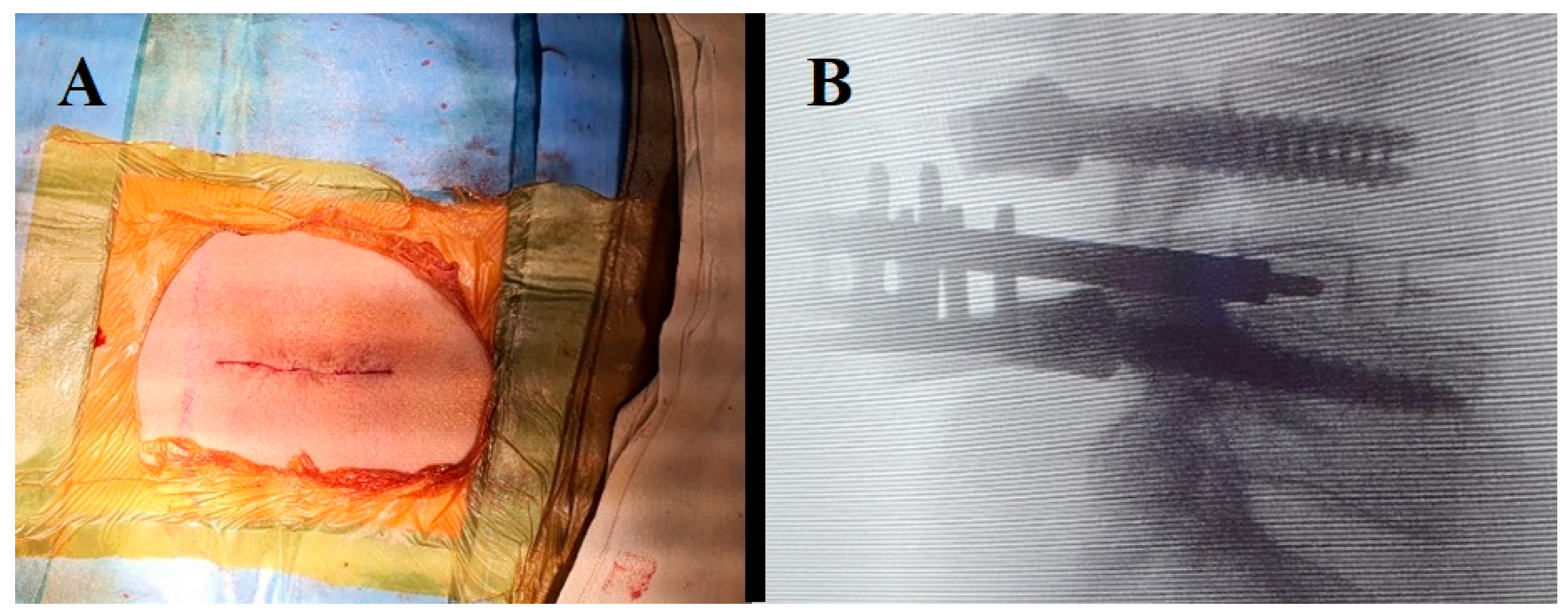
Figure 1. (A) Midline incision for open TLIF technique. (B) Intraoperative X-ray lateral view shows the placement of pedicle screws and a cage at L4–L5 level with open TLIF technique.
Surgeries 04 00060 g001
Figure 2. (A) In the Wiltse TLIF technique, the two vertical incisions are observed at 2.5 cm from the midline as well as the placement of the tubular retractor. (B) Intraoperative X-ray shows the tubular retractor placed in the decompression area, the needle to mark the level, and the percutaneously placed screws.
Surgeries 04 00060 g002
Table 1. Demographic data and intraoperative outcomes.
Open TLIF
Technique
Wiltse TLIF
Technique
p-Value
Number of patients300300-
Age (years, mean ± SD)56.1 ± 7.950.3 ± 8.60.23
Sex (M/F)160/140155/1450.78
Time of surgery (min)136.2 ± 24.3117.8 ± 19.6<0.01
Time of fluoroscopy (s)18. ± 324.6 ± 6.4<0.05
Blood loss (mL)467.8 ± 104.9226.4 ± 56.5<0.01
Table 2. Surgical outcomes of both techniques.
Outcome MeasureOpen TLIF
Technique
Wiltse TLIF
Technique
VAS pre-op7.2 ± 1.17.4 ± 1.2
VAS post-op2.0 ± 1.22.1 ± 1.4
p-value<0.05<0.05
ODI pre-op61.7 ± 9.560.3 ± 8.9
ODI post-op23.9 ± 5.925.1 ± 6.2
p-value<0.05<0.05
Table 3. Surgical complications.
ComplicationsOpen TLIF Technique (Patient n° (%))Wiltse TLIF Technique (Patient n° (%))p-Value
Infection3 (1)1 (0.3)0.616
Nerve damage2 (0.7)2 (0.7)1.000
Dural tear3 (1)3 (1)1.000
Implant malposition or failure1 (0.3)-1.000
Persistent pain1 (0.3)-1.000
Wound dehiscence3 (1)-0.247
Overall complications13 (4.3)7 (2.3)0.255
Disclaimer/Publisher’s Note: The statements, opinions and data contained in all publications are solely those of the individual author(s) and contributor(s) and not of MDPI and/or the editor(s). MDPI and/or the editor(s) disclaim responsibility for any injury to people or property resulting from any ideas, methods, instructions or products referred to in the content.