1. Introduction
Autologous fat grafting (AFG) is a powerful surgical means to augment the tissue volume. AFG is now widely indicated for reconstructive, aesthetic, and functional procedures, with applications across the breadth of medical and surgical care. While this technique has advanced rapidly in the last three decades, one of the key remaining limitations is unpredictable long-term retention. Unpredictable graft loss is a burden on both the patient and provider, with the risk of either repeat procedures or unsatisfactory outcomes. Improving this is a key focus in current AFG research. Historically, rodent models have been utilized to better understand the mechanisms underpinning graft survival and to test therapeutics. However, their dissimilarities to humans in terms of their size, physiology, and wound healing process limit the direct clinical applicability. Large animal models play a crucial role in preclinical research, bridging the gap between benchtop experiments and clinical trials; however, these experiments are more expensive, require more sophisticated surgical techniques, and preferably utilize longitudinal (i.e., nondestructive) outcome assessments in lieu of regular animal attrition. Here, we report our efforts to develop a robust, efficient, and reproducible swine fat grafting model with consideration toward the animal models available at our institution, the comparability of pig adipose tissue attributes to human tissue, and post-surgical approaches to minimize complications from autologous fat harvesting, improving the model’s safety.
Swine are a powerful model for translational research given their anatomic and physiologic similarity to humans. This is particularly well described in regard to their cutaneous biology [
1]. The comparable organ size, tissue characteristics, and wound healing process between swine and humans supports the hypothesis that pigs are well suited for evaluation of AFG [
2]. Moreover, the availability of established imaging modalities allows for the comprehensive evaluation of graft survival, vascularization, tissue integration, and potential complications. Consequently, swine have been utilized in prior fat transfer studies [
3,
4,
5,
6]. A close review of this literature, however, demonstrates a significant variability in the models, outcomes, and techniques [
3,
4,
5,
6]. There is a wide range of pig breeds available with significant anatomic, physiologic, and histologic variabilities between them. Furthermore, the geographic location plays a role, with some breeds available only in certain regions or in a very limited supply. From a technical perspective, we identified variabilities in the surgical approach, harvest, processing, and recipient sites [
3,
4,
5,
6], and although the extant literature does explore the anatomic and tissue qualities of porcine fat, it is often with a focus on the agricultural productivity and nutritional science rather than from a translational perspective [
3,
4,
5,
6].
In this descriptive report, we seek to provide information on critical attributes of swine fat grafting, including the following: (1) model selection; (2) donor site selection and harvest; (3) tissue processing; (4) recipient site preparation; (5) post-operative care; and (6) graft assessment. We provide insights into the challenges inherent across different breeds, the potential use of allogeneic approaches, methods to improve tissue processing and engraftment, and a comprehensive discussion of the advantages and disadvantages associated with each approach. We anticipate that the conclusions presented may be extrapolated to other breeds not included in the study due to regional differences in breeder availability (e.g., Göttingen minipig), but which may be preferred by other research groups.
2. Methods
All procedures were performed under the University of Pittsburgh IACUC approval #23032796, with 12 h light and dark cycles, water provided ab libitum, and food provided per the Department of Laboratory Animal Research (DLAR) schedule according to the pigs’ weights. All procedures were performed in compliance with the National Institute of Health guidelines for ethical animal treatment. The pigs were socially housed apart from the acute post-surgical periods, when single housing was required to avoid wound contamination and ensure adequate wound healing.
2.1. Pig Breed
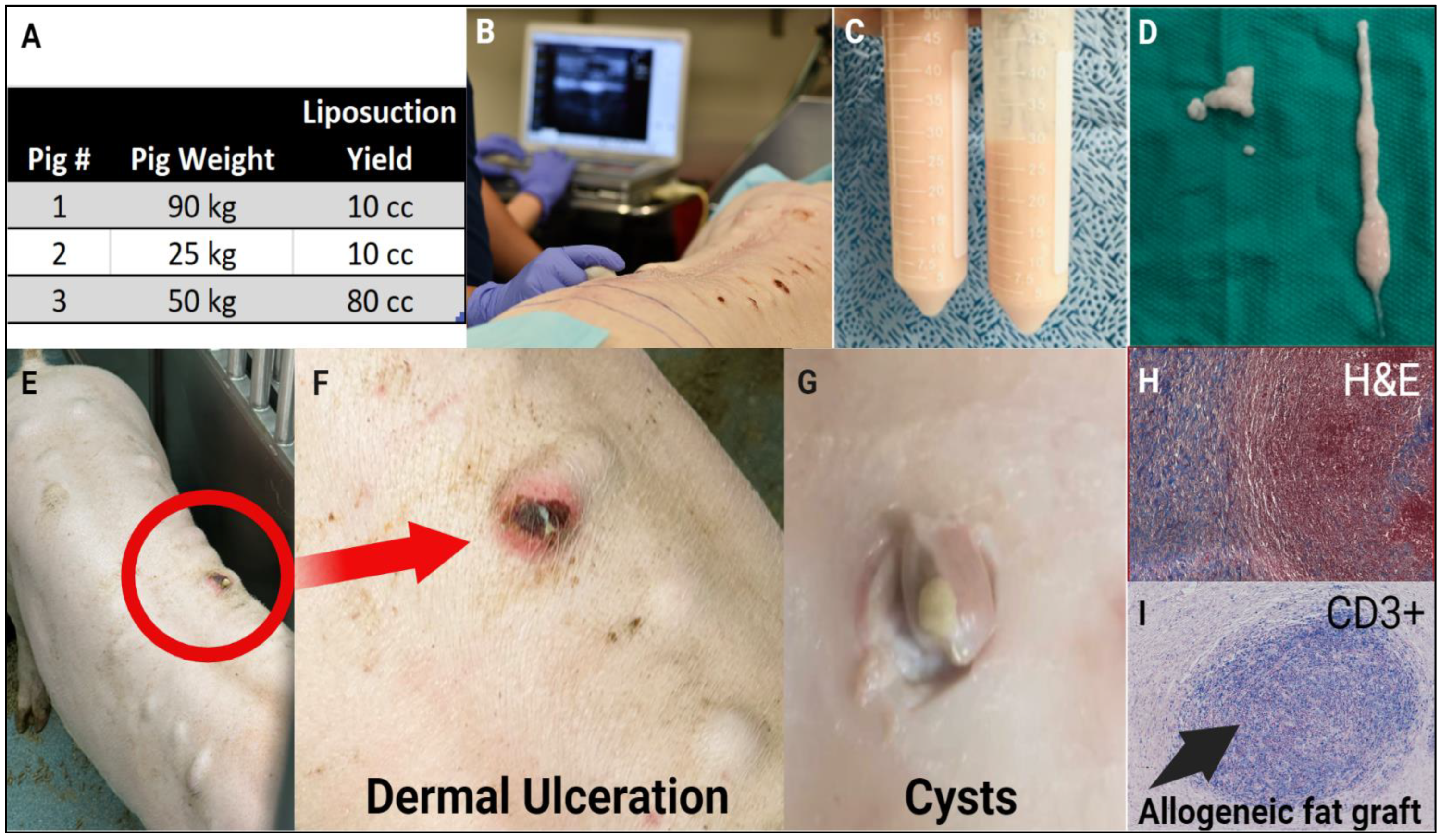
Yorkshire and Yucatan breeds were selected, as both breeds are regularly procured and maintained at our institution. The Yorkshire strain composition varies by vendor. The pigs utilized in our facility are estimated to be 75% Yorkshire crossbred with either Landrace (Premier BioSource, formerly S&S Farms, Ramona, CA, USA) or Duroc (Archer Farms, Inc, Darlington, MD, USA). Conversely, Yucatan minipigs are 100% pure. All animals used are specific-pathogen free (S.P.F.). The Yorkshire cohort consisted of 8 pigs (4 pigs per vendor), aged 3–6 months and 30–50 kg on delivery. The Yucatan cohort consisted of 4 pigs (Premier BioSource) aged 10–22 months and 60–70 kg on delivery. An additional 7 animals (3 Yorkshire and 4 Yucatan) were donated for the initial anatomic survey of adipose tissue distribution.
2.2. Adipose Donor Site Selection and Harvest
Nine donor sites were selected based on their similarity to mechanical properties relative to human adipose depots: nuchal, ventral cervical, paravertebral, mammary, axillary, abdominal, inguinal, omental, and rump. In our preliminary survey, we found that after the surgical lipectomy, post-harvest processing results in variable volume losses; therefore, to ensure a sufficient engraftment volume, we selected an a priori minimum of 150 cc per animal for harvesting across all sites. Prior to harvesting, a 3 MHz ultrasound (Terason 2000+) was used to compare the depth and volume of the adipose tissue in each area. Depending on the mechanical stiffness and fibrosity of the donor site, a combination of surgical and suction lipectomy was utilized.
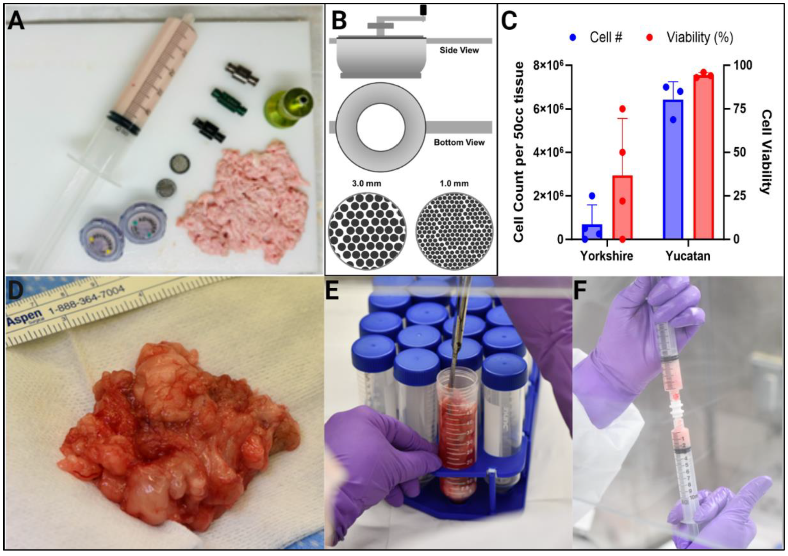
For the autologous harvest, pigs were first administered ketamine (IM, 20 mg/mL), weighed, placed under general anesthesia with isoflurane, and washed extensively with betadine, soap, and water. The inguinal fat pad thickness was visualized using an ultrasound, and markings were made to indicate the areas of high volume. The surgical field was scrubbed with betadine and draped in a sterile fashion. To excise the inguinal fad pads, an initial 3–5 cm incision was made with a #24 scalpel blade. The subcutaneous fat was excised using dissecting scissors until a total volume of 150 cc of adipose was obtained. Hemostasis was achieved with electrocautery, and lymph nodes were carefully avoided. Following excision, the dead space was minimized by placing tacking sutures to adhere the skin to the fascia, and the incision was closed with two additional layers of sutures. The pigs were draped with towels and warming blankets were placed underneath the sterile field to maintain normothermia. The suction lipectomy was performed as follows: Adipose was harvested by liposuction with a Gomco 4040 aspirator, a regular 1-liter OR suction canister, and either a Grainger Gast Vacuum Pump (Model 0523-V4-G588NDX) or Welch oil-free pump (Model WOB-L 2522). Multiple cannula types were investigated, including a 3 mm Mercedes, 3 mm Basket, and 3 mm and 6 mm Disrupter, all handheld on a 60 mL Toomey syringe. The remaining subcutaneous adipose tissue was dissected using an aseptic surgical technique and transferred for volume analysis.
To reduce the risks from extended anesthesia for large-volume surgical excision, we also assessed whether herd-matched allogeneic adipose was a viable alternative to autologous tissue. In three consecutive experiments, allogeneic Yorkshire adipose was obtained immediately after sacrifice, prepared as described below in the Tissue Processing subsection and aliquoted as 3 cc grafts.
2.3. Tissue Processing
The adipose tissues collected during the anatomic studies were assessed for their overall quality using mechanical manipulation for a surgical “feel” and ability to mince the tissue with scissors or cutting blades. Strips of excised tissue were then finely chopped using a mezzaluna knife to generate pieces of approximately 3–5 mm in diameter. Notably, these fragments often consisted of not only lobular adipose but included stromal and fascial elements as well. Milling was required to further disrupt the tissue into injectable fragments using a stainless-steel, plate-style, hand-powered food mill (Passetout, Rosle), which was cleaned and sterilized in an autoclave between each pig. Milling first with a 3 mm perforated plate followed by a 1 mm plate replicated the average aperture size of a 1 and 3 mm caliber cannula. This 3-stage process reliably generated adipose which could be injected through an 18G cannula. Finally, the viability of the processed adipose tissue was subsequently confirmed by digesting the tissue with a solution of 1 mg/mL collagenase type 2 in Hank’s or Earle’s balanced salt solution containing 3.5% BSA and quantifying the viability of the stromal cells through trypan blue exclusion and an automated cell counter (Invitrogen Countess C10281). All procedures performed on tissue intended to be injected were performed in a biosafety cabinet with sterilized surfaces and instruments.
The preparation of Yucatan adipose proved to be less resource intensive and more akin to processing human adipose. Yucatan inguinal fat pads were easily processed in a biosafety cabinet in an adjoining surgical suite, and the fat was transferred between rooms in a secondary container with a sterile interior; milling was not required. Immediately after excision, approximately 20–30 cc of adipose was placed into sterile 50 cc conical tubes, into which 20 cc of warm (37 °C) saline was added. Within the 50 cc conical tube, the tissue was finely minced using sterile surgical scissors and then slowly transferred into a capped 10 cc syringe with the plunger removed. The syringe was then placed inside a clean 50 cc conical tube, capped and centrifuged for 3 min at 1260× g. The bottom fluid layer was discarded into a sterile waste receptacle, and the plunger was carefully replaced without creating pressure. Finally, aliquots were prepared using a female-to-female Luer connector to slowly dispense consistent volumes of minced fat into new 5 cc syringes. Each prepared syringe was weighed before and after fat injection to confirm the graft weight and volume.
2.4. Development of the Recipient Site and Fat Grafting Procedure
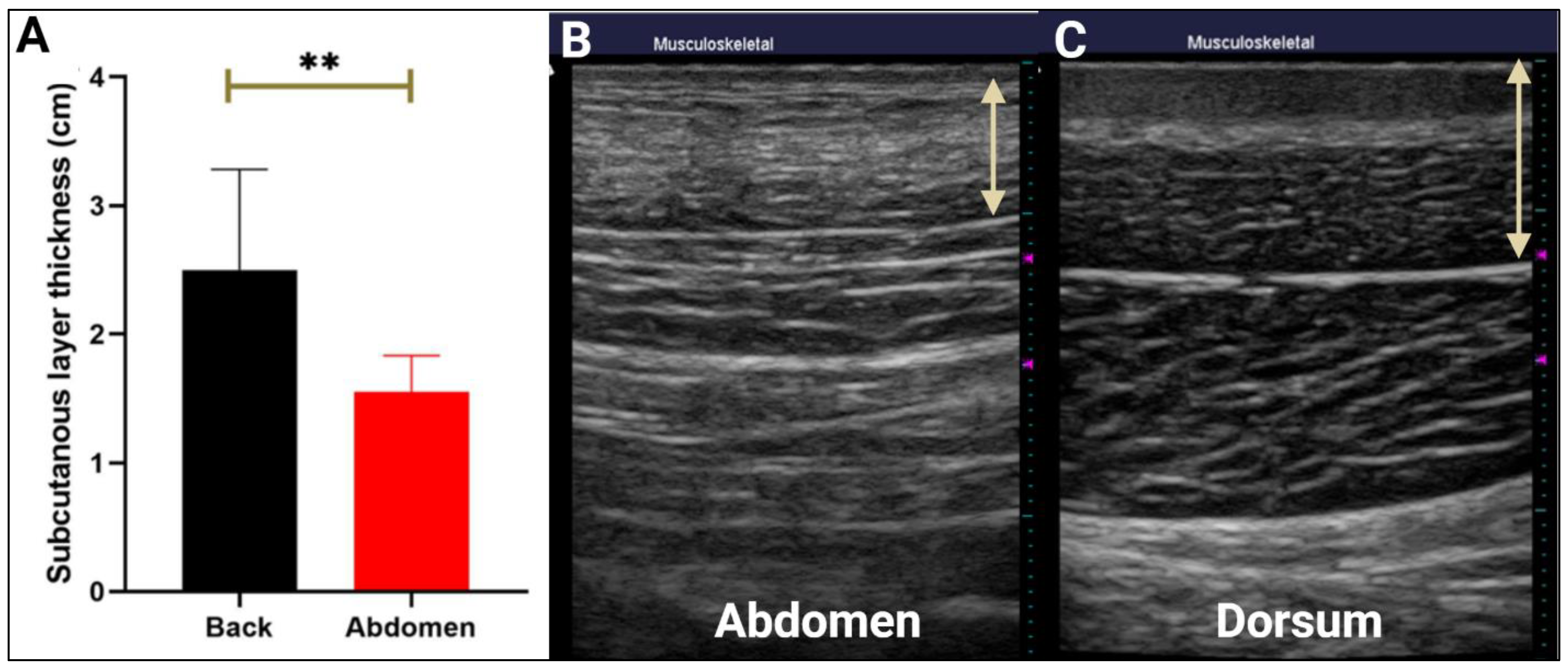
The selection of a subcutaneous recipient site was challenging in the porcine model given the relative inflexibility and mechanical resistance of the thick cutaneous tissue layer, further complicated by the likelihood of graft compression during rest positions. To determine the optimal recipient site for fat grafting, we first measured the thickness of both the abdominal and dorsal subcutaneous layers using an ultrasound. The pigs were sedated, weighed, and placed under general anesthesia as previously described in the Adipose Donor Site Selection and Harvest subsection. Next, an ultrasound (Terason Ultrasound Imaging System, Version 4.7.6; Terason, Burlington, MA, USA) was used to measure the average distance between the skin and fascia levels at 8–16 points on each surface. We determined the fat density at each point based on the radio-opacity of the subcutaneous region. For both measurements, the mechanical index (MI) was set to 0.3, the depth to 4 cm, and the gain was optimized.
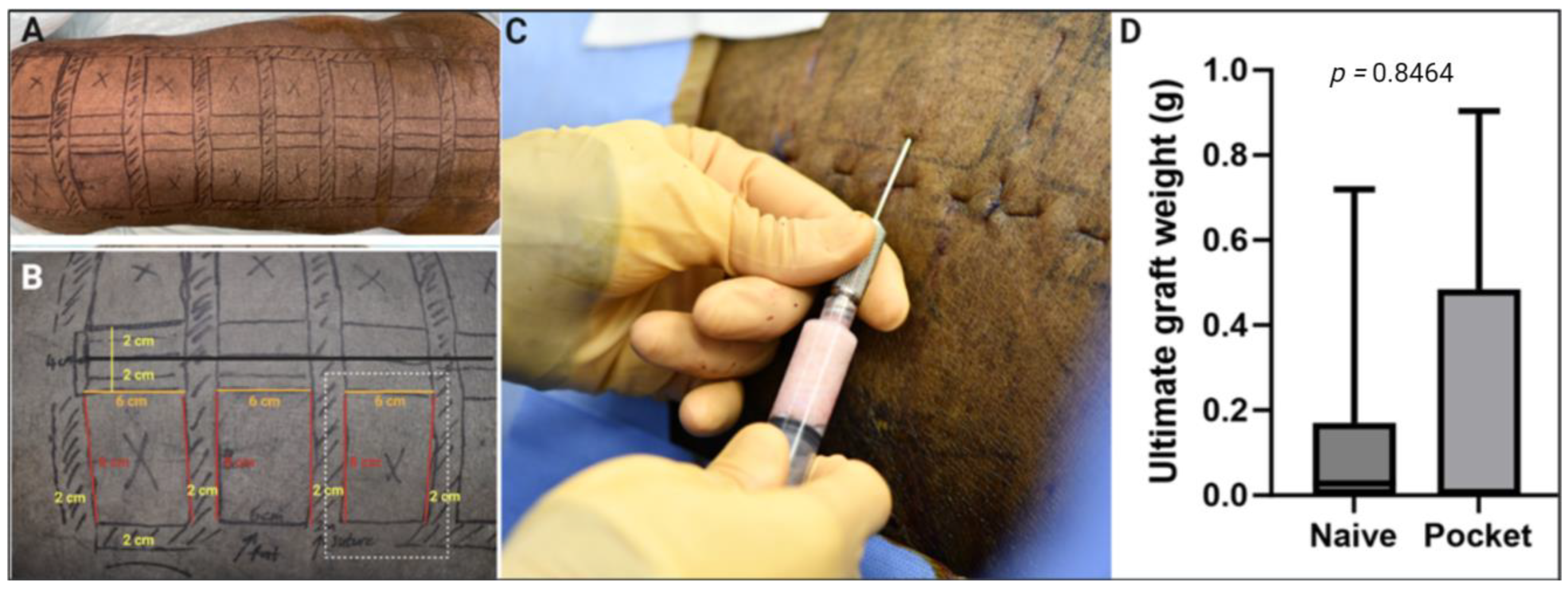
Next, we explored the use of sutured pockets at the recipient site to reduce graft flattening and control graft migration. Two Yorkshire pigs were anesthetized as previously described, transferred to the OR, and endotracheal intubation was performed in the supine position. The surgical field was scrubbed with betadine and draped in a sterile fashion. Identical surgical markings were made on the left (n = 8) and right (n = 8) dorsum, marking the edges of 6 × 8 cm rectangles spaced 2 cm apart. Suture pockets were then created on the left dorsum using transcutaneous 2-0 nylon sutures placed through the skin and into the deep trapezius or thoracolumbar fascia to create a predictable boundary. Sutures were placed in the anterior, posterior, and lateral aspects of the planned pockets in a continuous fashion through the middle of the 2 cm gap in between two injection sites. The sutures were tied loosely to avoid introducing localized mechanical stress or disrupting vascular circulation. A 1 cm distance was left while passing the continuous suture through the skin. The suture pocket was either created immediately prior to grafting or was permitted to mature for 2 weeks to allow for adhesion before engraftment. Fat grafts (3 cc) were placed into each of the 8 suture pockets on the left dorsum and into each surgically marked, but not sutured, rectangle on the right dorsum. The ultimate retention on the left and right sides was compared post-sacrifice to determine the pocket impact.
2.5. Post-Operative Care
Meloxicam and buprenorphine were provided prior to and post-surgical procedures for pain control. Post-operative pain control consisted of buprenex and meloxicam for 5–7 days or longer as needed as per the surgical and veterinary team evaluations.
Immediately following surgery, the pigs were allowed to fully recover prior to being transferred to their original caging. The animals were maintained in separate housing for approximately 2–3 weeks until the wounds were closed. Feeding was resumed as soon as possible after the surgery. To avoid post-surgical complications, we explored utilizing performance spandex goat tube jackets (Sullivan Supply, SKU: PGT) to provide compression, secure the dressings in place, and safeguard the wounds against contamination. Regular assessments were conducted to monitor their body temperature, appetite, general activity, and signs of infection or abscess formation, with particularly close monitoring during the first two post-operative days. On post-operative day three, the animals were placed under anesthesia once again to assess the donor site for potential complications such as a seroma, hematoma, or any issues related to wound healing. In the presence of significant fluid collection, needle drainage was performed promptly.
2.6. Outcomes and Endpoint
For all studies, graft biopsies were obtained using 6 mm hollow punch biopsy at 1-month and 2-months post-grafting. The pigs were sacrificed at 3 months by pentobarbital injection (IV, 120 mg/kg), and the entire dorsal skin was surgically removed. The retention outcomes were assessed monthly up to 3 months, a time point determined in clinical studies as representative of long-term outcomes [
7]. Residual fat grafts were meticulously dissected and recovered from the deep cutaneous tissues and measured using gas pycnometry (micromeritics AccuPyc II 1340, Norcross, GA, USA). The tissues were embedded for histology, sectioned, and visualized with H–E and immunohistochemistry. For allograft studies, graft rejection was assessed through clinical observation and the histologic presence of T cells, using a mouse anti-pig CD3 (BioRad #MCA 5951GA, Hercules, CA, USA) antibody.
2.7. Longitudinal Fat Graft Volume Estimation with Ultrasound
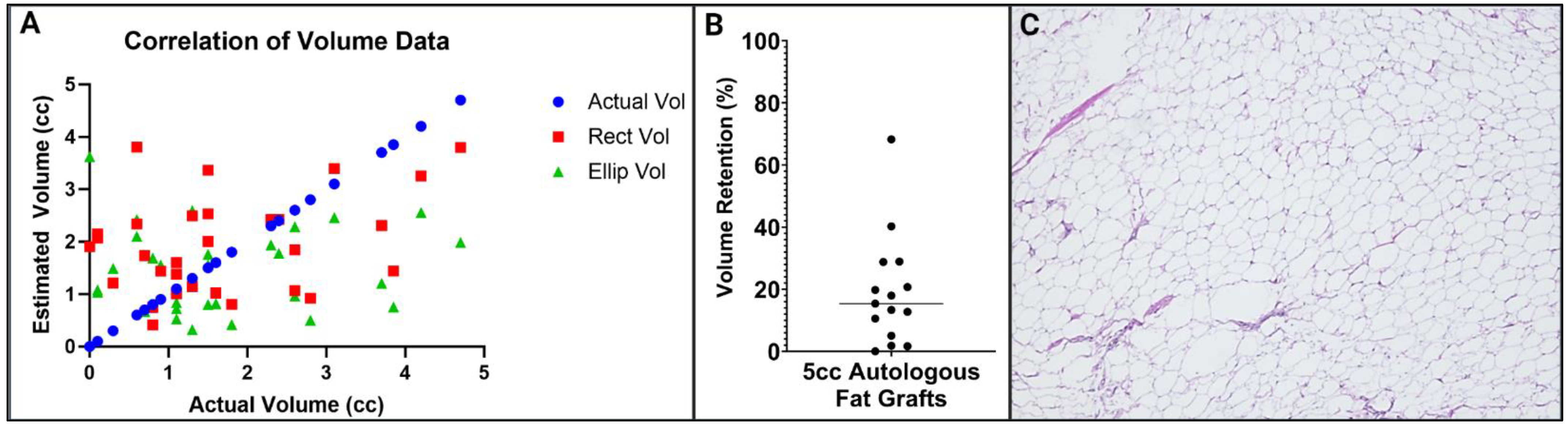
To determine if the ultrasound was able to accurately estimate the fat graft volume at intermittent time points, we compared the calculated graft volumes obtained from the ultrasound images taken immediately prior to sacrifice with the actual graft volumes measured using gas pycnometry. An ultrasound and palpation were used first to locate the irregular-shaped fat grafts in their outlined pockets, as they were easily distinguished from the surrounding tissues by their radiopaque appearance. Two ultrasound images were taken of each graft, in the sagittal and axial planes, to assess the graft length and width. ImageJ was used to measure the fat graft length, width, and depth on the ultrasound images. These measurements were then used to estimate the volume of each graft using two volumetric formulas: the rectangular volume (L×W×D) and generic ellipsoid volume (4/3×π×L×W×D). The outcomes were compared with the actual volume measurements of the dissected-out grafts obtained using gas pycnometry post-sacrifice, and a correlation analysis was performed using Prism Graphpad (v10).
2.8. Data Analysis and Statistical Methods
For the cell quantification studies, a minimum of three cell counts were performed per digested tissue sample. A minimum of eight ultrasound measurements of the adipose thickness were obtained for tissue assessments, and proprietary software (Terason Ultrasound Imaging System, Version 4.7.6; Terason, Burlington, Mass.) was used to obtain measurements to the nearest 0.1 cm. All statistical comparisons were performed using Prism GraphPad Software, Version 10.1.0 (Dotmatics, Boston, MA, USA). The data normality was assessed using the D’Agostino and Pearson normality test followed by two-sided t-tests to determine the difference in means between the parametric data, with the significance set to the level of p < 0.05, p < 0.01, or p < 0.001, as indicated.
4. Discussion and Conclusions
In this study, we sought to better understand the factors needed to model clinically relevant fat grafting in swine. Porcine models present several unique challenges which include highly variable mechanical characteristics and distributions of subcutaneous adipose. Furthermore, surgical and anesthetic challenges unique to pigs include sensitivity to hypothermia due to higher ambient temperature requirements and an increased risk of post-surgical complications from large-volume fat harvesting, resulting in a higher risk for developing seromas, infections, and blood loss. Consequently, we investigated several approaches to tissue harvesting, processing, and engraftment across both autologous and allogeneic tissues and multiple pig strains.
When developing a translational mode, careful consideration must be given to the anatomic and histologic variations in the anatomy, particularly as it differs between humans and swine. In human surgical practice zones of adhesion, points where the subcutaneous fascial system (SFS) is more firmly associated with the underlying muscle fascia are present, which alter the mechanical properties of the subcutaneous fat [
8]. A similar SFS in present in pigs, particularly evident in the ventral or abdominal regions, though dorsally, it is congruent with a more well-defined panniculus carnosus than found in humans. In swine, the panniculus carnosus is firmly attached to the dermis, with its origin in the superficial fascia of the back and insertion ventrally. Where the panniculus is present, this well-defined anatomic layer in swine separates the adipose into a dense, fibrous, and compact superficial abdominal layer and a more loose, areolar, and less-compact deep layer.
Additionally, the swine body composition varies highly with age. The Yorkshire swine, which we predominantly utilized in early experiments, is a late-maturing variety with a predominance of lean weight during adolescence and early adulthood. Given this, careful consideration was made relative to the donor site characteristics of the animal. Compact superficial subcutaneous adipose (nuchal, back/flank, jowl, etc.) is mechanically stiff, lacking pliability of deeper tissues, and is marked histologically by small, tightly packed adipocytes. Given the relative compactness of superficial adipose deposits, we found these tissues were not easily amenable to liposuction in the Yorkshire breed, and even with open harvest, required extensive post-processing to achieve a consistency suitable for syringe-mediated lipografting. In Yorkshire swine, we noted that soft, pliable adipose could be more readily harvested from deeper deposits not associated with the panniculus, such as the axillary and inguinal regions, with the inguinal region providing the bulk of the safe harvest. Utilizing our initial Yorkshire model, we were reliably able to surgically harvest between 100–200 cc of soft adipose under an open harvest from this region, which was amenable to rapid processing and re-engraftment. This site, however, is highly enriched in lymphatic and venous drainage, which generated a high risk of fluid collection in the post-harvest period. Notably, we found that in the open harvest of inguinal adipose in the Yorkshire strain, a post-operative seroma commonly accumulated, requiring post-procedural drainage alongside conservative management with pressure dressing.
An ultrasound assessment of the native subcutaneous adipose tissue thickness was used to identify the ideal location of recipient engraftment. Pigs tend to rest in either the sternal or lateral recumbent positions. This generally means that the dependent position of the animal will sustain the greatest pressure at rest. For engraftment studies, this is not ideal, as variability in the compression and mechanical stress/strain by location can affect graft revascularization, take, and consequently, resorption. To standardize this, we utilized a paraspinous position extending along the caudal trapezius and then along the thoracolumbar fascia. Fat grafts were placed suprafascially into the areolar space along the deep margin of the subcutaneous tissue, which presented the least mechanical resistance to engraftment. This allowed for reliable placement, easy visualization, and quick access with an ultrasound to determine the depth and volume of the graft. The dorsum of the pig had the additional benefit of minimizing the environmental and fecal/urine waste contamination found in ventral sites with the post-/peri-operative dressing approach we describe later in this text. Within the first month, however, we noted significant mobilization of the grafted fat and spreading in the horizontal axis along the plane to engraftment. This made the identification of the volume challenging, led to the merger of adjacent grafts, and ultimately represented a degree of mechanical shear that was not compatible with our goals. This contrasted with prior work that we had performed evaluating adipose tissue in the setting of cutaneous trauma, which did not demonstrate graft migration despite usage of a similar plane. Drawing from surgical experience, we developed a delayed surgical pocket model, utilizing deep transcutaneous nylon sutures through the skin and into trapezius or thoracolumbar fascia to generate a predictable boundary for fat to be placed. With this model in place, we identified a significant improvement in graft localization, and consequently, a much higher yield at the time of harvest, consistent with no significant impact on the graft viability.
While the long-term transplantation of allogeneic or xenogeneic tissues is commonly understood to require immunosuppression or similar immunomodulation, temporary engraftment of allogeneic tissues in the absence of systemic immunosuppression is a more well-accepted practice, being particularly common in the area of cadaveric skin grafting. What is less well known in the modern literature is the practice of allogeneic cadaveric fat grafting, which, although now is mostly abandoned in the setting of more reliable options, was an extant practice in the setting of breast reconstruction throughout the 1900s. The rationale viability of this practice is complex, given the traditional for HLA compatibility in tissue transplantation in the absence of immunosuppression. Fat grafting and fat graft viability, however, carries several characteristics which allow for graft growth and survival. First, successful fat transfer is not entirely dependent on the survival of the transplanted cells [
9], allowing for successful recellularization and graft formation in the setting of acellular allogeneic adipose matrix transfer. This is based in the now well-accepted host/recipient origin for retained grafts in the setting of allo- or xenotransplantation and the adipo-inductive properties of transplanted fat. Second, however, is that adipose and more specifically, adipose-derived stromal cells (ADSCs), carry known immunomodulatory properties which allow for greater flexibility in transplantation when compared with more immunogenic tissues such as skin. This has been demonstrated by the viability of allogeneic and xenogeneic ADSC transfer of porcine cells to immunocompetent recipients in the absence of immunosuppression [
10]. While this is a more common practice in lower animal models, particularly in the setting of syngeneic transfer within inbred species of rats and mice, the question of allograft survival is an active one. With this being primarily in the setting of highly inbred species, it was initially questionable whether allogeneic fat transfer would be viable. The genetic variability across pig breeds and disparate populations is quite low when compared with humans and other herd animals, given their long history of domestication. The Yorkshire or Large White breed has a particularly high degree of genomic and pedigree inbreeding when compared with similar domestic swine, although not the extent of breeds specifically inbred for xenotransplantation. Genetic variability can be further limited within a single population, particularly in the setting of same-breed, single-sourced, closed-herd animals and sibling-matched donor-recipient pairs. Given the logistical challenges we initially faced with obtaining sufficient adipose from a live animal for autotransplantation, this was the approach we undertook and demonstrated viable, live adipose allograft at 3-months post-transplant (
Figure 1). Over time, however, additional challenges arose. Chronic rejection appeared to be a consistent concern, and as it became challenging to guarantee single-supplier swine pairs, we transitioned to an autologous Yucatan model as described above.
The Yucatan pig is a naturally occurring miniature swine breed found in the Yucatan Peninsula and various regions of Mexico and Central America [
11]. Unlike other breeds in North America, the Yucatan breed is distinct, as it originates from a single genetic pool without the incorporation of genetic material from other breeds or strains [
12]. In our study, we observed several characteristics of Yucatan pigs that render them ideal for fat grafting procedures. In contrast to the Yorkshire breed, the inguino-abdominal area of Yucatan pigs exhibits a significant volume of subcutaneous fat, making it a highly suitable donor site that is capable of yielding up to 200 cc of fat. The surgical harvesting of fat from this region can be accomplished through smaller incisions and with minimal tissue dissection, thereby decreasing tissue trauma, saving time and effort, and reducing the duration of anesthesia. Furthermore, these advantages contribute to a decreased risk of post-operative complications such as a seroma, hematoma, and wound healing issues. Moreover, the fat obtained from Yucatan pigs exhibits a lower fibrous content and closely resembles human fat, simplifying the processing phase before engraftment. The Yucatan pig, known for its relatively hairless nature, exhibits higher ambient temperature requirements compared to other swine breeds. Having a sparser hair coat is advantageous in indoor environments and laboratory settings, as it reduces the typical odor associated with swine and facilitates various laboratory procedures, such as locating blood vessels, conducting pre- and post-surgical interventions, monitoring wound healing, applying and removing bandages, and reading ear notches. Not requiring hair removal also allows for the convenient application and evaluation of topical preparations without additional complications. Yucatan pigs exhibit gentle, docile behavior in laboratory environments and generally display a higher agreeability compared to other swine breeds [
11,
12].
An important consideration for any model utility is their clinical relevance. We found that the Yucatan model easily tolerated 5 cc grafts, with 16 grafts placed dorsally. This volume is associated with smaller fat grafts, not typically placed as a bolus in clinical practice, which is a study limitation. The ultimate volume retention in this model approximated 20% of the initial injection volume, which is considered a low outcome when compared to clinically reported values. The Department of Plastic Surgery at the University of Pittsburgh has completed multiple clinical studies and shown that volume retention is significantly impacted by the anatomic graft location, with 95% of craniofacial grafts retaining between 45% and 65% of the initial volume [
7], while high compression sites such as boney amputation surfaces and pedal fat pads have almost no volume retention [
13,
14]. Therefore, it is possible that fat grafts in the pig model are under increased compression compared to alternative recipient sites in humans, and therefore, have a reduced average retention. Furthermore, it is highly likely that grafts at different positions along the swine dorsum incur disparate compressive forces due to anatomical location; thus, a wide range of graft survival rates within a single animal may be unavoidable. Such variability may more accurately model the diversity observed in clinical applications of fat grafting. Despite this, the model remains valid for intervention studies, as differences in retention can be detected at three months if they exist.
We conclude that autologous fat grafting using surgically excised inguinal adipose in a Yucatan pig is a feasible approach for investigating fat graft survival or therapeutic interventions. The initial grafting procedure is well tolerated, and careful post-surgical monitoring of the donor site is paramount for avoiding serious post-operative complications. Mid-study monitoring of the fat graft volume is difficult to estimate using ultrasound imaging and did not correlate with the terminal graft volumes following sacrifice and dissection. Therefore, more robust modalities such as MRI are suggested, though this greatly increases study costs and may not translate to what is typically done in clinical practice. Finally, the recipient site can be readily modified with suture pockets to prevent graft flattening and migration to improve identification and recovery, but this does not significantly impact ultimate retention.


 ,
, 


