Review

Overview of Radiological Reporting and Data System (RADS) Guidelines Currently Applicable in Surgery

by
Marco Parillo
1,* and
Carlo Cosimo Quattrocchi
1,2
1
Radiology, Multizonal Unit of Rovereto and Arco, APSS Provincia Autonoma Di Trento, 38123 Trento, Italy
2
Centre for Medical Sciences—CISMed, University of Trento, 38122 Trento, Italy
*
Author to whom correspondence should be addressed.
Surgeries 2025, 6(1), 23; https://doi.org/10.3390/surgeries6010023
Submission received: 30 January 2025 / Revised: 11 March 2025 / Accepted: 13 March 2025 / Published: 14 March 2025

Abstract

:
Standardized frameworks for interpreting medical images, such as the radiological Reporting and Data Systems (RADS), are designed to improve the consistency and accuracy of radiological assessments across different imaging modalities, anatomical locations, and disease processes. Clear communication and information sharing between radiologists and referring physicians, including surgeons, is a key goal of the RADS guidelines. Therefore, familiarity with these guidelines is crucial for all physicians involved in patient care. This review synthesizes current RADS guidelines relevant to surgical practice. Our analysis identified 28 radiological RADS with potential applications in surgical workflows, primarily in oncology. Of the RADS examined, nine were validated by the American College of Radiology (ACR), one was validated through a collaboration between the ACR and other scientific societies, and seventeen were developed by other scientific organizations. Numerous surgical specialties may encounter RADS in clinical practice, including neurosurgery, head and neck surgery, cardiovascular surgery, thoracic surgery, endocrine surgery, breast surgery, gastrointestinal surgery, hepatobiliary surgery, gynecological surgery, urological surgery, orthopedic surgery, emergency surgery, and surgical oncology. The effective utilization and validation of RADS necessitates close collaboration between radiologists and surgeons, coupled with widespread education for all healthcare professionals involved in patient care. Artificial intelligence software will play an important role in facilitating the dissemination and use of RADS in clinical practice.

1. Introduction

A standardized system for reporting mammography findings, known as the Breast Imaging Reporting and Data System (BI-RADS), was introduced by the American College of Radiology in 1993. The primary goals of BI-RADS were to enhance the differentiation between benign and malignant breast conditions, minimize ambiguity in radiology reports, enable automated data analysis, and facilitate improved communication with referring physicians [1,2]. Following the introduction of BI-RADS, there has been a significant expansion of Reporting and Data Systems (RADS) in radiology. These guidelines aim to enhance image interpretation by providing standardized frameworks for various imaging techniques, anatomical regions, and diseases [3,4]. Each system is developed through a consensus process involving a panel of experts, not necessarily endorsed by the ACR, and may undergo periodic revisions to align with advancements within the specific medical field.
The primary objective of RADS is to enhance clear communication and information sharing between radiologists and referring physicians. Due to the complexity of many diseases, particularly cancer, a multidisciplinary approach involving surgeons and other specialists has become standard practice to optimize patient outcomes. Therefore, it is essential that all members of the multidisciplinary team are familiar with the RADS guidelines. Including a RADS score within a report may introduce more confusion than clarity if there is a lack of consensus among physicians regarding its interpretation.
This review presents a summary of the current radiological RADS guidelines as they relate to surgical applications. This analysis may serve as a starting point for future investigations into the clinical applicability in surgery of specific RADS across various institutions worldwide.

2. Materials and Methods
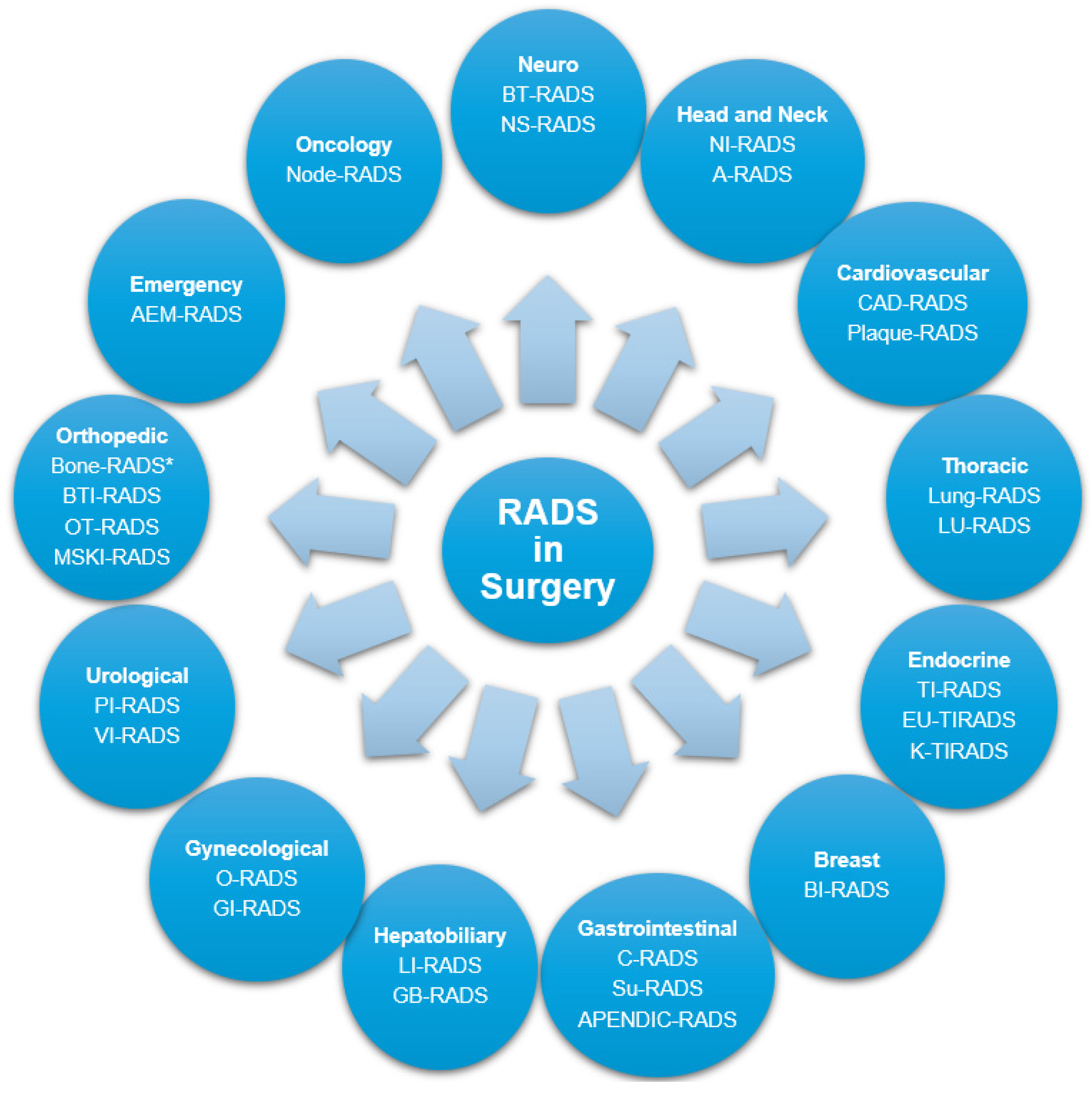
An initial search of official websites [1,2] identified 25 RADS. A subsequent literature review using PubMed (from 1 January 2005 to 27 January 2025) yielded an additional 16 RADS, resulting in a total of 41 identified RADS (see Appendix A for the search terms used). Following a thorough review, 13 RADS were excluded from further analysis due to their use in nuclear medicine or lack of significant surgical implications. Consequently, the final analysis included 28 radiological RADS with current surgical relevance (Figure 1).

3. Surgery and RADS

Nine RADS were validated by the ACR: BI-RADS [5,6], Bone Reporting and Data System (Bone-RADS) [7], CT Colonography Reporting and Data System (C-RADS) [8], Liver Imaging Reporting and Data System (LI-RADS) [9,10], Lung CT Screening Reporting and Data System (Lung-RADS) [11], Neck Imaging Reporting and Data System (NI-RADS) [12], Ovarian-Adnexal Reporting and Data System (O-RADS) [13,14], Prostate Imaging Reporting and Data System (PI-RADS) [15], and Thyroid Imaging Reporting and Data System (TI-RADS) [16]. One RADS was validated by the ACR in collaboration with the American College of Cardiology, the Society of Cardiovascular Computed Tomography, and the North America Society of Cardiovascular Imaging: Coronary Artery Disease-Reporting and Data System (CAD-RADS) [17]. Eighteen RADS were proposed by other scientific groups: ultrasound classification of cervical adenopathy (A-RADS) [18], Abdominal Emergency Reporting and Data System (AEM-RADS) [19], Appendix Imaging Reporting and Data System (APENDIC-RADS) [20], Bone-RADS [21,22], Brain Tumor Reporting and Data System (BT-RADS) [23], Bone Tumor Imaging Reporting and Data System (BTI-RADS) [24], European Thyroid Imaging and Reporting Data System (EU-TIRADS) [25], Gallbladder Reporting and Data System (GB-RADS) [26], Gynecologic Imaging Reporting and Data System (GI-RADS) [27], Korean Thyroid Imaging Reporting and Data System (K-TIRADS) [28], Lung Reporting and Data System (LU-RADS) [29], Musculoskeletal Infection Reporting and Data System (MSKI-RADS) [30], Node Reporting and Data System (Node-RADS) [31], Neuropathy Score Reporting and Data System (NS-RADS) [32], Osseous Tumor Reporting and Data System (OT-RADS) [33], Plaque-RADS [34], Stomach Ultrasound Reporting and Data System (Su-RADS) [35], and Vesical Imaging Reporting and Data System (VI-RADS) [36].

3.1. Neurosurgery

BT-RADS is a proposed framework for classifying and reporting post-treatment primary brain tumors using contrast-enhanced magnetic resonance imaging (MRI) scans [23] (as it is the main imaging modality for brain disorders [37,38,39]). This system categorizes brain MRI findings as follows: 0 baseline; 1 improvement (1a improved tumor response and 1b possible medication effect); 2 stable; 3 worsening (3a likely treatment-related changes, 3b possible combination of treatment effects and tumor progression, and 3c likely tumor progression); 4 definite tumor progression [23].
NS-RADS was developed to standardize the evaluation and reporting of peripheral neuropathy findings on MRI examinations. Distinct classes within the NS-RADS framework were established to encompass the diverse spectrum of underlying pathological conditions: injury (I from 1 to 5 according to Sunderland class), neoplasia (N1 definitely benign, N2 probably benign, N3 probably malignant, and N4 recurrent tumor), entrapment (E1 mild, E2 moderate, and E3 severe), diffuse neuropathy (D1 diffuse mononeuropathy and D2 diffuse polyneuropathy), not otherwise specified (NOS), and postintervention state (PI1 near normal, PI2 possible persistent neuropathy, and PI3 definitely persistent or worsening neuropathy) [32].

3.2. Head and Neck Surgery

NI-RADS was established by the ACR to provide a standardized framework for reporting and interpreting neck imaging studies in patients undergoing surveillance for head and neck cancer. Initially developed for contrast-enhanced computed tomography (CT) scans with or without positron emission tomography imaging, NI-RADS has since been expanded to include specific category descriptors and imaging findings for MRI studies. Scoring for imaging suspicion of recurrence is performed independently for the primary tumor site and neck lymph nodes: 0 incomplete; 1 no evidence of recurrence; 2 low suspicion of recurrence; 3 high suspicion of recurrence; 4 known recurrence. In particular, category 2 is further divided into 2a or 2b for the evaluation of the primary tumor site, indicating, respectively, an alteration that affects the superficial mucosa or the deeper submucosal tissues. Finally, only in the diagnostic algorithm used for MRI are categories 1f and 2f added to be applied to the first examination after treatment, indicating, respectively, no evidence of recurrence and low suspicion of recurrence at the primary tumor site [12,40,41].
A-RADS provides a framework for evaluating cervical lymph nodes affected by diverse pathological processes with ultrasound. A four-tiered classification system is used to categorize lymph nodes: 1 normal, 2 reactive, 3 suspicious for lymphoid disorders, and 4 metastatic [18].

3.3. Cardiovascular Surgery

CAD-RADS is a standardized framework for reporting and communicating findings from coronary CT angiography examinations. The clinical spectrum of CAD-RADS encompasses patients experiencing stable chest pain as well as those presenting with acute chest pain. CAD-RADS categories in patients with stable chest pain are as follows: 0 absence of coronary artery disease; 1 minimal nonobstructive coronary artery disease; 2 mild nonobstructive coronary artery disease; 3 moderate stenosis; 4 severe stenosis; 5 total coronary occlusion or sub-total occlusion. CAD-RADS categories in patients with acute chest pain are as follows: 0 acute coronary syndrome highly unlikely; 1 acute coronary syndrome unlikely; 2 acute coronary syndrome less likely; 3 acute coronary syndrome possible; 4 acute coronary syndrome likely; 5 acute coronary syndrome very likely. The CAD-RADS N category is assigned when the exclusion of acute coronary syndrome is not definitively possible. CAD-RADS incorporates an assessment of coronary plaque burden, designated as “P”, which can be categorized as follows: 1 mild amount of plaque; 2 moderate amount of plaque; 3 severe amount of plaque; 4 extensive amount of plaque [17].
Plaque-RADS is a standardized system for characterizing carotid plaque composition and morphology across various imaging modalities, including ultrasound, CT, and MRI. Plaque-RADS categories are associated with the attributable risk of developing of ipsilateral cerebrovascular event: 1 absent, 2 low, 3 moderate, and 4 high [34].

3.4. Thoracic Surgery

The ACR developed Lung-RADS to standardize the reporting and management of pulmonary nodules detected during lung cancer screening CT scans. This system categorizes chest CT findings as follows: 0 incomplete; 1 negative; 2 benign; 3 probably benign; 4A suspicious; 4B very suspicious; 4X category 3 or 4 nodules with additional features or imaging findings that increase suspicion of lung cancer [11].
LU-RADS categorizes chest CT findings as follows: 1 no nodule; 2 benign nodule; 3 indeterminate; 4 suspicious; 5 malignant by CT; 6 malignancy confirmed through biopsy [29].

3.5. Endocrine Surgery

TI-RADS, developed by the ACR, is a standardized system for evaluating thyroid nodules identified on ultrasound. This system utilizes a scoring system to categorize nodules based on their ultrasound characteristics, providing clinicians with recommendations for subsequent management, such as fine-needle aspiration biopsy or ultrasound follow-up. The nodules are classified as follows: 1 benign; 2 not suspicious; 3 mildly suspicious; 4 moderately suspicious; 5 highly suspicious [16].
The European Thyroid Association developed EU-TIRADS to standardize the assessment of thyroid nodules using ultrasound. This system helps determine the need for fine-needle aspiration and the risk of malignancy. EU-TIRADS classifies thyroid nodules into five categories: 1 absent; 2 benign; 3 low risk; 4 intermediate risk; 5 any high risk features [25].
The Korean Society of Thyroid Radiology developed K-TIRADS to standardize thyroid nodule ultrasound evaluation. This system provides a framework for determining the necessity of fine-needle aspiration and assessing malignancy risk. Additionally, K-TIRADS offers guidance on when lymph node sampling is indicated. The thyroid nodules are classified as follows: 1 absent; 2 benign; 3 low suspicion; 4 intermediate suspicion; 5 high suspicion [28].

3.6. Breast Surgery

The ACR BI-RADS atlas establishes a standardized framework for breast imaging reports, encompassing standardized terminology, report organization, assessment structure, and a classification system for ultrasound, mammography (including contrast-enhanced mammography), and MRI of the breast. Breast imaging studies are categorized into seven assessment categories with a progressively higher risk of cancer: 0 incomplete; 1 negative; 2 benign; 3 probably benign; 4 suspicious for malignancy; 5 highly suggestive of malignancy; 6 known malignancy confirmed by biopsy [5,6]. Moreover, mammographic breast density is categorized based on the proportion of fibroglandular tissue to fatty tissue. The BI-RADS classification uses four categories: almost entirely fatty (a), scattered fibroglandular densities (b), heterogeneously dense (c), and extremely dense (d) [42]. Heterogeneous and extreme density can potentially mask small masses and reduce mammographic sensitivity.

3.7. Gastrointestinal Surgery

C-RADS, supported by the ACR, serves as a comprehensive classification framework for characterizing findings detected during CT colonography examinations. Colonic findings are categorized as follows: 0 inadequate study or awaiting prior comparisons; 1 normal colon or benign lesion; 2a intermediate polyp or indeterminate finding; 2b likely benign diverticular finding; 3 polyp, possibly advanced adenoma; 4 colonic mass, likely malignant [8].
A system for reporting and categorizing gastric ultrasound findings, known as Su-RADS, was developed. This system was designed to facilitate the screening of gastric cancer using transabdominal ultrasound following the oral administration of an echogenic cellulose-based contrast agent. The examinations results are categorized as follows: 0 incomplete or needing additional examination; 1 almost normal finding; 2 low probability of malignancy; 3 moderate risk for malignancy; 4 high likelihood of malignancy; 5 extremely high probability of malignancy; 6 malignancy confirmed through biopsy [35].
APENDIC-RADS was developed to standardize the description of ultrasound findings related to the appendix. This system categorizes findings into five levels: 0 appendix not visualized; 1 normal appendix; 2 likely normal appendix with limited visualization; 3 appendicitis cannot be excluded due to equivocal findings; 4 acute appendicitis [20].

3.8. Hepatobiliary Surgery

ACR LI-RADS offers a consistent and standardized approach to interpreting and reporting liver imaging studies in patients at risk for hepatocellular carcinoma (HCC), namely, those with cirrhosis, chronic hepatitis B viral infection, or current or prior HCC, linking assessment categories to specific management recommendations. It also facilitates the evaluation of treatment responses to locoregional therapies. The scoring system is applicable to various imaging modalities, including contrast-enhanced MRI, contrast-enhanced CT, ultrasound, and contrast-enhanced ultrasound. Patients undergoing ultrasound screening can be classified into three main categories: 1 negative; 2 subthreshold; 3 positive [43]. The results of contrast-enhanced diagnostic imaging studies (CT, MRI, or ultrasound) can be classified into the following categories: 1 definitely benign, 2 probably benign, 3 intermediate malignancy probability, 4 probably HCC, 5 definitely HCC, M probably or definitely malignant, but not HCC-specific, and TIV tumor in vein [9,10].
GB-RADS was developed to enhance consistency in ultrasound interpretation, reporting, and risk assessment of gallbladder wall thickening in non-acute settings. Ultrasound evaluations of the gallbladder are categorized as follows: 0 inadequate evaluation due to technical limitations, patient factors, or gallbladder-related issues; 1 normal gallbladder; 2 benign findings; 3 equivocal findings; 4 likely malignant; 5 highly suggestive of malignancy [26].

3.9. Gynecological Surgery

O-RADS is a standardized framework supported by the ACR for assessing the risk of malignancy in ovarian and adnexal lesions. This system categorizes findings from ultrasound and contrast-enhanced MRI examinations, linking each category to specific management recommendations. Imaging findings can be interpretated as follows: 0 incomplete evaluation; 1 normal ovaries; 2 almost certainly benign; 3 low risk for malignancy; 4 intermediate risk of malignancy; 5 high risk of malignancy [13,14].
GI-RADS was developed for classifying adnexal masses based on findings observed during transvaginal sonography. This system utilizes pattern recognition analysis and the location of color Doppler blood flow to aid in determining the presumptive diagnosis. Transvaginal sonography findings are classified as follows: 1 definitively benign; 2 very probably benign; 3 probably benign; 4 probably malignant; 5 very probably malignant [27].

3.10. Urological Surgery

ACR PI-RADS is a standardized framework for reporting and interpreting multiparametric prostate MRI examinations in men suspected of having prostate cancer who have not yet undergone treatment. Each lesion is categorized into one of five risk categories based on the likelihood of clinically significant cancer: 1 very low; 2 low; 3 intermediate; 4 high; 5 very high [15].
VI-RADS is a standardized system for assessing muscle invasion in bladder cancer using multiparametric MRI. Each lesion is assigned a score from 1 to 5, indicating the following levels of muscle invasion risk: 1 very low likelihood of muscle invasion; 2 low likelihood of muscle invasion; 3 uncertain presence of muscle invasion; 4 high likelihood of muscle invasion; 5 very high likelihood of muscle invasion and potential extravesical extension [36].

3.11. Orthopedic Surgery

ACR Bone-RADS establishes a standardized framework for the evaluation of potentially neoplastic bone lesion on skeletal radiographs. A diagnostic radiography is categorized into five assessment categories: 0 incomplete; 1 very likely benign; 2 probably benign; 3 potentially malignant; 4 highly suspicious for malignancy [7].
BTI-RADS provides a standardized framework for classifying solitary bone lesions. This system utilizes a range of imaging characteristics to assess the likelihood of benign or malignant behavior, and it is applicable to both CT and contrast-enhanced MRI examinations. Imaging is classified into four categories: 1 benign; 2 likely benign; 3 suspicion of malignancy; 4 likely malignant [24].
Bone-RADS offers a diagnostic management approach for incidental solitary bone lesions detected in adults during CT or MRI examinations. Imaging is classified into four categories: 1 likely benign; 2 incompletely assessed on imaging; 3 indeterminate; 4 suspicious for malignancy or need for treatment [21,22].
OT-RADS assists in separating benign from malignant osseous tumors on contrast-enhanced MRI examinations. MRI categories are as follows: 0 incomplete imaging; 1 negative; 2 definitely benign; 3 probably benign; 4 suspicious for malignancy or indeterminate; 5 highly suggestive of malignancy; 6 known biopsy-proven malignancy or recurrent malignancy in the tumor bed [33].
MSKI-RADS is a classification framework specifically developed for evaluating MRI scans of suspected extremity infections in adult patients. The scoring system encompasses the following categories: 0 incomplete imaging; 1 negative for infection; 2 superficial soft tissue infection; 3 deeper soft tissue infection; 4 possible osteomyelitis; 5 highly suggestive of osteomyelitis and/or septic arthritis; 6 confirmed osteomyelitis; NOS (not otherwise specified), indicating nonspecific bone lesions [30].

3.12. Emergency Surgery

AEM-RADS was developed to address the need for standardized reporting in emergency abdominal CT imaging. CT scans are categorized as follows: 1 normal findings; 2 findings requiring non-urgent evaluation, including post-traumatic simple soft tissue injuries and non-traumatic incidental findings; 3 technical limitations or need for alternative imaging; 4 findings indicative of immediate medical emergency; 5 findings indicative of urgent medical condition [19]. Based on the specific organ affected, various surgical specialists may need to be involved.

3.13. Surgical Oncology

Many of the RADS discussed up until now have typically been tailored to specific oncological diseases and anatomical areas. Node-RADS, however, is a more versatile system that can be used for staging and follow-up across several types of cancers. In fact, the development of Node-RADS 1.0 addressed the need for a standardized approach to classifying and reporting lymph nodes on CT and MRI scans in the context of oncological imaging in all body regions. Node-RADS employs a five-point scale to assess lymph node malignancy risk: 1 very low suspicion, 2 low suspicion, 3 moderate suspicion, 4 high suspicion, and 5 very high suspicion [31].

4. Discussion

From our analysis, as many as 28 RADS emerged with potential surgical implications, most for oncological purposes (Table 1).
Table 1. Summary of the radiological Reporting and Data Systems (RADS) potentially applicable in surgery, listed in alphabetical order. CT, computed tomography; MRI, magnetic resonance imaging; US, ultrasound.
RADSIndication/Type of SurgeryImaging TechniquesScores
ACR BI-RADS [5,6]Breast cancer/Breast surgeryMammography, US, MRI0–6
ACR Bone-RADS [7]Neoplastic bone lesion/Orthopedic surgeryRadiograph0–4
ACR C-RADS [8]Colon cancer/Gastrointestinal surgeryCT colonography0–4
ACR LI-RADS [9,10]Hepatocellular carcinoma/Hepatobiliary surgeryUS, MRI, CT1–5, M, TIV
ACR Lung-RADS [11]Lung cancer/Thoracic surgeryCT0–4X
ACR NI-RADS [12]Head and neck cancer/Head and neck surgeryCT, MRI0–4
ACR O-RADS [13,14]Ovarian-adnexal mass/Gynecological surgeryUS, MRI0–5
ACR PI-RADS [15]Prostate cancer/Urological surgeryMRI1–5
ACR TI-RADS [16]Thyroid cancer/Endocrine surgeryUS1–5
A-RADS [18]Cervical adenopathy/Head and neck surgeryUS1–4
AEM-RADS [19]Acute abdomen/Emergency surgeryCT1–5
APENDIC-RADS [20]Appendicitis/Gastrointestinal surgeryUS0–4
Bone-RADS [21,22]Solitary bone lesion/Orthopedic surgeryCT, MRI1–4
BT-RADS [23]Primary brain tumor/NeurosurgeryMRI0–4
BTI-RADS [24]Solitary bone lesion/Orthopedic surgeryCT, MRI1–4
CAD-RADS [17]Coronary artery disease/Cardiovascular surgeryCT0–5
EU-TIRADS [25]Thyroid cancer/Endocrine surgeryUS1–5
GB-RADS [26]Gallbladder cancer/Hepatobiliary surgeryUS0–5
GI-RADS [27]Ovarian-adnexal mass/Gynecological surgeryUS1–5
K-TIRADS [28]Thyroid cancer/Endocrine surgeryUS1–5
LU-RADS [29]Lung cancer/Thoracic surgeryCT1–6
MSKI-RADS [30]Extremity infection/Orthopedic surgeryMRI0–6
Node-RADS [31]Lymph node in cancer/Surgical oncologyCT, MRI1–5
NS-RADS [32]Peripheral neuropathy/NeurosurgeryMRII 1–5, N 1–4, E 1–3, D 1–2, PI 1–3
OT-RADS [33]Neoplastic bone lesion/Orthopedic surgeryMRI0–6
Plaque-RADS [34]Cerebrovascular event in carotid artery disease/Cardiovascular surgeryUS, CT, MRI1–4
Su-RADS [35]Gastric cancer/Gastrointestinal surgeryUS0–6
VI-RADS [36]Bladder cancer/Urological surgeryMRI0–5
Figure 2 summarizes the RADS currently available for use in surgical workflows, grouped by subspecialty.

4.1. Potential Strengths, Limitations, and Requirements for RADS Implementation in Surgery

Key advantages of implementing a RADS include the following [44]:
  • Enhanced quality assurance: By establishing clear and standardized assessment criteria, RADS contributes to improved diagnostic accuracy and consistency.
  • Optimized imaging pathways: RADS facilitates the selection of the most appropriate and efficient imaging examinations for each patient, minimizing unnecessary procedures.
  • Precise diagnostic criteria: RADS provides well-defined imaging features and scoring systems, enabling more accurate and consistent interpretation of findings.
  • Standardized patient management: RADS outlines clear recommendations for patient follow-up, surgical procedures, and subsequent management strategies.
  • Improved interdisciplinary communication: RADS fosters better communication and collaboration among radiologists, as well as with other healthcare professionals involved in patient care, particularly within multidisciplinary teams.
  • Enhanced education and training: RADS serves as a valuable tool for educating and training radiologists on best practices in diagnostic imaging and patient management.
Furthermore, treatment planning, as dictated by RADS, might extend to non-invasive radiosurgical methods in addition to traditional surgery. For instance, stereotactic radiosurgery, characterized by its ability to focus a high dose of radiation on a defined target while sparing nearby vital structures, provides a treatment option for primary brain tumors [45,46]. In this scenario, BT-RADS could potentially be applied for monitoring the patient.
A potential limitation of the RADS approach is the proliferation of different systems, including multiple versions of the same RADS, which can lead to complexity and potential challenges for radiologists in clinical practice. Furthermore, the limited awareness and utilization of these systems among clinicians and surgeons may hinder their widespread adoption. For instance, since 2021, three distinct imaging-based RADS have been proposed for the evaluation of focal bone lesions with CT and/or MRI [21,22,24,33]. While this demonstrates a growing interest in this approach, it also highlights a potential challenge: the absence of a standardized system can lead to confusion and hinder the selection of the most effective approach.
Of the various RADS classifications available, those developed by the ACR are the most widely used and have the support of guidelines beyond the field of radiology. For instance, European and extra-European guidelines stipulate that breast density, determined via mammography screening, must be categorized using BI-RADS categories. This is crucial as it informs the decision to employ supplemental or alternative imaging techniques [47,48]. Similarly, the European Association of Urology advocates for the PI-RADS guidelines in prostate MRI acquisition and interpretation [49]. Furthermore, LI-RADS has been incorporated into the practice guidelines of the American Association for the Study of Liver Diseases, solidifying its position as a standard in the management and care of HCC [50]. In our clinical practice, we primarily encounter RADS endorsed by the ACR, such as the BI-RADS, PI-RADS, and TI-RADS. While other RADS hold potential, their consistent incorporation into radiology reports may require further time and broader adoption. It is crucial that the diagnostic and prognostic value of each RADS be tested through multicenter and prospective studies to demonstrate their actual utility in the clinical management of patients. Thus, a key step in validating RADS involves assessing their sensitivity, specificity, and the area under the curve. This evaluation should ideally include comparisons with alternative diagnostic tools, particularly those incorporating clinical data, such as clinical-imaging nomograms, tailored to the specific pathology under investigation. For instance, studies have demonstrated that a Likert-based scoring system exhibits superior specificity (0.77 vs. 0.66), a larger area under the curve (0.92 vs. 0.87, p = 0.002), and a higher positive predictive value (0.66 vs. 0.58) compared to PI-RADS in prostate cancer detection. Notably, both systems demonstrated equivalent negative predictive value (0.96) and sensitivity (0.94) [51]. Equally important is the validation of these RADS through intra- and inter-reader agreement studies to ensure that the consistency in assigning one score rather than another is sufficient even among different physicians and institutions [3,4]. Another point to consider is that almost all RADS have been proposed and tested on the adult population, underscoring the necessity for pediatric-focused validation to establish their utility in children. For example, the accurate classification of MRI ovarian masses in pediatric patients has been achieved through the use of O-RADS, with the added prospect of score simplification tailored to this population [52]. On the other hand, it was observed that 22.1% of pediatric thyroid cancers were missed at the initial visit when employing TI-RADS for nodule management, thereby questioning its reliability for this patient demographic [53]. Finally, the successful implementation of RADS in routine clinical practice necessitates significant investment in standardized protocols, structured reporting, and comprehensive training programs for radiologists. Moreover, widespread adoption requires educating a substantial number of clinicians and surgeons on the interpretation and utilization of this standardized terminology [54].

4.2. Artificial Intelligence and RADS in Surgery

In healthcare, artificial intelligence (AI) offers promising solutions by mimicking human thought processes through computational methods. Machine learning, a key AI technique, often utilizes image processing to extract features from medical data. These features are then used for classification via supervised learning (with labeled data) or unsupervised learning (without labels). Deep learning, a subfield of machine learning, has gained traction in the medical field, with convolutional neural networks being a commonly used architecture. Convolutional neural networks employ multiple layers for feature extraction and classification [55]. In radiology, deep learning applications span lesion detection, segmentation, classification, and longitudinal tracking, increasing efficiency by automating tasks that were previously performed manually. This automation helps reduce variability between and within readers, ultimately aiming to improve patient outcomes. A potential challenge for deep learning is its limited interpretability. Unlike some other machine learning approaches, the feature extraction process in deep learning is often opaque, making it difficult to understand how the model arrives at its conclusions [56]. In this scenario, AI software has also been applied to RADS guidelines, showing very promising results in identifying abnormalities and assigning a score, with high agreement compared to human readers [55,57,58,59,60]. While reported accuracy is promising, comprehensive multicenter, large-scale validation studies are required to rigorously assess the reproducibility and generalizability of deep learning models prior to widespread clinical adoption. It is reasonable to think that, in the near future, we will witness a large-scale use of such software, assisting physicians in image evaluation and greatly facilitating the application of RADS.
Natural language processing, a significant area within AI, aims to equip machines with the ability to understand, interpret, and produce human language. Large language models, a type of deep learning-based natural language processing model, are trained on massive text datasets from diverse sources like websites, books, social media, and articles. This training allows them to discern linguistic patterns and predict subsequent words in a sequence [61]. As a form of written communication, radiology reports are well suited for analysis by large language models. Their textual nature makes them a valuable resource for investigating potential clinical applications. It is therefore understandable why they have garnered significant attention from researchers in the past year. In this scenario, the application of large language models to the automated derivation of RADS classifications from radiology reports warrants further investigation. While promising, large-scale validation studies are required to establish their efficacy [62].

4.3. Educational Value and Limitations of This Overview

Our research distinguishes itself by having conducted an updated literature search of RADS in surgery, something that, to our knowledge, has not been accomplished by other reviews. We acknowledge that our work has limitations, as we have not conducted a detailed review for each individual RADS nor have we performed direct comparisons between systems applicable to the same pathology. Nevertheless, it is crucial to clarify that the purpose of this study is not to perform a meticulous literature analysis for each RADS, but rather to provide a list of existing systems and to discuss the potential implications, benefits, and limitations of the RADS approach in general, encouraging critical thinking and future investigations on their use in clinical practice. Depending on their area of pathological expertise, both surgeons and radiologists can utilize this summary to further explore specialized areas within RADS, construct validation trials, and even put forth new RADS that could prove beneficial in patient management.

5. Conclusions

Our analysis identified 28 radiological RADS with significant potential for surgical application. To maximize the benefits and validate these RADS, strong collaboration between radiologists and surgeons is crucial, along with the broad dissemination of this knowledge among all healthcare providers involved in patient management. AI software will play an important role in facilitating the dissemination and use of RADS in clinical practice.

Author Contributions

Conceptualization, M.P.; methodology, M.P.; software, M.P.; validation, C.C.Q.; formal analysis, M.P.; investigation, M.P.; resources, M.P.; data curation, M.P.; writing—original draft preparation, M.P.; writing—review and editing, C.C.Q.; visualization, C.C.Q.; supervision, C.C.Q.; project administration, M.P.; funding acquisition, M.P. All authors have read and agreed to the published version of the manuscript.

Funding

This research received no external funding.

Institutional Review Board Statement

Not applicable.

Informed Consent Statement

Not applicable.

Data Availability Statement

No new data were created or analyzed in this study. Data sharing is not applicable to this article.

Conflicts of Interest

Marco Parillo and Carlo Cosimo Quattrocchi are affiliated with APSS Provincia Autonoma Di Trento. The company had no role in the design of the study; in the collection, analyses, or interpretation of data; in the writing of the manuscript, or in the decision to publish the results.

Appendix A

Search terms used on PubMed: (Reporting and Data Systems) OR (RADS) OR (-RADS) OR (Reporting & Data Systems); (-RADS[Title]) NOT (C-RADS) NOT (Lung-RADS) NOT (PI-RADS) NOT (BI-RADS) NOT (LI-RADS) NOT (NI-RADS) NOT (O-RADS) NOT (TI-RADS) NOT (Bone-RADS) NOT (BT-RADS) NOT (BTIRADS) NOT (CAD-RADS) NOT (CO-RADS) NOT (COVID-RADS) NOT (GI-RADS) NOT (LU-RADS) NOT (MET-RADS) NOT (MY-RADS) NOT (NS-RADS) NOT (OT-RADS) NOT (RI-RADS) NOT (VIRADS) NOT (CAC-DRS).

References

  1. ACR Reporting and Data Systems. Available online: https://www.acr.org/Clinical-Resources/Clinical-Tools-and-Reference/Reporting-and-Data-Systems (accessed on 27 January 2025).
  2. Bell, D.J. Reporting and Data Systems. Available online: https://radiopaedia.org/articles/reporting-and-data-systems-disambiguation (accessed on 27 January 2025).
  3. Parillo, M.; Quattrocchi, C.C. Brain Tumor Reporting and Data System (BT-RADS) for the Surveillance of Adult-Type Diffuse Gliomas after Surgery. Surgeries 2024, 5, 764–773. [Google Scholar] [CrossRef]
  4. Parillo, M.; Quattrocchi, C.C. Node Reporting and Data System 1.0 (Node-RADS) for the Assessment of Oncological Patients’ Lymph Nodes in Clinical Imaging. J. Clin. Med. 2025, 14, 263. [Google Scholar] [CrossRef] [PubMed]
  5. D’Orsi, C.J.; Sickles, E.A.; Mendelson, E.B.; Morris, E.A. ACR BI-RADS® Atlas, Breast Imaging Reporting and Data System; American College of Radiology: Reston, VA, USA, 2013. [Google Scholar]
  6. Contrast Enhanced Mammography. Available online: https://www.acr.org/-/media/ACR/Files/RADS/BI-RADS/BIRADS_CEM_2022.pdf (accessed on 27 January 2025).
  7. Caracciolo, J.T.; Ali, S.; Chang, C.Y.; Degnan, A.J.; Flemming, D.J.; Henderson, E.R.; Kransdorf, M.J.; Letson, G.D.; Madewell, J.E.; Murphey, M.D. Bone Tumor Risk Stratification and Management System: A Consensus Guideline from the ACR Bone Reporting and Data System Committee. J. Am. Coll. Radiol. 2023, 20, 1044–1058. [Google Scholar] [CrossRef] [PubMed]
  8. Yee, J.; Dachman, A.; Kim, D.H.; Kobi, M.; Laghi, A.; McFarland, E.; Moreno, C.; Park, S.H.; Pickhardt, P.J.; Plumb, A.; et al. CT Colonography Reporting and Data System (C-RADS): Version 2023 Update. Radiology 2024, 310, e232007. [Google Scholar] [CrossRef]
  9. LI-RADS® CT/MRI. Available online: https://Www.Acr.Org/-/Media/ACR/Files/RADS/LI-RADS/LI-RADS-2018-Core.Pdf (accessed on 27 January 2025).
  10. LI-RADS® CEUS. Available online: https://Www.Acr.Org/-/Media/ACR/Files/RADS/LI-RADS/CEUS-LI-RADS-2017-Core.Pdf (accessed on 27 January 2025).
  11. Christensen, J.; Prosper, A.E.; Wu, C.C.; Chung, J.; Lee, E.; Elicker, B.; Hunsaker, A.R.; Petranovic, M.; Sandler, K.L.; Stiles, B.; et al. ACR Lung-RADS V2022: Assessment Categories and Management Recommendations. J. Am. Coll. Radiol. 2024, 21, 473–488. [Google Scholar] [CrossRef]
  12. Aiken, A.H.; Rath, T.J.; Anzai, Y.; Branstetter, B.F.; Hoang, J.K.; Wiggins, R.H.; Juliano, A.F.; Glastonbury, C.; Phillips, C.D.; Brown, R.; et al. ACR Neck Imaging Reporting and Data Systems (NI-RADS): A White Paper of the ACR NI-RADS Committee. J. Am. Coll. Radiol. 2018, 15, 1097–1108. [Google Scholar] [CrossRef]
  13. Sadowski, E.A.; Thomassin-Naggara, I.; Rockall, A.; Maturen, K.E.; Forstner, R.; Jha, P.; Nougaret, S.; Siegelman, E.S.; Reinhold, C. O-RADS MRI Risk Stratification System: Guide for Assessing Adnexal Lesions from the ACR O-RADS Committee. Radiology 2022, 303, 35–47. [Google Scholar] [CrossRef]
  14. Strachowski, L.M.; Jha, P.; Phillips, C.H.; Blanchette Porter, M.M.; Froyman, W.; Glanc, P.; Guo, Y.; Patel, M.D.; Reinhold, C.; Suh-Burgmann, E.J.; et al. O-RADS US V2022: An Update from the American College of Radiology’s Ovarian-Adnexal Reporting and Data System US Committee. Radiology 2023, 308, e230685. [Google Scholar] [CrossRef]
  15. Turkbey, B.; Rosenkrantz, A.B.; Haider, M.A.; Padhani, A.R.; Villeirs, G.; Macura, K.J.; Tempany, C.M.; Choyke, P.L.; Cornud, F.; Margolis, D.J.; et al. Prostate Imaging Reporting and Data System Version 2.1: 2019 Update of Prostate Imaging Reporting and Data System Version 2. Eur. Urol. 2019, 76, 340–351. [Google Scholar] [CrossRef]
  16. Tessler, F.N.; Middleton, W.D.; Grant, E.G.; Hoang, J.K.; Berland, L.L.; Teefey, S.A.; Cronan, J.J.; Beland, M.D.; Desser, T.S.; Frates, M.C.; et al. ACR Thyroid Imaging, Reporting and Data System (TI-RADS): White Paper of the ACR TI-RADS Committee. J. Am. Coll. Radiol. 2017, 14, 587–595. [Google Scholar] [CrossRef]
  17. Cury, R.C.; Leipsic, J.; Abbara, S.; Achenbach, S.; Berman, D.; Bittencourt, M.; Budoff, M.; Chinnaiyan, K.; Choi, A.D.; Ghoshhajra, B.; et al. CAD-RADSTM 2.0—2022 Coronary Artery Disease-Reporting and Data System: An Expert Consensus Document of the Society of Cardiovascular Computed Tomography (SCCT), the American College of Cardiology (ACC), the American College of Radiology (ACR), and the North America Society of Cardiovascular Imaging (NASCI). JACC Cardiovasc. Imaging 2022, 15, 1974–2001. [Google Scholar] [CrossRef] [PubMed]
  18. Alamdaran, S.A.; Randian, A.; Rasoulian, B.; Jafarian, A.H.; Aminzadeh, B.; Niroumand, S. Correlation of Sonographic Classification of Neck Adenopathy (A-RADS) and Malignancy. Iran. J. Otorhinolaryngol. 2023, 35, 39–47. [Google Scholar] [CrossRef] [PubMed]
  19. Erdemir, A.G.; Başaran, H.; İdilman, İ.S.; Onur, M.R.; Akpınar, E. Introducing AEM-RADS: A Novel Reporting and Data System for Abdominal Emergencies. Abdom. Radiol. 2024, 49, 4175–4184. [Google Scholar] [CrossRef]
  20. De Queiroz, M.R.G.; Jabour, V.A.; de Souza Junior, J.L.; Paixão, M.R.; da Silva, P.S.D.; Kang, D.W.W.; Barboza, G.C.Y.G.; Bourroul, G.M.; de Lamare, J.M.H.; de Macedo Pontes, I.C.; et al. APENDIC-RADS: An Ultrasound Reporting System for the Diagnosis of Acute Appendicitis. Einstein 2024, 22, eAO1164. [Google Scholar] [CrossRef]
  21. Chang, C.Y.; Garner, H.W.; Ahlawat, S.; Amini, B.; Bucknor, M.D.; Flug, J.A.; Khodarahmi, I.; Mulligan, M.E.; Peterson, J.J.; Riley, G.M.; et al. Society of Skeletal Radiology—White Paper. Guidelines for the Diagnostic Management of Incidental Solitary Bone Lesions on CT and MRI in Adults: Bone Reporting and Data System (Bone-RADS). Skelet. Radiol. 2022, 51, 1743–1764. [Google Scholar] [CrossRef]
  22. Haseli, S.; Park, C.; Azhideh, A.; Karande, G.; Chalian, M. Performance and Reliability Comparison: Original vs. Revised Bone Reporting and Data System (Bone-RADS). Skelet. Radiol. 2025; epub ahead of print. [Google Scholar] [CrossRef]
  23. Weinberg, B.D.; Gore, A.; Shu, H.-K.G.; Olson, J.J.; Duszak, R.; Voloschin, A.D.; Hoch, M.J. Management-Based Structured Reporting of Posttreatment Glioma Response with the Brain Tumor Reporting and Data System. J. Am. Coll. Radiol. 2018, 15, 767–771. [Google Scholar] [CrossRef]
  24. Ribeiro, G.J.; Gillet, R.; Hossu, G.; Trinh, J.-M.; Euxibie, E.; Sirveaux, F.; Blum, A.; Teixeira, P.A.G. Solitary Bone Tumor Imaging Reporting and Data System (BTI-RADS): Initial Assessment of a Systematic Imaging Evaluation and Comprehensive Reporting Method. Eur. Radiol. 2021, 31, 7637–7652. [Google Scholar] [CrossRef]
  25. Russ, G.; Bonnema, S.J.; Erdogan, M.F.; Durante, C.; Ngu, R.; Leenhardt, L. European Thyroid Association Guidelines for Ultrasound Malignancy Risk Stratification of Thyroid Nodules in Adults: The EU-TIRADS. Eur. Thyroid. J. 2017, 6, 225–237. [Google Scholar] [CrossRef]
  26. Gupta, P.; Dutta, U.; Rana, P.; Singhal, M.; Gulati, A.; Kalra, N.; Soundararajan, R.; Kalage, D.; Chhabra, M.; Sharma, V.; et al. Gallbladder Reporting and Data System (GB-RADS) for Risk Stratification of Gallbladder Wall Thickening on Ultrasonography: An International Expert Consensus. Abdom. Radiol. 2022, 47, 554–565. [Google Scholar] [CrossRef]
  27. Amor, F.; Vaccaro, H.; Alcázar, J.L.; León, M.; Craig, J.M.; Martinez, J. Gynecologic Imaging Reporting and Data System: A New Proposal for Classifying Adnexal Masses on the Basis of Sonographic Findings. J. Ultrasound Med. 2009, 28, 285–291. [Google Scholar] [CrossRef]
  28. Shin, J.H.; Baek, J.H.; Chung, J.; Ha, E.J.; Kim, J.-H.; Lee, Y.H.; Lim, H.K.; Moon, W.-J.; Na, D.G.; Park, J.S.; et al. Ultrasonography Diagnosis and Imaging-Based Management of Thyroid Nodules: Revised Korean Society of Thyroid Radiology Consensus Statement and Recommendations. Korean J. Radiol. 2016, 17, 370–395. [Google Scholar] [CrossRef] [PubMed]
  29. Manos, D.; Seely, J.M.; Taylor, J.; Borgaonkar, J.; Roberts, H.C.; Mayo, J.R. The Lung Reporting and Data System (LU-RADS): A Proposal for Computed Tomography Screening. Can. Assoc. Radiol. J. 2014, 65, 121–134. [Google Scholar] [CrossRef] [PubMed]
  30. Chhabra, A.; Alaia, E.F.; Ashikyan, O.; Wong, P.K.; Eajazi, A.; Taneja, A.K.; Colucci, P.; Bajaj, G.; Vossen, J.A.; Pezeshk, P.; et al. MSKI-RADS: An MRI-Based Musculoskeletal Infection Reporting and Data System for the Diagnosis of Extremity Infections. Radiology 2024, 312, e232914. [Google Scholar] [CrossRef] [PubMed]
  31. Elsholtz, F.H.J.; Asbach, P.; Haas, M.; Becker, M.; Beets-Tan, R.G.H.; Thoeny, H.C.; Padhani, A.R.; Hamm, B. Introducing the Node Reporting and Data System 1.0 (Node-RADS): A Concept for Standardized Assessment of Lymph Nodes in Cancer. Eur. Radiol. 2021, 31, 6116–6124. [Google Scholar] [CrossRef]
  32. Chhabra, A.; Deshmukh, S.D.; Lutz, A.M.; Fritz, J.; Andreisek, G.; Sneag, D.B.; Subhawong, T.; Singer, A.D.; Wong, P.K.; Thakur, U.; et al. Neuropathy Score Reporting and Data System: A Reporting Guideline for MRI of Peripheral Neuropathy with a Multicenter Validation Study. AJR Am. J. Roentgenol. 2022, 219, 279–291. [Google Scholar] [CrossRef]
  33. Chhabra, A.; Gupta, A.; Thakur, U.; Pezeshk, P.; Dettori, N.; Callan, A.; Xi, Y.; Weatherall, P. Osseous Tumor Reporting and Data System-Multireader Validation Study. J. Comput. Assist. Tomogr. 2021, 45, 571–585. [Google Scholar] [CrossRef]
  34. Saba, L.; Cau, R.; Murgia, A.; Nicolaides, A.N.; Wintermark, M.; Castillo, M.; Staub, D.; Kakkos, S.K.; Yang, Q.; Paraskevas, K.I.; et al. Carotid Plaque-RADS: A Novel Stroke Risk Classification System. JACC Cardiovasc. Imaging 2024, 17, 62–75. [Google Scholar] [CrossRef]
  35. Liu, Z.; Ren, W.; Guo, J.; Zhao, Y.; Sun, S.; Li, Y.; Liu, Z. Preliminary Opinion on Assessment Categories of Stomach Ultrasound Report and Data System (Su-RADS). Gastric Cancer 2018, 21, 879–888. [Google Scholar] [CrossRef]
  36. Panebianco, V.; Narumi, Y.; Altun, E.; Bochner, B.H.; Efstathiou, J.A.; Hafeez, S.; Huddart, R.; Kennish, S.; Lerner, S.; Montironi, R.; et al. Multiparametric Magnetic Resonance Imaging for Bladder Cancer: Development of VI-RADS (Vesical Imaging-Reporting And Data System). Eur. Urol. 2018, 74, 294–306. [Google Scholar] [CrossRef]
  37. Parillo, M.; Vaccarino, F.; Di Gennaro, G.; Kumar, S.; Van Goethem, J.; Beomonte Zobel, B.; Quattrocchi, C.C.; Parizel, P.M.; Mallio, C.A. Overview of the Current Knowledge and Conventional MRI Characteristics of Peri- and Para-Vascular Spaces. Brain Sci. 2024, 14, 138. [Google Scholar] [CrossRef]
  38. Parillo, M.; Vertulli, D.; Mallio, C.A.; Quattrocchi, C.C. Imaging Findings in Carcinomatous Encephalitis Secondary to Malignant Melanoma. Egypt. J. Neurol. Psychiatry Neurosurg. 2023, 59, 76. [Google Scholar] [CrossRef]
  39. Parillo, M.; Quattrocchi, C.C.; Pilato, F.; Di Lazzaro, V.; Beomonte Zobel, B. Whole-Body Computed Tomography as First-Line Imaging Procedure to Exclude Cancer in Patients with Neurological Suspicion of Paraneoplastic Syndromes: Shall Clinical Practice Adhere to Recommendations? Radiography 2023, 29, 8–13. [Google Scholar] [CrossRef] [PubMed]
  40. NIRADS® MRI. Available online: https://edge.sitecorecloud.io/americancoldf5f-acrorgf92a-productioncb02-3650/media/ACR/Files/RADS/NI-RADS/NIRADS-MRI-Management-Table.pdf (accessed on 27 January 2025).
  41. NIRADS® PET/CT. Available online: https://edge.sitecorecloud.io/americancoldf5f-acrorgf92a-productioncb02-3650/media/ACR/Files/RADS/NI-RADS/NIRADS-PET-CT-Management-Table.pdf (accessed on 27 January 2025).
  42. Pesce, K.; Orruma, M.B.; Hadad, C.; Bermúdez Cano, Y.; Secco, R.; Cernadas, A. BI-RADS Terminology for Mammography Reports: What Residents Need to Know. Radiographics 2019, 39, 319–320. [Google Scholar] [CrossRef] [PubMed]
  43. LI-RADS® Ultrasound Surveillance. Available online: https://Www.Acr.Org/-/Media/ACR/Files/RADS/LI-RADS/LI-RADS-US-Surveillance-V2024-Core.Pdf (accessed on 27 January 2025).
  44. Ribeiro, G.J.; Gillet, R.; Blum, A.; Teixeira, P.A.G. Imaging Report and Data System (RADS) for Bone Tumors: Where Do We Stand and Future Directions. Skelet. Radiol. 2023, 52, 151–156. [Google Scholar] [CrossRef]
  45. Ganau, M.; Foroni, R.I.; Gerosa, M.; Zivelonghi, E.; Longhi, M.; Nicolato, A. Radiosurgical Options in Neuro-Oncology: A Review on Current Tenets and Future Opportunities. Part I: Therapeutic Strategies. Tumori 2014, 100, 459–465. [Google Scholar] [CrossRef]
  46. Ganau, M.; Foroni, R.I.; Gerosa, M.; Ricciardi, G.K.; Longhi, M.; Nicolato, A. Radiosurgical Options in Neuro-Oncology: A Review on Current Tenets and Future Opportunities. Part II: Adjuvant Radiobiological Tools. Tumori 2015, 101, 57–63. [Google Scholar] [CrossRef]
  47. Marcon, M.; Fuchsjäger, M.H.; Clauser, P.; Mann, R.M. ESR Essentials: Screening for Breast Cancer—General Recommendations by EUSOBI. Eur. Radiol. 2024, 34, 6348–6357. [Google Scholar] [CrossRef]
  48. Isautier, J.M.J.; Houssami, N.; Hadlow, C.; Marinovich, M.L.; Hope, S.; Zackrisson, S.; Brennan, M.E.; Nickel, B. Clinical Guidelines for the Management of Mammographic Density: A Systematic Review of Breast Screening Guidelines Worldwide. JNCI Cancer Spectr. 2024, 8, pkae103. [Google Scholar] [CrossRef]
  49. EAU Prostate Cancer Guidelines. Available online: https://uroweb.org/guidelines/prostate-cancer/chapter/diagnostic-evaluation (accessed on 20 February 2025).
  50. Marrero, J.A.; Kulik, L.M.; Sirlin, C.B.; Zhu, A.X.; Finn, R.S.; Abecassis, M.M.; Roberts, L.R.; Heimbach, J.K. Diagnosis, Staging, and Management of Hepatocellular Carcinoma: 2018 Practice Guidance by the American Association for the Study of Liver Diseases. Hepatology 2018, 68, 723–750. [Google Scholar] [CrossRef]
  51. Zawaideh, J.P.; Sala, E.; Pantelidou, M.; Shaida, N.; Koo, B.; Caglic, I.; Warren, A.Y.; Carmisciano, L.; Saeb-Parsy, K.; Gnanapragasam, V.J.; et al. Comparison of Likert and PI-RADS Version 2 MRI Scoring Systems for the Detection of Clinically Significant Prostate Cancer. Br. J. Radiol. 2020, 93, 20200298. [Google Scholar] [CrossRef]
  52. Munari, A.M.; Monti, C.B.; Viglio, C.; Folco, G.; Rizzetto, F.; Zirpoli, S. MRI of Pediatric Ovarian Masses: Validation of the O-RADS Framework. Eur. Radiol. 2025. [Google Scholar] [CrossRef] [PubMed]
  53. Richman, D.M.; Benson, C.B.; Doubilet, P.M.; Wassner, A.J.; Asch, E.; Cherella, C.E.; Smith, J.R.; Frates, M.C. Assessment of American College of Radiology Thyroid Imaging Reporting and Data System (TI-RADS) for Pediatric Thyroid Nodules. Radiology 2020, 294, 415–420. [Google Scholar] [CrossRef] [PubMed]
  54. Balaji, R. The RADS-Panacea or Pain? Indian J. Radiol. Imaging 2022, 32, 149–150. [Google Scholar] [CrossRef]
  55. Saleh, G.A.; Batouty, N.M.; Gamal, A.; Elnakib, A.; Hamdy, O.; Sharafeldeen, A.; Mahmoud, A.; Ghazal, M.; Yousaf, J.; Alhalabi, M.; et al. Impact of Imaging Biomarkers and AI on Breast Cancer Management: A Brief Review. Cancers 2023, 15, 5216. [Google Scholar] [CrossRef]
  56. Shao, Y.; Cheng, Y.; Shah, R.U.; Weir, C.R.; Bray, B.E.; Zeng-Treitler, Q. Shedding Light on the Black Box: Explaining Deep Neural Network Prediction of Clinical Outcomes. J. Med. Syst. 2021, 45, 5. [Google Scholar] [CrossRef]
  57. Laino, M.E.; Viganò, L.; Ammirabile, A.; Lofino, L.; Generali, E.; Francone, M.; Lleo, A.; Saba, L.; Savevski, V. The Added Value of Artificial Intelligence to LI-RADS Categorization: A Systematic Review. Eur. J. Radiol. 2022, 150, 110251. [Google Scholar] [CrossRef]
  58. Tsuboyama, T.; Yanagawa, M.; Fujioka, T.; Fujita, S.; Ueda, D.; Ito, R.; Yamada, A.; Fushimi, Y.; Tatsugami, F.; Nakaura, T.; et al. Recent Trends in AI Applications for Pelvic MRI: A Comprehensive Review. Radiol. Med. 2024, 129, 1275–1287. [Google Scholar] [CrossRef]
  59. Alqahtani, S. Systematic Review of AI-Assisted MRI in Prostate Cancer Diagnosis: Enhancing Accuracy Through Second Opinion Tools. Diagnostics 2024, 14, 2576. [Google Scholar] [CrossRef]
  60. Verpalen, V.A.; Coerkamp, C.F.; Henriques, J.P.S.; Isgum, I.; Planken, R.N. Automated Classification of Coronary LEsions fRom Coronary Computed Tomography Angiography Scans with an Updated Deep Learning Model: ALERT Study. Eur. Radiol. 2025, 35, 1543–1551. [Google Scholar] [CrossRef]
  61. Bhayana, R. Chatbots and Large Language Models in Radiology: A Practical Primer for Clinical and Research Applications. Radiology 2024, 310, e232756. [Google Scholar] [CrossRef]
  62. Parillo, M.; Vaccarino, F.; Beomonte Zobel, B.; Mallio, C.A. ChatGPT and Radiology Report: Potential Applications and Limitations. Radiol. Med. 2024, 129, 1849–1863. [Google Scholar] [CrossRef] [PubMed]
Figure 1. Flow diagram of the search strategy and study selection. RADS, Reporting and Data System. The first 25 RADs were identified through websites [1,2].
Surgeries 06 00023 g001
Figure 2. Summary of Reporting and Data Systems (RADS) currently available for use in surgical workflows, grouped by subspecialty. * Two different Bone-RADS developed, respectively, by the American College of Radiology and the Society of Skeletal Radiology.
Surgeries 06 00023 g002
Disclaimer/Publisher’s Note: The statements, opinions and data contained in all publications are solely those of the individual author(s) and contributor(s) and not of MDPI and/or the editor(s). MDPI and/or the editor(s) disclaim responsibility for any injury to people or property resulting from any ideas, methods, instructions or products referred to in the content.

