1. Introduction
Gold nanoparticles (AuNP) are among the most widely used noble metal nanobodies due to their numerous and well-characterized surface functionalities, as well as the manifestation of Local Surface Plasmon Resonance (LSPR) effects that are tunable by the local environment [
1]. AuNPs can be combined with various biomolecules to form nano-biological assemblies, such as oligonucleotides, antibodies, enzymes, and other proteins, to extend or enhance their functionality [
2]. The immobilization of biomolecules on the AuNP surface changes the LSPR excitation conditions, and the electrical, optical, and chemical (such as the occurrence of redox reactions) properties of the system as a whole. Biomolecules can also act as active components in the process of nanoparticle synthesis: they can be both reducing and stabilizing agents for AuNP synthesis. In other words, biomolecules are capable of both reducing Au(III) ions to zero-valent gold and stabilizing the emerging metal phase by forming an organic coat on the outer surface of NPs [
3]. One class of such multifunctional biomolecules are polysaccharides—the monosaccharides that are linked together by glycosidic bonds. It has been shown that polysaccharides can serve as reagents in the reduction of Au(III) ions to the metallic state [
4].
AuNPs have the ability to bind to other materials through bonds that are responsive to external conditions or triggers. These factors, which may affect binding affinity, electrostatic or hydrophobic interactions, may control the strength of the non-covalent attachment of specific functional entities to AuNPs. This allows modifications to AuNPs that are suitable for drug delivery because they require an easy drug release upon reaching the target. For example, it was shown that the rate of release of drugs depends on the pH of the medium—it increased with a decrease in the pH level [
5]. This study demonstrated the possibility of developing smart carriers, where the amount of drug release is minimal during delivery (pH 7.2), and when the tumor area is reached (pH 4.5–6.5), the drug release rate increases dramatically. It is interesting to note that pH-dependent processes are not only characteristic of complexes of nanostructures with organic structures [
6], but are also observed for nonorganic polymer matrices, namely Carbon Nanotubes (CNTs) and Halloysite Nanotubes (HNTs) [
7,
8,
9]. These studies demonstrate a large class of pH-dependent processes involving nano-biological assemblies, which stimulate further research in this area.
In addition to practical interest in obtaining new materials with unique properties, the combination of nanostructures with macromolecules opens the way for the development of new methodological approaches for analyzing the structure and properties of such complex objects. Indeed, the dissolution of a biopolymer with ionogenic groups in an aqueous medium leads to the formation of various conformations of the macromolecule, which significantly depend, in particular, on the pH value [
10]. Due to the large size and the complex and labile structure of such macro-objects, the instrumental tools for studying their spatial conformation are extremely limited. The use of LSPR of gold nanoparticles embedded in a macromolecular structure can be an extremely useful tool for studying and, in fact, monitoring the conformational changes of a macromolecule in an aqueous solution.
The features of the manifestation of the effects of LSPR of gold nanoparticles embedded in labile organic matrices has not been studied in detail due to the complexity of the problem and the ambiguity in the interpretation of the results obtained. In this work, we studied the simultaneous influence of several factors accompanying the procedure of changing the pH level on the features of the manifestation of LSPR effects in such systems. We consider how the characteristics of LSPR change during successive cycles of changing the pH level (from less than 2 to more than 10 and vice versa) for the same sample (i.e., acid, then alkali, etc. are added to the initial sample without replacing it with a new one). A feature of this process is that with an increase in the number of cycles, the ionic strength of the solution increases, since the concentration of ions formed during the dissociation of hydrochloric acid (used to lower the pH level) and sodium hydroxide (used to increase the pH level) increases with each cycle. Thus, the aim of this work is to elucidate the role of ionic strength and the presence of sodium and chlorine ions potentially interacting with the components of the bio-nano-assembly on the nature of the pH dependence of the LSPR band. Establishing the features of this process will make it possible, in particular, to develop the basis for adequate procedures for the analysis of conformational changes in macromolecules using built-in probes based on plasmonic nanoparticles of noble metals.
In this work, we studied biocomposites of gold nanoparticles grown inside a polysaccharide matrix; polysaccharide glucuronoxylomannan (GXM), known by its antiviral activity [
11] extracted from the yellow brain fungus
Tremella mesenterica, was used both as a reducing and stabilizing agent in the formation of Au NPs [
12].
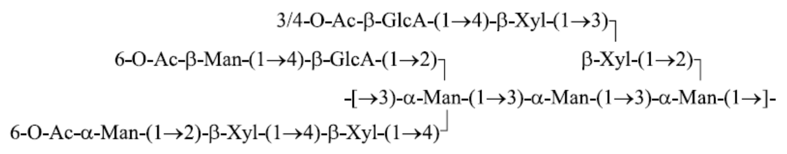
GXM consists of a linear backbone (1 → 3)-linked α-D-mannose with mainly xylose and glucuronic acid in the side chains (
Figure 1). Glucuronic acid contains a functional carboxylic acid, which at certain pH levels can stimulate the agglomeration of various polysaccharide assemblies due to the formation of intermolecular hydrogen bonds. The carboxyl group in the sugar backbone affects the intramolecular structure of the biocomposite, i.e., determines the conformation of individual polysaccharide macromolecules in an aqueous solution.
3. Results
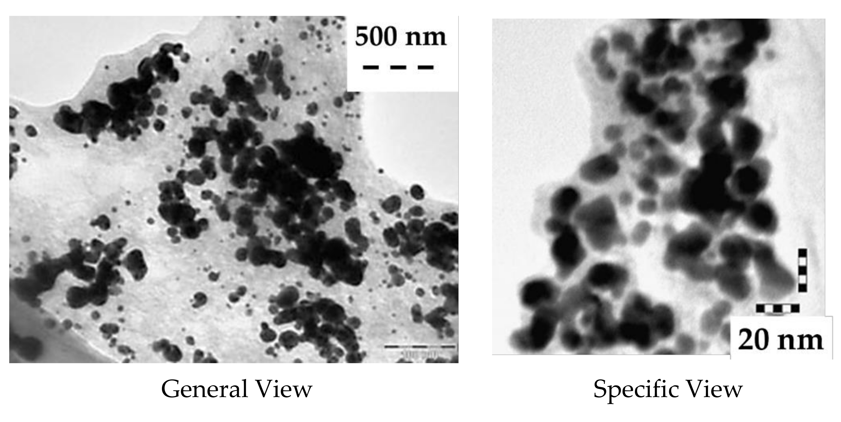
The morphological features of freshly prepared AuNP nanoparticles in a biopolymer polysaccharide matrix (Au-GXM), obtained using a transmission electron microscope, are shown in
Figure 2. Individual nanoparticles are characterized by a spherical geometry with a typical size ranging from 10 to 20 nm; most of the nanoparticles are separated from each other. The images are characterized by isolated groups of NPs with a size of about 100 nm × 500 nm, which may correspond to nanoparticles located inside the organic matrix of one or more polysaccharides—however, this observation requires additional special studies.
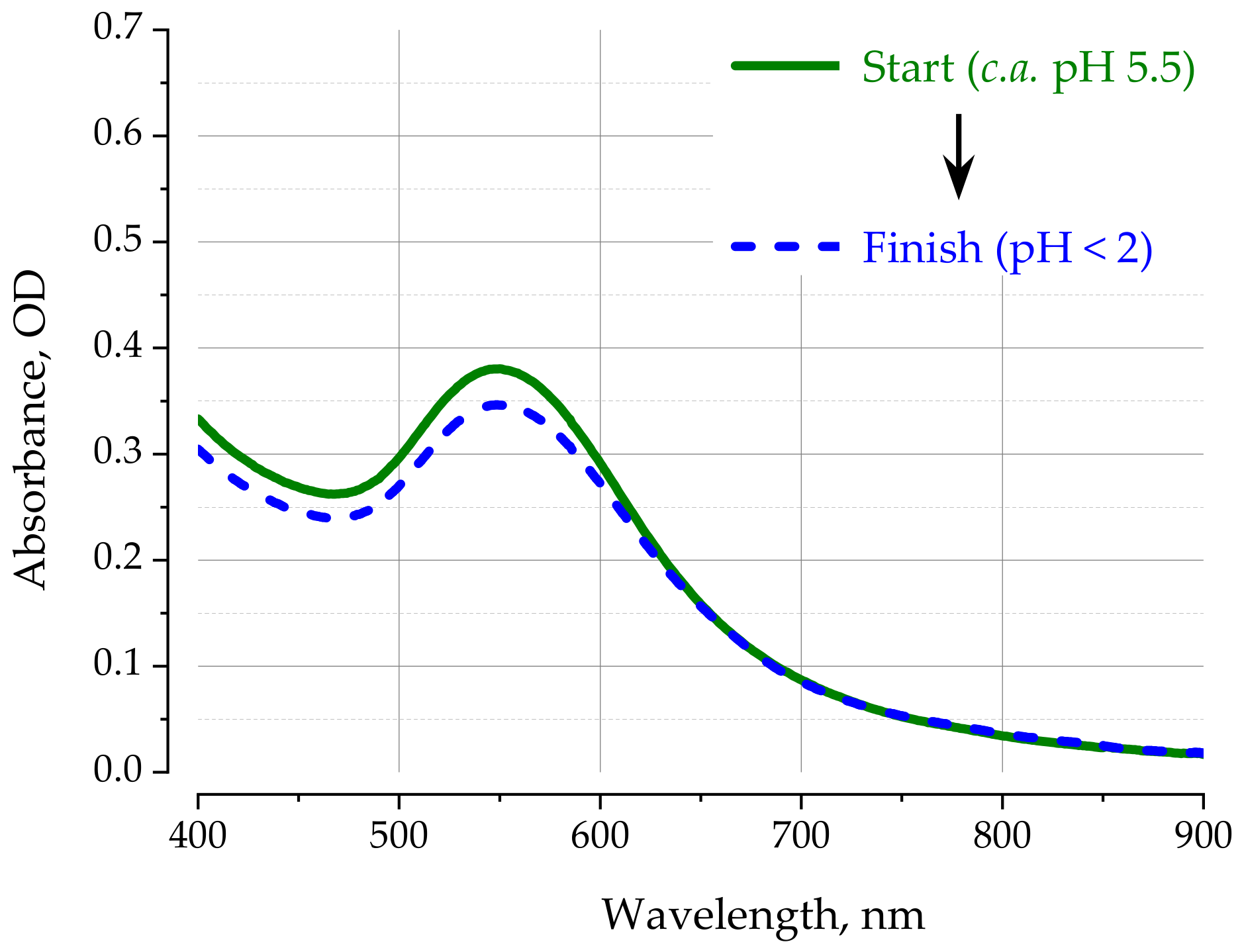
The absorption spectrum of the biocomposite in water is characterized by the LSPR band typical for individual gold nanoparticles in solutions (
Figure 3). The position of the absorption band with a maximum at about 550 nm indicates a dense organic environment of the metal core, which shifts the resonance conditions for gold nanoparticles 10–20 nm in size to lower energies. This fact confirms the assumption that gold NPs are formed inside the biopolymer matrix of the polysaccharide.
1 mL of the stock solution was subjected to successive acidification-alkalinization processes by sequentially adding a certain volume of 1 M HCl solution (acidification) and 1 M NaOH solution (alkalinization), respectively. Since the observed changes in the absorption spectra are characterized by irreversible changes, let us consider the changes typical for each of the cycles carried out.
Figure 3 shows the absorption spectra of the biocomposite at the first decrease in pH to a value less than 2, when all groups in the GXM macromolecule are protonated and electrostatic interactions do not prevent the formation of a dense globule. Taking into account the dilution effect caused by the addition of 100 μL of 1 M HCl to 1 mL of the initial solution, it can be concluded that a decrease in pH does not actually lead to any significant changes in the relative position of gold nanoparticles inside the polysaccharide matrix. Moreover, lowering the pH level below 5–6 also has no effect on the aggregation of biocomposite macromolecules.
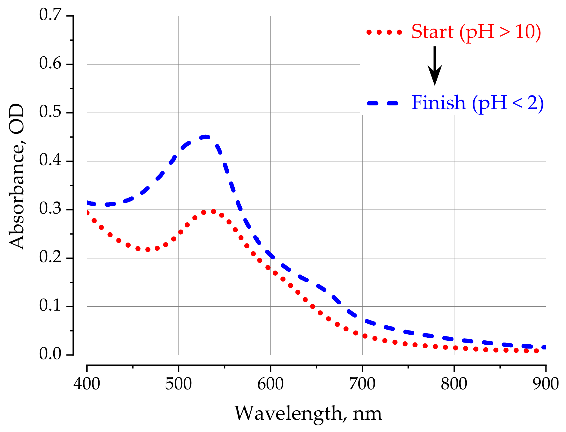
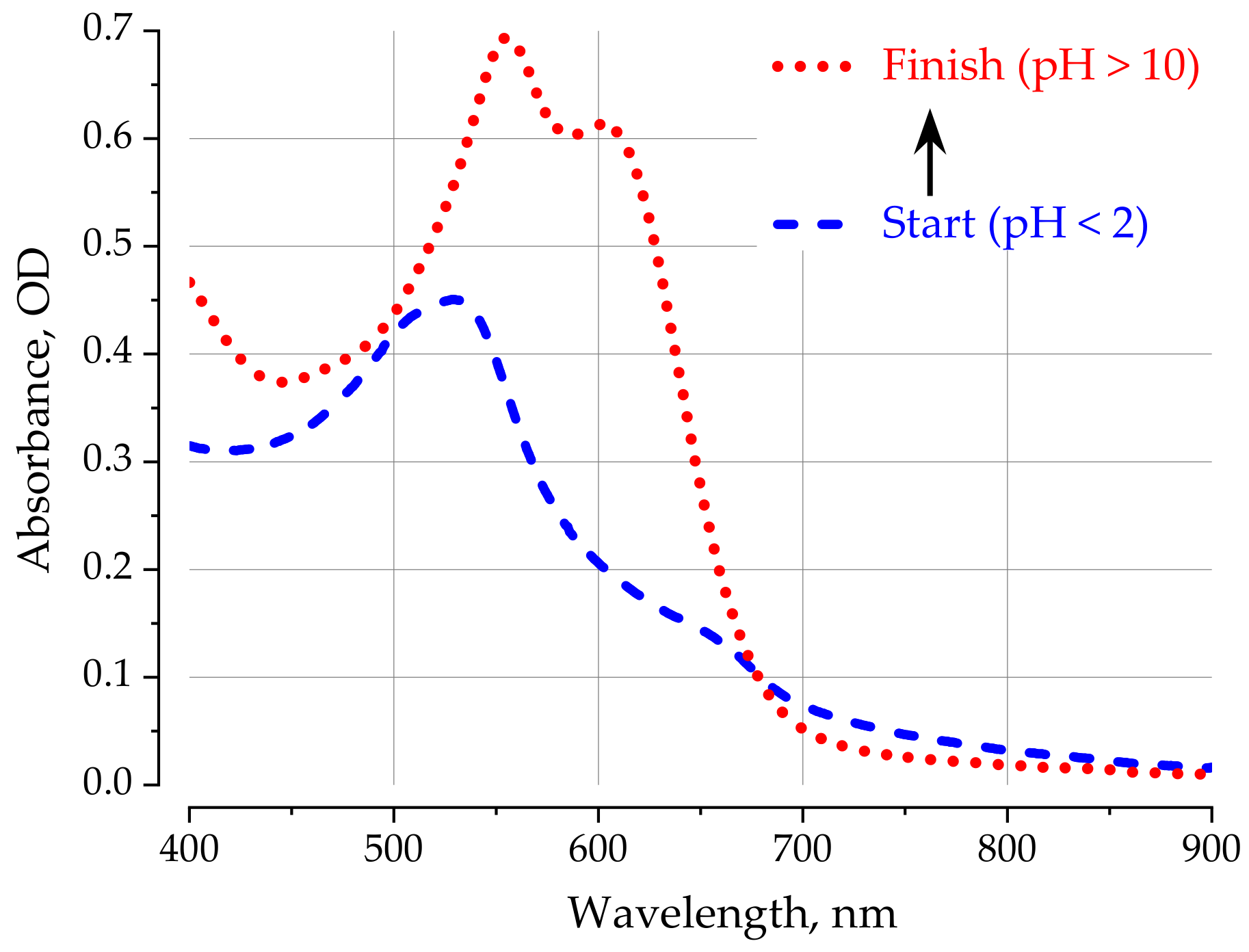
In contrast to acidification, alkalization of the solution to a pH level more than 10 leads to a significant change in the shape of the spectrum—the LSPR band shifts to the region of high energies (up to 535 nm), and its half-width decreases, significantly lowering the absorption in the region of 600–700 nm, which is typical for absorption by ensembles of nanoparticles (
Figure 4). This behavior may be associated with the “unfolding” (transformation to long linear chains) of polysaccharide molecules as a result of which the thickness of the organic coating decreases and the solvent can come closer to the metal surface, and the nanoparticles, respectively, disperse over long distances relative to each other.
Subsequent acidification (second cycle) leads to the appearance of a weak band in the region of 650 nm, despite the fact that the main LSPR band does not change its position (535 nm) (
Figure 5). This behavior indicates that, with a decrease in pH in the system, some densification is observed in the arrangement of nanoparticles, probably inside individual polysaccharide macromolecules, which manifests itself in an increase in the absorption of the band at 650 nm.
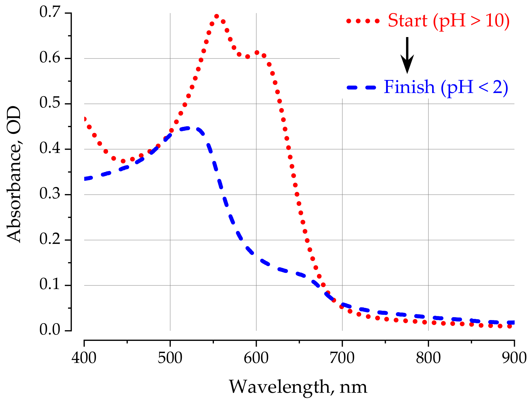
A qualitatively new picture is observed during subsequent alkalization in the second cycle. Indeed, when pH ~ 9 is reached (data not shown, see
Supplementary Materials for details), the previously observed absorption spectrum changes abruptly: the total absorption sharply increases, the main LSPR band shifts again to 550 nm (like the initial solution in water), and the intensity of the band at 600 nm almost equals in terms of intensity the LSPR band of single gold nanoparticles (
Figure 6). This pattern is qualitatively preserved with a further increase in pH more than 10. Moreover, a similar pattern with a sharp change in the spectrum is also observed with the subsequent acidification (cycle 3) of the solution (
Figure 7).
4. Discussion and Conclusions
An analysis of the experimental results presented above on the nature of the change in the optical characteristics of the local plasmon resonance excitations of gold nanoparticles embedded in a labile organic polysaccharide matrix allows us to draw the following main conclusions.
- (1)
The synthesized biocomposite is a pH-sensitive structure, whose changes in the features of the local plasmon resonance spectrum are associated both with a change in the internal structure of the polysaccharide and with its ability to form multimolecular aggregates.
- (2)
The initial solution of the biocomposite synthesized in water is partially aggregated; this aggregation can be destroyed by increasing the pH above 8–9, when the linkage of polysaccharide chains of different macromolecules is broken and the macromolecule tends to realize a linear chain conformation.
- (3)
Changes in the optical spectra of LSPR and, accordingly, the conformation and nature of the intermolecular association of the biocomposite change during the first cycles of acidification-alkalinization. This is due to the process of self-organization of the internal structure of the polysaccharide under conditions of repeated cycles of folding-unfolding of the macromolecular globule. An increase in the ionic strength of the solution stimulates the process of achieving the optimal three-dimensional packing of chains by suppressing electrostatic interactions that prevent the implementation of the optimal conformation specified by the primary structure of the macromolecule.
- (4)
After several cycles of acidification-alkalinization, a stable spatial configuration is achieved, in which a reversible process of “folding” (globular like conformation) and “unfolding” (linear chain conformation) of the biocomposite occurs when the pH level changes.
The results observed in this work are typical for polysaccharides isolated by alkaline extraction from extracts of Heterobasidiaceae mushrooms. It should be noted that the reason for the observed regularities can be not only the properties of the polysaccharide itself, but also other biocomponents (likely polysaccharide-associated proteins) released together with the main macromolecule in the process of isolation. It is quite possible that the observed irreversible changes in the structure of the bionanocomposite during cyclic changes in pH accompanied by an increase in the ionic strength of the solution are associated, among other things, with the separation of proteins initially associated with the polysaccharide. However, the verification of this hypothesis and the establishment of the details of the observed processes require additional research and will be considered by us in further studies.
Finally, gold nanoparticles embedded in polysaccharide chains are a useful tool for observing and studying the conformational features of a polysaccharide in an aqueous solution. In addition, AuNP makes it possible to determine transition points from one conformation to another. These considerations open the way to the creation of an adaptive natural nanomachine whose properties can be controlled by external influences.


