3. Results and Discussion
Interplay between husbandry, veterinary, behavioral, and welfare considerations occurs daily for the PCR.
Maintaining water husbandry for a tank this size is an ongoing task. The production of seawater and maintaining the appropriate parameters has presented unique challenges. The City and County of San Francisco makes seasonal changes to the sources for their primary water supply, with seasonal shifts in alkalinity from as low as 0.25–0.3 mEq/L in the summer to as high as 1.0–1.2 mEq/L in the winter. Incoming water has to be regularly tested, and the salt formulation is adjusted frequently in order to account for these changes. The contamination of salt bags with calcium phosphate, a non-clumping agent for table salt, presented as spikes in the saltwater PO43− as high as 1.0 mg/L. Now, the aquarium implements a rigorous protocol of analyzing every bag of salt that arrives, with unacceptable bags being returned to the manufacturer for exchange.
Within the first year of operation, it quickly became evident that the climate of San Francisco was not ideal for natural light-based coral growth. Light intensity should be between 150 and 1000 µE·m
−2·s
−1 for optimum coral health and growth [
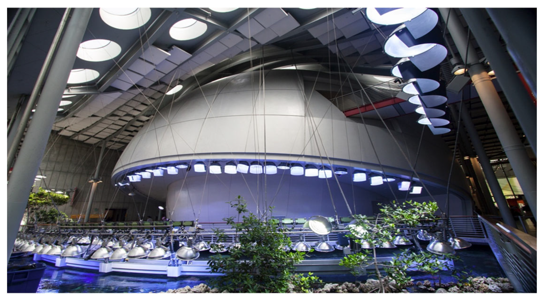
2]; measurements in both light intensity and photosynthetically active radiation (PAR) using sensors and data loggers (LI-193SA spherical quantum sensor and Li-1400 datalogger, both from Li-Cor Inc., Lincoln, NE, USA) found inconsistencies throughout the PCR due to wide variation in light intensity throughout the day and year, as the number of sunny days and angle of the sun changed. Signs of ailment such as bleaching (the expulsion of the symbiotic algae) was seen in corals throughout the tank, and focal death of corals occurred in areas of the PCR where the sun entered the circular skylights. Despite the original building design, a reliance on artificial light of a suitable intensity and color spectrum would be needed. A team of coral reef biologists, light designers, exhibit designers, architects, and outside consultants were therefore tasked with designing the PCR lighting to ensure that it both appropriately adhered to the needs of the reef while staying true to the design aesthetics of the building. A systematic assessment of light penetration by different light fixtures and at various depths was conducted in downtown San Francisco in the temporary coral reef tank that had been built for interim CAS use while the new academy was being built. Using these data and given the design and aesthetic requirements, a compromise design was settled upon: a lighting catwalk to mount the lamps needed closer to the water surface, allowing for ease of maintenance but also to meet the animals’ needs and maintain their welfare. Ultimately, seventy-six 1 kW metal halide fixtures would be needed to provide the appropriate spectrum and intensity that the PCR corals would need to thrive but with minimal energy expenditure (
Figure 3). In order to provide the habitat with a more natural light spectrum and promote photosynthesis in zooxanthellate corals, anemones, and clams, an emphasis on the shorter blue wavelengths indicative of natural coral reef waters was achieved via the distribution of both 10,000 K and 14,000 K lamps. Lastly, a translucent light film was applied to cover the building skylights that directly impacted the PCR, cutting out 80% of the light. Improvement in coral appearance was seen shortly thereafter, and over the next few years, the vertical glass walls on the west side of the habitat were also covered with this material.
As with water quality, maintaining the lighting for this tank is an ongoing task. Light-emitting diode (LED) lights are more energy-efficient than the metal halide fixtures that have been used over the PCR. Currently, Steinhart Aquarium is replacing the metal halide fixtures with greener LED-based options. As these changes are made, husbandry teams continue to track PAR throughout the PCR tank to ensure that they meet the corals’ light requirements (
Figure 4). Sites and depths are pre-assigned throughout the tank for consistency. Coral presence at each site is recorded. PAR is measured at varying intervals throughout the year, with the frequency of measurement being increased if major husbandry changes, such as lighting alterations, are made or poor animal health is noted. Meticulous spreadsheets detail dates, weather conditions, and if any lights are broken for each site. In general, PAR is taken during times of peak sun exposure, usually between 11:00 and 15:00. Steinhart Aquarium aims to keep PAR at the PCR sites at 200–1000 µE·m
−2·s
−1 for optimum coral health and growth. However, the spectrum varies based on location and depth in the tank, and some coral colonies have been seen to thrive at the extremes of measurements.
As the aquarium adds fauna to the collection in the PCR, the veterinary team has become even more involved in the health management of this habitat. Often, husbandry parameters alone can facilitate improvement in sick corals. For example, colonies with visible bleaching have been relocated to other parts of the PCR and have improved with no further interference. The ultimate cause of malaise may never be known, but husbandry factors are assumed because the animals’ statuses improved with only this change. Although there are currently limited resources for veterinarians working with corals, standard medical philosophies and approaches may be utilized. For the individuals that can be isolated, active diagnostics and treatment are pursued as they would be for domestic dogs and cats. For fish, physical examination, radiography, ultrasonography, and tissue sampling are performed under anesthesia [
6]. Animals can be kept in a hospital tank, where they undergo immersive, injectable, or oral therapy depending on the condition being treated, stress of handling, and the efficacy of the administration of medication. Invertebrates such as corals, anemones, urchins, or clams are either hospitalized or isolated in floating baskets that are still submerged within the PCR (
Figure 5). If a treatment is indicated, animals are removed and placed into medicated baths. Steinhart Aquarium has had success treating its aquatic invertebrates both with aggressive therapy, such as via daily antibiotic baths, but also with simple nutraceutical approaches, such as Revive Coral Cleaner (Two Little Fishies Inc., Miami Gardens, FL, USA) or Reef Primer (Polylab, Montreal, QC, Canada) dips for 10 min. In-depth descriptions of veterinary examinations and diagnostic methods are beyond the scope of this paper and can be found in the references [
1,
6]. However, especially for invertebrates, the value of microscopic analysis as a diagnostic tool cannot be overstated. Bacterial, fungal, or ciliate overgrowth as a cause of coral illness can be diagnosed via a simple skin scrape of tissue and microscopic analysis [
1,
10]. Corals have a rich endolithic community, and together, they are known as the coral holobiont [
1,
10,
11]. Veterinarians that work frequently with coral colonies may benefit from submitting tissues of healthy colonies for histopathology in order to understand this endolithic community of their animals, which may differ significantly from those of other aquariums, between coral species at the same facility, or even among healthy versus sick colonies of the same species [
10]. This would provide baseline information similar to that of “healthy animal” physical examinations, imaging, and blood values. Ill-animal biopsies can identify pathogens causing malaise, while shifts from the normal endolithic makeup can be indicative of the health of the colony, sometimes irrespective of gross appearance [
10]. For example, although initially thought to be an overgrowth of microorganisms and a possible cause of malaise in colonies from the PCR, a layer of specific algal hyphae has now been accepted as an appropriate finding for corals from this habitat, as it is seen repeatedly in animals with no evidence of gross nor histopathological abnormalities [
10]. Veterinarians working with systems such as the PCR are encouraged to communicate with colleagues regarding the successful or unsuccessful therapeutics used for corals at an individual or population level in an ongoing expansion of the medical body of knowledge available for these animals.
One of the most frustrating disease syndromes seen with captive coral care is Brown Jelly Syndrome (BJS). The underlying pathogenic trigger of BJS is still unknown, but a multifactorial cause, such as weakening immunity secondary to water stressors, has been considered, and the presence of the
Philaster ciliate has been associated with the condition [
1,
2]. Multifocal outbreaks of BJS have been noted in the PCR. With each outbreak, different factors were explored in an effort to understand why they were happening. Fish displaying coral-aberrant behaviors or targeted coral were removed from the tank. Particularly affected colonies were treated medically with Revive or Reef Primer dips or medicated paste [
12]. The PCR tank has a history of pycnogonid sea spider presence, and colonies noted to be harboring heavy populations of these arthropods (primarily
Pocillopora damicornis) were removed to prevent an overflow of sea spiders damaging the other corals [
10]. Underwater photographic monitoring of segments of the habitat was utilized to track outbreaks, and although resolution would spontaneously occur in some areas, new outbreaks of BJS continued. Water quality is closely monitored in all of Steinhart Aquarium’s aquatic habitats, but in late 2022, with a new BJS outbreak in the PCR and previous methods of interference proving unsuccessful, focus again shifted to environmental parameters as an underlying cause. Orthophosphate levels have historically been high in the PCR, sometimes reaching 0.8 mg/L PO
43−. In late 2022, the aquarium modified its water parameter testing procedures, shifting from manual to automated method-based technology, and discovered that phosphate values in the PCR were much higher than previously reported, sometimes up to ten times higher. With ongoing outbreaks in the corals, the aquarium increased the rate of PO
43− removal to bring levels to 0.2 mg/L, targeting < 0.15 mg/L. After this change was made, the incidence of disease declined significantly. One species of coral—
Echinopora—was almost extirpated from the habitat due to BJS during a 2020 outbreak, but this species has since been reestablished. Currently, testing for phosphate levels (as well as other water quality parameters) in the PCR is performed weekly on a Gallery Discrete Analyzer spectrophotometer (Thermo Fisher Scientific Inc., Waltham, MA, USA).
It is not always possible to hospitalize ailing PCR fish or invertebrates—some fish cannot be caught, some coral colonies are too large, some anemones are too established to move, etc. For these animals, medical interference is limited but on-exhibit treatment can still be pursued. For example, coral colonies diagnosed via skin scrape with Brown Jelly Syndrome (BJS) have benefited from a treatment regimen involving a coral-safe paste (CoralCure Ointment Base2B, Ocean Alchemists LLC, Largo, FL, USA) mixed with amoxicillin (Sandoz Inc., Princeton, NJ, USA), which is applied to affected tissues 1–3 times a week during dives (
Figure 5a) [
12]. Successful biological control methods have also been employed. Intermittent outbreaks of acoel flatworms, such as the recently described species
Amakusaplana acroporae, are addressed with freshwater flushes during dives [
13]. The control of
Phestilla subodiosus, nudibranchs that feed exclusively on
Montipora folliosa coral, was achieved via the addition of their predator, yellow-brown wrasse (
Thalassoma lutescens). Canary wrasse (
Halichoeres chrysus) are utilized to depopulate dwarf brittlestars (
Amphipholis spp.).
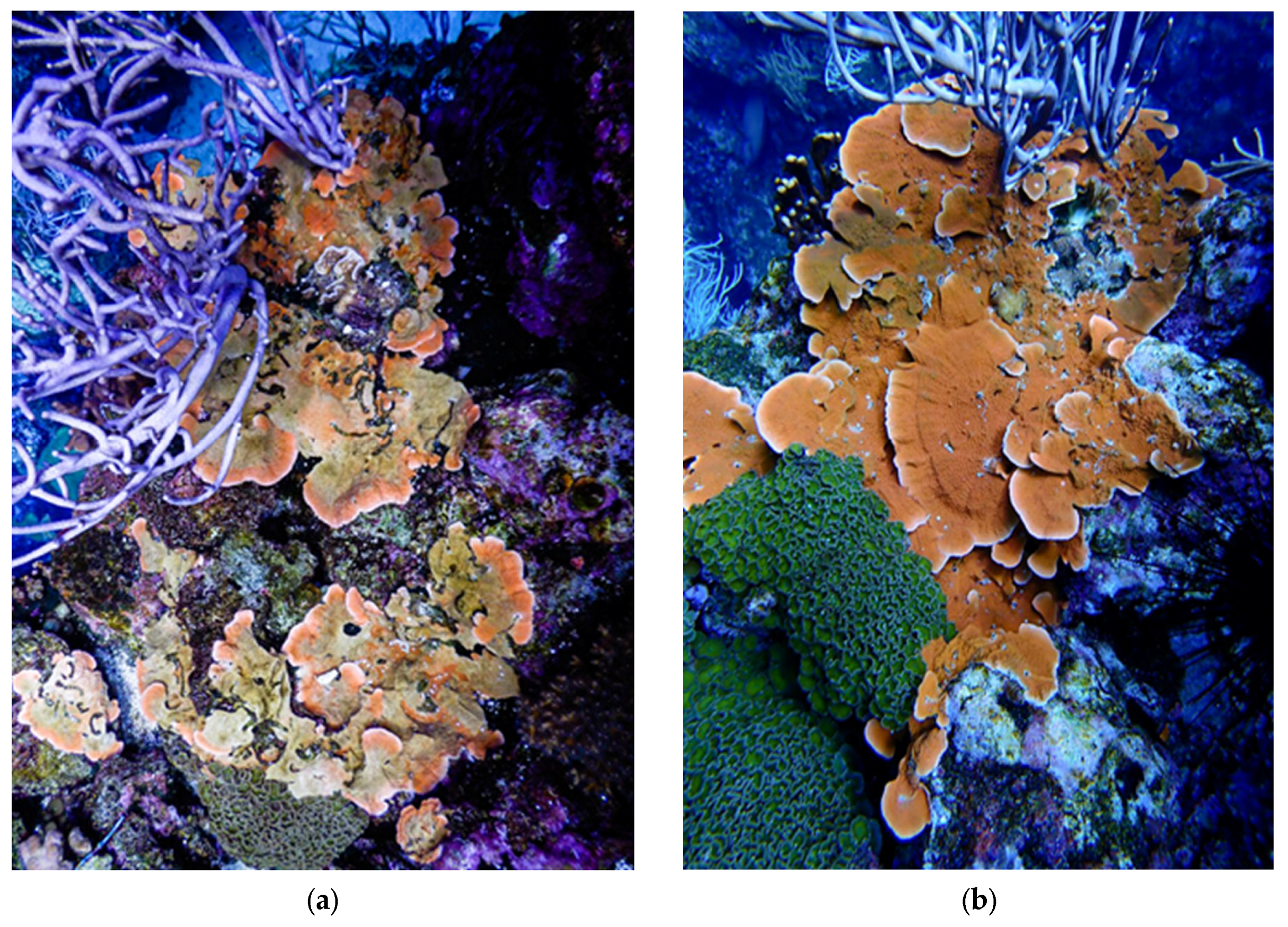
When welfare assessments indicate criteria that need improvement, processes are in place to address these. Examples of such criteria include exhibit conditions, diet presentation, collection planning, or providing more opportunities for animals to express agency. In cases of worsening welfare scores, often due to geriatric status or chronic medical conditions, animals are assessed more frequently via a separate Quality of Life Assessment. This assessment, completed by the biologists, curators, and veterinarians at the aquarium, aims to determine if improvements in welfare and quality of life can be achieved for the individual via husbandry, nutrition, or medical care changes. Underwater photography has been an immensely helpful tool for the welfare management of the PCR and the animals within it. Repeated photographs of sectors of the exhibit or particular animals allow for more consistent monitoring of the progression or improvement of subjects (
Figure 2). Objective quantification of concerns is performed using rulers and underwater photography during regularly scheduled tank dives. The PCR, in its entirety, is also photographed in set sections so that macro changes to the habitat can be appreciated and compared. Light measurements with handheld PAR meters and inductively coupled plasma-mass spectrometry (ICP-MS) analysis of water from systems with ailing colonies have not only shed light on potential causes of malaise but also helped in identifying changes in certain water quality parameters by which the animals begin to improve. If all courses of action have been exhausted, veterinarians determine if euthanasia is appropriate. If so, euthanasia is performed following the American Veterinary Medical Association (AVMA) and American Association of Zoo Veterinarians (AAZV) guidelines for the euthanasia of vertebrate and invertebrate animals [
14,
15].
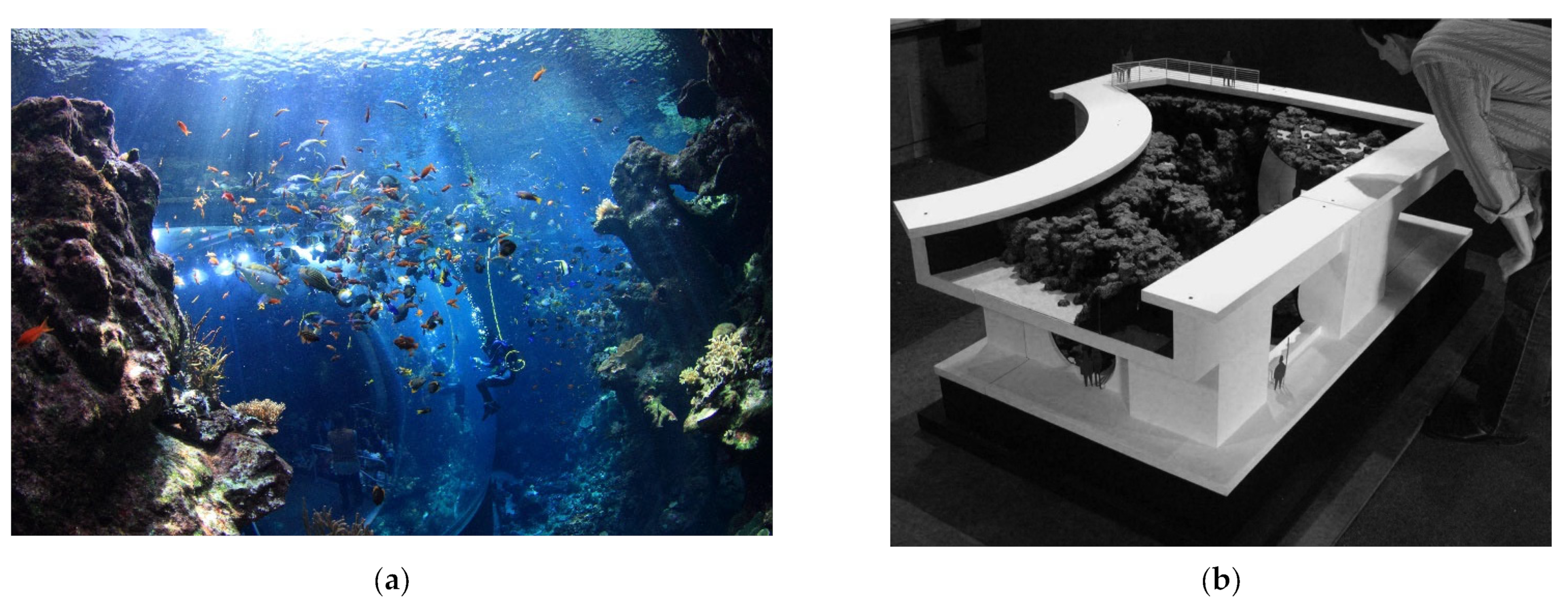
It is important to point out that the causes of many coral ailments in closed systems are still very much a “black box” despite husbandry or medical interference. Often, several changes must be instituted to arrest a suspected disease process. Abiotic factors such as lighting, water flow, and water chemistry can act synergistically, and changes to more than one need to be made in order to cause positive change. The willingness to reevaluate “what works” and adjust it for the benefit of the exhibit and its animals is a key approach for a captive habitat such as the PCR. Ultimately, the interplay of husbandry factors, animal behaviors, and health management contributes to a positive welfare experience and the vibrant growth of all tank animals, leading to the aquarium’s excellent, aesthetically pleasing appearance (
Figure 6).
A major negative aspect of a mixed-species exhibit is that, despite everyone’s best efforts, some species-specific behaviors may be detrimental to the other animals in the habitat [
8]. Abrupt changes in animal behavior can occur. For example, the naturally planktivorous pyramid butterflyfish (
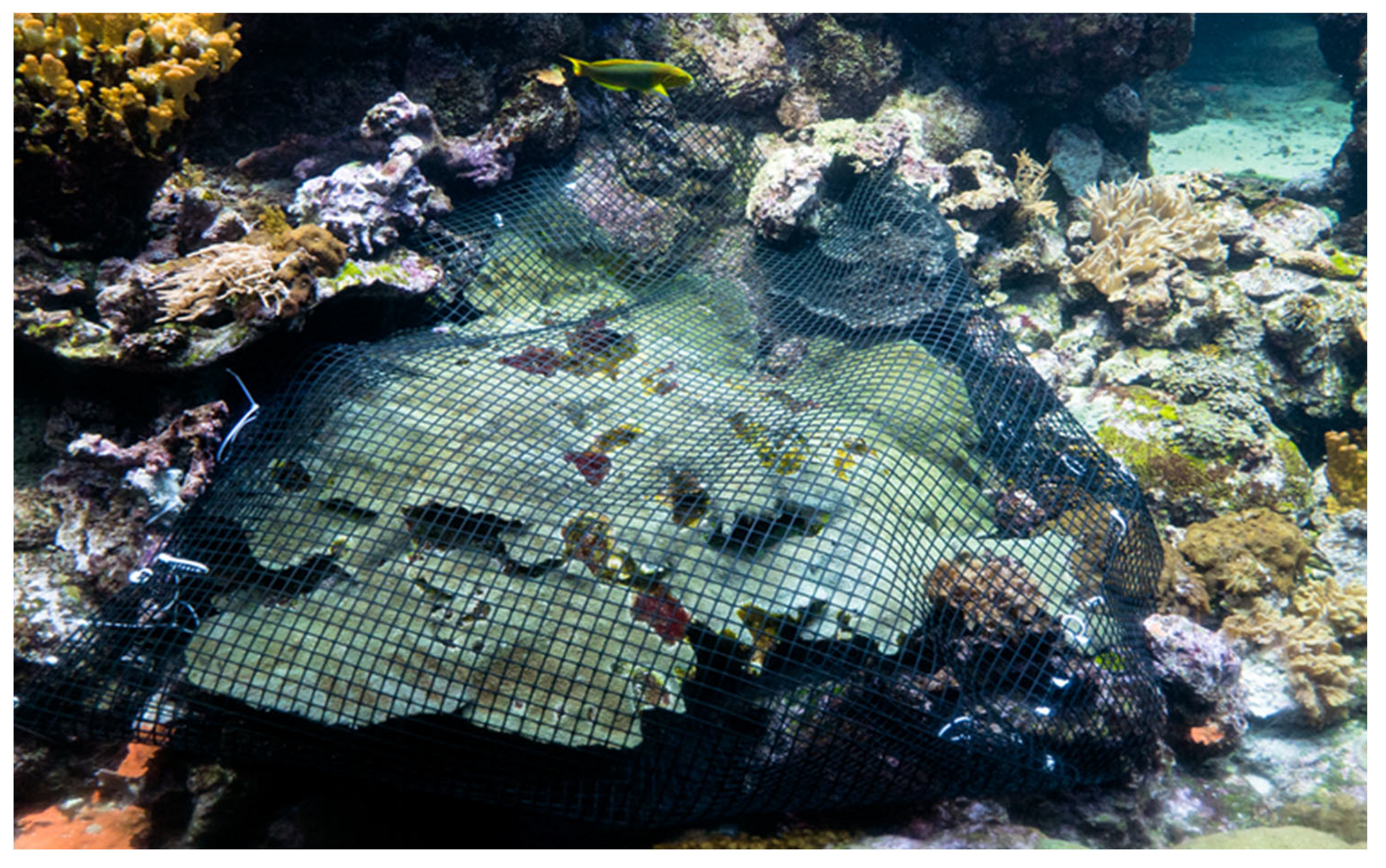
Hemitaurichthys polylepis) in the PCR suddenly began to eat soft corals after 8 years in the system. Steinhart Aquarium addresses such potentially detrimental behaviors by redirecting them to more appropriate outlets. Heads of lettuce or broccoli are lowered into the PCR twice a week in order to provide for herbivorous fish that might otherwise resort to picking at corals for food, redirecting this behavior away from affecting the invertebrates. Fish that continue aberrant behaviors despite husbandry alterations are moved to other habitats within the aquarium or transferred to other aquariums. Similarly, invertebrates that are repeatedly targeted are also either moved or temporarily shielded within a plastic mesh cage to protect them from predation until the fish in question can be removed or the behavior ceases (
Figure 7). Failing this, the coral can be relocated to areas of the habitat that can be shielded from the fish species in question.
Many aquariums offer the public an opportunity to experience fish feeding behaviors up close via scheduled dive shows and feeds involving in-water divers feeding the fish. Steinhart Aquarium has not allowed diver feedings during scheduled dive shows since the PCR was debuted in 2008. It is important to note that such opportunities would not occur in the wild and that there is potential for negative behaviors to develop from animals when these types of feeds occur, as fish will quickly associate divers with food and swarm them as soon as they enter the water [
16]. In addition, some of the fish (e.g., Niger triggerfish,
Odonus niger) have substantial dentition that can cause damage to fingers, and there has been a history of this in aquaria, including in the old Steinhart Aquarium facility (B. Shepherd, pers. comm.). Therefore, in keeping with the goal of presenting the most natural coral reef system possible, Steinhart Aquarium has chosen not to have divers feed the fish by hand during PCR dives. Instead, food is added from the surface by top-side staff at the tail end of the show to allow for interpretation by a dry-side presenter.


