Case Report

Rectal Spacer Placement for Anorectal Reirradiation of De Novo Rectal or Anal Cancer Following Prostate Radiation Therapy

1
Department of Radiation Oncology, Memorial Sloan Kettering Cancer Center, New York, NY 10065, USA
2
Department of Medical Physics, Memorial Sloan Kettering Cancer Center, New York, NY 10065, USA
3
New York Proton Center, New York, NY 10035, USA
*
Author to whom correspondence should be addressed.
Radiation 2024, 4(3), 242-252; https://doi.org/10.3390/radiation4030019
Submission received: 15 July 2024 / Revised: 29 August 2024 / Accepted: 4 September 2024 / Published: 6 September 2024

Abstract

:

Simple Summary

Pelvic reirradiation of de novo rectal or anal cancer after prior prostate cancer radiation carries several risks, including urinary and rectal fistula. Because of this, radiation is often omitted in these patients despite being an important component of standard of care therapy, leading to compromised outcomes and inferior quality of life. We therefore aimed to implement and evaluate the safety and feasibility of a novel approach to pelvic reirradiation of de novo rectal or anal cancer after prior prostate radiation, involving the placement of a rectal spacer prior to intensity-modulated radiation therapy or proton beam therapy. In this case series, we demonstrated excellent dosimetry and minimal toxicity with this approach, offering a feasible, promising treatment approach that can optimize patient outcomes, preserve quality of life, and maintain radiation therapy as a treatment option in patients with a history of prior pelvic radiation.

Abstract

Background: Pelvic reirradiation of de novo rectal or anal cancer after prior prostate cancer RT poses a significant risk of urinary and rectal fistula. In this report we describe the use of a rectal spacer to improve dosimetry and reduce this risk. Methods: Patients undergoing anorectal radiotherapy (RT) after prior prostate RT who had a rectal spacer placed prior to RT were identified in a prospective database. Patient, disease, and treatment characteristics were collected for these patients. Survival data were calculated from the end of RT. Radiation was delivered with intensity-modulated radiation therapy (IMRT) or proton beam therapy (PBT) following rectal spacer placement. Results: Rectal spacer placement with hydrogel injected transperineally under transrectal ultrasound guidance was successful in all five patients. MR/CT simulation 1–2 weeks post-spacer placement and IMRT or PBT delivered to a dose of 36–50 Gy in 24–30 fractions once or twice daily were tolerated well by all patients. The V100% of the PTV ranged from 62–100% and mean rectal and bladder dose ranged from 39–46 Gy and 16–40 Gy, respectively. At the last follow-up, three patients were alive and without evidence of disease up to 48 months out from treatment. There were no acute or late grade 3 or higher toxicities observed, but acute grade 2 proctitis was observed in all patients. Conclusions: The use of a rectal spacer placement to improve dosimetry of IMRT and PBT after prior prostate RT is safe and feasible in appropriately selected anorectal cancer patients.

1. Introduction

For locally advanced rectal cancer, standard of care includes combination therapy with radiation treatment (RT), surgery, and chemotherapy [1]. For nonmetastatic anal cancer, standard of care is definitive chemoradiation [2]. These treatment approaches have demonstrated marked benefits in disease control and survival [3,4,5]. However, for a subset of patients, the feasibility and safety of delivering RT as a component of treatment is sometimes limited by prior cancers and their associated treatments. In particular, patients with a history of prostate cancer treated with external beam RT or brachytherapy have historically been treated for subsequent rectal or anal cancers without RT in an attempt to avoid the potential increased risk of treatment complications associated with pelvic reirradiation. This limitation in treatment has become particularly important for this patient population, as prostate cancer treatment with RT has been shown to be associated with an increased risk of developing secondary malignancies, the most common being rectal cancer [6,7,8]. Additionally, with continued improvements in prostate RT and associated gains in survival, the incidence of rectal and anal cancers requiring treatment with RT has only become increasingly common.
For pelvic RT specifically, sparing of bladder and rectum is of primary concern, as the development of a fistula has been a well-documented complication of RT [9,10,11,12,13]. The use of more advanced radiation techniques such as intensity-modulated radiation therapy (IMRT) or proton beam therapy (PBT) has been one way in which this limitation is routinely addressed. Another way to address this limitation involves the use of a rectal spacer to increase the separation between the rectum and prostate/urinary tract. This approach has been shown to significantly improve dosimetry and toxicity profiles and is now FDA approved for radiation treatment of the prostate [14,15,16].
To our knowledge, there are no reports on the use of a rectal spacer to decrease toxicity in patients undergoing rectal or anal cancer reirradiation after prior prostate RT. Here, we report a novel approach to reirradiation involving rectal spacer placement followed by RT, with the potential to afford patients the best chance at disease control and survival while minimizing toxicity and preserving quality of life.

2. Methods

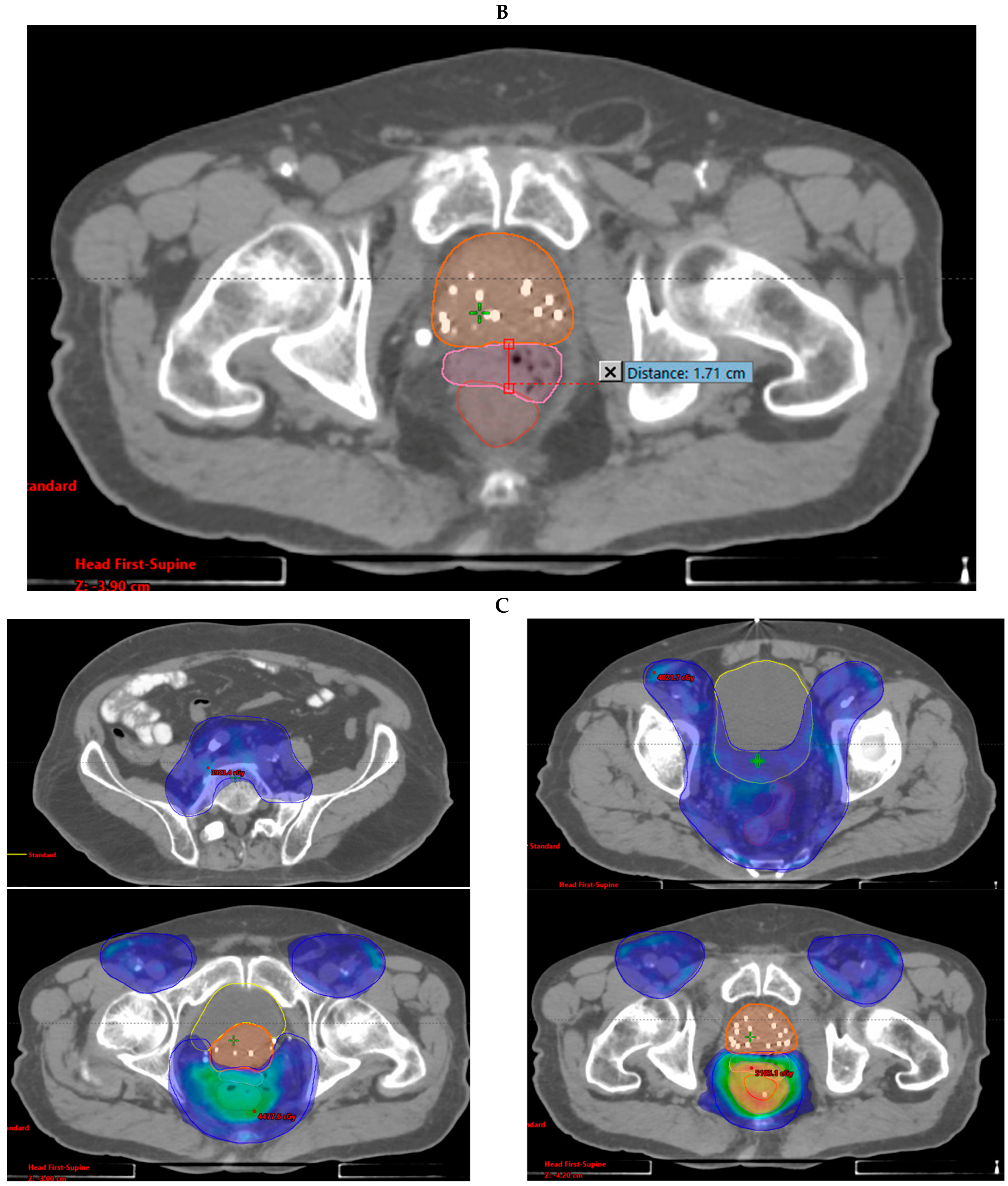
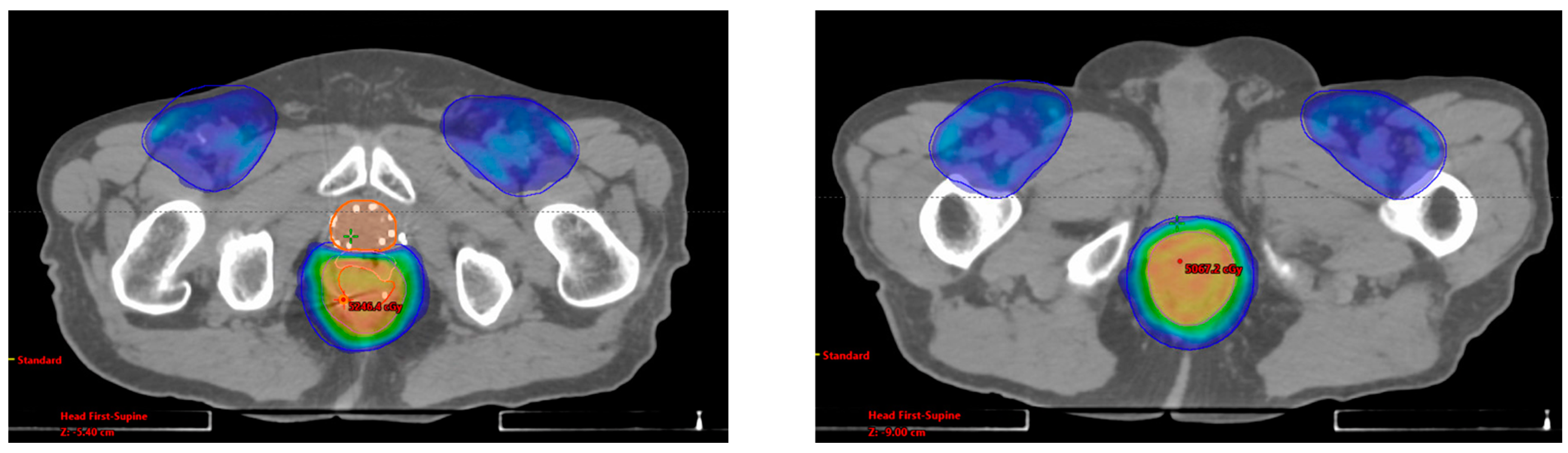
2.1. Rectal Spacer Placement

Rectal spacer insertion was performed in an outpatient setting in five patients according to previously reported protocols [17,18]. Briefly, the hydrogel was injected transperineally under transrectal ultrasound (TRUS) guidance with the patient in the lithotomy position. Hydrodissection was implemented for each patient to facilitate placement. Under real-time TRUS guidance in the sagittal plane, the injection needle (18G × 15 cm), with an attached syringe containing saline or lidocaine 0.5–1.0% diluted in 15 mL saline, was inserted approximately 1 cm above the TRUS probe through the perineum, and steadily advanced to the pelvic floor muscles. Once the rectourethralis muscle was penetrated, the tip was positioned inferior to the prostatic apex between Denonvilliers’ fascia and the anterior rectal wall. Hydrodissection was then implemented using small volumes of fluid to open the potential space between Denonvilliers’ fascia and the anterior rectal wall. The needle was subsequently advanced into the created space, with saline injection and needle advancement continuing until the needle tip was at mid-gland position, confirmed by ultrasound. With the needle in place, the syringe was then removed so that the syringe assembly could then be attached, and 10 mL of hydrogel was inserted slowly over 8–10 s. Patients with anterior tumors were excluded due to challenging spacer placement and risk of tissue abutting the tumor.

2.2. Radiation Therapy

MR/CT simulation was performed in each patient approximately 1–2 weeks after the placement of the rectal spacer. Simulation was performed with the patient in either a supine or prone position, and with a relatively full bladder. Immobilization systems used included Aquaplast, Alphacradle, or Bellyboards. The Eclipse treatment planning system was used for both CT-based IMRT and PBT planning. The rectal spacer gel was delineated on the MR simulation scan. The gross tumor volume (GTV) was delineated based on all simulation images; fusions with diagnostic imaging such as PETCT scans were also used to help better delineate disease. The clinical target volume (CTV) was constructed using institutional guidelines, and the planning target volume (PTV) was then constructed by expanding the CTV by 5 mm in all directions to account for uncertainties in both setup and beam range. The treating radiation oncologist determined radiation dose and fractionation, which ranged from 36–50 Gy delivered in 24–30 fractions delivered once or twice daily.
Quality assurance CBCT images were obtained throughout the course of therapy, with imaging frequency varying depending on the particular case and clinical discretion. Institutional dose constraints were adhered to during the planning process. Toxicities were recorded according to the Common Terminology Criteria for Adverse Events, version 5.0 (CTCAE) [19].

3. Results

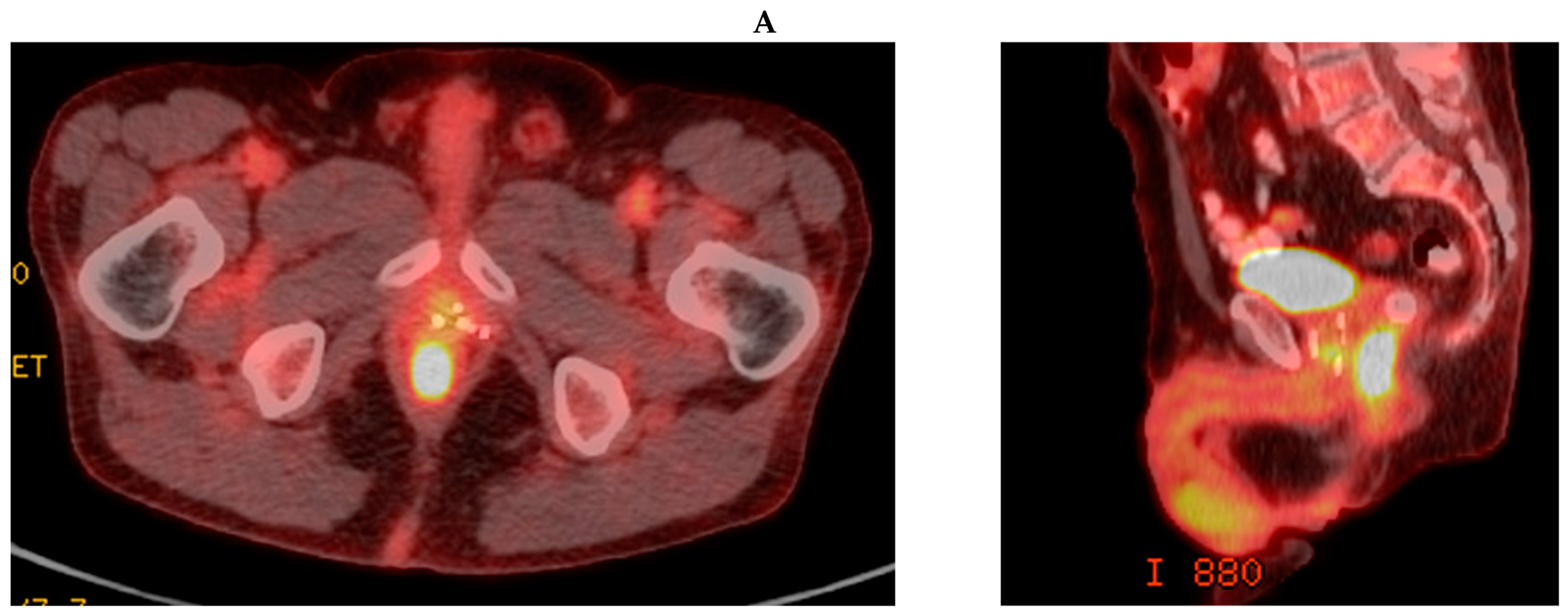
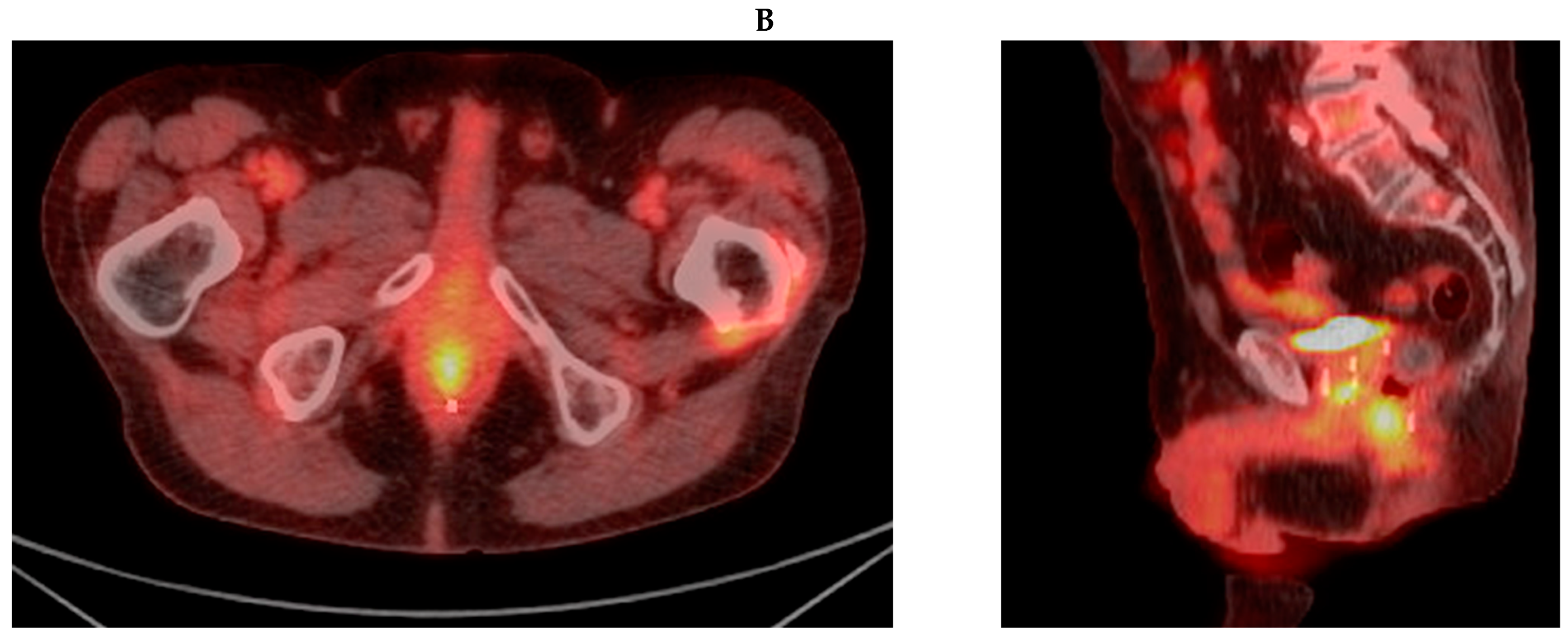
3.1. Case Report 1: Anal Squamous Cell Carcinoma

A 75-year-old man with a past medical history of HIV diagnosed in 1994, well-controlled on highly active antiretroviral therapy (HAART), and prostate cancer treated with low dose rate (LDR) brachytherapy at an outside institution in 2006 (records not available), presented to our institution for surgical evaluation after self-palpating an anal mass in 2018. A physical exam did not reveal any lymphadenopathy and a digital rectal exam showed normal external skin, but a 3 cm anal mass in the posterolateral position fixed to the anal sphincter muscles, 3 cm from the anal verge, was revealed. Flexible sigmoidoscopy was performed and confirmed a right posterior lateral anal mass approximately 3 cm from the anal verge involving the sphincter complex. Biopsy of the suspicious mass showed p16-positive invasive squamous cell carcinoma with basaloid features. Subsequent staging positron emission tomography/computed tomography (PET/CT) showed segmental hypermetabolism at the anus without any other areas of FDG avidity, and his disease was staged as clinical T2N0M0. Figure 1A shows pretreatment PET/CT images. He was then referred to Medical and Radiation Oncology and recommended treatment with definitive chemoradiation.
Given his prior history of prostate radiation, he first underwent rectal spacer placement per our institution’s protocol with same-day MR simulation thereafter (Figure 2A). RT was initiated 10 days after spacer placement, and he completed treatment with Capecitabine/Mitomycin and external beam RT to the anal canal (48 Gy in 24 fractions) and inguinal lymph nodes (36 Gy in 24 fractions). Representative images of the RT plan are shown in Figure 2B,C. The V100% of the GTV and PTV of the anal canal were 100% and 96% of the prescription dose, respectively. The mean dose to the rectum and bladder were 4024.7 cGy (3765 cGy EQD2) and 2730.1 cGy (2259 cGy EQD2), respectively. The patient tolerated treatment well overall with no grade 3 or higher toxicities. He did experience grade 2 proctitis characterized by intermittent rectal urgency and loose stool (attributed to RT) and grade 1 radiation dermatitis, fatigue, diarrhea, and abdominal pain during treatment, all of which resolved by his first post-treatment follow-up visit two months later. He did not experience any urinary symptoms or cystitis.
Unfortunately, the patient’s first post-treatment PET/CT revealed a 1.6 cm liver nodule, which was biopsied and consistent with his anal squamous cell primary. He was treated with 50 Gy in 10 fractions to the liver metastasis without complication but experienced a second liver recurrence along with a peritoneal recurrence several months later which were treated with Carboplatin and Paclitaxel. A first post-treatment evaluation with imaging revealed that the patient was in remission, and on his most recent flexible sigmoidoscopy and PET/CT 3.5 years post-anal and pelvic RT, the patient remained without evidence of disease (Figure 1B).

3.2. Case Report 2: Rectal Adenocarcinoma

A 75-year-old man with a past medical history of low-risk prostate cancer treated with external beam RT to 81 Gy in 45 fractions in 2013 presented to our institution with newly diagnosed adenocarcinoma of the distal rectum in 01/2019. He initially presented to an outside institution in 2018 with left lower quadrant pain for which CT of the abdomen and pelvis was performed and showed a suspicious left inguinal node. The lymph node was excised, and pathology showed metastatic poorly differentiated carcinoma with mucinous and neuroendocrine features. On physical examination, there was no noted lymphadenopathy, but a digital rectal exam revealed a 2 mm tender, sessile firm tumor at the dentate line in the left lateral position. Colonoscopy was then performed and showed a firm submucosal mass approximately 2–2.5 cm above the dentate line, in the left lateral position, with biopsy of the mass showing poorly differentiated rectal mucinous adenocarcinoma with neuroendocrine features. An MRI of the rectum was not performed as the patient had a pacemaker which was not MRI-compatible. Subsequent PET/CT did not show any foci of neoplastic disease but did show a 9–10 mm nonmetabolic aortocaval node and a new 3–4 mm lymph node just superior to the previously enlarged left inguinal lymph node. Finally, an endoscopic rectal ultrasound was performed and demonstrated a hypoechoic mass invading through the muscularis propria measuring 15 mm × 8 mm without any perirectal lymphadenopathy, and he was staged as T3N + M1 disease.
He was evaluated by colorectal surgery at our institution who felt that surgery would require abdominal perineal resection for complete resection and would be unlikely to be curative given the metastasis to the left inguinal and aortocaval lymph nodes as well as his high-risk pathology. Surgery was therefore not recommended, and he was referred to Medical and Radiation Oncology. After multidisciplinary evaluation, he was recommended induction Leucovorin, Fluorouracil, and Oxaliplatin (FOLFOX) and definitive chemoradiation. He underwent eight cycles of FOLFOX followed by RT to the rectum and lymph nodes to a dose of 45 Gy delivered in 30 fractions twice daily with concurrent Capecitabine. The V100% of the GTV and PTV of the rectum were 62% and 73% of the prescription dose, respectively. This coverage was the result of limiting the dose to the GTV abutting the prostate to a 100 Gy EQD2 cumulative dose; the rectal treatment delivered a max point dose of 1875.5 cGy to the prostate. The mean dose to the rectum and bladder were 4462.5 cGy (4006 cGy EQD2) and 3957.6 cGy (3419 cGy EQD2), respectively. RT was preceded by rectal spacer placement 12 days prior to treatment start. At the time of simulation, the distance created by the spacer was 1.1 cm.
The patient tolerated treatment well overall with no grade 3 or higher toxicities. He did experience grade 2 radiation proctitis involving mucous discharge and minimal rectal bleeding which largely resolved within 5 months post-treatment, and grade 1 cystitis, dehydration, radiation dermatitis, diarrhea, and fatigue. Since completing chemoradiation he has remained without evidence of disease clinically or radiographically through the last follow-up 36 months post-rectal RT.

3.3. Remaining Patients

We have subsequently treated three more patients (for a total of five) according to the protocol outlined and with successful rectal spacer placement. Details of treatment and disease-control outcomes are shown in Table 1. Of the five patients, three are currently alive with no evidence of disease, one died of rectal cancer progression, and one died of prostate and anal cancer progression (patient with BRCA1 germline mutation). Both deceased patients were found to have local progression of disease on their first post-treatment imaging. Of the surviving patients only one had an in-field recurrence at 14 months post RT; this recurrence was treated with low anterior resection (LAR), previously on non-operative management. For the two deceased patients, the GTV/PTV V100% was 56%/62% and 85%/81%, respectively. The surviving patients had the following GTV/PTV V100%: 100%/96%, 62%/73%, and 100%/100%, respectively. Additional dosimetric details for the five patients’ treatment plans are shown in Table 2.
In terms of toxicity, the patients tolerated chemoradiation well, where no one experienced grade 3 or higher acute or late toxicities. All patients had acute grade 2 proctitis which was characterized by intermittent rectal discomfort and pressure in one patient, small amounts of rectal bleeding in a second patient, and rectal pain with associated blood and mucous in stool in the remaining patient, which were all felt to be due to the RT. Symptoms eventually improved and were mostly resolved within several months, either without medical intervention or with over-the-counter pain medication and bowel regimens. Two patients experienced acute grade 1 cystitis, but none experienced urinary or recto-vesicular fistulas. Other toxicities included the following: grade 1–2 fatigue, diarrhea, fecal incontinence, radiation dermatitis, and dehydration. No toxicities associated with rectal spacer placement were observed.

4. Discussion

With the development of more sophisticated radiation technologies, reirradiation has become an increasingly used treatment strategy for a variety of clinical scenarios and can be a safe and effective treatment option in carefully selected patients [20,21,22]. Still, treatment options for patients who develop anorectal cancers following RT for prostate cancer have traditionally been limited to non-RT-based approaches due to concern for increased toxicity, specifically urinary and rectal, which can significantly impact quality of life. Here, we report our institutional experience treating five patients with secondary anorectal cancers after RT for prostate cancer with reirradiation, using a novel approach involving the use of a rectal spacer followed by IMRT or PBRT. In our current series, rectal spacer placement was successful in all five patients and no patients experienced any complications associated with the procedure. The spacer placement was feasible in those reported select cases because of the location of the anorectal tumors (posterior/lateral) and little fibrosis in the space between the rectum and prostate. All patients experienced grade 2 proctitis (attributable to RT and not to other therapies), making it difficult to assess whether this toxicity was associated with rectal spacer placement quality or cumulative RT dose received. Nevertheless, none of the patients experienced grade 3 or higher acute or late toxicities and treatment with RT afforded durable local control in three patients.
To date, several studies have reported on toxicity profiles associated with different dosing and fractionation regimens of anorectal RT following pelvic RT, most commonly in the setting of reirradiation for gastrointestinal malignancies [23,24,25,26,27]. In these reports, reirradiation regimens typically involved hyperfractionated RT courses of 1.5 Gy delivered twice daily to a dose of approximately 40 Gy, with grade 3–4 toxicity rates as high as 35%. For example, a recent retrospective study from MD Anderson Cancer Center evaluated fifty patients with a history of pelvic RT (14% for cancer other than rectal) treated with hyperfractionated accelerated RT for primary or recurrent rectal adenocarcinoma and observed two grade 3 acute toxicities and a 3-year rate of grade 3–4 toxicity of 35%, with the most notable toxicities including the following: cystitis, bowel obstruction, rectovaginal or vesicovaginal fistulas, and pelvic abscess formation. While such studies have demonstrated a role for reirradiation in improving local control of disease, they also demonstrate the importance of minimizing RT toxicity. Moreover, as the majority of pelvic reirradiation data pertain to patients who initially received RT to the GI tract, data on the feasibility and safety associated with anorectal RT after prostate RT specifically are lacking.
We recently evaluated and reported our institutional experience treating twenty-six patients with de novo anorectal cancers following RT for prostate cancer [28]. We observed a 5-year local progression and overall survival of 30% and 31% for rectal cancer, and 35% and 49% for anal cancer patients, respectively, with a median follow-up time of 84.4 months. Additionally, we evaluated dosimetric parameters in eleven patients with data available, and found that target coverage met our institutional goals, with a median GTV V100% of 100% (68.1–100%) and PTV V100% of 97.5% (89.6–100%). Sparing of adjacent normal structures was also met, with a median EQD2 cumulative dose to the rectum and bladder of 11764 cGy and 11540 cGy, respectively. While toxicity was acceptable in that no patients experienced grade > 3 acute toxicities, two patients did develop fistulae (urinary-cutaneous and recto-vesicular) which required extensive surgical repair, both after LDR for prostate cancer and 45–50 Gy in 25 fractions for anal or rectal cancer.
These findings motivated the subsequent use of rectal spacer placement to mitigate some of the toxicities observed in these patients. A recent large multi-institutional clinical trial of rectal spacers for prostate RT demonstrated positive outcomes in reducing low- and high-grade toxicity events following IMRT, and the safety of this approach has also been reported [12,13]. Additionally, while typically more challenging, rectal spacer placement in patients who have received prior RT has been shown to be feasible, with one study of 11 patients undergoing salvage brachytherapy after having undergone prior RT reporting successful spacer placement in eight patients (73%) [29,30].
Although limited by small sample size, our results reported here suggest that the incorporation of rectal spacer placement in reirradiation approaches is feasible and safe for select anatomically favorable (posterior/lateral) tumors with little urinary toxicity risk, as we did not observe any grade 3 or higher toxicities in patients with rectal spacers placed. This novel approach to reirradiation may afford a safe and effective treatment option for this unique patient population. Additional studies involving larger cohorts with longer follow-up would be useful in enhancing the clinical impact of these early results, with cost-effectiveness analyses, patient-reported outcomes, and dosimetric comparison between IMRT and PBT being key areas for further study.

5. Conclusions

This is the first report in the literature to describe the placement of a rectal spacer prior to pelvic reirradiation to improve dosimetry and mitigate toxicity. Our results suggest that this approach is safe and feasible, and that it can offer durable local control in appropriately selected patients. Continued implementation of this treatment approach in larger cohorts should be considered for further study.

Author Contributions

Conceptualization: M.J.Z., C.H.C. and C.H.; methodology: A.D.D., J.P.N., N.K., A.S., M.J.Z., C.H.C. and C.H.; software: J.P.N., N.K. and A.S.; validation: A.D.D., J.P.N., N.K., A.S., P.B.R., M.R., M.J.Z., C.H.C. and C.H.; formal analysis: A.D.D., A.D.D., J.P.N., N.K. and A.S.; investigation: P.B.R., M.R., M.J.Z., C.H.C. and C.H.; resources: A.D.D., J.P.N., N.K., A.S., P.B.R., M.R., M.J.Z., C.H.C. and C.H.; data curation: A.D.D., J.P.N., N.K. and A.S.; writing—original draft preparation: A.D.D., J.P.N. and N.K.; writing—review and editing: A.D.D., J.P.N., N.K., A.S., P.B.R., M.R., M.J.Z., C.H.C. and C.H.; visualization: P.B.R., M.R., M.J.Z., C.H.C. and C.H.; supervision: M.J.Z., C.H.C. and C.H.; project administration: C.H.; funding acquisition: M.J.Z. and C.H. All authors have read and agreed to the published version of the manuscript.

Funding

This research received no external funding.

Institutional Review Board Statement

The study was conducted in accordance with the Declaration of Helsinki and approved by the Institutional Review Board (or Ethics Committee) of Memorial Sloan Kettering Cancer Center (protocol code 16-370, date of approval 4/26/2016).

Informed Consent Statement

The request to waive the requirement to obtain written informed consent and a research authorization has been granted as per 45 CFR 46.116(c)(d) and 45 CFR 164.512(i)(1)(ii).

Data Availability Statement

Research data are stored in an institutional repository and will be shared upon request to the corresponding author.

Conflicts of Interest

The authors report no conflicts of interest.

References

  1. NCCN, Rectal Cancer. 2020. Available online: https://www.nccn.org/patients/guidelines/content/PDF/rectal-patient.pdf (accessed on 1 April 2024).
  2. NCCN, Anal Cancer. 2020. Available online: https://www.nccn.org/patients/guidelines/content/PDF/anal-patient.pdf (accessed on 1 April 2024).
  3. Folkesson, J.; Birgisson, H.; Pahlman, L.; Cedermark, B.; Glimelius, B.; Gunnarsson, U. Swedish Rectal Cancer Trial: Long lasting benefits from radiotherapy on survival and local recurrence rate. J. Clin. Oncol. 2005, 23, 5644–5650. [Google Scholar] [CrossRef]
  4. O’Connell, M.J.; Martenson, J.A.; Wieand, H.S.; Krook, J.E.; Macdonald, J.S.; Haller, D.G.; Mayer, R.J.; Gunderson, L.L.; Rich, T.A. Improving adjuvant therapy for rectal cancer by combining protracted-infusion fluorouracil with radiation therapy after curative surgery. N. Engl. J. Med. 1994, 331, 502–507. [Google Scholar] [CrossRef]
  5. Wolmark, N.; Wieand, H.S.; Hyams, D.M.; Colangelo, L.; Dimitrov, N.V.; Romond, E.H.; Wexler, M.; Prager, D.; Cruz, A.B., Jr.; Gordon, P.H.; et al. Randomized trial of postoperative adjuvant chemotherapy with or without radiotherapy for carcinoma of the rectum: National Surgical Adjuvant Breast and Bowel Project Protocol R-02. J. Natl. Cancer Inst. 2000, 92, 388–396. [Google Scholar] [CrossRef]
  6. Baxter, N.N.; Tepper, J.E.; Durham, S.B.; Rothenberger, D.A.; Virnig, B.A. Increased risk of rectal cancer after prostate radiation: A population-based study. Gastroenterology 2005, 128, 819–824. [Google Scholar] [CrossRef]
  7. Boue-Rafle, A.; Briens, A.; Supiot, S.; Blanchard, P.; Baty, M.; Lafond, C.; Masson, I.; Crehange, G.; Cosset, J.M.; Pasquier, D.; et al. Does radiation therapy for prostate cancer increase the risk of second cancers? Cancer Radiother. 2024, 28, 293–307. [Google Scholar] [CrossRef] [PubMed]
  8. McPartland, C.; Salib, A.; Banks, J.; Mark, J.R.; Lallas, C.D.; Trabulsi, E.J.; Gomella, L.G.; Goldberg, H.; Leiby, B.; Den, R.; et al. Risk of Secondary Malignancies After Pelvic Radiation: A Population-based Analysis. Eur. Urol. Open Sci. 2024, 63, 52–61. [Google Scholar] [CrossRef] [PubMed]
  9. Chrouser, K.; Leibovich, B.; Bergstralh, E.; Zincke, H.; Blute, M. Bladder cancer risk following primary and adjuvant external beam radiation for prostate cancer. J. Urol. 2005, 174, 107–110; discussion 110–111. [Google Scholar] [CrossRef]
  10. Ma, J.L.; Hennessey, D.B.; Newell, B.P.; Bolton, D.M.; Lawrentschuk, N. Radiotherapy-related complications presenting to a urology department: A more common problem than previously thought? BJU Int. 2018, 121 (Suppl. 3), 28–32. [Google Scholar] [CrossRef] [PubMed]
  11. Marguet, C.; Raj, G.V.; Brashears, J.H.; Anscher, M.S.; Ludwig, K.; Mouraviev, V.; Robertson, C.N.; Polascik, T.J. Rectourethral fistula after combination radiotherapy for prostate cancer. Urology 2007, 69, 898–901. [Google Scholar] [CrossRef]
  12. Stone, N.N.; Stock, R.G. Complications following permanent prostate brachytherapy. Eur. Urol. 2002, 41, 427–433. [Google Scholar] [CrossRef]
  13. Zoubek, J.; McGuire, E.J.; Noll, F.; DeLancey, J.O. The late occurrence of urinary tract damage in patients successfully treated by radiotherapy for cervical carcinoma. J. Urol. 1989, 141, 1347–1349. [Google Scholar] [CrossRef] [PubMed]
  14. Song, D.Y.; Herfarth, K.K.; Uhl, M.; Eble, M.J.; Pinkawa, M.; van Triest, B.; Kalisvaart, R.; Weber, D.C.; Miralbell, R.; Deweese, T.L.; et al. A multi-institutional clinical trial of rectal dose reduction via injected polyethylene-glycol hydrogel during intensity modulated radiation therapy for prostate cancer: Analysis of dosimetric outcomes. Int. J. Radiat. Oncol. Biol. Phys. 2013, 87, 81–87. [Google Scholar] [CrossRef] [PubMed]
  15. Uhl, M.; Herfarth, K.; Eble, M.J.; Pinkawa, M.; van Triest, B.; Kalisvaart, R.; Weber, D.C.; Miralbell, R.; Song, D.Y.; DeWeese, T.L. Absorbable hydrogel spacer use in men undergoing prostate cancer radiotherapy: 12 month toxicity and proctoscopy results of a prospective multicenter phase II trial. Radiat. Oncol. 2014, 9, 96. [Google Scholar] [CrossRef] [PubMed]
  16. Folkert, M.R.; Zelefsky, M.J.; Hannan, R.; Desai, N.B.; Lotan, Y.; Laine, A.M.; Kim, D.W.N.; Neufeld, S.H.; Hornberger, B.; Kollmeier, M.A.; et al. A Multi-Institutional Phase 2 Trial of High-Dose SAbR for Prostate Cancer Using Rectal Spacer. Int. J. Radiat. Oncol. Biol. Phys. 2021, 111, 101–109. [Google Scholar] [CrossRef]
  17. Hatiboglu, G.; Pinkawa, M.; Vallee, J.P.; Hadaschik, B.; Hohenfellner, M. Application technique: Placement of a prostate-rectum spacer in men undergoing prostate radiation therapy. BJU Int. 2012, 110, E647–E652. [Google Scholar] [CrossRef]
  18. Pinkawa, M.; Corral, N.E.; Caffaro, M.; Piroth, M.D.; Holy, R.; Djukic, V.; Otto, G.; Schoth, F.; Eble, M.J. Application of a spacer gel to optimize three-dimensional conformal and intensity modulated radiotherapy for prostate cancer. Radiother. Oncol. 2011, 100, 436–441. [Google Scholar] [CrossRef]
  19. Cancer Therapy Evaluation Program (CTEP). Common Terminology Criteria for Adverse Events (CTCAE)v.5.0 [5x7]. In Cancer Ther Aval Progr 155 (2017). Available online: https://ctep.cancer.gov/protocolDevelopment/electronic_applications/ctc.htm#ctc_50 (accessed on 1 April 2024).
  20. Kamran, S.C.; Zelefsky, M.; Nguyen, P.L.; Lawton, C.A.F. To Radiate or Not to Radiate-The Challenges of Pelvic Reirradiation. Semin. Radiat. Oncol. 2020, 30, 238–241. [Google Scholar] [CrossRef]
  21. Chung, S.Y.; Koom, W.S.; Keum, K.C.; Chang, J.S.; Shin, S.J.; Ahn, J.B.; Min, B.S.; Lee, K.Y.; Kim, N.K.; Yoon, H.I. Treatment Outcomes of Re-irradiation in Locoregionally Recurrent Rectal Cancer and Clinical Significance of Proper Patient Selection. Front. Oncol. 2019, 9, 529. [Google Scholar] [CrossRef]
  22. Zilli, T.; Benz, E.; Dipasquale, G.; Rouzaud, M.; Miralbell, R. Reirradiation of Prostate Cancer Local Failures After Previous Curative Radiation Therapy: Long-Term Outcome and Tolerance. Int. J. Radiat. Oncol. Biol. Phys. 2016, 96, 318–322. [Google Scholar] [CrossRef]
  23. Chang, D.T.; Koay, E.J.; Herman, J.M.; Hong, T.S.; Das, P. Abdominal and Pelvic Reirradiation for Recurrent Gastrointestinal Cancers. Semin. Radiat. Oncol. 2020, 30, 232–237. [Google Scholar] [CrossRef]
  24. Das, P.; Delclos, M.E.; Skibber, J.M.; Rodriguez-Bigas, M.A.; Feig, B.W.; Chang, G.J.; Eng, C.; Bedi, M.; Krishnan, S.; Crane, C.H. Hyperfractionated accelerated radiotherapy for rectal cancer in patients with prior pelvic irradiation. Int. J. Radiat. Oncol. Biol. Phys. 2010, 77, 60–65. [Google Scholar] [CrossRef] [PubMed]
  25. Mohiuddin, M.; Marks, G.; Marks, J. Long-term results of reirradiation for patients with recurrent rectal carcinoma. Cancer 2002, 95, 1144–1150. [Google Scholar] [CrossRef]
  26. Tao, R.; Tsai, C.J.; Jensen, G.; Eng, C.; Kopetz, S.; Overman, M.J.; Skibber, J.M.; Rodriguez-Bigas, M.; Chang, G.J.; You, Y.N.; et al. Hyperfractionated accelerated reirradiation for rectal cancer: An analysis of outcomes and toxicity. Radiother. Oncol. 2017, 122, 146–151. [Google Scholar] [CrossRef] [PubMed]
  27. Valentini, V.; Morganti, A.G.; Gambacorta, M.A.; Mohiuddin, M.; Doglietto, G.B.; Coco, C.; De Paoli, A.; Rossi, C.; Di Russo, A.; Valvo, F.; et al. Preoperative hyperfractionated chemoradiation for locally recurrent rectal cancer in patients previously irradiated to the pelvis: A multicentric phase II study. Int. J. Radiat. Oncol. Biol. Phys. 2006, 64, 1129–1139. [Google Scholar] [CrossRef] [PubMed]
  28. Hilal, L.; Wu, A.J.; Reyngold, M.; Cuaron, J.J.; Navilio, J.; Romesser, P.B.; Dreyfuss, A.; Yin, S.; Zhang, Z.; Bai, X.; et al. Radiation therapy for de novo anorectal cancer in patients with a history of prostate radiation therapy. Front. Oncol. 2022, 12, 975519. [Google Scholar] [CrossRef]
  29. Mahal, B.A.; Ziehr, D.R.; Hyatt, A.S.; Neubauer-Sugar, E.H.; O’Farrell, D.A.; O’Leary, M.P.; Steele, G.S.; Niedermayr, T.R.; Beard, C.J.; Martin, N.E.; et al. Use of a rectal spacer with low-dose-rate brachytherapy for treatment of prostate cancer in previously irradiated patients: Initial experience and short-term results. Brachytherapy 2014, 13, 442–449. [Google Scholar] [CrossRef]
  30. Nguyen, P.L.; Devlin, P.M.; Beard, C.J.; Orio, P.F., 3rd; O’Leary, M.P.; Wolfsberger, L.D.; O’Farrell, D.A.; Sweeney, C.M.; Hadaschik, B.A. High-dose-rate brachytherapy for prostate cancer in a previously radiated patient with polyethylene glycol hydrogel spacing to reduce rectal dose: Case report and review of the literature. Brachytherapy 2013, 12, 77–83. [Google Scholar] [CrossRef]
Figure 1. (A) Pretreatment PET/CT images demonstrating a hypermetabolic anal mass. (B) Most recent post-treatment PET/CT images demonstrating post-treatment nonspecific anal hypermetabolism without evidence of disease.
Radiation 04 00019 g001aRadiation 04 00019 g001b
Figure 2. (A) Representative axial T1 post-contrast and T2 images from MR simulation showing enhancement in the anal canal and hyperintense rectal spacer gel, respectively. (B) Rectal spacer creating 1.71 cm between the prostate and rectum shown on CT planning scan. (C) Representative slices of RT plan for a patient who received 48 Gy to the anal canal and 36 Gy to the pelvic nodes in 24 fractions following rectal spacer placement.
Radiation 04 00019 g002aRadiation 04 00019 g002bRadiation 04 00019 g002c
Table 1. Treatment characteristics and disease-control outcomes in patients treated with reirradiation for anorectal cancers following prostate cancer RT 1.
Primary DiseaseProstate RTAnorectal RTInitial ResponseCurrent StatusIn-Field
POD
(mo)
Out-of-Field POD
(mo)
OS from RT End
Anal2006:
-LDR
2018:
-Anal canal + LNs
36–48 Gy/24 fx QD
-POD; liver
-CR; local
NEDNo POD2 mo; liver treated with SBRT48 mo
Rectal2013:
-EBRT 81 Gy/45 fx
2019:
-Rectum + LNs
45 Gy/30 fx BID
CRNEDNo PODNo POD36 mo
Rectal1998:
-Brachy 2
2020:
-Rectum + LNs
36 Gy/24 fx QD
-Rectum CD
39 Gy/26 fx QD
CRNED14 mo; treated with LAR6 and 9 mo; liver and lungs treated with IR ablation24 mo
Rectal2010:
-EBRT
2020:
-Rectum + LNs
43.5 Gy/29 fx QD
POD; rectumDied of disease5 mo; treated with chemoNo POD11 mo
Anal2018:
-LDR 2,3
2020:
-Anal canal + LNs 4
43–50 Gy/25 fx QD
POD; anusDied of prostate/anal disease3 mo; treated with diverting ostomy + PembroNo POD10 mo
RT: radiation therapy, POD: progression of disease, mo: months, OS: overall survival, LDR: low dose rate brachytherapy, LNs: lymph nodes, Gy: Gray, fx: fraction, QD: once daily, CR: complete response, NED: no evidence of disease, SBRT: stereotactic body radiation therapy, EBRT: external beam RT, BID: twice daily, Brachy: brachytherapy, CD: cone down, LAR: lower anterior resection, chemo: chemotherapy, Pembro: pembrolizumab. 1 Anal cancer patients received concurrent Xeloda/Mitomycin; rectal cancer patients received induction FOLFOX and concurrent Xeloda. 2 Patients also treated for recurrences with androgen deprivation therapy +/− Abiraterone. 3 Patient also received 25 Gy/17 fractions to a right testicular seminoma in 2006 and 45 Gy/15 fractions to the left pelvis for a squamous cell carcinoma of unknown primary involving the left pelvic nodes in 2017. 4 Treated with proton therapy.
Table 2. Dosimetric details for the five radiation treatment plans.
DiseaseGTV V100%PTV V100%Mean Rectal Dose (cGy)Mean Bladder Dose (cGy)Max Distance of Spacer (cm)
Anal100%96%4024.72730.11.71
Rectal62% 173% 24462.53957.61.10
Rectal100%100%3920.61840.91.76
Rectal56% 162% 24091.6 (29/30 fx delivered)1592.7 (29/30 fx delivered)0.72
Anal85% 181% 24574.32046.01.94
1 Dose carved out by prostate. 2 Radiation was delivered to the rectum and/or anal canal and lymph nodes; PTV V100% for the total volume was 96%, 78%, and 96%, respectively (the primary PTV reported in the table (PTV rectum) lost coverage disproportionately).
Disclaimer/Publisher’s Note: The statements, opinions and data contained in all publications are solely those of the individual author(s) and contributor(s) and not of MDPI and/or the editor(s). MDPI and/or the editor(s) disclaim responsibility for any injury to people or property resulting from any ideas, methods, instructions or products referred to in the content.

