Case Report

Implementing Neurosurgery and Cesium-131 Brachytherapy in Veterinary Medicine: A Veterinary Case Study with a Review of Clinical Usage of Cesium-131 for Brain Tumors in Human Patients and Opportunities for Translational Research

1
Department of Radiation Oncology, Medical Center, University of Southwestern, Dallas, TX 75390, USA
2
School of Veterinary Science, University of Liverpool, Neston CH64 7TE, UK
3
Sink Cancer Physics, Columbus, OH 47203, USA
4
College of Veterinary Medicine, University of Minnesota, St. Paul, MN 55108, USA
5
Environmental Health and Safety, University of Yale, New Haven, CT 06510, USA
6
College of health and Human Sciences, Purdue University, Lafayette, IN 47906, USA
7
College of Veterinary Medicine, Purdue University, Lafayette, IN 47906, USA
*
Author to whom correspondence should be addressed.
Radiation 2025, 5(2), 13; https://doi.org/10.3390/radiation5020013
Submission received: 25 September 2024 / Revised: 5 April 2025 / Accepted: 8 April 2025 / Published: 15 April 2025
(This article belongs to the Topic Innovative Radiation Therapies)

Simple Summary

The authors outline the adaptation of the Cesium-131 brachytherapy technique for a canine glioma patient, presenting preliminary feasibility results. They describe the primary indications and outcomes of Cesium-131 for human brain tumors and explore future directions for advancing veterinary neuro-brachytherapy. Establishing a veterinary program could translate to human applications, addressing key questions about the benefits of Cesium-131 treatment for primary tumors and its potential in combination therapies, such as immunotherapy and targeted therapy.

Abstract

This article explores the implementation of Cesium-131 brachytherapy in veterinary academia, challenging the prevailing use of external beam therapy for small animal brain tumors. The authors report on the first ever canine patient treated with Cesium-131. While recent advances like intensity-modulated and stereotactic radiation therapies gain ground, brachytherapy remains underutilized in veterinary practice, primarily due to regulatory hurdles. In contrast, Cesium-131 brachytherapy, widely adopted in human medicine for neoplasia within the brain, presents advantages such as a short half-life, low kilovolt emission, and enhanced safety. Motivated by the need to eliminate post-surgery radiation delays, our academic center undertakes Cesium-131 brachytherapy for small animals, aiming to gather preliminary clinical data on disease-free intervals and survival rates. Comparative analyses against historical external beam therapy data may offer insights applicable to the human neuro-radiation community. Additionally, the technique’s implementation could initiate preclinical platform for combined intracavitary treatments, particularly immunotherapy, leveraging brachytherapy’s spatial dose distribution heterogeneity to influence the tumor microenvironment and enhance the immune response. The authors outline the adaptation of the technique on a canine glioma patient to provide preliminary feasibility results, describe the principal indications and outcomes of Cesium-131 for human brain tumors, and discuss prospects for advancing veterinary neuro-brachytherapy.

1. Introduction

The treatment of canine brain tumors is a prevalent issue for veterinary radiation oncologists. In our radiation service, approximately 20% of our radiation-treated patients are referred for brain tumors. Veterinarians have reported an incidence rate for brain tumors of 14–20 per 100,000 canines [1,2]. The most frequently treated tumors are meningiomas and gliomas (including astrocytomas and oligodendrogliomas). Oligodendrogliomas are the most prevalent type of glioma tumor in canines and serve as a relevant model for translating research [3,4,5]. Canine patients benefit from therapeutic options like humans, such as neurosurgery and radiation therapy. The standard treatment for companion animals with radiation is external beam therapy which is performed under general anesthesia. Today, veterinary radiation centers are equipped with technology similar to human hospitals. Modern linear accelerators have shifted the practice from 3D radiation to techniques such as intensity-modulated radiation therapy (IMRT) and stereotactic techniques. Advances in collimators and modern treatment planning systems optimize better conformation around tumors. Imaging and couch precision also allow for the delivering of a higher dose to the tumor while sparing critical organs. The trend is to reduce the number of treatments while increasing the dose per session, thereby reducing anesthesia events and better accommodating owners. Life expectancy for canine patients with glioma is, on average, one year [6,7]. However, veterinary radiation facilities, including linear accelerators are scarce on a global scale, partly due to the initial and maintenance costs, and less than 140 diplomates of the American College Of Radiology in 2023 recognized as veterinary radiation oncologist specialists. Surprisingly, neuro-brachytherapy, a frequent radiation therapy for people affected by brain tumors, is not currently practiced in the veterinary world. There are no reports on the clinical usage of radioisotope seeds such as Cesium-131, Palladium-103, Iodine-125, or liquid brachytherapy (I 125-GliaSite). This technique is mainly used in the human world to control brain metastasis. Further, human neurosurgeons and radiation oncologists consider brachytherapy a potential option for recurrent meningiomas and gliomas. However, the technique is not used for treating primary glioblastoma.
A case of the first veterinary treatment with cesium 131 for a recurrent canine oligodendroglioma is presented. We discuss the potential advantages of neuro-brachytherapy for veterinarians, acknowledging that our perspective may be biased by the rapid advancements in human medicine. Additionally, canine patients could serve as preclinical models for research on combination therapy, such as immunotherapy. The heterogeneity of spatial dose distribution associated with brachytherapy needs to be better understood for its impact on the tumor microenvironment and the immune response.
While interstitial brachytherapy has been utilized for canine soft tissue sarcoma nasal tumors [8,9], the application for neuro-brachytherapy has been neglected in the veterinary world. Only research centers have used Iodine-125 on experimental beagles for safety studies. These studies involve the usage of Iodine-125 for preclinical evaluation of an inflatable balloon loaded with Iodine-125 solution; however, this approach is not currently implemented in clinical practice [10,11]. This case report describes the implementation of the first Brachytherapy Unit in a Neurology Veterinary Service.

2. Methodology

2.1. Licensing and Ethical Approval

In 2022, Purdue University Radiation Safety Staff worked with the NRC to obtain license approval for utilizing Cesium-131 at the Purdue Veterinary Hospital. The NRC license amendment was acquired in January 2023, permitting a maximum activity of 500 mCi for veterinary medicine therapy use in canine manual brachytherapy. (NRC_ 13-02812-04). In addition, ethical committee approval (IAUCUC 0123002343) was obtained by the Purdue Veterinary Hospital for a feasibility and safety study on three canines with glioma.

2.2. Isotope Considerations

General considerations considered in the approach were as follows: Cesium-131 is a low-energy radionuclide emitter (30.4 keV) with a half-life of 9.7 days (90% of the dose decays in 33 days). Thus, the radioactivity is no longer detectable after 97 days. Due to its fast decay and low energy activity, Cesium-131 is considered a safe treatment choice compared to other radionuclides (Table 1).

2.3. Protocol and Radiation Safety

Cesium-131 seeds are ordered by the physicist (AG) and the principal investigator (IV) one week before surgery from GT Medical Technologies (GT Medical Technologies Inc., Tempe, AZ, USA). Delivery is typically 48 h before the procedure via specially accredited transport. Upon arrival at the hospital’s nuclear medicine facility, the package undergoes a leakage test performed by the radiation safety officer. The facility is secured and restricted by the primary radiology supervisor. The seeds remain untouched until the day of surgery. Each seed is intended to have an activity of 2.4 U or 3.80 mCi at implantation.
Surgical theater access is limited to approved Radiation Safety Office (RSO) personnel, including the PI, Co-PI, a surgery resident, and an anesthesia technician. Pregnant women are excluded from participation in any aspect of surgical implantation or post-operative care. All personnel wear lead aprons, thyroid protection, and individual body dosimeters during implantation. Surgeons, physicists, and radiation oncologists wear finger dosimeters. Movement within the OR is restricted to authorized personnel, with in-and-out movements monitored by radiation safety officers using a survey meter (Ludlum model 3; 44-9 probe; Ludlum Measurement, 501 Oak Street, Sweetwater, TX 79556, USA). After the procedure, the radiation safety officers survey the theater and all instruments, including surgical waste. The number of implanted seeds is verified by comparing the initial count with the remaining seeds, which are then sealed by the physicist and returned to the company under the care of the radiation safety officer. Each personal dosimeter is returned and verified by radiation safety staff.
Before releasing the patient from the OR, the physicist and radiation safety officers measure the patient’s dose emission directly on the skull and at a one-meter distance. The patient undergoes imaging before waking up and being transferred to the ICU. Post-operative care follows standard procedures for brain tumor surgery. Basic precautions are instructed by the ICU technicians, such as avoiding unnecessary exposure and maintaining as much distance from the patient’s head as possible. Requirements for aprons and personal dosimetry were not mandatory for the first case based on the low emission at the skull level immediately after surgery. The RSO conducts patient surveys daily, and the RSO authorizes discharge when the patient fulfills the release criteria. The maximum exposure rate at one foot from the pet shall not exceed 2 mR/h. When these criteria are observed and complied with, the patient’s owner will not be exposed to more than 25 mrem.
In the event of death during hospitalization, the RSO requires the brachytherapy team to safely remove the seeds from the carcass and return them safely to the RSO before incinerating the body.
After discharge, patients are required to wear a radioactive tag or ID collar on their harness for two weeks. They cannot travel by plane. Owners are given special instructions to avoid exposure to children, pregnant women, and people undergoing chemotherapy for a month (10% of the residual activity). Owners are informed that there is no radioactive emission with urine, feces, blood, or saliva. The following results describe the first case enrolled in this protocol.

3. Results

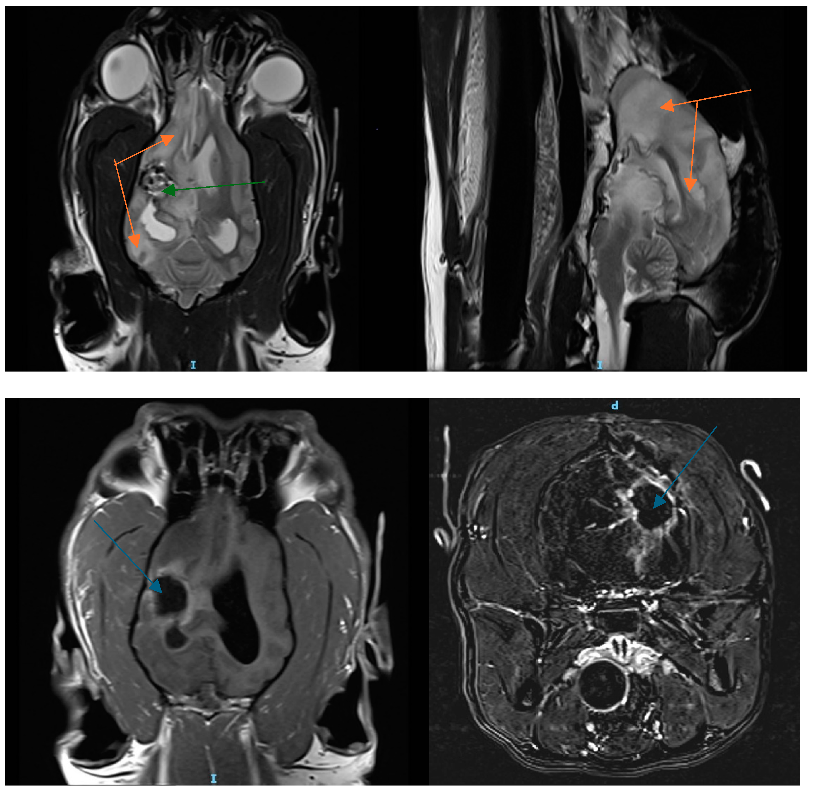
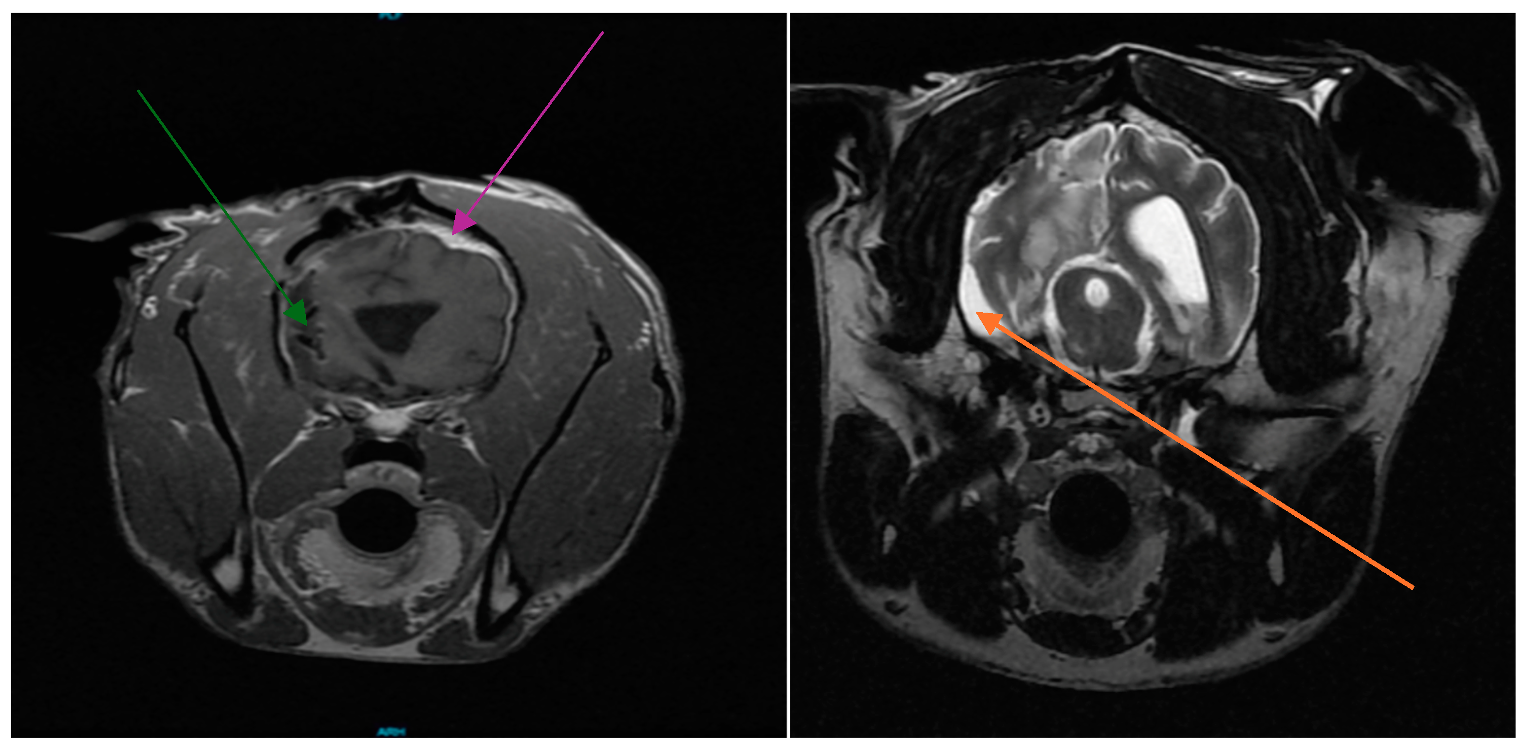
3.1. Patient History

A 10-year-old castrated male Australian shepherd was presented to the University of Minnesota Veterinary Medical Center (UMN VMC) in July 2023 to address a suspected right temporal lobe glioma that was diagnosed via MRI at a local neurology practice, following an acute onset of generalized seizures. The dog was subsequently enrolled in a clinical trial currently underway at the UMN VMC, consisting of surgical resection followed by the intradermal administration of autologous tumor lysate vaccines plus an immune checkpoint inhibitor against the CD200 checkpoint. The resected tumor was submitted to a board-certified veterinary pathologist with expertise in canine brain tumor histopathology and was confirmed as a high-grade oligodendroglioma. An immediate post-operative MRI confirmed gross total resection. As part of the immunotherapy treatment, the dog received weekly injections for three weeks, then monthly until a recheck MRI was performed at four months post operation.
This rechecks MRI confirmed substantial tumor recurrence in the right temporal lobe within the surgical cavity, presenting as an enhancing caudo-dorsal mass on T1 FLAIR imaging. The mass localized near the lateral ventricle displayed posterior–medial thalamic thickening (Figure 1). The dog was prescribed an anti-inflammatory dose of steroids, and his phenobarbital dosage was increased to 4 mg/kg BID in combination with levetiracetam (68 mg/kg BID) and zonisamide (10 mg/kg BID), as his seizures were not well-controlled. He was then referred to Purdue University for Cesium brachytherapy as part of a clinical trial (IACUC 0123002343), where the neurology service received him on 27 February 2024.
During the physical examination, hemiparesis was noted on his left side, along with a strong left-sided proprioceptive deficit, a moderate proprioceptive deficit on the right side, and an absence of menace response and visual deficit on the left side. The dog tended to circle to the right while walking. Blood work revealed that his phenobarbital level was within the normal range, but there was a moderate increase in liver values (ALT and ALP).

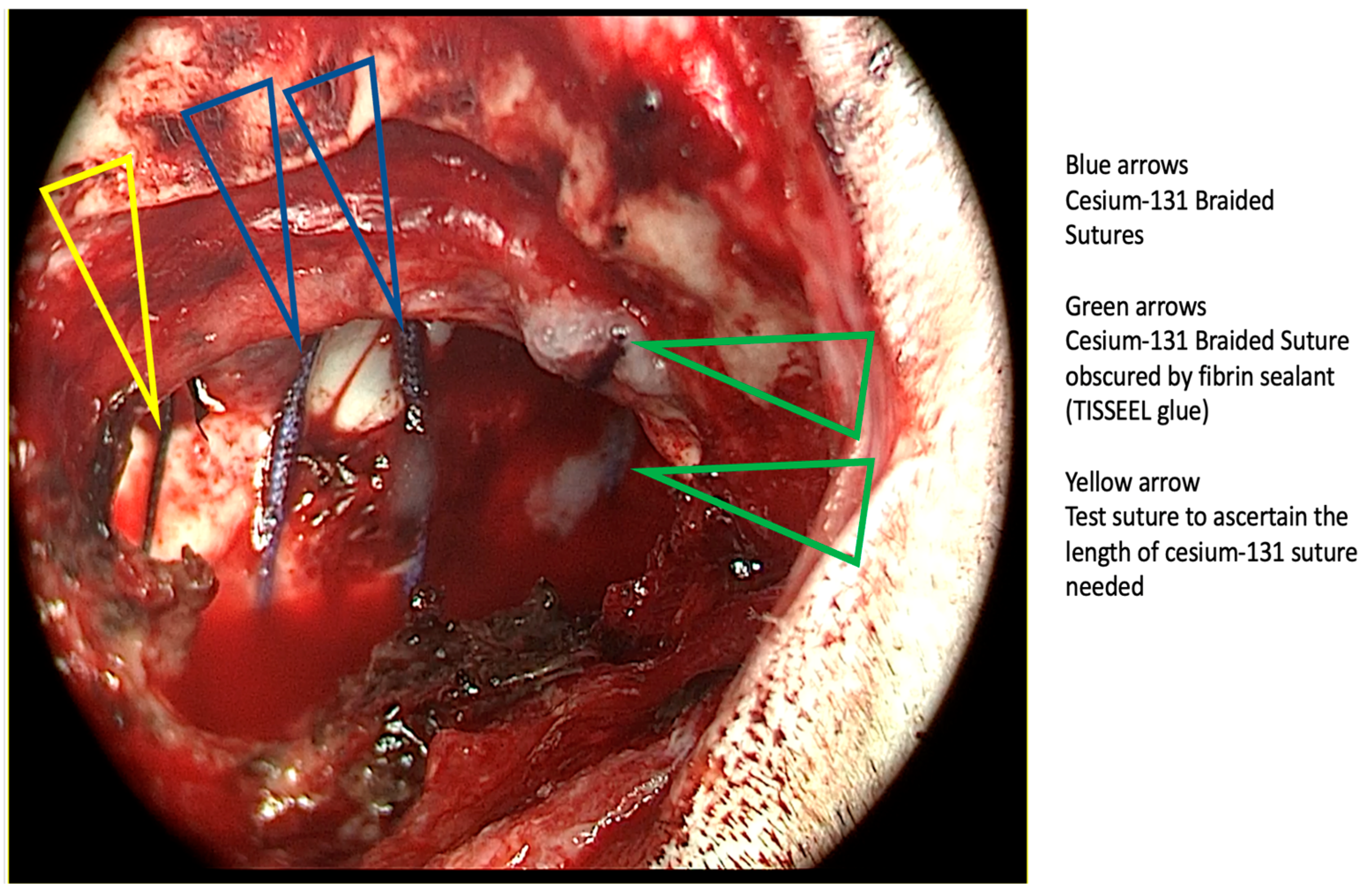
3.2. Cesium Brachytherapy

A second craniotomy was performed, during which a right parietal bone flap was removed rostromedially to the right lateral ventricle tumor, and a 4 × 3 cm defect was created to remove the visible parts of the tumor. A small cerebrospinal fluid leakage was noted, and the remaining cavity was implanted with 20 Cesium-131 radioactive seeds (2.4 U, 3.80 mCi), with a prescription dose of 70 Gy at 5 mm beyond the cavity. Pre-implantation dosimetry plans, realized by the radiation oncologist and the physicist (Figure 2), provided an estimate of the number of seeds necessary. A template grid supplied by the company confirmed the forecast based on implantation volume, total dose, and individual seed activity.
The authors referenced Cornell University methodology for Cesium seed positioning [12]. During the implantation, the radiation oncologist (IV) and physicist (AG) guided the neurosurgeon in placing parallel tracks of the braided suture with Cesium-131 seeds (0.7 cm to 1 cm apart), starting at the level of the tumor recurrence (Figure 3). Each track corresponded to an inserted polyglactin braided suture with seeds spaced longitudinally, center to center, at 1 cm intervals. The surgeons (TB, JK) used dummy silk sutures on each track before implantation to determine the appropriate polyglactin length to cut. Seeds were securely affixed using fibrin glue Tisseel ® and Surgicel® to prevent cavity collapse and ensure optimal seed positioning as the seeds decayed over time. The flap was securely fastened using bone screws, providing stability through layers of platysma, subcutaneous tissue, and skin closure (Figure 4). Of the twenty-four seeds ordered, four were retrieved post-procedure. They were returned to the radiation safety officers, who conducted surveys of the staff and the operating room for radiation safety clearance before the patient was transferred to CT and ICU. The CT allowed for post-implantation dose verification. Since the patient had been under anesthesia for almost six hours, clinicians opted for a CT scan recheck over an MRI to reduce time under anesthesia.
Postoperative care included monitoring for seizures, fluid therapy, pain management, precautionary measures to prevent aspiration pneumonia, and rehabilitation nursing. This encompassed assistance with elimination needs and preliminary walking after 24 h. The recovery proceeded smoothly without complications or seizures (Figure 5). Radiation safety officers granted hospital clearance after 48 h, as the patient’s emission dose at one meter remained below 1.2 mrem/h. The patient was subsequently discharged four days post procedure.

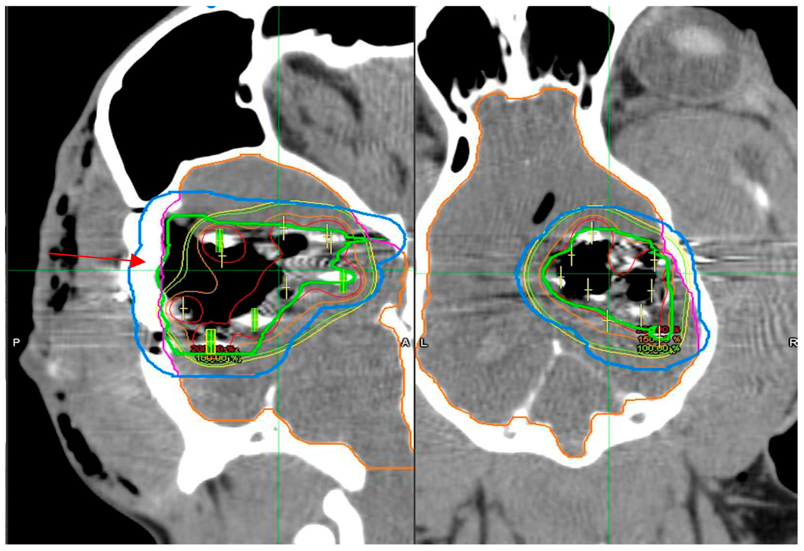
3.3. Dosimetry Verification
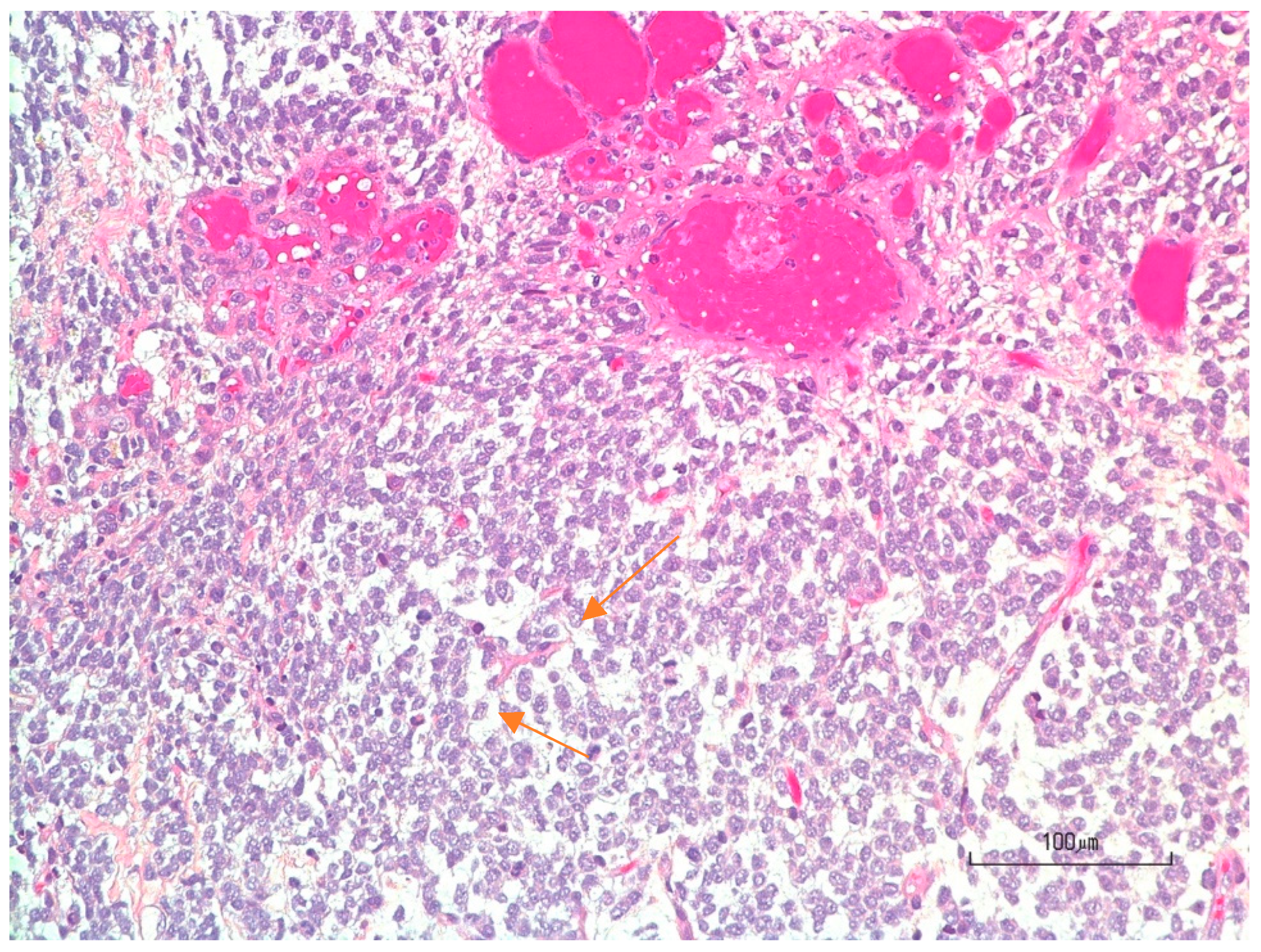
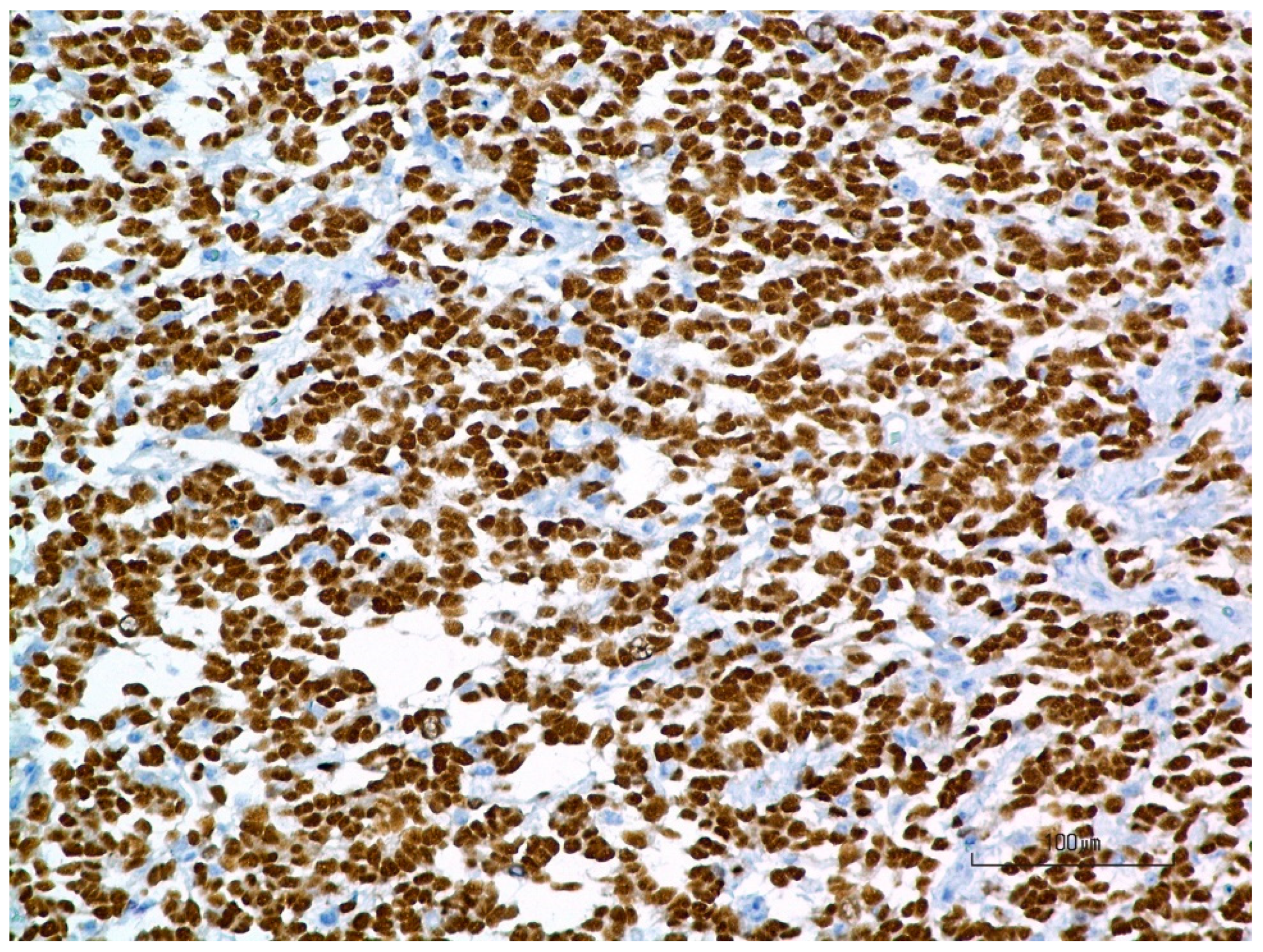
We verified the position of seeds and screws with a CT scan (Figure 6). Dose distribution analysis was conducted using the MIM Symphony Software treatment plan. The tumor coverage within the cavity and the 5 mm post-cavity region was generally satisfactory, except for insufficient coverage at the flap area, leading to underdosing at the bone level (Figure 7 red arrow).

3.4. Pathology

The Purdue University pathologist’s report indicated a high-grade oligodendroglioma with a high mitotic index and positive Olig2 immunostaining (Figure 8 and Figure 9).

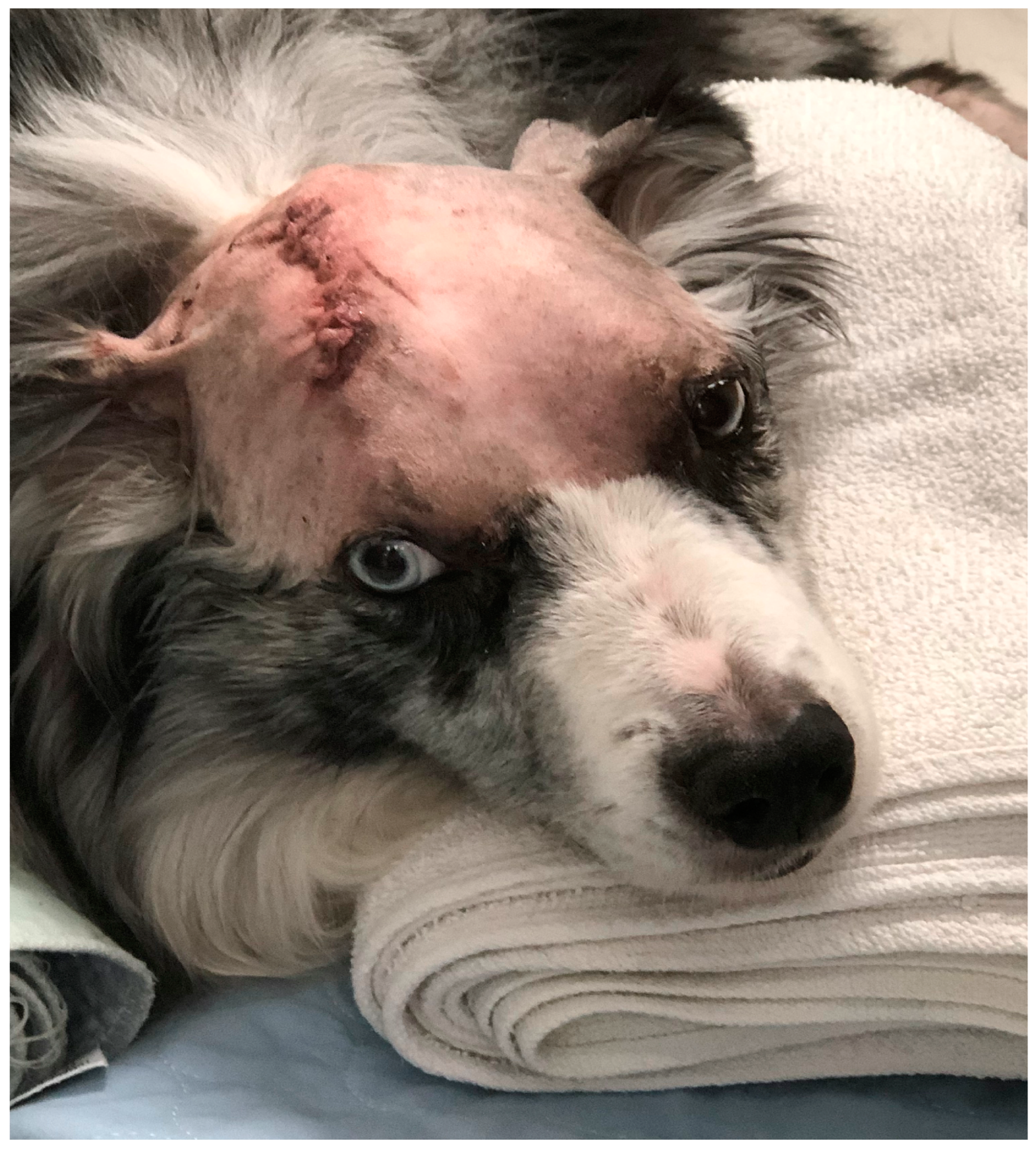
3.5. Patient Follow-Up

The patient returned to Minnesota under the care of the University of Minnesota neurology team (SA). The phenobarbital dose was decreased to 4 mg/kg BID, and steroids were prescribed for three weeks at a dosage of 0.5 mg/kg with a progressive reduction over two weeks. He had no seizures for six weeks and began to regain his ability to walk under the care of a rehabilitation and physical therapy service.
In April 2024, the patient was presented to Purdue University for his first clinical trial recheck, with the only complaint being intermittent vomiting. The steroids prescribed post-surgery had been tapered off. The patient made daily progress in recovering to a regular walk, had normal mentation, and experienced no seizures. Therefore, the Purdue University neurology team decided to reduce the level of anti-seizure drugs after six weeks. After an attempt to taper off phenobarbital, the patient experienced focal seizures and was returned to the 4 mg/kg BID dosage of phenobarbital.
Ten weeks after surgery, the owners sent a video to the medical teams showing the patient could run in a park. However, the patient was still leaning to the right side.
Twelve weeks after the procedure, the owners reported that the dog was experiencing balance deficits. The patient had been weaned off steroids. He was presented to the University of Minnesota for a neurology evaluation. The clinical exam revealed ataxia, left-sided postural reaction deficits, and propulsive circling in rightward circles. An MRI exam revealed the presence of severe generalized brain edema, likely vasogenic in nature. The surgical cavity was contrast-enhanced, but tumor recurrence could not be identified (Figure 10 bottom level). The radiologist and neurologist favored an inflammatory response to radiation. The patient was placed back on steroids at 0.5 mg/kg and showed improvement after ten days. After eight weeks, the steroids were tapered off, and the patient’s walking began to deteriorate, showing difficulties with his balance. The patient was placed back on 0.5 mg/kg of steroids. The patient improved after four days and was rechecked by the Purdue neurology team. His neurological exam was consistent with a right forebrain ablation: mentation was quiet, alert, and responsive. Left cranial nerve deficits (visual menace) and left proprioceptive deficits (anterior and posterior) were noted. The patient was ambulatory, walking in the hospital with a slight lean to the right side.
The patient was enrolled for his six-month MRI recheck at Purdue University. On his scan (Figure 11) we observed the missing part of his right frontal lobe, a shift with cerebrum parenchyma distortion, and cerebral fluid shifted toward the skull. There was moderate parenchymal and surgical cavity enhancement and strong meningeal enhancement, but the extent of edema was improved compared to the last MRI. No apparent tumor mass was observed. The conclusion was the persistence of inflammation post-surgery and radiation, and the patient remained dependent on anti-inflammatory steroids at this stage to maintain his quality of life.

4. Discussion

With six months of follow-up, this patient represents the first attempt to demonstrate the feasibility of Cesium-131 brachytherapy in the veterinary field. We acknowledge two limits of the present study: (1) additional patients are required to allow us to better assess the technique and optimize dose selection and seed activity based on the volume, pathology, and location of the tumor and (2) a longer follow-up would also allow for a better assessment of the potential risks associated with this technique. In this specific case, the patient exhibited inflammatory changes post-intervention, though we could not determine whether they were due to radiation, surgical intervention, or microscopic tumor persistence. Acute radiation injury typically presents as edema caused by increased blood–brain barrier permeability. These side effects can usually be managed with steroids or anti-angiogenic therapy. In contrast, late-stage radiation damage is characterized by vascular thrombosis, hyalinization, fibrosis, glial cell depletion, and neuronal rarefaction—collectively known as radiation necrosis—which is often poorly responsive to treatment [13]. To mitigate these adverse effects, future studies should aim to limit radiation dose hot-spots within the brain by staying below thresholds linked to late-stage radiation damage. This could be accomplished using treatment planning software in conjunction with follow-up dose verification using imaging. In addition, similar to human treatments, close collaboration between physicists, oncologists, and the surgical team is necessary to ensure that the brachytherapy is positioned in the correct locations. An additional discussion of human experiences related to mitigating adverse side-effects of brachytherapy is outlined in the subsequent sections.

4.1. Future Considerations for Veterinary Brain Brachytherapy with Cesium-131

Future studies should aim to enroll canine patients in a pilot study to compare brachytherapy to historical data using an external beam (fractionated and hypo-fractionated protocols with stereotactic techniques). Although there would be potential bias comparing outcomes with historical external beam data, this type of study would provide an initial indication of the potential benefit of a brachytherapy approach. An additional MRI scan (one MRI sequence) immediately after the surgery could be helpful for checking for complete tumor resection, if the total anesthesia time allows. Also, there is the consideration of needing to switch the current technique with braided sutures for tiles (GammaTile™) during implantation. This is expected to provide better radiation techniques and reduce anesthesia time for the patient. The Cesium-131 brain brachytherapy technique could offer promise in the veterinary world, overcoming the global issue of the limited number of veterinary radiation facilities equipped with a linear accelerator (<100, according to the Veterinary Oncology Society website). Moreover, based on the authors’ experience, dogs are often referred numerous weeks after surgery for external beam therapy, allowing cancer cell repopulation. The delivery of intraoperative radiation could result in a therapeutic advantage as there is no time delay for the adjuvant treatment.

4.2. The Human Experience in Neuro-Brachytherapy with Cesium-131: What Can We Learn as Veterinarians from Their Experience

Neurosurgeons have over a decade of experience treating patients in the OR with Cesium-131. The following sections describe these prior experiences as historical background.
Treatment of brain metastasis with Cesium-131 seeds on braided polyglactin suture: In 2014, the Cornell Team led by AG. Wernicke and T. Schwartz reported the usage of the radioisotope CS-131 in a cohort of 24 patients immediately following the surgical resection of a brain metastasis [12]. The primary origins were lung, kidney, melanoma, colon, and cervix. On average, twelve seeds were directly implanted, with a median cavity size of 10.3 ccs pre-implantation and a dose prescription of 80 Gy at a 5 mm depth. The median patient follow-up was 19.3 months. Locally, there was no recurrence; however, one patient experienced regional recurrence (leptomeningeal), and 48.4% of patients developed distant metastases.
The authors highlighted several advantages of the technique: no treatment time delay (the doubling time of glioblastoma is 17 days) and allowing for direct irradiation during surgery. Tumor repopulation becomes a concern for four weeks after surgery, and patients undergoing external beam radiation therapy may experience delays beyond this timeframe, compromising the chance for local tumor control. No cases of radiation necrosis were observed, unlike with the GliaSite technique or Iodine-125 seeds. The only complications observed were one case of seizure and a post-operative infection [12,13,14]. Compared to whole-brain radiation therapy (WBT), patients did not experience alterations in their quality of life and maintained their cognitive function. Another consideration is the moderate cost of this technology in comparison to similar treatment with radiosurgery techniques [15].
In 2023, the Cornell University team released findings on the most significant retrospective series treated with intraoperative brachytherapy using Cesium-131 from 2010 to 2021, encompassing 119 patients [16]. The local control rate for brain metastasis reached 85%, with auspicious outcomes observed in non-small cell lung tumors (NSCLCs). Notably, when brachytherapy was administered during the initial surgery, the local control rate surged to 91%, an improvement compared to the 64% rate for re-intervention cases. This 91% local control rate for newly diagnosed patients surpasses rates achieved with post-surgery stereotactic radiosurgery (SRS), typically ranging from 72% to 87%.
Larger tumor size did not emerge as a statistically significant prognostic variable for brain metastasis. Complications were relatively rare, with less than 8% experiencing radiation necrosis at a median time of 13.9 months post treatment. This low incidence of radiation necrosis contrasts with rates associated with I125 (16–40%) and SRS (5–17%). Radiation necrosis was more common in cases with smaller initial tumor diameters, potentially attributed to the close proximity of seeds within a confined space. Surgical complications, including infection and wound breakdown, were observed in 12% of cases. Additionally, new neurological deficits or strokes occurred in 9% of patients, with seizures documented in 3%.
Expanding their series, the team began including recurrent high-grade gliomas and meningiomas. One-year local control rates were 83% for meningiomas (primarily Grade II and III, with 100% local control for Grade I and II) and 34% for recurrent gliomas. The median survival for gliomas in this series stood at 10.8 months.
Introduction to collagen tiles with embedded seeds—GammaTile™: The GammaTile™ option, named Surgically Targeted Radiation Therapy (STaRT), represents significant progress in bioengineering. The GammaTile™ inventors, Dr. Brachman and colleagues, focused on simplifying and standardizing brain radionuclide seeds implantation. They designed and patented 2 cm diameter collagen-embedded tiles, each containing 4 Cesium-131 seeds with a spacing of 1 cm. The FDA approved this device in July 2018 for treating recurrent tumors such as brain metastases, meningiomas, and gliomas.
The University of Minnesota has released a first report on 23 patients treated with GammaTile™ following FDA approval [17]. The study outlines a comprehensive procedure for brain implantation; covering prescription recommendation; dose verification with treatment plan software; and safety considerations for patients, staff, and the public. When ordering tiles, the team selects tile numbers based on pre-MRI surgery assessments, calculating the tumor area relative to tile dimensions. To capture dynamic changes in the cavity, they recommend conducting MRI no later than three weeks before surgery. Dosimetry verification occurs 24–48 h post-surgery using CT or MRI scans with a slice thickness of 1 mm. Variseed™ dosimetry software 7.1 from Varian™ assists with the procedure. The targeted tumor volume encompasses the cavity plus any residual suspected area enlarged by 5 mm. The prescription dose is 60 Gy, with a strength kerma of 3.5 U for each seed. Radiation safety criteria dictate emissions at a 1 m distance should be less than 6 mRem/h. The highest recorded emission on a patient was 3.2 mRem/h. The clinical outcomes of using Cesium-131 radiation during intra-operative procedures over a decade after its initial use have shown promising results.
Studies and reports indicate several benefits of this modality. (1) Time efficiency: Neurosurgeons can cover the tumor cavity with pre-ordered titanium collagen embedded tiles in just a 5 min procedure following tumor ablation, demonstrating significant time savings compared to traditional methods. (2) Reduced Risk: The collagen carrier used in the procedure prevents direct contact between the radioactive seeds and brain tissue, thereby reducing the risk of hot spots and brain necrosis. (3) FDA Approval: In January 2020, the FDA granted an extension for treating newly diagnosed tumors, indicating confidence in the safety and efficacy of this approach. (4) GT medical technologies, the company behind the treatment, reported the significant adoption of the technique. As of November 2020, 535 neurosurgery centers provided this treatment, highlighting its growing acceptance within the medical community [18].
One key driver behind the adoption of this technique is its ability to address the disparity in access to radiosurgery centers, particularly in remote rural areas. Traditional stereotactic radiation therapy can require patients to travel hundreds of miles, posing challenges such as job absences and the need for travel assistance. The brachytherapy technique using tiles offers a more accessible option for patients in these regions.
Furthermore, the lower upfront investment required for establishing brachytherapy practices compared to traditional external beam ration therapy and radiosurgery centers makes it a more feasible option for many neurosurgery centers. This is because it does not require expensive infrastructure like bunkers or specialized machinery and software, and it is not as technically challenging for neurosurgeons trained in craniotomy.
Overall, the adoption of Cesium-131 radiation during intra-operative procedures has led to improved access to radiation therapy, streamlined procedures, and promising clinical outcomes for patients with brain tumors.
GammaTile™ Outcomes: When analyzing the outcomes associated with the GammaTile™ implementation, it is essential to differentiate the results based on the primary pathology. For brain metastasis treatment, Brachman et al. [19] report on the advantages of STaRT for large recurrent or newly diagnosed brain metastases compared to standard techniques. In their initial study, they examined 11 patients with 16 metastases. Seven originated from breast, six from lungs, and three from sarcoma, with a prevalence of female patients over males (64%). The average diameter of the pre-operative cavity was 3.1 cm. Twelve lesions were recurrent, heavily pretreated brain metastasis with an average of 1.5 prior surgeries and 1.5 pre-radiation treatments (EBRT). Their average time to recurrence was 4.8 months. Four lesions had no previous treatment history. The dose prescription was 60 Gy at 5 mm for the tumor-targeted volume. Considering the recurrence rate for large cavity metastases (e.g., >2–3 cm) of 40–60% with surgery alone and 20% or more with adjuvant radiation [20,21,22], they highlight favorable outcomes with a local control rate of 100% for de novo metastases and 80% for recurrent metastases. Minimal complications were observed, with only two patients requiring dexamethasone for 2–4 weeks due to brain lesions; these patients had prior radiation therapy before STaRT.
The same group analyzed outcomes for 20 patients with recurrent meningioma who had previously undergone irradiation: a series from 2013 to the end of 2016, published in 2019 [23]. These patients underwent an average of two surgeries and received one round of radiation before brachytherapy implantation. The median dimension of the treated cavity was 11.3 cc. Among the patients, 20% had Grade 1 tumors, 70% had Grade 2 tumors, and 10% had Grade 3 tumors; additionally, 10% underwent subtotal tumor resection. This technique was considered potentially less risky and more effective than classical treatments for recurrent resection and external beam radiation therapy (EBRT). The median prescription dose was 63 Gy at 5 mm from the resected cavity, with an average implantation time of 6 min and using 22 seeds on average.
The median for progression-free survival interval with brachytherapy at 15.4 months was not reached in the brachytherapy series. In comparison, 50% of patients undergoing prior therapy (surgery and external beam radiation) had progression at 18.3 months. Of the two patients who experienced early local recurrence after receiving brachytherapy, both had subtotal tumor resection with Grade 2 and Grade 3 meningiomas. The overall survival for patients with brachytherapy was 26 months, with a radiation necrosis rate of 10%. One limitation of the study is the absence of a control group; however, the authors compared it favorably to historical research using external beam radiation therapy for recurrent meningioma, which reported a median progression-free interval of 8 months and a one-year survival rate of 17% [24].
Glesser et al. from the University of Minnesota described the initial series of IDH wild-type recurrent glioblastoma treated with GammaTile™ [25]. Among the fourteen patients included, eight were experiencing their second recurrence, and six were at their third recurrence. To mitigate radiation toxicity, patients underwent a minimum 6-month delay after initial radiation–temozolomide treatment. Safety outcomes revealed one minor complication involving CSF leakage in a patient and one bleeding resulting in a fatality in a patient being treated for an ischemic leg while on anticoagulants. The latter complication was determined to be unrelated to the surgical technique. Notably, no instances of radiation necrosis were observed as a secondary complication to radiation therapy. The prescribed dose was targeted to the cavity, with two patients undergoing subtotal resection. A dose of 60 Gy was prescribed with a 5 mm cavity expansion. The average cavity dimension was 22.18 cc.
The one-year disease-free interval reached 81%, while overall survival varied based on the methylated versus unmethylated status of 6-O-6 Methylguanine-DNA transferase (MGMT) status, which reflects the capacity to repair DNA damage associated with alkyl agents such as temozolomide. Overall survival was 20 months for unmethylated status and 37.4 months for methylated status. The authors validated this technique to escalate radiation dose for recurrent patients safely and demonstrated its potential benefits on survival, acknowledging the limitations of a small series and preliminary data.
In 2021, the University of Indianapolis described a small series of seven patients treated with STaRT for recurrent glioma [26]. The prescription was 60 Gy at 5 mm beyond the resection cavity, with an average prescribed volume (PTV) of 48.4 cc. The authors utilized MIM treatment plan software to verify post-implantation dosimetry. Different pathologies were reported: three cases of GBM, two of anaplastic astrocytoma, one of Grade 2 oligodendroglioma, and one of anaplastic oligodendroglioma. Gross resection was achieved in four patients, while subtotal resection occurred in three. Only two complications were observed: one patient required an ICU stay due to cerebral edema, which was present before surgery, and this patient had undergone subtotal resection. The second patient experienced aphasia and weakness expected based on the tumor location and required treatment for cerebral edema. The authors only reported on the procedure’s feasibility and safety data. No significant complications were noted. The study’s limitations lie in the small sample size and the diversity of pathology.
Brachman et al. [27] reported their initial investigation on recurrent glioblastoma from 2013 to 2018 through a prospective single-arm trial (NCT#0308857) involving 28 patients, with 20 patients experiencing the first recurrence. The prescribed radiation dosage ranged between 60 and 80 Gy at a 5 mm distance to the cavity. Among these patients, 17 received chemotherapy alone or in combination, with temozolomide administered to 12, lomustine to 8, and bevacizumab to 15 individuals. Radiological assessments revealed a local control duration of 8.5 months after the first recurrence. The overall survival rate was 15.1 months for patients receiving combination therapy, including the oncological recycling of chemotherapy at minimum intervals. This study highlights the potential efficacy of the treatment of recurrent glioblastoma. Consequently, following this study, the FDA approved de novo or recurrent intracranial neoplasm therapy with this technique.
In summary, brachytherapy with Cesium-131 for human patients has become standard for adjuvant intraoperative radiation therapy for brain metastases. With recent advancements in bioengineering of GammaTiles, this technique has also gained recognition as a second- or third-line treatment option for recurrent gliomas or meningiomas. It is worth noting that brachytherapy with Cesium-131 is currently not the first choice as a first-line radiation treatment following surgery for aggressive meningiomas and gliomas. Experts suggest that further studies should be conducted to explore the potential of Cesium-131 as primary radiation or neoadjuvant therapy, either alone or in combination with chemotherapy or immunotherapy for primary brain neoplasia. Neuro-oncologists need to address this gap in knowledge. Utilizing Cesium-131 in small animal companions affected by gliomas as a preclinical model could provide valuable insights for human colleagues regarding its efficacy as a first-line therapy during surgery or in combination with other treatments such as chemotherapy or immunotherapy. Collaboration between neurologists, radiation oncologists, and veterinarians for preclinical studies in brain tumors has become a reality due to NCI programs.

4.3. Opportunity for Translational Research (NCI Brain Comparative)

Preclinical data for Cesium-131 as primary radiation treatment post-glioma resection could yield valuable insights for human neurologists. This technique, not commonly used as a first-line radiation procedure in the human world, holds promise for glioblastoma patients. Dogs represent a promising avenue for translational research in radiation oncology [28]. They develop spontaneous diseases that resemble those found in human patients and possess robust immune systems. Recognizing the potential, the National Cancer Institute (NCI) has identified certain canine cancers as valuable preclinical study models, supporting research through initiatives like the Comparative Oncology Program (COP) and the Preclinical Cancer Immunotherapy Network for Canine Trials (PRECINCT). Notably, dog gliomas are included in this list. Introducing brachytherapy veterinary platforms could significantly enhance our understanding of fundamental research questions related to this radiation technique. Moreover, it could result in the opportunity to gather preclinical data on combination therapies, ultimately saving time and substantial financial resources before advancing to clinical trials.

4.4. Advancing Understanding Through Fundamental Research

The Radiation Spatial Dose Heterogeneity Impact: The inherent spatial dose distribution heterogeneity associated with brachytherapy LDR plans underscores the need for comparative studies. Such investigations could be conducted using preclinical models, starting with syngeneic rodents harboring orthotopic tumors. Serial biopsies and advanced imaging techniques could then be employed to monitor the impact on the tumor microenvironment. Furthermore, studies enrolling companion animals could provide valuable insights into the effects of dose heterogeneity associated with brachytherapy plans and the patient’s immune response or changes in the tumor microenvironment [29]. Dose heterogeneity is considered a factor modulating the immune response for other radiation modalities [30,31].
Overcoming Local Tumor Recurrence with Photodynamic Therapy: Despite advancements in neurosurgical techniques, such as intraoperative MRI guidance, neuro-navigation, and intraoperative fluorescent dye usage, local tumor relapse remains a significant challenge, particularly in patients with glioblastoma. Combining radiation therapy with hyperthermia, such as LITT, and other modalities like laser therapy (PDT) presents an opportunity to enhance local tumor control [32,33]. However, there is a notable absence of ongoing trials combining these techniques with Cesium implantation. Utilizing canine models could offer insights into treatment efficacy, immune responses, and toxicity profiles, thus streamlining the transition to human clinical trials and optimizing therapeutic outcomes.
Clinical Research on Combined Therapies with Drugs: Current treatment approaches for glioblastoma with Cesium-131 often involve intraoperative radiation therapy, followed by adjuvant systemic therapies like temozolomide or lomustine upon recurrence. Companion animal models hold promise for investigating combined therapeutic approaches, including nanoparticle injections, targeted therapies, vaccines, and immunotherapy. Moreover, combining therapies during surgery within the resected cavity presents an attractive synergistic potential facilitated by advancements in intra-tumoral drug delivery techniques [34]. Veterinary research leveraging techniques such as stereotactic injections and convention-enhanced delivery offers a fertile ground for exploring innovative treatments, including cytokine therapy and immunotherapy [35,36]. Veterinary neurosurgeons, familiar with intra-brain parenchymal delivery techniques, are well-positioned to contribute to translational research efforts. Furthermore, the availability of IDO inhibitors and TLR [37,38,39] agonists in the veterinary domain underscore the potential for cross-species collaboration and knowledge exchange. The USDA approval of the first canine anti-PD1 antibody, Gilvetmab, highlights the translational potential of veterinary research in advancing glioblastoma treatment strategies (https://www.merck-animal-health-usa.com/species/canine/products/gilvetmab-product-overview) (accessed on 25 September 2024). By harnessing the resources and expertise within the veterinary community, preclinical investigations into combined therapies with Cesium-131 brachytherapy can be effectively advanced, offering new avenues for improving patient outcomes in glioblastoma management.

5. Conclusions

Cesium-131, coupled with collagen tiles, has emerged as a prominent therapy for treating brain metastasis and recurrent primary brain tumors. Many reputable US hospitals, known for their expertise in neurosurgery, offer this technology due to its safety, efficacy, and minimal risk of brain damage associated with radiation. However, this technique is not typically used for treating primary aggressive tumors like glioblastoma. The authors report on the initial steps taken to introduce this technique to the veterinary field and its application in a dog suffering from recurrent glioblastoma with a six-month follow-up. This patient maintained a decent quality of life six months after the recurrence of a high-grade glioma. If further studies are needed to refine dose prescriptions and to conduct extended follow-ups to assess tumor control and the long-term effects of radiation, the Cesium-131 technique could be promising in the veterinary world for clinical application due to its relatively low initial investment cost compared to external beam radiation. Moreover, veterinary teams could be involved in research to address questions relevant to human patients, ranging from its efficacy as a first-line treatment for glioma to its potential in combination therapies such as laser therapy, hyperthermia, local chemotherapy, or immunotherapy. The NCI has underscored the benefits of including dogs with similar cancers to humans in preclinical trials, highlighting the potential for advancements in veterinary and human medicine.

Author Contributions

Conceptualization, I.F.V., R.T.B. and M.L.S.; methodology, I.F.V. and A.G.; validation, I.F.V., R.T.B., M.L.S., S.A., A.G., N.C. and J.A.Y.; formal analysis, I.F.V., A.G. and M.L.S.; investigation, I.F.V. and R.T.B.; resources, L.D.; data curation, I.F.V., R.T.B., A.G., J.B.K., S.A. and L.D.; writing—original draft preparation, I.F.V.; writing—review and editing, I.F.V., R.T.B. and M.L.S.; visualization, I.F.V. and R.T.B.; supervision, I.F.V. and M.L.S.; project administration, I.F.V., R.T.B. and M.L.S.; funding acquisition, I.F.V. All authors have read and agreed to the published version of the manuscript.

Funding

This research received no external funding.

Institutional Review Board Statement

The study was conducted in accordance with the Declaration of Helsinki, and approved by the Institutional Review Board of Purdue Veterinary Hospital (protocol code IAUCUC 0123002343).

Data Availability Statement

The raw data supporting the conclusions of this article will be made available by the authors on request.

Acknowledgments

The authors thank Ed Rappel and Troy Sienko (Isoray, now GT Medical Technologies) for their support to the project; Emily Molinich, Julie Cortino, Kyle Kiffner, and MIM software company) for their support of the project; and P. Devlin, Dana Farber Institute, for the guidance regarding the prescription for our patient Finn.

Conflicts of Interest

The authors declare no conflicts of interest.

References

  1. Moore, M.P.; Bagley, R.S.; Harrington, M.L.; Gavin, P.R. Intracranial tumors. Vet. Clin. N. Am. Small Anim. Pract. 1996, 26, 759–777. [Google Scholar] [CrossRef] [PubMed]
  2. Dobson, J.M.; Samuel, S.; Milstein, H.; Rogers, K.; Wood, J.L. Canine neoplasia in the UK; estimates of incidence rates from a population of insured dogs. J. Small Anim. Pract. 2002, 43, 240–246. [Google Scholar] [CrossRef] [PubMed]
  3. Bentley, R.T.; Ahmed, A.U.; Yanke, A.B.; Cohen-Gadol, A.A.; Dey, M. Dogs are man’s best friend: In sickness and in Health. Neuro-Oncology 2017, 19, 312–322. [Google Scholar] [CrossRef] [PubMed]
  4. Hicks, J.; Platt, S.; Kent, M.; Haley, A. Canine brain tumours: A model for the human disease? Vet. Comp. Oncol. 2017, 15, 252–272. [Google Scholar] [CrossRef]
  5. Hu, H.; Barker, A.; Harcourt-Brown, T.; Jeffery, N. Systematic review of brain tumor treatment in dogs. J. Vet. Intern. Med. 2015, 29, 1456–1463. [Google Scholar] [CrossRef]
  6. Spugnini, E.P.; Thrall, D.E.; Price, G.S.; Sharp, N.J.; Munana, K.; Page, R.L. Primary Irradiation of Canine Intracranial Masses. Veter-Radiol. Ultrasound 2000, 41, 377–380. [Google Scholar] [CrossRef]
  7. Targeser, E.; Martin, T.T.; Burkedin, B.; Hart, C.; Leary, D.; Larue, S.; Boss, M.K. Efficacy of stereotactic radiation therapy for the treatment of confirmed presumed canine glioma. Vet. Com. Oncol. 2023, 21, 578–586. [Google Scholar] [CrossRef]
  8. Walker, M.A. Interstitial implant brachytherapy in small animals. Veter-Clin. North Am. Small Anim. Pract. 1997, 27, 59–71. [Google Scholar] [CrossRef]
  9. Klueter, S.; Krasteld, D.; Ludewig, E.; Reischauer, A.; Henichke, F.; Pohlmann, S. High-dose-rate brachy-therapy for intranasal tumours in dogs, results of a pilot study. Vet. Comp. Oncol. 2006, 4, 218. [Google Scholar] [CrossRef]
  10. Groothuis, D.R.; Wright, D.C.; Ostertag, C.B. The effect of 125I interstitial radiotherapy on blood-brain barrier function in normal canine brain. J. Neurosurg. 1987, 67, 895–902. [Google Scholar] [CrossRef]
  11. Stubbs, J.B.; Frankel, R.H.; Schultz, K.; Crocker, I.; Dillehay, D.; Olson, J.J. Pre-Clinical Evaluation of a Novel Device for Delivering Brachytherapy to the Margins of Resected Brain tumor cavities. J. Neurosurg. 2002, 96, 335–343. [Google Scholar] [CrossRef] [PubMed]
  12. Wernicke, A.G.; Yondorf, M.Z.; Peng, L.; Trichter, S.; Nedialova, L.; Sabbas, A.; Khulidzhanov, F.; Pararshar, B.; Nori, D.; Clifford Chao, K.S.; et al. Phase I/II study of resection and intraoperative cesium-131 radioisotope brachytherapy in patients with newly diagnosed brain metastases. J. Neurosurg. 2014, 121, 338–348. [Google Scholar] [CrossRef]
  13. Rahmathulla, G.; Marko, N.F.; Weil, R.J. Cerebral radiation necrosis: A review of the pathobiology, diagnosis and management considerations. J. Clin. Neurosci. 2013, 20, 485–502. [Google Scholar] [CrossRef]
  14. Wernicke, A.G.; Lazow, S.P.; Taube, S.; Yondorf, M.Z.; Kovanlikaya, I.; Nori, D.; Christos, P.; Boockvar, J.A.; Pannullo, S.; Stieg, P.E.; et al. Surgical Technique and Clinically Relevant Resection Cavity Dynamics Following Implantation of Cesium131(CS-131) Brachytherapy in patients with Brain Metastases. Oper. Neurosurg. 2016, 12, 49–60. [Google Scholar] [CrossRef] [PubMed]
  15. Wernicke, A.G.; Yondorf, M.Z.; Parashar, B.; Nori, D.; Chao, K.S.C.; Boockvar, J.A.; Pannullo, S.; Stieg, P.; Schwartz, T.H. The cost-effectiveness of surgical resection and cesium-131 intraoperative brachytherapy versus surgical resection and stereotactic radiosurgery in the treatment of metastatic brain tumors. J. Neuro-Oncol. 2016, 127, 145–153. [Google Scholar] [CrossRef] [PubMed]
  16. Bander, E.D.; Kelly, A.; Ma, X.; Christos, P.J.; Wernicke, A.G.; Stieg, P.E.; Trichter, S.; Knisely, J.P.; Ramakrishna, R.; Schwartz, T.H. Safety and efficacy of Cesium-131 brachytherapy for brain tumors. J. Neuro-Oncol. 2023, 163, 355–365. [Google Scholar] [CrossRef]
  17. Ferreira, C.; Sterling, D.; Reynolds, M.; Dusenbery, K.; Chen, C.; Alaei, P. First clinical implementation of Gammatile permanent brain implants after FDA clearance. Brachytherapy 2021, 20, 673–685. [Google Scholar] [CrossRef]
  18. Choi, M.; Martin, B. Gammatile Surgically Targeted Radiation Therapy Improving Access Care Without Compromising Treatment Outcomes; Clinical White Paper; Gammatile Company: Richland, WA, USA, 2020. [Google Scholar]
  19. Nakaji, P.; Smith, K.; Youssef, E.; Thomas, T.; Pinnaduwage, D.; Rogers, L.; Wallstrom, G.; Brachman, D. Resection and Surgically Targeted Radiation Therapy for the treatment of larger recurrent or Newly diag-nosed Brain Metastasis: Results from a prospective trial. Cureus 2020, 12, e11570. [Google Scholar] [CrossRef]
  20. Mahajan, A.; Ahmed, S.; Aleer, M.F.; Weinberg, J.S.; Li, J.; Brown, P.; Settle, S.; Prabhu, S.S.; Lang, F.F.; Levine, N.; et al. Post operative radiosurgery versus observation for completely resected brain metastasis; a single center, randomized, controlled, phase 3 trial. Lancet Oncol. 2017, 18, 1040–1048. [Google Scholar] [CrossRef]
  21. Ling, D.C.; Vargo, J.A.; Wegner, R.E.; Flickinger, J.C.; Burton, S.A.; Engh, J.; Amankulor, N.; Ozhasoglu, C.; Quinn, A.E.; Heron, D.E. Postoperative stereotactic radiosurgery to the resection cavity for large brain metastases; Clinical outcomes, predictors of intracranial failure, and implications for optimal patient selection. Neuroseurgery 2015, 76, 150–157. [Google Scholar] [CrossRef]
  22. Brown, P.D.; Balkman, K.V.; Cerhan, J.H.; Anderson, S.K.; Carrero, X.W.; Whitton, A.C.; Greenspoon, J.; Parney, I.F.; Laack, N.N.; Ashman, J.B.; et al. Postoperative stereotactic radiosurgery compared with whole brain radiotherapy for resected metastatic brain disease (NCCTG N107c/CEC.3) a multicentre, randomized controlled, phase 3 trial. Lancet Oncol. 2017, 18, 1049–1060. [Google Scholar] [CrossRef] [PubMed]
  23. Brachman, D.G.; Youssef, E.; Dardis, C.J.; Sanai, N.; Zabramski, J.; Smith, K.A.; Little, A.S.; Shetter, A.G.; McBride, H.L.; Sorensen, S.; et al. Resection and Permanent intracranial brachytherapy using modular, biocompatible cesium 131 implants results in 20 recurrent previously irradiated meningiomas. J. Neurosurg. 2019, 131, 1819–1828. [Google Scholar] [CrossRef] [PubMed]
  24. Wojcieszynski, A.P.; Ohri, N.; Andrews, D.W.; Evans, J.J.; Dicker, A.P.; Werner, M. Wasik reirradiation of re-current meningioma. J. Clin. Neurosci. 2012, 19, 1261–1264. [Google Scholar] [CrossRef] [PubMed]
  25. Gessler, D.G.; Neil, E.C.; Shah, R.; Levine, J.; Shanks, J.; Wilke, C.; Reynolds, M.; Zhang, S.; Ozutemiz, C.; Genturk, M.; et al. GammatiIe TM brachytherapy in the treat-ment of recurrent glioblastomas. Neuro-Oncol. Adv. 2022, 4, vdab185. [Google Scholar] [CrossRef]
  26. Budnick, H.C.; Richardson, A.M.; Shiue, K.; Watson, G.; Ng, S.K.; Le, Y.; Shah, M.V. Gamma Tile for Gliomas: A Single-Center case series. Cureus 2021, 13, e19390. [Google Scholar] [CrossRef]
  27. Nakaiji, P.; Brachman, D.; Smith, P.K.; Thomas, T.; Daris, C.; Pinnaduwage, D.; Wallsrrom, G.; Rogers, C.L.; Youssef, E.F. Resection and surgically targeted radiation therapy for locally recurrent GBM. J. Clin. Oncol. 2021, 39, 2054. [Google Scholar] [CrossRef]
  28. Vanhaezebrouck, I.F.; Scarpelli, M.L. Companion Animal as the key to success translating radiation. Cancers 2023, 15, 3377. [Google Scholar] [CrossRef]
  29. Parel, R.B.; Baniel, C.C.; Sriramaneni, R.N.; Bradley, K.; Markovina, S.; Morris, Z.S. Combining brachytherapy and immunotherapy to achieve in situ tumor vaccination; A review of cooperative mechanisms and clinical opportunities. Brachytherapy 2018, 17, 995–1003. [Google Scholar] [CrossRef]
  30. Lukas, L.; Zhang, H.; Cheng, K. A Epstein Immune Priming with spatially fractionated Radiation therapy. Curr. Oncol. Rep. 2023, 12, 1483–1496. [Google Scholar] [CrossRef]
  31. Cho, Y.B.; Yoon, N.; Suh, J.H.; Scott, J.G. Radio-immune response modeling for spatially fractionated radiotherapy. Phys. Med. Biol. 2023, 68, 165010. [Google Scholar] [CrossRef]
  32. Cramer, S.W.; Chen, C.C. Photodynamic therapy for treating Glioblastoma. Front. Surg. 2020, 6, 81. [Google Scholar] [CrossRef] [PubMed]
  33. Nakahara, Y.; Ito, H.; Masuoka, J.; Abe, T. Boron Neutron Capture Therapy and Photodynamic Therapy for High-grade Meningiomas. Cancers 2020, 12, 1334. [Google Scholar] [CrossRef] [PubMed]
  34. Van Solinge, T.S.; Nieland, L.; Chiocca, E.A.; Broekman, M.L. Advances in local therapy for glioblastoma—Taking the fight to the tumour. Nat. Rev. Neurol. 2022, 18, 221–236. [Google Scholar] [CrossRef] [PubMed]
  35. Rossmeisl, J.H.; Herpai, D.; Quigley, M.; Cecere, T.E.; Robertson, J.L.; D’Agostino, R.B.; Hinckley, J.; Tatter, S.B.; Dickinson, P.J.; Debinski, W. Phase I trial of convection-enhanced delivery of IL13RA2 and EPHA2 receptor targeted cytotoxins in dogs with spontaneous intracranial gliomas. Neuro Oncol. 2021, 23, 422–434. [Google Scholar] [CrossRef] [PubMed] [PubMed Central]
  36. Rosenbluth, K.H.; Martin, A.J.; Mittermeyer, S.; Eschermann, J.; Dickinson, P.J.; Bankiewicz, K.S. Rapid inverse planning for pressure-driven drug infusions in the brain. PLoS ONE 2013, 8, e56397. [Google Scholar] [CrossRef] [PubMed Central]
  37. Monjazeb, A.M.; Kent, M.S.; Grossenbacher, S.K.; Mall, A.E.; Zamora, C.; Mirsoian, A.; Chen, M.; Kol, A.; Shiao, S.L.; Reddy, A.; et al. Blocking Indolamine-2-3 dioxygenase rebound immune suppression boosts antitumor effects of radioimmunotherapy in murine models and spontaneous canine malignancies. Clin. Cancer Res. 2016, 22, 4328–4340. [Google Scholar] [CrossRef]
  38. Boss, M.K.; Watts, R.; Harrison, L.G.; Hopkins, S.; Chow, L.; Trageser, E.; Eaton, C.; Larue, S.M.; Regan, D.; Dewhirst, M.W.; et al. Immunologic effects of stereotactic radiation therapy in dogs with spontaneous tumors and the impact of intratumora OX40/TLR agonist immunotherapy. Int. J. Mol. sci. 2022, 23, 826. [Google Scholar] [CrossRef]
  39. Litak, J.; Grochowski, C.; Litak, J.; OsuschowskaI, I.; Gisk, K.; Radzikowska, E.; Kmaieniak, P.; Rolinski, J. TLR4 signaling bs Immune checkpoints, miRNAs molecules, cancer stem cells and wingless signaling interplay in glioblastoma multiforme-future perspectives. Int. J. Mol. Sci. 2020, 21, 3114. [Google Scholar] [CrossRef]
Figure 1. Purdue University MRI before Cesium-implantation: recurrent tumor (red arrow) in the right parietal lobe (blue arrow) and the lateral ventricle (green arrow). Upper left axial view, T1 contrast flair; Upper right Coronal view, T1 with contrast; bottom view sagittal view, T1 contrast.
Radiation 05 00013 g001
Figure 2. Dosimetry plan, pre-implantation (MIM Symphony Software Inc., Cleveland, OH, USA). Blue, resected cavity (CTV). Pink, GTV. Red, isodose 200%. Orange, isodose 150%. Green, isodose 100%. Yellow, isodose 90%. Green dots are brachytherapy seeds. Bottom right: the coronal view, A blue dots spacing grid provided by the software assist for the seeds placement.
Radiation 05 00013 g002
Figure 3. Implantation methodology after tumor removal: dummy sutures (yellow arrow) braided sutures (blue arrows) and fibrin glue (green arrows).
Radiation 05 00013 g003
Figure 4. Closure—bone flap.
Radiation 05 00013 g004
Figure 5. Dog one day post operation. Head in an elevated position to prevent aspiration pneumonia.
Radiation 05 00013 g005
Figure 6. CT scan for dosimetry post operation. Sagittal view with seeds and bone screws.
Radiation 05 00013 g006
Figure 7. Post-surgery dosimetry: sagittal and coronal view. Dosimetry—Thick green line. Surgical cavity—Thick blue line. Surgical cavity plus 5 mm Isodoses fine lines: red, 200%; orange, 150%; green, 100%; yellow, 90%. green rectangle parts: Cesium-131 seeds. Crosses: Seeds emplacement.
Radiation 05 00013 g007
Figure 8. Pathology sample. HES coloration: observable mitosis (orange arrows), magnification ×20.
Radiation 05 00013 g008
Figure 9. Immunostaining (Olig2) ×20 magnification.
Radiation 05 00013 g009
Figure 10. MRI at 3 months post-operation. Upper left Titanium seeds (green arrow) and edema (orange arrows), T2 pre-flair coronal view; Upper right T2 pre-flair sagittal view, edema (orange arrow); Bottom left T1 coronal post flair; bottom right 3-month control cavity transverse view, flair removed (blue arrow indicates surgical cavity with no residual tumor visible).
Radiation 05 00013 g010
Figure 11. MRI at 6-month control. (T1 transverse post-contrast. Pink arrow indicates meningeal enhancement (green arrow indicate seeds with no tumor visible); and 6-month control, T2 contrast transverse view. Orange arrow shows cerebrospinal fluid.
Radiation 05 00013 g011
Table 1. Radionuclides comparison.
Radionuclide TypeProviderEmitted Energy (Kev)Decay Properties—Half-Life
Cesium-131(CS-131)GammaTile Therapy/GT Medical Technologies
https://gtmedtech.com (accessed on 25 September 2024)
30.49.7 days
Palladium-107 (Pd-103)
Theraseed™
BD-850 W Rios Salado Pkwy, Tempe, AZ 85281
USA
https://www.bd.com (accessed on 25 September 2024)
20.717.0 days
Iodine-125 (I-125)
Isoseed ® I-125
Eckert & Ziegler
https://Medical.ezag.com (accessed on 25 September 2024)
Robert-Rossle-Strasse 10
13125 Berlin, Germany
28.459.4 days
Disclaimer/Publisher’s Note: The statements, opinions and data contained in all publications are solely those of the individual author(s) and contributor(s) and not of MDPI and/or the editor(s). MDPI and/or the editor(s) disclaim responsibility for any injury to people or property resulting from any ideas, methods, instructions or products referred to in the content.

