Article

Revisit of the Photoirradiation of α-Lipoic Acid—Role of Hydrogen Sulfide Produced in the Reaction

Faculty of Biological Science and Technology, Institute of Science and Engineering, Kanazawa University, Kanazawa 920-1192, Japan
*
Author to whom correspondence should be addressed.
BioChem 2021, 1(3), 148-158; https://doi.org/10.3390/biochem1030012
Submission received: 30 August 2021 / Revised: 28 September 2021 / Accepted: 30 September 2021 / Published: 2 October 2021

Abstract

:
α-Lipoic acid (LA) has the specific absorption band at 330 nm and is quite vulnerable to UV irradiation, affording a variety of compounds including polymeric materials and hydrogen sulfide. A better understanding of the photochemical reaction of LA has already been carried out focusing mainly on the reaction product analysis derived from LA. We re-investigated the photochemical reaction of LA focusing our attention on the fate of hydrogen sulfide (H2S) produced in the photochemical reaction procedure. The photoirradiation of LA in the presence of oxidized glutathione (GSSG) formed glutathione trisulfide (GSSSG) and a reduced form of glutathione (GSH). Similar results were obtained in the co-presence of cystine and dimethyl disulfide. The concentration of H2S was reaching the maximum concentration, which was gradually decreasing within 10 min after photoirradiation, while the concentration of GSSSG was increasing with the decrease of H2S concentration. The structural confirmation of GSSSG and the plausible mechanism for the formation of GSSSG are proposed based on the time-dependent and pH-dependent profile of the photoirradiation.

1. Introduction

The redox potential of the α-lipoic acid (LA) and dihydrolipoic acid (DHLA) pair is strong enough to reduce the oxidized form of glutathione (GSH) to its reduced form [1]. The administration of lipoic acid to cultured cells increases the amount of glutathione which can regenerate various antioxidants such as ascorbic acid, ubiquinone, and vitamin E present in our cells [2,3,4,5]. This antioxidant recycling system is recognized as an antioxidant network [6]. Namely, LA and DHLA are easily converted in the cell with the help of the NADH-NAD system. In this procedure DHLA is oxidized to LA, which makes it possible to regenerate other oxidized antioxidants to their reduced states.
The LA and DHLA system itself is a powerful antioxidant system, which acts not only to scavenge a wide range of reactive oxygen species (ROS), including singlet oxygen, superoxide anion radical (O2•–), hydrogen peroxide (H2O2), and lipid hydroperoxides [7] but also to intervene the signal transduction pathway directly or indirectly [8]. LA is an important player in this antioxidant network; however, the usage of LA is quite limited to its vulnerability against physical stimuli such as ultra-violet light (UVL) and heat [9]. Trials to form the inclusion complexes of LA with cyclodextrins have been carried out to overcome this weakness [10,11,12].
We also focused our attention on the crosstalk of LA with several biothiols under UVL-irradiation conditions to expand the potentiality of LA [13,14]. The addition of a biothiol such as cysteine to the photoreaction process involving LA led to the selective formation of DHLA, which can be explained considering the hydrogen abstraction from biothiols by thiyl radical produced by the photoirradiation of LA [13]. In the absence of any biothiols, the photoreaction of LA gave a series of decomposed products, including LA-based disulfide polymer, which could be de-polymerized with biothiols to generate DHLA [14]. In both cases, the total loss of LA could be suppressed by its chemical reaction with the biothiols, which provided a protection system for LA against photodecomposition. Up to 70% of the LA initially loaded into the reaction system could be protected, although the missing 30% of the LA had most likely decomposed to produce a variety of different chemical species.
Historically, Calvin and Barltrop reported the energy transfer from the excited state of plant pigment (chlorophyll) to LA, which produces the thiyl radicals of LA. These thiyl radicals might be a trigger in the following physiological reaction [15]. In 1969, Brown and Edwards carried out the photodecomposition of LA. They reported the generation of hydrogen sulfide (H2S) during the photodecomposition of LA [16]. The main focus of their research was product analysis, and they reported the formation of some organic compounds and polymers. At that time, H2S was considered to be a toxic gas and not much attention was paid to it. Recent studies have shown that H2S can behave as a unique signaling molecule [17,18], which provides the possibility of an H2S-releasing drug [19]. In this study, we re-examined the photochemical reaction of LA under photoirradiated conditions. We also re-confirmed the formation of H2S under the same conditions and clarified the role of H2S. For this purpose, we carried out the photoirradiation of LA in the presence of oxidized glutathione (GSSG) and cystine (CysSSCys). We found a stocking mechanism for the sulfur atoms generated by the UVL-decomposition of LA, which takes the form of the corresponding trisulfides of GSSG and CysSSCys.

2. Results

2.1. Generation of Glutathione Trisulfide (GSSSG) by Ultra-Violet Light (UVL) Irradiation of α-Lipoic Acid (LA) in the Presence of Oxidized Glutathione (GSSG)

The UVL-irradiation of LA in the presence of GSSG afforded many reaction products, which were analyzed by high-performance liquid chromatography (HPLC). The retention time of GSSG and LA were 7.8 and 18.3 min, respectively (Figure 1A). By UVL-irradiation, the peak intensity of LA significantly decreased by 20%, which coincided with the appearance of numerous other peaks in the chromatogram (Figure 1B). These peaks were attributed to the photo-decomposed derivatives of LA. GSSG did not prevent the photodecomposition of LA, while we observed a new clear peak at a 10.1 min retention time (Figure 1B). The analysis of this new peak by electrospray ionization-mass spectrometry (ESI-MS) revealed a protonated molecular ion peak ([M+H]+) with an m/z value of 645.3, which was 32 mass units bigger than that of GSSG (Figure 1C). The 1H-NMR profile showed a similar spectral pattern as that of GSSG (Figure S1). The molar ratio of C, N, and S atoms in this compound was 26:6:3, which was in good agreement with the tris-trifluoroacetic acid (TFA) salt of glutathione trisulfide (GSSSG). The new peak assignable to GSSSG was obtained only in the case of LA by UVL-irradiation in the presence of GSSG. UVL-irradiation of LA or GSSG did not afford this new peak in their HPLC chromatograms (Figure S2), which suggested the one sulfur atom in GSSSG might come from the sulfur atom of LA.

2.2. UVL Irradiation of LA in the Presence of Cystine (CysSSCys) and Dimethyldisulfide (DMDS)

The reaction was also conducted in the presence of other disulfides, DMDS, and CysSSCys. The reaction progress was monitored by HPLC. The UVL irradiation of LA in the presence of DMDS gave a new peak at a retention time of 10.5 min (Figure S3A,B). The retention time of this new peak was found to be the same as that of dimethyl trisulfide (DMTS). The co-injection of a commercially available DMTS and the new peak showed a single peak at the HPLC conditions employed. The concentration of DMTS increased in a time-dependent manner (Figure S3C) and reached ca. 40 μM after 7 h. The final yield of DMTS was 4 mol% based on the initial concentration of LA, which was lower than GSSSG. The UVL irradiation of LA in the presence of CysSSCys also showed a similar pattern; namely, a new peak was observed at a retention time of 10.4 min (Figure S3D,E). The ESI-MS analysis exhibited a characteristic peak with an m/z value of 273, which is larger than that of cystine by 33 mass units and is attributed to the additional S and H+ atoms. The peak area of this new peak also increased in a time-dependent manner (Figure S3F). These results suggested the generality of the trisulfides formation from the corresponding disulfides, and their generation was strongly related to the UVL irradiation of LA.

2.3. Quantification of Hydrogen Sulfide (H2S)

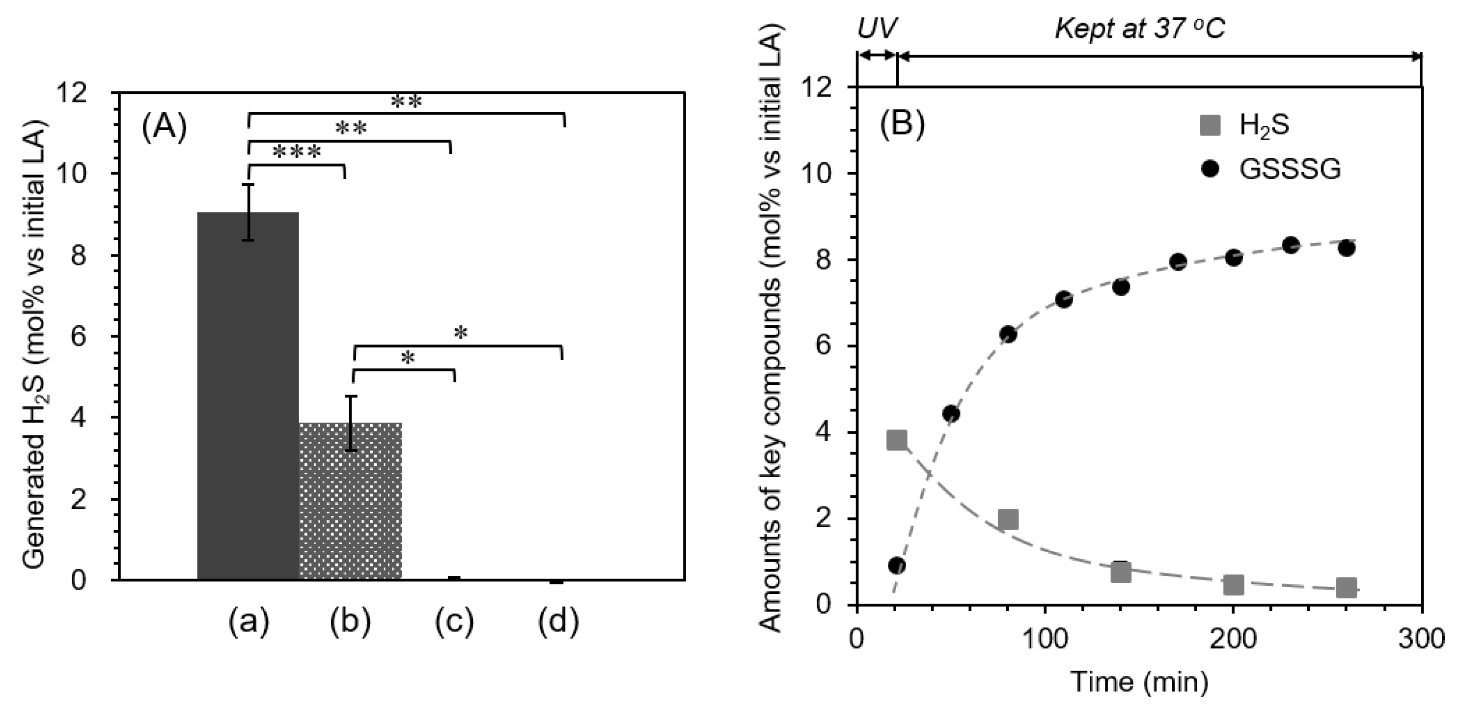
In the earlier study of Brown and Edwards, H2S was produced in the photodecomposition of LA; however, the precise reaction mechanism and the amount of H2S were not described in the literature [16]. We tried to measure the amount of H2S produced in the reaction procedure using the colorimetric titration (zinc acetate method) with slight modification [20]. The amount of H2S gas generated by the photodecomposition of LA was determined to be 9.1 ± 0.7 mol% of the initial LA amount (Figure 2A(a)). In contrast, the amount of H2S generated by the reaction decreased considerably (3.9 ± 0.7 mol% of LA) when the photochemical reaction was carried out in the presence of GSSG (p < 0.001) (Figure 2A(b)). The UVL-irradiated GSSG and non-irradiated LA solutions did not show a detectable amount of H2S (Figure 2A(c,d)). These results indicate that the source of the sulfur atom in the H2S molecule must come from the photo-decomposed product(s) of LA. Furthermore, the significant decrease of H2S amount observed in the presence of GSSG supported the idea that H2S was consumed by the interaction of H2S with GSSG, which might be responsible for the formation of GSSSG. We also carried out the time-dependent concentration changes of H2S and GSSSG, shown in Figure 2B. As a result, the formation of H2S decreases with time following the increased formation of GSSSG.

2.4. pH-Dependent Formation of GSSSG

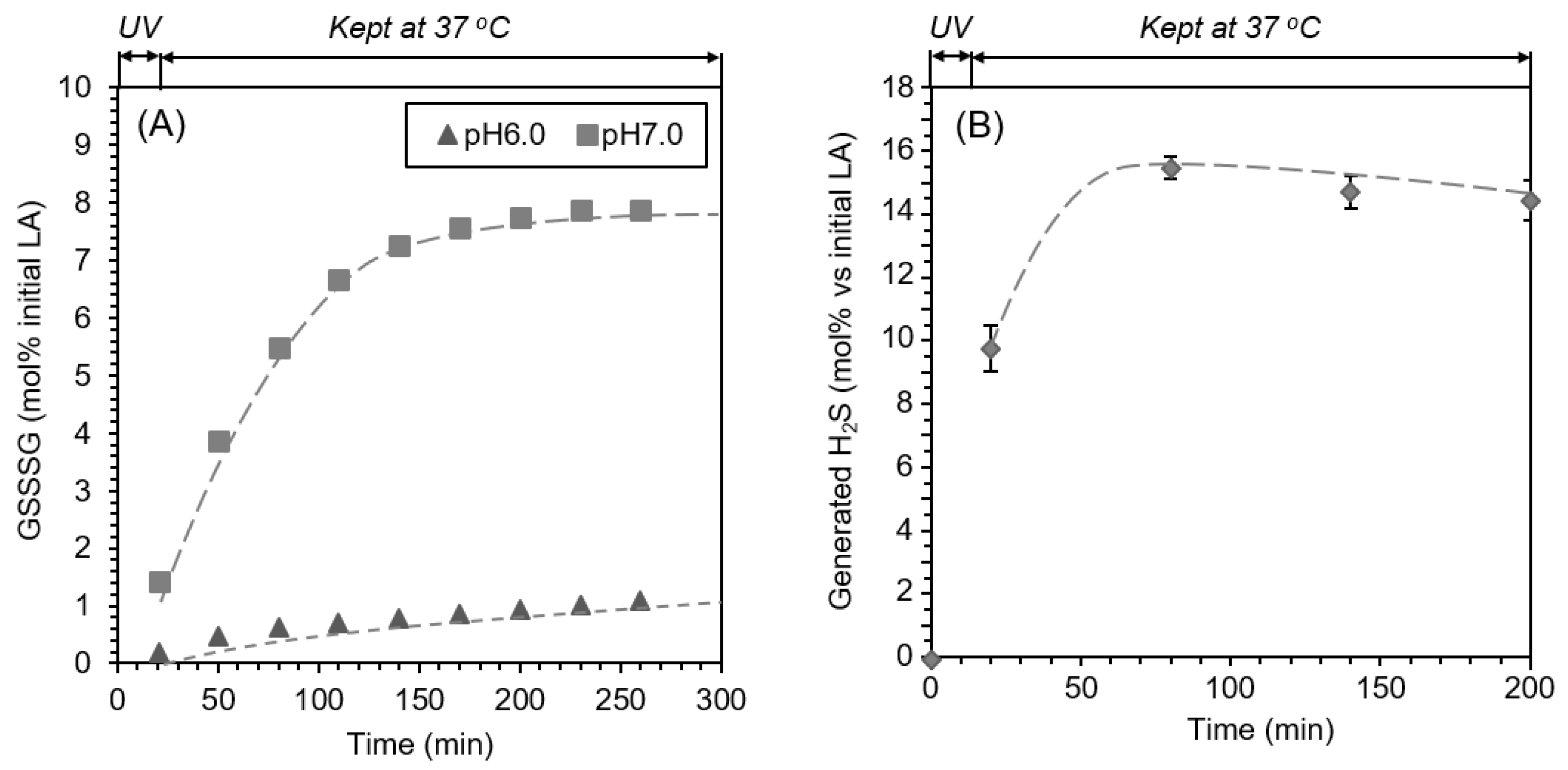
We carried out the experiments using different pH conditions. When the reaction was carried out at pH 6, the formation of GSSSG was quite slow, while at pH 7, the formation of GSSSG increased to approximately 8 mol% of initial LA. This marked pH dependency suggests that the deprotonation of SH (SSH) group might play an important role in the formation of GSSSG (Figure 3A). Next, we examined the time-course for the formation of H2S. The amount of generated H2S gas gradually increased up to 15.5 ± 0.36 mol% after the UVL irradiation was ceased (Figure 3B).

2.5. Reaction of GSSG with Na2S

The reaction of GSSG with Na2S was examined and analyzed by HPLC (eluent condition: H2O–MeOH gradient, 0.05% trifluoroacetic acid (TFA), Figure S4). The time-course for the formation of GSSSG and GSH (GSSH) from GSSG was investigated by the peak heights corresponding to GSH (GSSH), GSSG, and GSSSG. The retention time of GSH and GSSH (both compounds were in protonated form in the eluent conditions employed), however, was very close and could not be separated. The result revealed that the formation of GSH (GSSH) increased time-dependently and reached a maximum level at 30 min after the reaction (Figure 4) while the amount of GSSG was decreasing with time and reached approximately 50% of the amount from the initial stage. The formation of GSSSG gradually increased from 10 min and reached 8 mol% of initial GSSG. The reaction was also carried out in the degassed (anaerobic) conditions; however, we did not find any significant differences in the product formation (Figure S5). This result suggests that the oxygen molecule is not involved in the formation of GSSSG from the reaction of GSSG with Na2S.

2.6. Reaction of GSSSG with Glutathione (GSH) (Interaction and Cross-Talk of Biothiols)

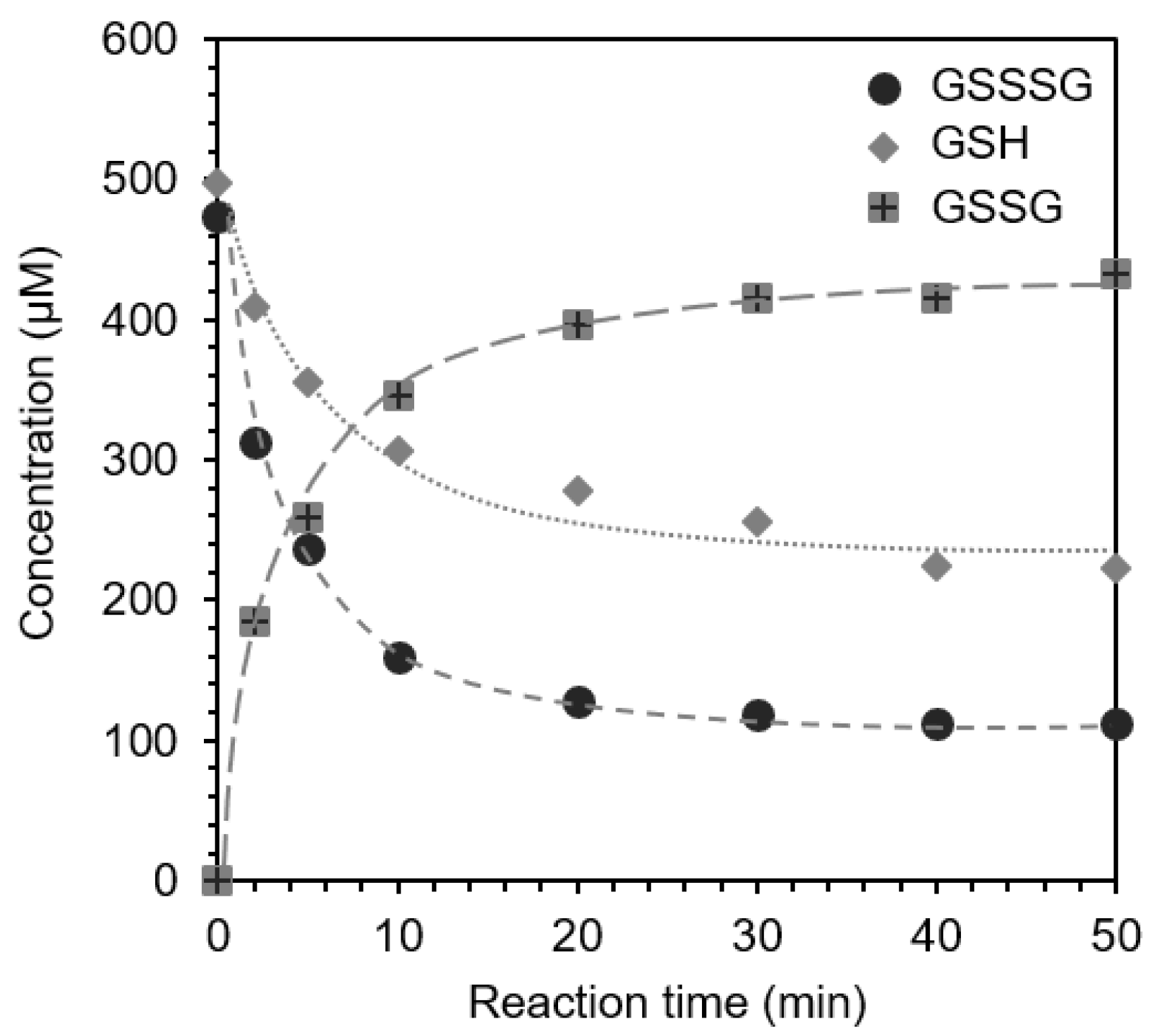
It is quite important to clarify the physiological role of polysulfides. In this sense, the investigation of the interaction between GSH with GSSSG is a good model. We analyzed HPLC profile time-dependently to follow the changes in the concentration of GSSSG, GSH, and GSSG during the dark reaction at 37 °C (Figure 5). The concentrations of GSSSG and GSH both decreased in a time-dependent manner, while the concentration of GSSG increased correspondingly. Furthermore, an inseparable peak appeared with a similar retention time to that of the GSH peak, which might be related to GSSH and/or one of its relatives (GSSnH). The concentrations of the reactants (GSSSG and GSH) and product (GSSG) became almost constant after 30 min. The decreased amount of GSSSG (approximately 80 mol%) was almost the same as that of the GSSG generated during the reaction. On the other hand, the decreased concentration of GSH was almost two-thirds of the consumption of GSSSG.

3. Discussion

The accumulated quantity of H2S generated by the UVL irradiation of LA was approximately 15 mol% of initial LA (Figure 3B), which may cause the temporal excess of H2S concentration if LA was photo-decomposed in vivo. This suggests there must be a novel adaptation mechanism to control the concentration of H2S. The amount of H2S generated during the UVL irradiation process (approximately 9 mol% of initial LA, Figure 2A(a)) was almost two-fold greater than the amount generated in the coexistence of GSSG (approximately 4 mol% of initial LA, Figure 2A(b)). In contrast, the amount of GSSSG generated from the UVL irradiation process (only 1 mol% of initial LA, Figure 2B) was much smaller than that of H2S. These results, therefore, implied the occurrence of other pathways for the consumption of H2S, such as the reaction with polymeric materials derived from LA photoirradiation. Notably, the UV reaction of the highly concentrated LA solution resulted in the formation of a slightly turbid solution, which might be the formation of S8 through complicated reduction-oxidation processes. It is noteworthy that the concentration of H2S continued to increase for over 1 h after UVL irradiation was ceased, while it gradually decreased for over 4 h when the process was conducted in the presence of GSSG (Figure 2B). The final yield of GSSSG (approximately 8 mol%, Figure 2B) was almost half of the total amount of H2S, which suggests that the reaction of H2S with GSSG to afford GSSSG.
The sulfur atom of H2S more than likely became the center sulfur atom of GSSSG, because it is more reasonable to consider the S–S bond scission than to consider C–S bond scission. Namely, H2S might attack the S–S bond of GSSG to afford GSSH and GSH (Equation (1)) [21]. GSSH generated as a result of Equation (1) reacts with another GSSG to afford GSSSG and GSH (Equation (2)):
H2S + GSSG ⇄ GSSH + GSH
GSSH + GSSG ⇄ GSSSG + GSH
This mechanism is supported by the enhanced formation of GSSSG at pH 7 conditions compared with pH 6 conditions (Figure 3A). To support this hypothesis, we summarized pKa values of various thiol-containing compounds (Table 1) [22,23,24]. Based on the pKa value of GSSH, 99% of GSSH is dissociated at pH 7, while more than 99% of GSH is not dissociated at pH 7. This pKa value of GSSH is strongly related with the rapid reaction of GSSH at neutral pH conditions, while the nucleophilic attack of GSH is quite limited at neutral pH conditions.
Based on these results, we proposed the mechanism as shown in Figure 6. The photoirradiation of LA at pH 7 provided many compounds including thioketones (2, 2′) and polymeric materials bearing S-S bond (3, other polymers). The hydrolysis of thioketone (2, 2′) provides hydrogen sulfide and ketone derivatives (4). The reaction of H2S with GSSG will take place to afford GSSH and GSH. Deprotonated GSSH reacts smoothly with another GSSG to afford GSSSG and GSH as final products. We also confirmed the generality of this reaction using cystine and DMDS. In these cases, we observed trisulfides compounds under the same reaction conditions employed in the case of GSSG. In all cases, we could not find the formation of LA trisulfide; this can be explained by the residual LA in the reaction conditions, however, further studies of the fate dithiyl radical of LA under various conditions might be interesting. For example, Self et al. demonstrated the synthesis of selenotrisulfide-derivatives of lipoic acid and lipoamide and examined their stability and some biological activities [25]. Considering the physiological importance of rare metal ion such as Se, the capture of Se by dithiyl radical of LA might provide another role for LA.
We also observed the recovered formation of LA under photoirradiated conditions in the presence of biothiols, which can be explained by the nucleophilic attack of thiol (in this case GSH) on the S–S bond of the polymeric materials to afford a GS-attached polymer (5) and thiol-containing polymer (6). The intramolecular cyclization of thiol to another S-S bond takes place to afford LA and thiol-binding polymer (7) as final products.
The administration of LA to cells increases the intracellular glutathione level [1,5], which is the antioxidant network present in the cell. In summary, we have revealed a stocking mechanism for the sulfur atoms generated by the UVL-decomposition of LA, which takes the formation of trisulfide (GSSSG). The physiological role of GSSSG is not well understood; however, GSSSG might have the potential to become the source of GSH or GSSG. Some natural products bearing multiple sulfide atoms have been reported to possess physiological activities including antioxidant activities [26,27,28]. Synthetic organosulfur compounds bearing di-, tri-, and tetra-sulfides bearing tyrosol moiety have been prepared to examine their structure–activity relationship focusing on the number of sulfur atoms [29]. This observation will also provide the new antioxidant network of glutathione and LA. When the concentration of H2S exceeds a toxic level, an excess amount of H2S might be stored in some form of sulfane sulfur species [30,31]. It is interesting to clarify the precise relationship of H2S, LA and sulfane sulfur in detail. Bilska-Wilkosz et al. set up a hypothesis based on some experimental results in vitro and in vivo [32,33]. In this study, we have investigated the production of H2S via the UVL-mediated decomposition of LA and the crosstalk of LA with disulfide biomolecules such as glutathione disulfide (GSSG), cystine (CysSSCys), and dimethyl disulfide (DMDS) and clarified one pathway to store one sulfur atom inserting into the disulfide S–S bond. This mechanism might provide the new possibility for other polysulfide stocking systems and also for understanding the physiological roles of polysulfides.

4. Materials and Methods

4.1. Materials

All chemicals were used without further purification after being purchased from a chemical company. Phosphate buffer at pH 7.0 (100 mM) was used as a reaction medium throughout this study. UVL irradiation was performed on transilluminator NTM-10 (Funakoshi, Tokyo, Japan). The wavelength range of UVL was 280–390 nm, and the peak maximum appeared at 310 nm. HPLC analysis was performed on a reversed-phase column 5C18-MS (4.6 × 150 mm, Nacalai tesque, Kyoto, Japan). The ESI-MS experiment was performed on a JMS-T100TD mass spectrometer (JEOL, Tokyo, Japan), and nuclear magnetic resonance (NMR) spectra were also recorded on an ECS-400 (JEOL, Tokyo, Japan). FT-IR spectrum was recorded on a Nicolet 6700 (Thermo Fisher Scientific, Waltham, MA, USA). Elemental analysis was performed at the Organic Elemental Analysis Research Center of Kyoto University.

4.2. Photoreaction of LA in the Presence of Disulfides (GSSG, CysSSCys, DMDS)

A reaction solution (3 mL) containing LA (2 mM) and GSSG (10 mM) in PB was charged into a screw cap vial (Φ18 mm in diameter). The concentration of LA and GSSG were higher than the expected concentration in vivo because of the limitation of detector sensitivity in HPLC analysis. The vial was then sealed and irradiated with UVL (11 W/m2 at 310 nm) from the bottom of the vial at ambient temperature for 20 min. A small portion of the reaction mixture (20 μL) was injected into the HPLC system to analyze the reaction progress at determined intervals. The HPLC system was operated with a linear gradient elution program at a constant flow rate of 0.7 mL/min using water and methanol as a mobile phase. Both contained 0.05% (v/v) TFA. The percentage of methanol was changed as follows: 0–15% from 0–5 min; 15–80% from 5–15 min; and 80% from 15–18 min. The photo reaction of LA in the presence of CysSSCys was conducted at a much lower final concentration (0.1 mM for LA, and 0.5 mM for CysSSCys) because of the very low solubility of CysSSCys. Reaction progress was monitored with ion-paired HPLC analysis. Water containing two kinds of salt, sodium dodecyl sulfate 5 mM and sodium sulfate 25 mM, was adjusted at pH 3.0 with hydro sulfuric acid and flew at a constant rate (0.6 mL/min) as an eluent. The photo reaction of LA in the presence of DMDS was performed (1 mM for LA, and 5 mM for DMDS). Eluent condition for HPLC analysis was isocratic (60% (v/v) aqueous methanol containing 0.05% TFA, 0.7 mL/min).

4.3. Quantification of H2S Using a Methylene Blue Method

The concentration of H2S was determined using a modified version of the methylene blue method [20]. Briefly, a reaction mixture (3 mL) containing LA (2 mM) and/or GSSG (10 mM) in PB was loaded into a screw cap vial (Φ18 mm in diameter). The vial was then sealed and subjected to the UVL irradiation conditions described above. Upon completion of the UVL reaction, the sample solution (120 µL) was mixed with zinc acetate (1 w/v %, 150 µL) and PB (330 µL) to trap the H2S. A coloring reagent consisting of N,N-dimethyl-1,4-phenylenediamine dihydrochloride (20 mM in 7.2 N HCl, 100 µL) and iron (III) chloride (30 mM in 1.2 N HCl, 100 µL) was then added to the solution, and the resulting mixture was allowed to stand at room temperature for 15 min. The absorbance of the mixture was then measured at 670 nm. This experiment was repeated three times, and the H2S concentration in the sample solution was calculated using a calibration curve, which was made using sodium sulfide nonahydrate.

4.4. GSSSG Formation at Different pH Conditions

A phosphate buffer (100 mM at pH 6.0) was prepared, and 3 mL of a solution with an initial LA and GSSG concentration of 2 mM and 10 mM were prepared for the UV irradiation experiment. All experimental conditions other than pH were the same as pH 7.0. After the UVL irradiation, the reaction solution was analyzed by HPLC.

4.5. The Reaction of GSSG with Na2S at Air-Saturated and Degassed Conditions

The air-saturated stock solution of GSSG (208 μM, 1.44 mL) was prepared in PB (pH 7.0, 100 mM). Fresh Na2S solution (5.0 mM, 60 μL) in PB was prepared and immediately mixed with GSSG solution. The final concentration of these compounds was set as 200 μM each. The reaction progress at 37 °C was monitored by HPLC analyses up to 150 min after starting the reaction.
PB was degassed by a repeated freeze-thaw method and charged with N2 gas to establish the anaerobic reaction condition. GSSG solution (208 μM, 1.44 mL) in PB was prepared by this degassed PB, and this stock solution was degassed again. Na2S solution was also prepared in degassed PB and mixed with degassed GSSG solution. HPLC analyses were conducted to quantify the GSSG, GSSSG, and GSH.

4.6. The Reaction of GSSSG with GSH

The same volume of GSSSG (1 mM) and GSH (1 mM) solutions in PB (100 mM, pH 7.0) were mixed and stored at 37 °C. The progress of the reaction was monitored by HPLC analysis for over 50 min. Peaks corresponding to GSSSG, GSH, and GSSG were identified by comparing their retention times with those of the corresponding standards. The concentrations of these components were determined using calibration curves based on the peak areas of the different peaks.

Supplementary Materials

The following are available online at https://www.mdpi.com/article/10.3390/biochem1030012/s1. Figure S1: (A) 1H- and (B) 13C-NMR spectra of glutathione trisulfide (GSSSG). Figure S2: HPLC chromatograms of the reaction solutions containing (A) LA (5 mM) and (B) GSSG (25 mM) after UVL-irradiation. Figure S3: HPLC chromatograms of the reaction solutions containing LA (1 mM) and dimethyldisulfide (DMDS) (5 mM). (A) before and (B) after UVL-irradiation. (C) Time course for the amount of Dimethyltrisulfide (DMTS) at 37 °C after the UVL-irradiation. HPLC chromatograms of the reaction solutions containing LA (0.1 mM) and cystine (CysSSCys) (0.5 mM) (D) before and (E) after UVL-irradiation. (F) Time course for the peak area assignable to CysSSSCys at 37 °C after the UVL-irradiation. Figure S4: HPLC profile for the reaction of GSSG with Na2S -Formation of GSSSG-. Eluent condition: H2O–MeOH gradient with 0.05v% TFA. Figure S5: Time dependence of GSSSG formation in the aerobic and anaerobic conditions.

Author Contributions

The first author N.W. designed this study, collected and interpreted the experimental data sets, and wrote the first draft of this manuscript. S.M. discussed the experimental results and critically revised the paper including the discussion part. All authors have read and agreed to the published version of the manuscript.

Funding

This research received no external funding.

Institutional Review Board Statement

Not applicable.

Informed Consent Statement

Not applicable.

Acknowledgments

NMR and ESI-MS analyses were carried out at the Advanced Science Research Center of Kanazawa University.

Conflicts of Interest

The authors declare no conflict of interest.

References

  1. Han, D.; Tritschler, H.J.; Packer, L. Alpha-lipoic acid increases the intracellular glutathione in a human T-lymphocyte in Jurkat cell line. Biochem. Biophys. Res. Commun. 1995, 207, 258–264. [Google Scholar] [CrossRef]
  2. Packer, L.; Wutt, H.H.; Tritschler, H.J. Alpha-lipoic acid as a biological antioxidant. Free Rad. Biol. Med. 1995, 19, 227–250. [Google Scholar] [CrossRef]
  3. Jones, D.P. Redefining oxidative stress. Antioxid. Redox Signal. 2006, 8, 1865–1879. [Google Scholar] [CrossRef]
  4. Kemp, M.; Go, Y.M.; Jones, D.P. Nonequilibrium thermodynamics of thiol/disulfide redox systems: A perspective on redox system biology. Free. Radic. Biol. Med. 2008, 44, 921–937. [Google Scholar] [CrossRef] [Green Version]
  5. Yamada, T.; Hashida, K.; Takarada-Iemata, M.; Matsugo, S.; Hori, O. a-Lipoic acid enantiomers protect SH-SY5Y cells against glutathione depletion. Neurochem. Int. 2011, 59, 1003–1009. [Google Scholar] [CrossRef] [Green Version]
  6. Moni, H.; Packer, L.; Saris, N.E.L. Antioxidant and prooxidant activities of alpha-lipoic acid and dihydrolipoic acid. Toxicol. Appl. Pharmacol. 2002, 182, 84–90. [Google Scholar] [CrossRef] [Green Version]
  7. Packer, L.; Cadenas, E. Lipoic acid: Energy metabolism and redox regulation of transcription and cell signaling. J. Clin. Biochem. Nutr. 2011, 48, 26–32. [Google Scholar] [CrossRef] [Green Version]
  8. Dorsam, B.; Fahrer, J. The disulfide compound a-lipoic acid and its derivatives: A novel class of anticancer agents targeting mitochondria. Cancer Lett. 2016, 371, 12–19. [Google Scholar] [CrossRef]
  9. Matsugo, S.; Han, D.; Tritschler, H.J.; Packer, L. Decomposition of alpha-lipoic acid derivatives by photoirradiation- formation of dihydrolipoic acid from alpha-lipoic acid-. Biochem. Mol. Biol. Int. 1996, 38, 51–59. [Google Scholar]
  10. Ikuta, N.; Sugiyama, H.; Shimosegawa, H.; Nakane, R.; Ishida, Y.; Uekaji, Y.; Nakata, D.; Pallauf, K.; Rimbach, G.; Terao, K.; et al. Analysis of the enhanced stability of R(+)-alpha lipoic acid by the complex formation with cyclodextrins. Int. J. Mol. Sci. 2013, 14, 3639–3655. [Google Scholar] [CrossRef] [Green Version]
  11. Ikuta, N.; Tanaka, A.; Otsubo, A.; Ogawa, N.; Yamamoto, H.; Mizukami, T.; Arai, S.; Okuno, M.; Terao, K.; Matsugo, S. Spectroscopic studies of R(+)-alpha-lipoic acid complexes. Int. J. Mol. Sci. 2014, 15, 20469–20485. [Google Scholar] [CrossRef] [Green Version]
  12. Ikuta, N.; Endo, T.; Hosomi, S.; Setou, K.; Tanaka, S.; Ogawa, N.; Yamamoto, H.; Mizukami, T.; Arai, S.; Okuno, M.; et al. Structural analysis of crystalline R(+)-alpha-lipoic acid-alpha-cyclodextrin complex based on microscopic and spectroscopic studies. Int. J. Mol. Sci. 2015, 16, 24614–24628. [Google Scholar] [CrossRef] [Green Version]
  13. Wada, N.; Wakami, H.; Konishi, T.; Matsugo, S. The effect of biothiol on UV irradiated α–lipoic acid. Biofactors 2008, 34, 285–292. [Google Scholar] [CrossRef]
  14. Wada, N.; Wakami, H.; Konishi, T.; Matsugo, S. The degradation and regeneration of α–lipoic acid under the irradiation of UV light in the existence of Homocysteine. J. Clin. Biochem. Nutr. 2009, 44, 218–222. [Google Scholar] [CrossRef] [Green Version]
  15. Calvin, M.; Barltrop, J.A. A possible primary quantum conversion act of photosynthesis. J. Am. Chem. Soc. 1952, 74, 6153–6154. [Google Scholar] [CrossRef] [Green Version]
  16. Brown, P.R.; Edwards, J.O. Effect of solvent on the photolysis of α–lipoic acid. J. Org. Chem. 1969, 34, 3131–3135. [Google Scholar] [CrossRef]
  17. Nishida, M.; Sawa, T.; Kitajima, N.; Ono, K.; Inoue, H.; Ihara, H.; Motohashi, N.; Yamamoto, M.; Suematsu, M.; Kurose, H. Hydrogen sulfide anion regulates redox signaling via electrophile sulfhydration. Nat. Chem. Biol. 2012, 8, 714–724. [Google Scholar] [CrossRef]
  18. Ono, K.; Akaike, T.; Sawa, T.; Kumagai, Y.; Wink, D.A.; Tantillo, D.J.; Hobbs, A.J.; Nagy, P.; Xian, M.; Lin, J.; et al. Redox chemistry and chemical biology of H2S, hydropersulfides, and derived species: Implications of their possible biological activity and utility. Free. Radic. Biol. Med. 2014, 77, 82–94. [Google Scholar] [CrossRef] [Green Version]
  19. Wallace, J.L. Hydrogen sulfide-releasing anti-inflammatory drugs. Trends Pharmacol. Sci. 2007, 28, 501–505. [Google Scholar] [CrossRef]
  20. Li, L.; Bhatia, M.; Zhu, Y.Z.; Zhu, Y.C.; Ramnath, R.D.; Wang, Z.J.; Mohammed Anuar, F.B.; Whiteman, M.; Salto-Tellez, M.; Moore, P.K. Hydrogen sulfide is a novel mediator of lipopolysaccharide-induced inflammation in the mouse. FASEB J. 2005, 19, 1196–1198. [Google Scholar] [CrossRef]
  21. Ida, T.; Sawa, T.; Ihara, H.; Tsuchiya, Y.; Watanabe, Y.; Kumagai, Y.; Suematsu, M.; Motohashi, H.; Fujii, S.; Matsunaga, T.; et al. Reactive cysteine persulfides and S-polythiolation regulate oxidative stress and redox signaling. Proc. Natl. Acad. Sci. USA 2014, 111, 7606–7611. [Google Scholar] [CrossRef] [Green Version]
  22. Tajc, S.G.; Tolbert, B.S.; Basavappa, R.; Miller, B.L. Direct determination of thiol pKa by isothermal titration microcalorimetry. J. Am. Chem. Soc. 2004, 126, 10508–10509. [Google Scholar] [CrossRef]
  23. Cuevasanta, E.; Lange, M.; Bonanta, J.; Coitino, E.L.; Ferrer-Sueta, G.; Filipovic, M.R.; Alvarez, B. Reaction of hydrogen sulfide with disulfide and sulfenic acid to form the strongly nucleophilic persulfide. J. Biol. Chem. 2015, 290, 26866–26880. [Google Scholar] [CrossRef] [Green Version]
  24. Cotton, F.; Wilkinson, G. Advanced Inorganic Chemistry; John Wiley Sons, Inc.: New York, NY, USA, 1988. [Google Scholar]
  25. Self, W.T.; Tsai, L.; Stadtman, C. Synthesis and characterization of selenotrisulfide-derivatives of lipoic acid and lipoamide. Proc. Natl. Acad. Sci. USA 2000, 97, 12481–12486. [Google Scholar] [CrossRef] [Green Version]
  26. Munday, M.; Munday, J.S.; Munday, C.M. Comparative mono-, di-, tri-and tetrasulfides derived from plants of the Allium family: Redox cycling in vitro and hemolytic activity Phase 2 enzyme induction in vivo. Free. Radic. Biol. Med. 2003, 34, 1200–1211. [Google Scholar] [CrossRef]
  27. Kim, J.-M.; Chang, H.J.; Kim, W.-K.; Chang, N.; Chun, H.S. Structure-activity relationship of neuroprotective and reactive oxygen species scavenging activities for allium organosulfur compounds. J. Agric. Food Chem. 2006, 54, 6547–6553. [Google Scholar] [CrossRef]
  28. Jacob, J. Ascent of therapy: Pharmacological implications of natural products containing redox-active sulfur atoms. Nat. Prod. Rep. 2006, 23, 851–863. [Google Scholar] [CrossRef]
  29. Panzella, L.; Verotta, L.; Goya, L.; Ramos, S.; Martin, M.A.; Bravo, L.; Napolitano, A.; d’Lschia, M. Synthesis and bioactivity profile of 5-s-lipoylhydroxytyrosol-based multidefense antioxidants with sizable(poly)sulfide chain. J. Agric. Food Chem. 2013, 61, 1710–1717. [Google Scholar] [CrossRef] [Green Version]
  30. Iciek, M.; Włodek, L. Biosynthesis and biological properties of compounds containing highly reactive, reduced sulfate sulfur. Pol. J. Pharmacol. 2001, 53, 215–225. [Google Scholar]
  31. Park, C.M.; Weerasinghe, L.; Day, J.J.; Fukuto, J.M.; Xian, M. Persulfides: Current knowledge and challenges in chemistry and chemical biology. Mol. Biosyst. 2015, 11, 1775–1785. [Google Scholar] [CrossRef] [Green Version]
  32. Bilska, A.; Dudek, M.; Iciek, M.; Kwiecien, I.; Sokotowska-Jezewicz, M.; Filipek, B.; Wtodek, L. Biological actions of lipoic associated with sulfane sulfur metabolism. Pharmacol. Rep. 2008, 60, 225–232. [Google Scholar]
  33. Bilska-Wilkosz, A.; Iciek, M.; KowalczykpPachel, D.; Gorny, M.; Sokolowska-Jezewicz, M.; Wlodek, L. Lipoic acid as a possible pharmacological source of hydrogen sulfide/sulfane sulfur. Molecules 2017, 22, 388. [Google Scholar] [CrossRef] [Green Version]
Figure 1. Typical high-performance liquid chromatography (HPLC) chromatograms of the reaction solutions containing α-lipoic acid (LA, 5 mM) and oxidized glutathione (GSSG, 25 mM) (A) before and (B) after ultra-violet light (UVL) irradiation. (C) Mass spectrum (electrospray ionization-mass spectrometry, ESI-MS) of the isolated glutathione trisulfide (GSSSG).
Biochem 01 00012 g001
Figure 2. Amounts of (A) H2S generated immediately after UVL reaction. Reaction solutions contain (a) 2 mM LA, (b) 2 mM LA and 10 mM GSSG, (c) 10 mM GSSG and (d) 2 mM LA in PB (pH 7.0). UVL was used to irradiate solutions of (ac). All of these data represent the mean values of three experiments ±S.D. and statistical differences have been shown as *** (p < 0.001), ** (p < 0.005), * (p < 0.01). (B) Time course for the amount of H2S, GSSSG at 37 °C after the UVL irradiation to a mixture of LA (2 mM) and GSSG (10 mM).
Biochem 01 00012 g002
Figure 3. (A) Dependence of GSSSG generation rate on a pH value. (B) Time course for the H2S production after terminating UVL irradiation to LA. Except for (A), the experiments were conducted in triplicate, and data are represented as mean ±S.D.
Biochem 01 00012 g003
Figure 4. Time-dependent increase of GSSSG and glutathione (GSH) concomitant with the decrease of GSSG.
Biochem 01 00012 g004
Figure 5. Time dependence for the reaction of GSSSG with GSH.
Biochem 01 00012 g005
Figure 6. A plausible reaction mechanism for the formation of GSSSG by the photoirradiation of LA in the presence of GSSG.
Biochem 01 00012 g006
Table 1. Equilibrium of Dissociation of thiol derivatives.
pH56789
HS/H2S1/1001/101/110/1100/1
GS/GSH1/10,0001/10001/1001/101/1
GSS/GSSH1/110/1100/11000/110,000/1
pKa (ca.) GSSH:5.24, H2S:7.0, GSH:9.24 ± 0.15.
Publisher’s Note: MDPI stays neutral with regard to jurisdictional claims in published maps and institutional affiliations.