Article

Hydrogels Made with Tilapia Fish Skin Increase Collagen Production and Have an Effect on MMP-2/MMP-9 Enzymes in Burn Treatment

1
Department of Medicinal Biochemistry, School of Medicine, Aydin Adnan Menderes University, Aydin 09100, Turkey
2
Department of Molecular Biotechnology, Graduate School of Health Sciences, Aydin Adnan Menderes University, Aydin 09100, Turkey
*
Author to whom correspondence should be addressed.
Submission received: 15 February 2025 / Revised: 11 April 2025 / Accepted: 18 April 2025 / Published: 22 April 2025
(This article belongs to the Special Issue Feature Papers in BioChem)

Abstract

:
Background/Objectives: Burns are a prevalent health concern that manifest on the skin’s surface or within organs due to various traumas and necessitate prompt intervention. The healing process of the skin involves a sequence of time-dependent events, commencing with the activation of growth factors and culminating in the expression of various genes. To expedite the healing process of burn wounds, there is a need to develop biodegradable materials and new technologies that are compatible with the skin. Methods: In this study, the roles of tilapia (TL, Oreochromis niloticus) fish skin in burn wound treatment processes were investigated. TL or TL-alginate hydrogels (AGTL) were applied to a burn wound created in Sprague Dawley rats for 7 and 14 days. Following the administration of treatment, the levels of hydroxyproline, a critical element in tissue reorganization, along with the gene expression levels of COL1A1, COL3A1, MMP-2, and MMP-9, and the protein expression levels of MMP-2 and MMP-9 were evaluated. Results: Wound closure processes were faster in AGTL-groups compared to TL-groups, and hydroxyproline levels were found to be higher. While the increase in MMP-2 levels was less, the increase in MMP-9 gene and protein levels was greater in the AGTL-group. Concurrently, COL1A1 levels decreased over 14 days, while COL3A1 levels increased in the AGTL-group. Conclusions: Consequently, it was determined that the biological substances in the TL structure, in conjunction with alginate, were effective in the healing and reorganization of the wound tissue. This finding suggests that tilapia may provide a valuable source of insights for future studies aimed at developing effective wound dressings for wound tissues.

1. Introduction

Burns are the most common type of injury in everyday life, and they are caused primarily by heat or contact with chemicals, electricity, friction, and radioactivity on the outer surface tissue of organs located on or under the skin. Burns are critical and costly problems that require care depending on the type and extent of the material in contact. In cases of burns, depending on the degree of severity, they can be fatal depending on the complications that occur. On the other hand, non-fatal burns are one of the leading causes of morbidity, including long-term hospitalization, deformity and disability, as well as stigma and rejection [1,2]. In recent years, there has been an increasing tendency to utilize biological resources in the development of dressing materials with the objective of accelerating the healing process of patients and reducing the duration of hospital stay for burn treatment. These resources encompass both plant-based compounds and proteins derived from animal sources, which are engineered into forms compatible with various types of wounds [3,4].
Tilapia (Oreochromis niloticus) is a freshwater fish species belonging to the Cichlidae family. It is notable for its high economic and nutritional value. Tilapia is able to tolerate elevated water temperatures and wide pH ranges, which makes it a resilient species. The skin of tilapia fish has been utilized in burn treatment due to its reported wound healing properties [5,6]. These properties are attributed to the presence of unique biological components in the healing process. Recent studies have demonstrated a clear increase in the utilization of biological materials in treatment methods. Fish skin grafts, in particular, have been shown to accelerate wound healing due to their content. These grafts have demonstrated promising results in the treatment of diabetic foot ulcers and venous leg ulcers, suggesting their potential for use in the treatment of various other acute and chronic wounds [7]. Collagen, a protein that has the potential to support wound healing, is found in large amounts in fish skin [8]. Research findings have demonstrated that collagen, when dissolved using a tilapia fish source, has enhanced efficacy in promoting wound healing processes [9,10]. Tilapia skin has been reported to contain a collagen content exceeding 40% on a dry weight basis, and it is also used in the production of collagen peptides [11,12]. An analysis of the amino acid composition of collagen obtained by means of enzymatic hydrolysis from tilapia revealed that glycine, proline, and hydroxyproline, as the predominant amino acids, accounted for 20.92%, 11.32%, and 10.28%, respectively [9]. Research has demonstrated that collagen in tilapia comprises the following same four subunits: β chains, γ chains, and two α chains (α1 and α2). The fibrillar structure of collagen is responsible for its significant enhancement of cell adhesion capacities [13]. Therefore, it has been suggested that it is safe to use in the production of collagen-containing biomaterials and as a dressing material. Tilapia has been reported to be biosafe in studies conducted on patients, with accelerated wound healing being a notable outcome [14]. It has been documented that the addition of silver nanoparticles to tilapia skins via polyaniline coating enhances antibiofilm activity. Moreover, it is postulated that this process may play a pivotal role in combating contamination by Staphylococcus aureus [15,16]. Additionally, the potential of silver nanoparticles to mitigate collagen loss during the sterilization of tilapia skin grafts has been underscored. The development of modified tilapia skin grafts that exhibit antimicrobial properties is also recommended [17].
The process of wound healing is accompanied by a series of molecular mechanisms in the physiological process, which consists of four stages. Collagen, a structural protein, plays a critical role in wound healing; however, its production and reorganization are subject to rigorous regulation by enzymes such as matrix metalloproteinases (MMPs). Among the MMPs, MMP-2 and MMP-9 are of particular significance, as they play a pivotal role in the degradation of collagen, particularly type I and type III collagen. MMP-2 interacts with basement membrane structures, such as type IV and type V collagen, while MMP-9 targets collagens located more in the connective tissue and outer cell matrix [18]. In the post-burn healing process, the excessive activation or inhibition of these MMPs can determine the speed and quality of healing. Therefore, the balanced activities of MMP-2 and MMP-9 in burn healing are important for both optimal wound healing and the prevention of scar formation [19,20]. Collagen’s function is most significant in the remodeling stage, which is the third stage of the process [21,22]. The remodeling stage is to restore normal tissue structure and ensure the formation of scar tissue with collagen [23]. The function of collagen in this stage is particularly important, as it acts as a biodegradable protein for cellular migration and capillary growth, which are essential for the repair process [24].
In this study, a hydrogel form of tilapia skin was developed for use in burn cases, and its role in wound healing processes in rats was investigated. The aim was to determine changes in markers such as hydroxyproline, MMP-2, MMP-9, COL1A1, and COL3A1, which play important roles in collagen production and destruction, in tilapia skin hydrogels in burn wounds created in rats and to investigate whether they could be a dressing material that could be used in the clinic.

2. Materials and Methods

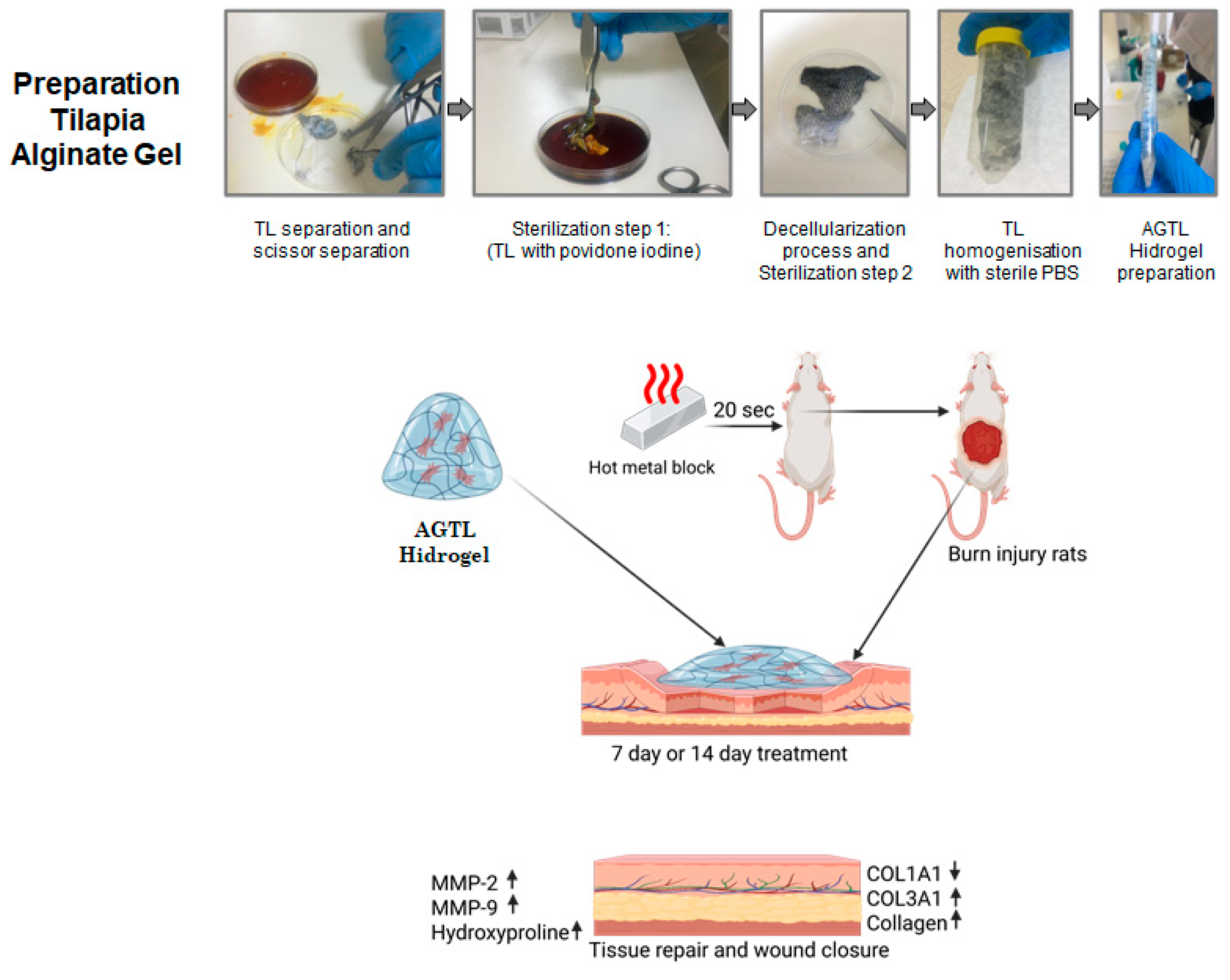
2.1. Preparation of Hydrogels from Tilapia and Alginate

Oreochromis niloticus, commonly known as the tilapia (TL) fish, were procured from a cultivation facility (Konya, Turkey). The skins of these fish were meticulously separated through mechanical means on ice using a sharp knife. The process of sterilization entailed the meticulous removal of the tilapia skin, which was then immersed in a solution of povidone iodine for a period of 10 min. This was followed by a thorough cleansing with NaCI (0.1%), which was repeated three times. For the decellularization process [25], the skin sample was subjected to an incubation in a phosphate-buffered saline (PBS) solution comprising 0.5% Triton X and 0.02% sodium azide at a rotary speed of 100 rpm for a duration of 1 h at ambient temperature. Subsequently, the sample underwent two washes with phosphate-buffered saline (PBS), each lasting 5 min and conducted at 100 rpm at room temperature. Subsequently, the decellularized skin sample was subjected to incubation with a trypsin solution (0.05 g/mL trypsin in 1 M Tris-HCl, pH 8.5) for a duration of 1 h at room temperature. Following this, the sample was thoroughly washed with PBS to ensure the removal of any residual enzymes. Subsequently, a sterilization solution was formulated containing 50% glycerol, 1% penicillin streptomycin, and an antifungal agent (2.5 μg/mL Amphotericin B), with the objective of ensuring long-term sterilization. Sodium alginate (AG W201502, Sigma-Aldrich Saint Louis, MO, USA,) was dissolved (0.1 g/mL) in sterile PBS and filtered through a 0.45 micron sterile filter. Sterile calcium chloride solutions (2% w/v) were prepared to reduce the agent. Then, the solution was prepared at a concentration of 1.25% AG. After the tilapia skin (1 g) was homogenized in the KCL solution (10 mL), it was mixed with alginate in equal proportions (10 mL homogenate and 10 mL alginate solution), and the calcium solution (2 mL) was added and kept for 5 min for the formation of the alginate complex. Then, TL-alginate hydrogels (AGTL) were washed with a sterilization solution (50% glycerol, 1% penicillin streptomycin, and 2.5 μg/mL Amphotericin B). Alginate was selected due to its non-toxic composition and substantial water retention capabilities. The decision was made to employ alginate in its hydrogel form to facilitate the adhesion of tilapia skin to the wound surface. The resultant AGTL hydrogel was transferred into a sterile PBS container and subsequently stored in capped falcons. The alginate hydrogels were meticulously shaped into dimensions commensurate with the size of the wound. While AGLT was being placed on the wound surface, it was placed on a dressing to prevent slipping, and the back area of the animals was covered with a plaster. Tilapia hydrogel production stages and experimental design for applications are included in Figure 1.

2.2. Characterization of AGLT Hydrogels

The rheological behaviors of the hydrogels were measured using a rheometer (HAAKE MARS 40 Rheometer, Thermo Fisher Scientific Inc., Waltham, MA, USA). AG and AGTL viscosity and shear rate were measured every 10 s for 1 min.
Since the wound surface is acidic, the swelling properties of AG or AGLT-containing hydrogel samples were investigated at different pH values. For swelling tests, hydrogel samples were dried in a vacuum oven at 40 °C, and their weights were measured. The dried hydrogels were placed in PBS (pH: 7.4) or acetate buffer (acetic acid/sodium acetate, pH: 4.5) at 25 °C, and the time of placement was taken as t = 0 min. At specified time intervals, the hydrogel sample was removed from the buffer, weighed, and returned to the buffer medium. This process continued until the hydrogel reached its equilibrium swelling value. The swelling values of the hydrogels were calculated by subtracting the mass of the swollen hydrogel from the initial mass of the dry hydrogel.

2.3. Burn Model Creation and Treatment Groups

This study was approved by the Ethical Committee for Animal Experiments at the Aydin Adnan Menderes University, approval number (2023/75). All experiments were conducted according to the Guide for the Care and Use of Laboratory Animals of the National Institutes of Health [26], and the experiments were reported according to the ARRIVE guidelines [27], Aydin Experimental. The burn model was created by anesthetizing the Sprague Dawley Rats (250–300 g, 3 months old) with 75–90 mg/kg of ketamine (5%; Pfizer Inc., New York, NY, USA) and 5–8 mg/kg of xylazine (2%; Bayer Health Care AG, Leverkusen, Germany) intraperitoneally, and then catheterization was performed from the lateral tail vein with a 24 Gauge intravenous catheter. The dorsal areas of the anesthetized rats were shaved and cleansed with povidone iodine for 10 min [28]. A 4 cm × 4 cm metal plate was heated by immersing it in boiling water (100 °C) for 15 s. Then, the heated metal plate was placed on the shaved back of the rat without applying pressure and held for 20 s with a stopwatch, and the same amount of time was applied to all rats. In the experimental group, 6 were used in each group. The following experiments were conducted: A 7-day or 14-day group was created for the burn models, then and a group with no treatment for 7 or 14 days was created. A TL-7-day or TL-14-day burn model was created, and then tilapia skin was applied for 7 or 14 days. An AGTL-7-day or AGTL-14-day burn model was created and then tilapia-containing hydrogel skin was applied for 7 or 14 days. Animals were placed in each cage individually. Infection monitoring and dressing monitoring were performed daily, and no infection was observed during the experiment. During the creation of the burn model and the treatment period, no rats were lost. The experiment concluded with the survival of all rats. The wound size was measured with a caliper and evaluated through the documentation of photographs. At the end of the experiment, rats were euthanized, and burn tissues were excised and utilized for biochemical, molecular, and histological analyses.

2.4. Gene Expression with qPCR

The isolation of RNA was conducted in accordance with the stipulated recommendations of the total RNA purification kit (GeneAll, Hybrid-RTM 305-101, Seoul, Republic of Korea). To this end, 100 mg of skin tissue was homogenized with the RiboEx RNA cell lysis buffer, which was included in the aforementioned kit. Thereafter, the homogenate was subjected to incubation for a period of five minutes at ambient temperature. The mixture was then subjected to a centrifugation process at 12,000× g for 10 min at a temperature of 4 °C. The resultant pellet was transferred to a fresh tube and utilized for RNA isolation. The quantity of RNA was subsequently quantified using nanodrops. To synthesize cDNA from the RNA samples, a commercial kit containing a reverse transcriptase enzyme was employed, following the manufacturer’s recommended protocol (Applied Biosystems Inc., High Capacity cDNA Reverse Transcription Kit, Catalog no. 4368813, Vilnius, Lithuania). Subsequently, a mixture of random primer and RNA samples was prepared and maintained at 25 °C for a duration of 10 min. Subsequently, the samples were subjected to incubation at 37 °C for two hours and at 85 °C for five minutes to facilitate the synthesis of cDNA. The quantity of cDNA obtained was then measured using nanodrops. To perform quantitative PCR (qPCR) reactions, 100 ng of cDNA was utilized, along with primers specific to MMP2 and MMP9 genes. GAPDH was selected as the housekeeping gene for normalization purposes. Primer sequences were used for MMP-2 Forward 5′-AGCAAGTAGACGCTGCCTTT-3′, Reverse 5′-ACCTGGGATCCCCTTACCTC-3′; MMP-9 Forward 5′-CTTCTCTTTCGTAGGGCGCA-3′, Reverse 5′-TAGGGCTCCTCCTACTGGTG-3′; COL1A1 Forward 5′-ATGGATCAGGCCAATGGCAA-3′, Reverse 5′-TGTTACTTACAGTGCAGCCA-3′; COL3A1 Forward 5′-GCGAAGGCAACAGTCGATTC-3′, Reverse 5′-GGACCTGGTCTGGGGATACT-3′; and GAPDH Forward 5′-TTCCACCTTTGATGCTGGGG-3′, Reverse 5′-AGAGGGCACCAAACCTTCAG-3′. The cDNA was subsequently diluted for qPCR, and the reaction mixture was prepared using a sybr green master mix (Applied Biosystem Inc., Vilnius, Lithuania, Catalog no. 4309155). The prepared mixture was configured to yield a total reaction volume of 20 µL per well. It was then subjected to the following thermal cycling conditions: 95 °C for 10 min (1 cycle), 95 °C for 15 s, and qPCR experiments were subsequently conducted on the qPCR device (Applied Biosystems StepOnePlus Real-Time PCR System) in accordance with the following program: 57 °C for 20 s, 72 °C for 30 s (40 cycles), 95 °C for 2 min (1 cycle).

2.5. Protein Expression with Western Blotting

Skin tissues were lysed in homogenization buffer containing 1% Triton X-100 and NP40, along with a protease inhibitor cocktail. The samples were then kept on ice to allow cell lysis. Subsequently, the samples were centrifugated at 12,000× g at 4 °C for 10 min, after which Western blot studies were performed on the upper phase. Samples were boiled at 95 °C for 5 min with sample loading buffer prepared at a 1:1 ratio. Some 12% SDS-PAGE gels were prepared, and samples containing 25 µg/mL protein were loaded into each well. Immunoblotting was performed with a semi-dry system to transfer proteins in the gel to the PVDF membrane. Following this, the PVDF membrane was blocked with 3% BSA for 2 h at room temperature and then incubated with primary antibody (1:500 dilution, MMP-2 sc-10736, MMP-9 sc-10737, GAPDH sc-32233) overnight. After this step, the membrane was washed 3 times with TBST and treated with a secondary antibody (1:2000 dilution, anti-rabbit-HRP sc-2357, and anti-mouse-HRP sc-2060). Then, it was incubated with a solution containing chemiluminescence substrate (Santa Cruz, Dallas, TX, USA, Western Blotting Luminol Reagent: sc-2048) for 1 min in the dark. The bands obtained in the imaging system (Syngene GBox Chemi XRQ) were evaluated by densitometric analysis, with GAPDH serving as the reference protein in the densitometric analysis and calculations to eliminate changes in the samples. Densitometric analysis of the bands was performed in the ImageJ 1.54 software (NIH, Bethesda, MD, USA) program.

2.6. Hydroxyproline Assay

Hydroxyproline (4-hydroxyproline) is a non-proteinogenic amino acid that is formed through the process of the post-translational hydroxylation of proline. Hydroxyproline is the predominant component of collagen and is employed as an indicator of collagen content due to its ability to stabilize the helical structure. To ascertain the impact of tilapia hydrogels on wound healing, the levels of hydroxyproline in the tissue were quantified by ELISA, a technique that utilizes a commercial kit for homogenized skin tissues. The Hydroxyproline Quantification Kit (Sigma-Aldrich MAK569, Saint Louis, MO, USA) was utilized in accordance with the protocol established by the kit’s manufacturer. The measurement of colorimetric color changes at 560 nm in the tissue sample was performed.

2.7. Histological Staining

To examine cell activities, hematoxylin-eosin (HE) staining was performed on biopsy samples taken from the rat burn model [29]. To observe the collagen factor during the healing process, Masson trichrome staining was performed [30]. After cervical dislocation, the skin tissues of the animals in the groups were placed in 4% paraformaldehyde solution and kept at 4 °C for histological examination. Tissue tracking was performed with a tissue tracking device, and the tissues were embedded in paraffin. This process includes the following steps: tissue collection, fixation, dehydration, transparency, impregnation, and embedding. Each paraffin block was sectioned at a thickness of 5 µm, and HE staining was performed on a number of sections for light microscope (Zeiss Axio Vert.A1, Jena, Germany).

2.8. Statistical Analysis

The data are presented as the means  ±  standard deviations. Statistical analyses were conducted using GraphPad Prism 7. p values less than 0.05 were considered to be statistically significant. The normality of distributions between groups was analyzed using one-way analysis of variance (ANOVA). Additionally, the Bonferroni and Tukey post-hoc tests were employed to further explore the data.

3. Results

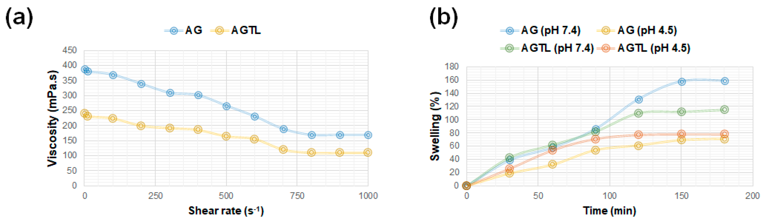
3.1. AG and AGTL Hydrogel Viscosity and Swelling Rate

The viscosity was measured as a function of shear rate at 25 °C using a rheometer setup. The hydrogel exhibited shear-thinning behavior, with viscosity decreasing as shear rate increased, indicating its suitability for extrusion-based applications, such as dressing material for injury. AG and AGTL viscosity were measured in a shear rate range of 1–1000 s−1 by increasing the shear rate every 10 s for one minute. The resultant shear viscosity values for 1.25% alginate at 25 °C were found to be 381 mPa s, while AGTL shear viscosity exhibited a value of 241 mPa s at the same temperature. In the rheological analysis of hydrogels, an examination was conducted of the relationship between shear rate and viscosity. It was observed that, as the shear rate increased, viscosity decreased. It was determined that the gel exhibited a certain degree of strength at shear rates ranging from 250 to 500 s per minute, as indicated by the shear stress, indicating that the gel was not highly fluid (see Figure 2a).
The swelling ratio (%) was measured at regular time intervals to evaluate the hydrogel’s responsiveness to different pH conditions. The percentage swelling values of hydrogels in pH 7.4 and pH 4.5 buffer environments are illustrated in Figure 2b. It was determined that the water retention capacity of hydrogels decreased in AG at a pH of 4.5; however, this decrease was less in AGTL. The buffering effect of collagen and proteins present in the AGTL structure contributes to its resistance against changes in acidic environments, thereby minimizing water loss. This behavior highlights the pH-dependent nature of alginate hydrogels, which is relevant for applications in dressing material for burn injury.

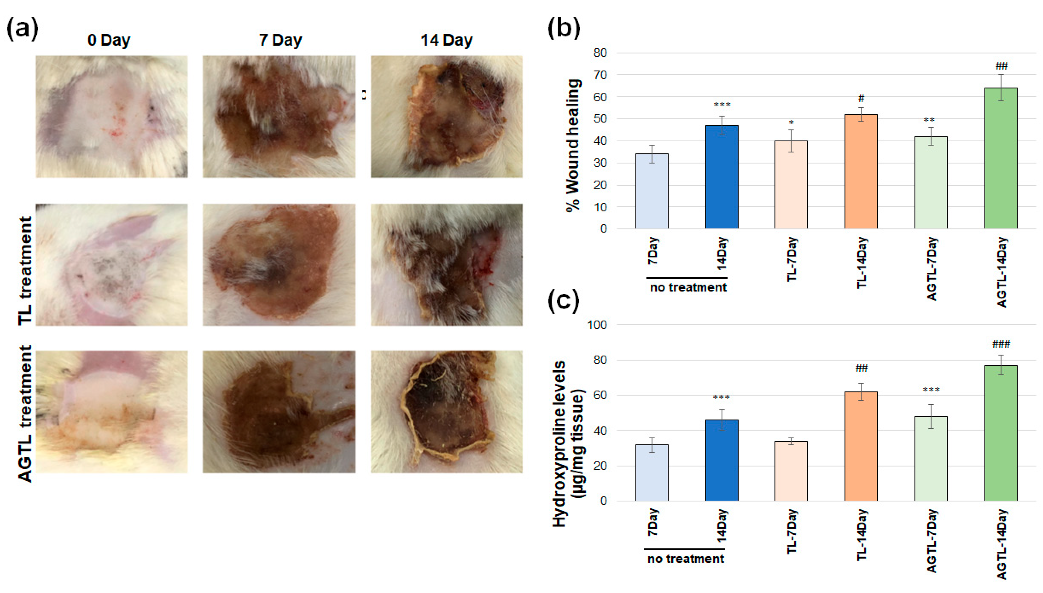
3.2. TL and AGTL Treatment Effects on Wound Closure

As illustrated in Figure 3a, images of the burn wounds were captured on the 7th and 14th days post-treatment in rats. Figure 3b presents the calculated wound closure rates, indicating a statistically significant difference (p < 0.001) in the burn wound closure between the untreated group on the 14th day and the 7th day. The 7-day TL treatment administered to the burn wound exhibited a significant increase in comparison to the 7-day untreated group (p ˂ 0.05). Similarly, the 14-day TL treatment demonstrated a significant increase in comparison to the 14-day untreated group (p ˂ 0.05). Furthermore, the 7-day AGTL treatment administered to the burn wound exhibited a significant increase in comparison to the 7-day untreated group (p < 0.01). Similarly, the 14-day AGTL treatment demonstrated a significant increase in comparison to the 14-day untreated group (p < 0.01).

3.3. TL and AGTL Treatment Effects on Hydroxyproline

Changes in hydroxyproline levels, a critical indicator of wound healing, are demonstrated in Figure 3c. A significant increase in hydroxyproline levels was observed in the untreated group on day 14 compared to the untreated group on day 7 (p < 0.001). However, no significant change was detected when TL treatment applied to the burn wound on day 7 was compared to the untreated group on day 7. A similar outcome was observed when TL treatment for 14 days was compared to the untreated group on day 14 (p < 0.01). AGTL treatment applied to the burn wound on day 7 demonstrated a significant increase in hydroxyproline levels compared to the untreated group on day 7 (p < 0.001). Conversely, no significant increase in hydroxyproline levels was observed when 14 days of AGTL treatment was compared to the 14-day untreated group (p < 0.001).

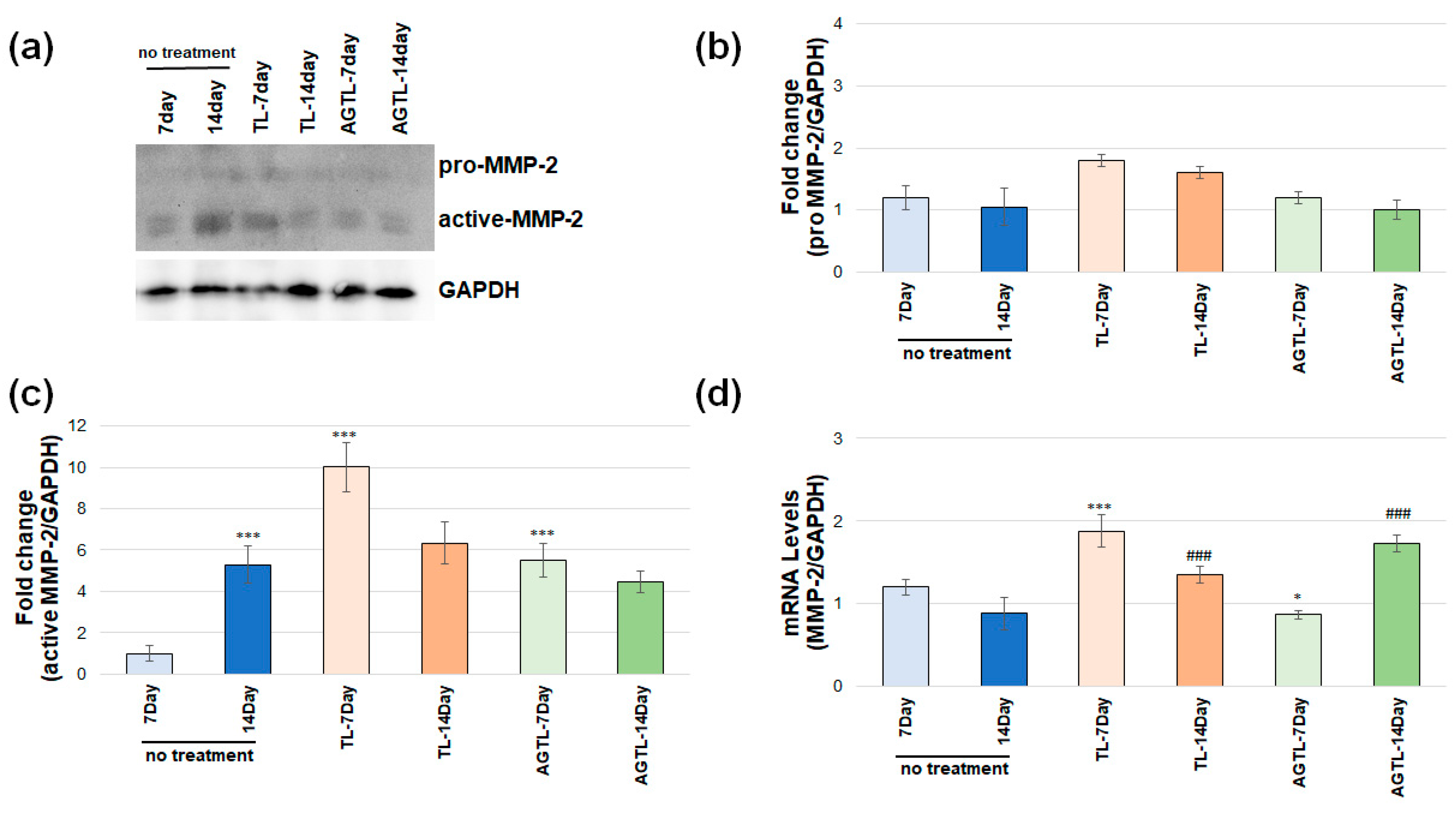
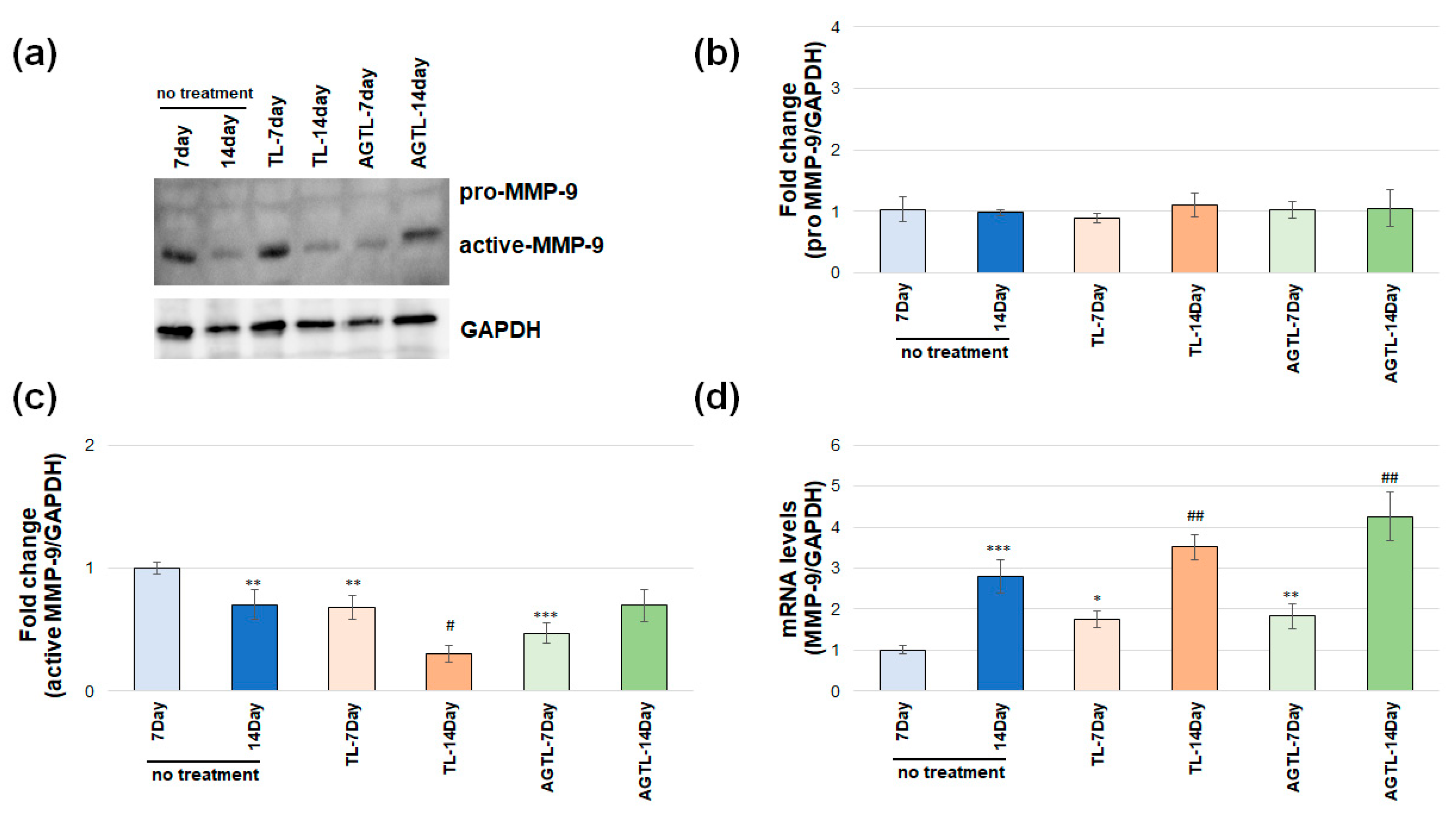
3.4. TL and AGTL Treatment Effects on MMP-2 and MMP-9 Gene and Protein Expression

Western blot gel images of the groups belonging to MMP-2 are shown in Figure 4a. No significant change was observed in the pro-MMP-2 protein expression levels in burn wounds on rats on day 7 and 14 (Figure 4b). Changes in active MMP-2 protein expression levels after TL and AGTL application on day 7 and 14 in burn wounds on rats are shown in Figure 4c. On day 14, the untreated group exhibited a substantial increase in active-MMP-2 protein expression levels in comparison to the untreated group on day 7 (p < 0.001). Similarly, TL treatment administered on day 7 resulted in a significant increase in MMP-2 protein expression levels when compared to the untreated group on the same day (p < 0.05). No significant change was observed when the 14 days of TL treatment group was compared with the 14 days of untreated group. A significant increase was observed when group with 7 days of AGTL treatment applied to the burn wound was compared with the 7 days untreated group (p < 0.001). No significant change was observed in active-MMP-2 protein levels when 14 days of AGTL treatment was compared to the 14 days of untreated group.
As illustrated in Figure 4d, the present study examined alterations in active MMP-2 gene expression levels following TL and AGTL application on the 7th and 14th days in burn wounds in rats. The investigation revealed no statistically significant change in MMP-2 gene expression levels on the 14th day in the untreated group of the burn wound compared to the 7-day untreated group. A significant increase was observed in the 7-day TL treatment applied to the burn wound compared to the 7-day untreated group (p < 0.001). However, no significant change was observed in the 14-day TL treatment compared to the 14-day untreated group. A significant increase was observed in the 7-day AGTL treatment applied to the burn wound compared to the 7-day untreated group (p < 0.05). However, no significant increase was observed in the MMP-2 gene expression levels when compared to the 14-day AGTL treatment compared to the 14-day untreated group (p ˂ 0.001).
Western blot gel images of the groups belonging to MMP-9 are shown in Figure 5a. No statistically significant alterations were observed in the protein levels of pro-MMP-9 (see Figure 5b). The alterations in active MMP-9 protein expression levels subsequent to TL and AGTL application on the 7th and 14th days in burn wounds in rats are presented in Figure 5c. A significant decrease in active-MMP-9 protein expression levels was observed on the 14th day in the untreated group of the burn wound compared to the 7th day in the untreated group (p < 0.01). A significant decrease was also observed when the 7-day TL treatment was applied to the burn wound compared to the 7-day untreated group (p < 0.01). However, no significant change was observed when 14 days of TL treatment was compared to the 14-day untreated group (p < 0.05). A significant decrease was observed when the 7-day AGTL treatment was compared to the 7-day untreated group (p < 0.001). However, no significant change was observed in active-MMP-9 protein levels when the 14-days AGTL treatment was compared to the 14-day untreated group. As illustrated in Figure 5d, the application of TL and AGTL on days 7 and 14 post-burn in rat models resulted in alterations in active MMP-9 gene expression levels. A notable increase in MMP-9 gene expression levels was observed in the untreated group on day 14 compared to the untreated group on day 7 (p < 0.001). A significant increase was observed in the 7-day TL treatment applied to the burn wound compared to the 7-day untreated group (p < 0.05). No significant change was observed in the 14-day TL treatment compared to the 14-day untreated group. A significant increase was observed in the 7-day AGTL treatment applied to the burn wound compared to the 7-day untreated group (p < 0.01). However, no significant increase was observed in the MMP-9 gene expression levels when compared to the 14-day AGTL treatment versus the 14-day untreated group (p < 0.01). Similarly, no significant increase was observed in the MMP-9 gene expression levels when compared to the 14-day AGTL treatment versus the 14-day untreated group (p < 0.01).

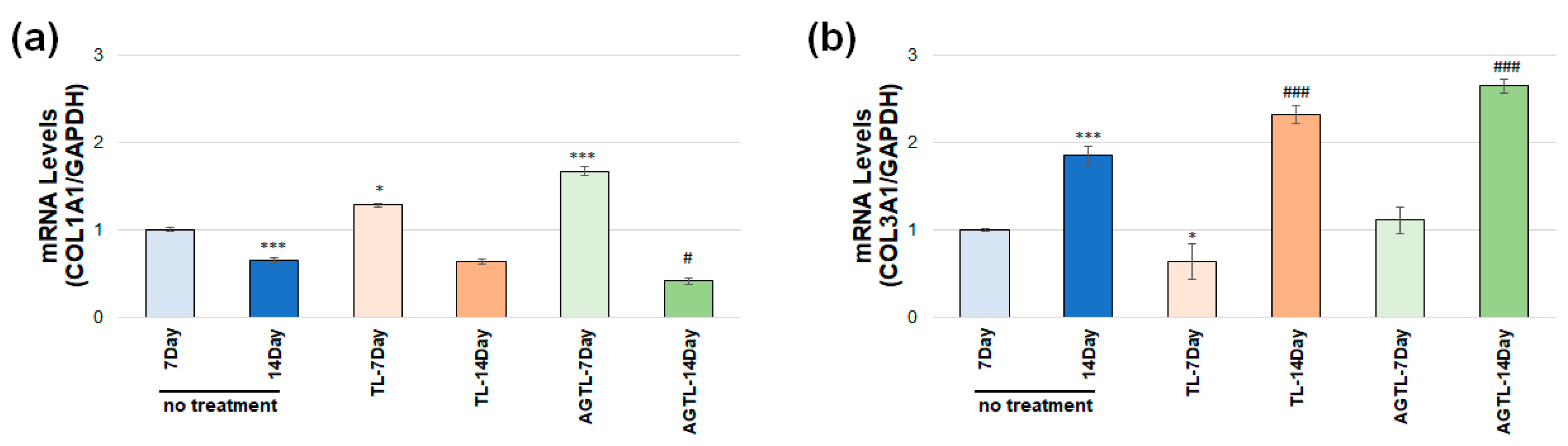
3.5. TL and AGTL Treatment Effects on COL1A1 and COL3A1 Gene Expression

As illustrated in Figure 6a, the data demonstrate a notable decrease in COL1A1 gene expression levels on the 14th day in the untreated group of the burn wound compared to the 7th day of the untreated group (p < 0.001). In contrast, a significant increase in COL1A1 gene expression levels was observed in the 7-day TL treatment group compared to the 7-day untreated group (p < 0.05). However, no significant change was observed when the 14-day TL treatment was compared to the 14-day untreated group. A significant increase in COL1A1 gene expression levels was also observed in the 7-day AGTL treatment group compared to the 7-day untreated group (p < 0.001). However, no significant increase was observed in COL1A1 gene expression levels when the 14-day AGTL treatment was compared to the 14-day untreated group (p < 0.05).
As illustrated in Figure 6b, the data demonstrate a notable increase in COL3A1 gene expression levels in the untreated group of the burn wound on the 14th day compared to the 7th day of the untreated group (p < 0.001). A significant change was observed when the 7-day TL treatment was compared to the 7-day untreated group (p < 0.05). A significant increase was observed when the 14-day TL treatment was compared to the 14-day untreated group (p < 0.001). No significant change was observed when the 7-day AGTL treatment was compared to the 7-day untreated group. However, a significant increase in COL3A1 gene expression levels was observed when the 14-day AGTL treatment was compared to the 14-day untreated group (p < 0.001).

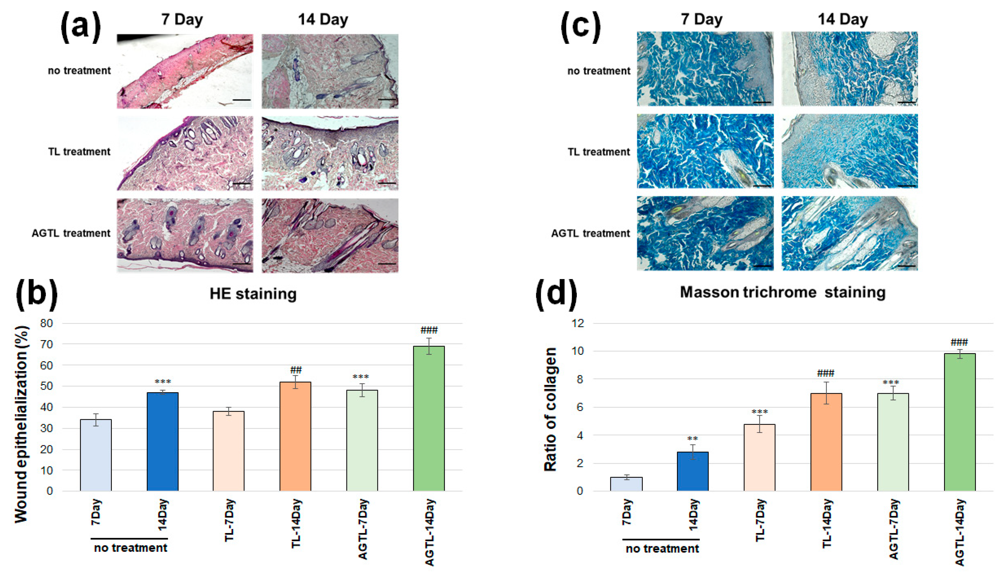
3.6. TL and AGTL Treatment Effects on Skin Tissue Epithelization and Collagen Fibers

In histological analyses performed on tissues, an evaluation was made in terms of skin epithelialization and hair follicles in HE staining (Figure 7a). On the seventh day, the absence of treatment resulted in an increase in inflammatory cells and the initiation of epithelization in the skin tissue. On the seventh day, the TL-treated groups exhibited progenitor cell formation, hair follicles, and an augmented epithelium layer. On the seventh day, the AGTL-treated groups demonstrated adipocytic tissue that was clearly discernible and dense epithelization. On the seventh day, the groups that were not treated exhibited light epithelization and hair follicles. On the fourteenth day, the groups that were treated exhibited dense cell formations and increased epithelization. On the fourteenth day, the groups that were treated exhibited dense hair follicles, aggregated cells, epithelium layer, and regeneration of the epidermis. Wound epithelization data (Figure 7b) demonstrated a notable increase in the untreated group of the burn wound on the 14th day compared to the 7th day (p < 0.001). No significant change was observed when the 7-day TL treatment was compared to the 7-day untreated group, but the 7-day AGTL group increased compared to the untreated group (p < 0.001). A significant increase was observed when the 14-day TL or AGTL treatment was compared to the 14-day untreated group (p < 0.01, p < 0.001).
Histological analyses were performed with Masson trichrome staining to evaluate the amount and distribution of connective tissue in the burn area. Blue staining of collagen fibers indicates the presence and distribution of connective tissue. The results showed that blue staining was scattered in the untreated groups, while in TL and AGTL treatment, blue staining was found to be more intense and healing in terms of providing connective tissue integration. Additionally, it was observed that the connective tissue of the skin increased, and fibrosis occurred, especially in 14-day treatments (Figure 7c). The collagen ratio (Figure 7d) increased in the untreated group of the burn wound on the 14th day compared to the 7th day (p < 0.01). A significant change was observed when the 7th day of the TL or AGTL treatment was compared to the 7th day of the untreated group (p < 0.001). A significant increase was observed when the 14th day of the TL or AGTL treatment was compared to the 14th day of the untreated group (p < 0.001).

4. Discussion

Wound healing consists of the following three basic phases: inflammatory, proliferative, and maturation. The inflammatory phase is clot formation followed by inflammatory cell formation at the wound or injury site. This phase lasts 2–5 days and includes vasoconstriction, platelet aggregation, and clot formation followed by vasodilation and phagocytosis. The second phase, consisting of granulation, contraction, and epithelialization, lasts approximately 2–3 weeks. Then comes the maturation phase, where new tissue is formed, and this phase lasts for about 3 weeks. When a superficial wound in the skin is damaged, it quickly passes the threshold of hemostasis, which is the primary phase of the healing process. Excessive bleeding is prevented by the onset of coagulation in the blood vessels, which results in the accumulation of platelets around the endothelial region and the formation of a plug. This is how platelets form a blood clot. The formation of a clot not only stops bleeding, but it also serves as a temporary matrix for cell migration [31,32]. The primary events contributing to wound closure are epithelialization, wound contraction, and collagenous scar formation, which are the results of wound healing. MMPs are proteolytic enzymes that hydrolyze the tissue matrix. MMPs facilitate keratinocyte migration by destroying structures in the extracellular matrix that impede keratinocyte migration [33]. MMP-2 and MMP-9 facilitate the separation of keratinocytes in the basal layer from the basement membrane and their movement [34].
Research conducted in various laboratories has demonstrated that matrix metalloproteinases (MMPs) derived from epithelial cells regulate a series of events during the inflammatory phase of wound healing. These events include the transepithelial migration of leukocytes and the emergence of signaling proteins, such as chemokines. It has been confirmed that MMPs are prominently expressed in inflammatory cells and that stromal and epithelial cells in the wounded area also express multiple MMPs, including MMP 1, 2, 3, 7, 9, 10, and 28 [35]. It has been reported that MMP-2 can be secreted from macrophages during the wound healing process and plays a significant role in gene regulation in inflammatory regions [36]. With the exception of MMP-2 and MMP-9, which have been observed to have negative or inhibitory effects on cell proliferation, the vast majority of MMPs have been reported to positively influence the wound healing process [37]. During the initial phases of wound healing, there is an increase in collagen synthesis, followed by a subsequent decrease as the wound heals. MMP-2 and MMP-9, classified as gelatinase enzymes, are responsible for the degradation of collagen on the wound surface. This process involves the conversion of collagen fibrils, which are insoluble in nature, into soluble gelatin peptides [38]. During the normal wound healing process, the synthesis of type III collagen occurs initially, followed by type I collagen synthesis, which occurs later. As the wound heals, a process known as maturation occurs, which involves the reorganization of the wound healing process. This maturation results in a decrease in the ratio of type III collagen to type I collagen. Consequently, the amount of the extracellular matrix (ECM), water, and glycosaminoglycan also decreases [39,40].
In the present study, it was determined that there was a significant increase in hydroxyproline levels, which serves as an important marker for collagen levels, in the TL and AGTL groups. Furthermore, it was determined that epithelialization increased even more with AGTL, and that the humidity in the environment accelerated collagen formation. In terms of changes in collagen gene levels, it was observed that the 7-day AGTL application increased COL1A1 levels, which decreased in the 14-day application. This phenomenon can be attributed to the destruction of collagen scar tissue, facilitated by MMP-2 and MMP-9 activation on the 14th day, which enabled keratinocytes to approach each other. COL3A1, a pivotal regulator of fibroblasts, exhibited an increase in gene levels following 14-day TL and AGTL treatments. This increase indicates that fibroblasts are directed towards the surface in the proliferation phase and that tissue healing is in a permanent phase. Although tilapia fish skins have recently been used in clinical applications for wound treatment in many countries, they have not become widespread due to some limitations. However, the presence of collagen and peptides that contribute to collagen production in the structure of tilapia indicates its continued utilization in traditional burn treatment practices. The lack of experimental studies in the literature on this subject is limited in understanding how effective tilapia is in burn treatment. A smart dressing material made from tilapia fish skin containing allopurinol nanoparticles coated with silver nanoparticles has been shown to accelerate wound healing and suppress inflammation in rats [41]. In a separate study, silver nanoparticles (AgNPs) obtained from aloe vera via green synthesis were expressed as a suitable dressing material for burn wound healing because scaffolds decellularized with tilapia skin showed antimicrobial properties on the skin [25]. A study was conducted on the basis of the high collagen content of tilapia skin, and it was reported that collagen nanoparticles obtained in rats caused wound closure as of the 8th day, and the wound closed at the end of the 28th day [42]. It was ascertained that collagen, when present in a gel-like form and extracted from tilapia skin, promoted expedited wound closure and elevated relative gene expressions of transforming growth factor-beta 1 (TGF-β1), basic fibroblast growth factor (bFGF), and α-smooth muscle actin (αSMA) in rats with induced wounds over a period of 12 days [43]. Nanofiber scaffolds made with collagen purified from tilapia have been demonstrated to promote cell migration in wounds and to augment the expression of involucrin, filaggrin, and transglutaminase 1 (TGase1) in skin cells [44]. In the present study, we demonstrated that AGLT enhanced the healing of burn wounds in tilapia by increasing collagen levels, and it was effective in increasing COL3A1 levels in the wound in the 14-day post-burn period. The primary objective of the studies was to obtain collagen from the tilapia structure and subsequently develop formulations and products for utilization in wound healing. In our study, we demonstrated that the alginate hydrogel mixture we obtained by using the tilapia skin as a whole and containing both collagen and other molecules that support collagen production can be used in burn treatment.
It has been reported that alginate-based hydrogels are suitable for clinical applications in terms of holding and stabilizing protein molecules [45]. Alginate hydrogels have a high potential for use in burn treatments or wound dressings, especially because they are biodegradable [46,47]. A comparison of alginate hydrogels with traditional medical dressings reveals several notable advantages. Firstly, alginate hydrogels are non-toxic and highly water absorbent, qualities that enable them to form a hydrogel network on the wound surface, thereby maintaining a moist environment [48]. Secondly, alginate hydrogels do not adhere to wound tissues, and their removal does not cause secondary injury to the wound surface [49]. In this study, we hypothesized that protein-based molecules found in the tilapia structure that are effective in wound healing can accelerate the healing process in cells by providing both moisture and the stabilization of proteins with the help of alginate hydrogels. The treatment with tilapia skin resulted in less scar tissue formation. Thanks to the moisture provided by the hydrogel, collagen and the healing compounds in its structure contributed to the scar tissue formed during wound healing being more flexible and smoother. This may contribute to the scars remaining lighter by regular collagen production in the wound in skin grafts.

5. Conclusions

Our study demonstrated that the preparation of alginate-based tilapia fish skin forms and their subsequent storage in a humid environment over an extended period of time holds promise for facilitating wound healing. This study’s limitations include its exclusive investigation of a single type of tilapia skin and its lack of experimentation with diverse hydrogel forms. Consequently, further research is necessary to investigate the effects of varying collagen amounts and hydrogel forms in different tilapia species. Such findings could contribute to the development of novel dressing materials for burn treatment. This finding is particularly significant in cases requiring prolonged use, such as for deep burns, where the ready availability of these products could significantly enhance the utilization of clinical treatments. Our study may contribute to researchers working in this field in the planning of dressing materials planned to be developed for burn wounds in the future and new biomaterials for the treatment of tilapia skin.

Author Contributions

Conceptualization, B.B. and O.C.; methodology, B.B., A.K., B.K., M.E. and B.I.A.; validation, B.B. and A.K.; formal analysis, B.B. and A.K.; investigation, B.B., A.K., B.K., M.E. and B.I.A.; resources, B.B. and O.C.; data curation, B.B. and B.I.A.; writing—original draft preparation, B.B. and O.C.; writing—review and editing, O.C.; visualization, B.B. and O.C.; supervision, O.C.; project administration, B.B. and O.C.; funding acquisition, O.C. All authors have read and agreed to the published version of the manuscript.

Funding

This research was funded by TUBITAK grant number 1919B012302802 and TUBA-GEBIP.

Institutional Review Board Statement

The animal study protocol was approved by the Experimental Animal Ethics Committee of Aydin Adnan Menderes University (2023/75 and 18 May 2023).

Informed Consent Statement

Not applicable.

Data Availability Statement

Data are contained within the article.

Acknowledgments

The authors would like to thank ODC Research and Development Inc. for providing access to facilities and supplying the reagents.

Conflicts of Interest

The authors declare no conflicts of interest.

References

  1. Nişancı, M.; Öztürk, S. Yanık Fizyopatolojisi. Turk. Klin. Plast. Surg.-Spec. Top. 2010, 2, 8–14. [Google Scholar]
  2. Hankins, C.L.; Tang, X.Q.; Phipps, A. Hot Beverage Burns: An 11-Year Experience of the Yorkshire Regional Burns Centre. Burns 2006, 32, 87–91. [Google Scholar] [CrossRef] [PubMed]
  3. Elfawy, L.A.; Ng, C.Y.; Amirrah, I.N.; Mazlan, Z.; Wen, A.P.Y.; Fadilah, N.I.M.; Maarof, M.; Lokanathan, Y.; Fauzi, M.B. Sustainable Approach of Functional Biomaterials–Tissue Engineering for Skin Burn Treatment: A Comprehensive Review. Pharmaceuticals 2023, 16, 701. [Google Scholar] [CrossRef]
  4. Davison-Kotler, E.; Marshall, W.S.; García-Gareta, E. Sources of Collagen for Biomaterials in Skin Wound Healing. Bioengineering 2019, 6, 56. [Google Scholar] [CrossRef]
  5. Costa, B.A.; Júnior, E.M.L.; Filho, M.O.d.M.; Fechine, F.V.; de Moraes, M.E.A.; Júnior, F.R.S.; Soares, M.F.A.D.N.; Rocha, M.B.S. Use of Tilapia Skin as a Xenograft for Pediatric Burn Treatment: A Case Report. J. Burn Care Res. 2019, 40, 714–717. [Google Scholar] [CrossRef]
  6. Mukherjee, S.; Bhattacherjee, S.; Keswani, K.; Nath, P.; Paul, S. Application of Tilapia Fish Skin in Treatment of Burn Patients. Biocatal. Agric. Biotechnol. 2024, 59, 103254. [Google Scholar] [CrossRef]
  7. Fiakos, G.; Kuang, Z.; Lo, E. Improved Skin Regeneration with Acellular Fish Skin Grafts. Eng. Regen. 2020, 1, 95–101. [Google Scholar] [CrossRef]
  8. Ibrahim, A.; Soliman, M.; Kotb, S.; Ali, M.M. Evaluation of Fish Skin as a Biological Dressing for Metacarpal Wounds in Donkeys. BMC Vet. Res. 2020, 16, 472. [Google Scholar] [CrossRef]
  9. Hu, Z.; Yang, P.; Zhou, C.; Li, S.; Hong, P. Marine Collagen Peptides from the Skin of Nile Tilapia (Oreochromis niloticus): Characterization and Wound Healing Evaluation. Mar. Drugs 2017, 15, 102. [Google Scholar] [CrossRef]
  10. Ge, B.; Wang, H.; Li, J.; Liu, H.; Yin, Y.; Zhang, N.; Qin, S. Comprehensive Assessment of Nile Tilapia Skin (Oreochromis niloticus) Collagen Hydrogels for Wound Dressings. Mar. Drugs 2020, 18, 178. [Google Scholar] [CrossRef]
  11. Mei, F.; Liu, J.; Wu, J.; Duan, Z.; Chen, M.; Meng, K.; Chen, S.; Shen, X.; Xia, G.; Zhao, M. Collagen Peptides Isolated from Salmo salar and Tilapia nilotica Skin Accelerate Wound Healing by Altering Cutaneous Microbiome Colonization via Upregulated NOD2 and BD14. J. Agric. Food Chem. 2020, 68, 1621–1633. [Google Scholar] [CrossRef] [PubMed]
  12. Zeng, S.K.; Zhang, C.H.; Lin, H.; Yang, P.; Hong, P.Z.; Jiang, Z. Isolation and Characterisation of Acid-Solubilised Collagen from the Skin of Nile Tilapia (Oreochromis niloticus). Food Chem. 2009, 116, 879–883. [Google Scholar] [CrossRef]
  13. Shalaby, M.; Agwa, M.; Saeed, H.; Khedr, S.M.; Morsy, O.; El-Demellawy, M.A. Fish Scale Collagen Preparation, Characterization and Its Application in Wound Healing. J. Polym. Environ. 2020, 28, 166–178. [Google Scholar] [CrossRef]
  14. Miranda, M.J.B.D.; Brandt, C.T. Nile Tilapia Skin Xenograft versus Silver-Based Hydrofiber Dressing in the Treatment of Second-Degree Burns in Adults. Rev. Bras. Cir. Plástica 2023, 34, 79–85. [Google Scholar] [CrossRef]
  15. Guimarães, M.L.; da Silva, F.A.G.; de Souza, A.M.; da Costa, M.M.; de Oliveira, H.P. All-Green Wound Dressing Prototype Based on Nile Tilapia Skin Impregnated with Silver Nanoparticles Reduced by Essential Oil. Appl. Nanosci. 2022, 12, 129–138. [Google Scholar] [CrossRef]
  16. Guimarães, M.L.; da Silva, F.A.G., Jr.; da Costa, M.M.; de Oliveira, H.P. Coating of Conducting Polymer-Silver Nanoparticles for Antibacterial Protection of Nile Tilapia Skin Xenografts. Synth. Met. 2022, 287, 117055. [Google Scholar] [CrossRef]
  17. Elshahawy, A.M.; Mahmoud, G.A.E.; Mokhtar, D.M.; Ibrahim, A. The Optimal Concentration of Silver Nanoparticles in Sterilizing Fish Skin Grafts. Sci. Rep. 2022, 12, 19483. [Google Scholar] [CrossRef]
  18. Hingorani, D.V.; Lippert, C.N.; Crisp, J.L.; Savariar, E.N.; Hasselmann, J.P.C.; Kuo, C.; Nguyen, Q.T.; Tsien, R.Y.; Whitney, M.A.; Ellies, L.G. Impact of MMP-2 and MMP-9 Enzyme Activity on Wound Healing, Tumor Growth and RACPP Cleavage. PLoS ONE 2018, 13, e0198464. [Google Scholar] [CrossRef]
  19. Fanjul-Fernández, M.; Folgueras, A.R.; Cabrera, S.; López-Otín, C. Matrix Metalloproteinases: Evolution, Gene Regulation and Functional Analysis in Mouse Models. Biochim. Biophys. Acta Mol. Cell Res. 2010, 1803, 3–19. [Google Scholar] [CrossRef]
  20. Feng, C.; Chen, X.; Yin, X.; Jiang, Y.; Zhao, C. Matrix Metalloproteinases on Skin Photoaging. J. Cosmet. Dermatol. 2024, 23, 3847–3862. [Google Scholar] [CrossRef]
  21. Wu, M.; Sapin-Minet, A.; Stefan, L.; Perrin, J.; Raeth-Fries, I.; Gaucher, C. Heparinized Collagen-Based Hydrogels for Tissue Engineering: Physical, Mechanical and Biological Properties. Int. J. Pharm. 2025, 670, 125126. [Google Scholar] [CrossRef] [PubMed]
  22. Gardeazabal, L.; Izeta, A. Elastin and Collagen Fibres in Cutaneous Wound Healing. Exp. Dermatol. 2024, 33, e15052. [Google Scholar] [CrossRef]
  23. Ghosh, S.; Haldar, S.; Gupta, S.; Chauhan, S.; Mago, V.; Roy, P.; Lahiri, D. Single Unit Functionally Graded Bioresorbable Electrospun Scaffold for Scar-Free Full-Thickness Skin Wound Healing. Biomater. Adv. 2022, 139, 212980. [Google Scholar] [CrossRef] [PubMed]
  24. Cao, L.; Zhang, Z.; Yuan, D.; Yu, M.; Min, J. Tissue Engineering Applications of Recombinant Human Collagen: A Review of Recent Progress. Front. Bioeng. Biotechnol. 2024, 12, 1358246. [Google Scholar] [CrossRef] [PubMed]
  25. Adhikari, S.P.; Paudel, A.; Sharma, A.; Thapa, B.; Khanal, N.; Shastri, N.; Rai, S.; Adhikari, R. Development of Decellularized Fish Skin Scaffold Decorated with Biosynthesized Silver Nanoparticles for Accelerated Burn Wound Healing. Int. J. Biomater. 2023, 2023, 8541621. [Google Scholar] [CrossRef]
  26. Council, N.R. Guide for the Care and Use of Laboratory Animals, 8th ed.; The National Academic Press: Washington, DC, USA, 2011. [Google Scholar]
  27. Kilkenny, C.; Browne, W.J.; Cuthill, I.C.; Emerson, M.; Altman, D.G. Improving bioscience research reporting: The ARRIVE guidelines for reporting animal research. PLoS Biol. 2010, 8, e1000412. [Google Scholar] [CrossRef]
  28. Herndon, D.N. Total Burn Care; Elsevier Health Sciences: Amsterdam, The Netherlands, 2007. [Google Scholar]
  29. Kocabiyik, B.; Gumus, E.; Abas, B.I.; Anik, A.; Cevik, O. Human Wharton-Jelly Mesenchymal Stromal Cells Reversed Apoptosis and Prevented Multi-Organ Damage in a Newborn Model of Experimental Asphyxia. J. Obstet. Gynaecol. 2022, 42, 3568–3576. [Google Scholar] [CrossRef]
  30. Oh, E.J.; Gangadaran, P.; Rajendran, R.L.; Kim, H.M.; Oh, J.M.; Choi, K.Y.; Chung, H.Y.; Ahn, B.-C. Extracellular Vesicles Derived from Fibroblasts Promote Wound Healing by Optimizing Fibroblast and Endothelial Cellular Functions. Stem Cells 2021, 39, 266–279. [Google Scholar] [CrossRef]
  31. Toriseva, M.; Kähäri, V.-M. Proteinases in Cutaneous Wound Healing. Cell. Mol. Life Sci. 2009, 66, 203–224. [Google Scholar] [CrossRef]
  32. Schultz, G.S.; Wysocki, A. Interactions Between Extracellular Matrix and Growth Factors in Wound Healing. Wound Repair Regen. 2009, 17, 153–162. [Google Scholar] [CrossRef]
  33. Erfan, G. Sıçanlarda ER: Yağ ile Oluşturulmuş Yarada Bitki Ekstrelerinin Karışımı Topikal Hemostatik Bir Ajanın Yara İyileşmesine Etkisi; İstanbul İl Sağlık Müdürlüğü: Istanbul, Turkey, 2009. [Google Scholar]
  34. Han, Y.-P.; Tuan, T.-L.; Hughes, M.; Wu, H.; Garner, W.L. Transforming Growth Factor-β- and Tumor Necrosis Factor-α-Mediated Induction and Proteolytic Activation of MMP-9 in Human Skin. J. Biol. Chem. 2001, 276, 22341–22350. [Google Scholar] [CrossRef] [PubMed]
  35. Parks, W.C.; Wilson, C.L.; López-Boado, Y.S. Matrix Metalloproteinases as Modulators of Inflammation and Innate Immunity. Nat. Rev. Immunol. 2004, 4, 617–629. [Google Scholar] [CrossRef] [PubMed]
  36. Eerola, L.M.; Alho, H.S.; Maasilta, P.K.; Inkinen, K.A.; Harjula, A.L.; Litmanen, S.H.; Salminen, U.-S. Matrix Metalloproteinase Induction in Post-Transplant Obliterative Bronchiolitis. J. Heart Lung Transplant. 2005, 24, 426–432. [Google Scholar] [CrossRef]
  37. Bullard, K.M.; Lund, L.; Mudgett, J.S.; Mellin, T.N.; Hunt, T.K.; Murphy, B.B.; Ronan, J.B.; Werb, Z.; Banda, M.J. Impaired Wound Contraction in Stromelysin-1–Deficient Mice. Ann. Surg. 1999, 230, 260. [Google Scholar] [CrossRef]
  38. Carrilho, M.R.O.; Geraldeli, S.; Tay, F.; de Goes, M.; Carvalho, R.; Tjäderhane, L.; Reis, A.; Hebling, J.; Mazzoni, A.; Breschi, L.; et al. In Vivo Preservation of the Hybrid Layer by Chlorhexidine. J. Dent. Res. 2007, 86, 529–533. [Google Scholar] [CrossRef]
  39. Monaco, J.L.; Lawrence, W.T. Acute Wound Healing: An Overview. Clin. Plast. Surg. 2003, 30, 1–12. [Google Scholar] [CrossRef]
  40. Niessen, F.B.; Spauwen, P.H.; Schalkwijk, J.; Kon, M. On the Nature of Hypertrophic Scars and Keloids: A Review. Plast. Reconstr. Surg. 1999, 104, 1435–1458. [Google Scholar] [CrossRef]
  41. Javed, A.; Tariq, A.; Khan, M.F.A.; Mirza, R.; Usman, M.; Nadir, A.; Khan, A. Multifunctional Tilapia Skin Based Smart Dressing of Silver Capped Allopurinol Nanoparticles for Treatment of Infectious Burn Wounds. J. Drug Deliv. Sci. Technol. 2025, 107, 106804. [Google Scholar] [CrossRef]
  42. Shalaby, M.; Hamouda, D.; Khedr, S.M.; Mostafa, H.M.; Saeed, H.; Ghareeb, A.Z. Nanoparticles Fabricated from the Bioactive Tilapia Scale Collagen for Wound Healing: Experimental Approach. PLoS ONE 2023, 18, e0282557. [Google Scholar] [CrossRef]
  43. Elbialy, Z.I.; Atiba, A.; Abdelnaby, A.; Al-Hawary, I.I.; Elsheshtawy, A.; El-Serehy, H.A.; Abdel-Daim, M.M.; Fadl, S.E.; Assar, D.H. Collagen Extract Obtained from Nile Tilapia (Oreochromis niloticus L.) Skin Accelerates Wound Healing in Rat Model via Up Regulating VEGF, bFGF, and α-SMA Genes Expression. BMC Vet. Res. 2020, 16, 352. [Google Scholar] [CrossRef]
  44. Zhou, T.; Wang, N.; Xue, Y.; Ding, T.; Liu, X.; Mo, X.; Sun, J. Electrospun Tilapia Collagen Nanofibers Accelerating Wound Healing via Inducing Keratinocytes Proliferation and Differentiation. Colloids Surf. B 2016, 143, 415–422. [Google Scholar] [CrossRef] [PubMed]
  45. Shao, X.; Tian, M.; Yin, J.; Duan, H.; Tian, Y.; Wang, H.; Xia, C.; Wang, Z.; Zhu, Y.; Wang, Y.; et al. Biofunctionalized Dissolvable Hydrogel Microbeads Enable Efficient Characterization of Native Protein Complexes. Nat. Commun. 2024, 15, 8633. [Google Scholar] [CrossRef] [PubMed]
  46. Chang, R.; Gruebele, M.; Leckband, D.E. Protein Folding Stability and Kinetics in Alginate Hydrogels. Biomacromolecules 2023, 24, 5245–5254. [Google Scholar] [CrossRef] [PubMed]
  47. Somsesta, N.; Jinnapat, A.; Fakpiam, S.; Suksanguan, C.; Wongsan, V.; Ouneam, W.; Wattanaeabpun, S.; Hongrattanavichit, I. Antimicrobial and Biodegradable Hydrogel Based on Nanocellulose/Alginate Incorporated with Silver Nanoparticles as Active Packaging for Poultry Products. Sci. Rep. 2024, 14, 27135. [Google Scholar] [CrossRef]
  48. Zhang, M.; Zhao, X. Alginate Hydrogel Dressings for Advanced Wound Management. Int. J. Biol. Macromol. 2020, 162, 1414–1428. [Google Scholar] [CrossRef]
  49. Gumus, E.; Abas, B.I.; Cevik, E.; Kocabiyik, B.; Cenik, M.; Cevik, O. Alginate Encapsulation Induces Colony Formation with Umbilical Cord-Derived Mesenchymal Stem Cells. Exp. Biomed. Res. 2021, 4, 113–121. [Google Scholar] [CrossRef]
Figure 1. Tilapia hydrogel production stages and experimental design.
Biochem 05 00008 g001
Figure 2. (a) Rheological characterization of the alginate hydrogel with a shear rate vs. viscosity curve. (b) Swelling behavior of alginate hydrogel in buffer solutions with a pH of 7.4 and a pH of 4.5 over time.
Biochem 05 00008 g002
Figure 3. (a) Macroscopic wound images of TL and AGTL treatment in rats on day 0, day 7, and day 14. (b) The wound closure percentages. (c) Hydroxyproline levels of TL and AGTL treatment on day 7 and day 14 (* p ˂ 0.05, ** p ˂ 0.01,*** p ˂ 0.001 day 7, # p ˂ 0.05, ## p ˂ 0.01, ### p ˂ 0.001 day 14 compared with no treatment group).
Biochem 05 00008 g003
Figure 4. (a) MMP-2 western blot image of TL and AGTL treatment on day 7 and day 14, TL and AGTL treatment on day 7 and day 14 (b) pro-MMP-2 band and (c) active MMP-2 band (d) MMP-2 gene expression analyses of TL and AGTL treatment on day 7 and day 14 (* p < 0.05, *** p < 0.001 on day 7, ### p < 0.001 on day 14 compared with no treatment group).
Biochem 05 00008 g004
Figure 5. (a) MMP-9 western blot image of TL and AGTL treatment on day 7 and day 14, and TL and AGTL treatment on day 7 and day 14; (b) pro-MMP-9 band and (c) active MMP-9 band; (d) MMP-9 gene expression analyses of TL and AGTL treatment on day 7 and day 14 (* p ˂ 0.05, ** p ˂ 0.01, *** p ˂ 0.001 on day 7, # p ˂ 0.05, ## p ˂ 0.01 on day 14 compared with no treatment group).
Biochem 05 00008 g005
Figure 6. (a) COL1A1 and (b) COL3A1 gene expression of TL and AGTL treatment on day 7 and day 14 (* p ˂ 0.05, *** p ˂ 0.001 on day 7, # p ˂ 0.05, ### p ˂ 0.001 on day 14 compared with no treatment group).
Biochem 05 00008 g006
Figure 7. Microscopic images of (a) HE and (b) Masson trichrome staining (Scale bar: 100 µm); (c) wound epithelialization percentage; and (d) collagen ratio of TL and AGTL treatment on day 7 and day 14 (** p ˂ 0.01, *** p ˂ 0.001 on day 7, ## p ˂ 0.01, ### p ˂ 0.001 on day 14 compared to the no treatment group).
Biochem 05 00008 g007
Disclaimer/Publisher’s Note: The statements, opinions and data contained in all publications are solely those of the individual author(s) and contributor(s) and not of MDPI and/or the editor(s). MDPI and/or the editor(s) disclaim responsibility for any injury to people or property resulting from any ideas, methods, instructions or products referred to in the content.

