1. Introduction
The postmortem pink discoloration of teeth was reported in humans as early as 1829, and since then there have been multiple reports of pink teeth in humans [
1,
2,
3,
4,
5,
6]. Various factors have been reported to influence the postmortem development of pink teeth, especially when the body is in a moist (humid) environment [
1,
7,
8,
9,
10]. Death due to asphyxiation, including drowning, was originally proposed as the cause of the pink discoloration of teeth; however, more recently it has been shown that pink discoloration is not pathognomonic for a specific cause of death, but is most often associated with water immersion [
1,
8,
9].
In dogs, postmortem pink discoloration of teeth is rarely and poorly documented in the scientific literature. In a decomposition study of euthanized beagle dogs, pink teeth were noted to be observed at 23 days postmortem [
11]. In another report, the teeth of three dogs had experimentally been discolored pink under various storage conditions during the postmortem period [
8]. Pink discoloration of teeth in cats during the postmortem period has not been reported. Causes of discoloration of dog and cat teeth include porphyria (red to brown), enamel hypoplasia (pale yellow), and treatment of a dog with tetracycline during the puppy phase of life (bright yellow) [
2,
12].
Extremely limited research has been done in the field of postmortem pink discoloration of teeth in animals; we hypothesize that the occurrence of pink teeth is under reported in veterinary pathology. The objective of this study was to characterize dog and cat cases that had postmortem pink discoloration of teeth.
3. Results
A review of the veterinary autopsy reports identified four dogs and one cat with pink discoloration of teeth, and decomposition study records identified five dogs with pink discoloration of teeth (
Table 2). The number of discolored teeth and time of observations of pink teeth varied between cases. Storage conditions varied, and included refrigeration, outdoors (buried and on the ground surface), and frozen. The nature of the decomposition studies meant that the dogs in decomposition study A could only be manipulated on a predetermined date, and that the dogs in studies B and C could not be manipulated throughout their respective studies’ entireties, which limited visualization of all teeth.
3.1. Case 1: Burial
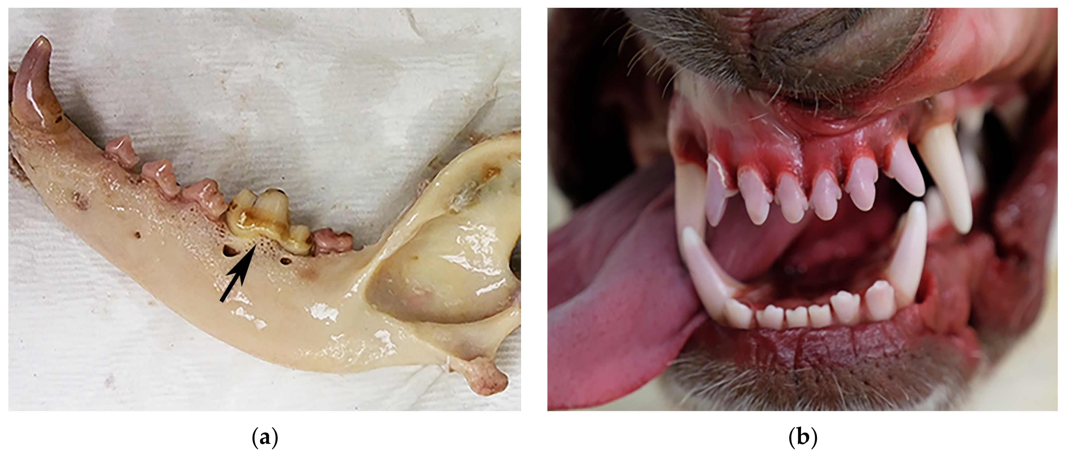
The almost completely defleshed skull of an adult dog was examined. The deceased dog was buried for an unknown period, and found with a wire around its neck. The skull was frozen prior to submission to the laboratory. There was bright pink discoloration of the crowns of multiple incisor (10), canine (3), premolar (13), and molar (7) teeth (
Figure 1a). The skull was refrozen after initial examination for storage purposes and reexamined 2 weeks later. When reexamined, the lower incisors’ color had changed from pink to red. One incisor was removed from the maxilla and there was light pink discoloration of the root. In contrast, a non-discolored premolar was removed and the root was not discolored. Five teeth were missing. Examination did not reveal significant dental disease. The cause of death was unknown.
3.2. Case 2: Frozen
The oral cavity of a 1.5-year-old intact female Husky-mix dog was examined. The body was initially frozen (9 days) and was thawed (1 day) prior to forensic autopsy. There was pink discoloration of the crowns of multiple incisor (7) and canine (3) teeth (
Figure 1b). Oral examination did not reveal significant dental disease. The dog died naturally due to multicentric lymphoma and hemoabdomen.
3.3. Case 3: Refrigeration
The oral cavity of a euthanized adult, intact female, pit bull type dog was examined. The dog was refrigerated for 28 days (range 6.1–13.7 °C) (decomposition study A). There was slight to light pink discoloration of the crowns of multiple incisor (8), canine (3), premolar (7), and molar (4) teeth. Oral examination did not reveal significant dental disease. Two teeth were missing.
3.4. Case 4: Refrigeration
The oral cavity of a euthanized adult, intact male dog was examined. The dog was refrigerated for 30 days (range 6.1–13.7 °C) (decomposition study A). There was slight to light pink discoloration of the crowns of multiple incisor (4), canine (4), premolar (13), and molar (6) teeth. Two canine teeth and a premolar were removed from the maxilla and there was pink to light red discoloration of the roots. Oral examination did not reveal significant dental disease. Three teeth were missing.
3.5. Case 5: Outdoors (on Ground Surface)
The oral cavity of an adult, intact male, pit bull type dog was examined. The dog was found deceased on the side of a road. Based on entomological evidence recovered from the dog, the time of colonization was 5 days prior to recovery and the mean temperature was 22.2 °C (range 15–30 °C). After the body was retrieved from the environment, it was refrigerated for 2 days prior to examination. There was light pink discoloration of the crowns of four incisors. Oral examination did not reveal significant dental disease. The cause of death was not determined; however, vehicular trauma was ruled out.
3.6. Case 6: Outdoors (on Ground Surface)
The oral cavity of an adult, intact male, pit bull type dog was examined. The dog was found deceased in a backyard. Based on entomological data, the dog was deceased for a minimum of 5 days and the mean temperature was 24.4 °C (range 20–32.7 °C). After the body was retrieved from the environment, it was refrigerated for 2 days prior to examination. There was pink discoloration of the crowns of multiple incisor (3), premolar (11), and molar (8) teeth. Oral examination did not reveal significant dental disease. The cause of death was complications of starvation due to exogenous circumstances.
3.7. Case 7: Outdoors (on Ground Surface)
The oral cavity of a euthanized adult, female (unknown spay status) Shih-Tzu dog was examined. The dog was placed outdoors in a wooded area on the ground surface (decomposition study B) in a cage to prevent scavenging. The dog was evaluated daily for various decompositional changes; pink discoloration of teeth was first observed on day 16 and ceased to be observed on day 19. The mean temperature was 22.3 °C (range 1.7–32.8 °C). The nature of this study meant that the dog was unable to be manipulated; only those teeth that could be visualized without manipulation were examined. There was pink discoloration of the crowns of one incisor, two canines, and one premolar tooth.
3.8. Case 8: Outdoors (on Ground Surface)
The oral cavity of a euthanized adult, male pit bull type dog was examined. The dog was placed outdoors in a wooded area on the surface (decomposition study B) in a cage to prevent scavenging. The dog was evaluated daily for various decompositional changes; pink discoloration of teeth was first observed on day 6 and ceased to be observed on day 9. The mean temperature was 27.3 °C (range 21–33.8 °C). The nature of this study meant that the dog was unable to be manipulated; only those teeth that could be visualized without manipulation were examined. There was slight to light pink discoloration of the crowns of one incisor, three canines, and three premolar teeth.
3.9. Case 9: Outdoors (on Ground Surface)
The body of a euthanized adult, female (unknown spay status) pit bull type dog was examined. The dog was placed outdoors in a wooded area on the surface (decomposition study C) in a cage to prevent scavenging. The dog was evaluated daily for various decompositional changes; pink discoloration of teeth was first observed on day 6 and ceased to be observed on day 10. The mean temperature was 27.3 °C (range 21–33.8 °C). The nature of this study meant that the dog was unable to be manipulated; only those teeth that could be visualized without manipulation were examined. There was slight pink discoloration of the crowns of three premolar teeth.
3.10. Case 10: Frozen
The defleshed skull of an adult domestic shorthaired cat was examined. The body (including the skull) of the cat was frozen and thawed prior to the autopsy being performed. There was pink discoloration of a single canine tooth. Oral examination did not reveal significant dental disease. The cat was reported to have died naturally due to hepatic lipidosis.
4. Discussion
The pink to red discoloration of teeth in humans has been reported in multiple scientific publications [
1,
2,
3,
4,
5,
6,
8,
9,
10] in all teeth types (incisors, canines, premolars, and molars) [
5]. At one point, it was thought that the development of pink teeth was indicative of asphyxiation, including drowning. In a retrospective review of cases of asphyxia (drowning, strangulation, and hanging), only a single occurrence of pink teeth was recorded [
10]. Although the occurrence of pink teeth is higher in cases of water immersion, it is generally accepted that the postmortem occurrence of pink teeth is not specific to any cause of death; rather it reflects postmortem decomposition, often in a humid environment [
1,
7]. A 2020 systematic review of the scientific literature found that the pink tooth phenomenon is unspecific and was more common in bodies found in wet and moist environments [
9]. Pink teeth and fingernails were described in a woman who died of a combination of trimipramine intoxication, hypothermia, and pneumonia [
6].
The coloration of human teeth observed in the phenomenon varies from slight to bright pink to shades of red, with red discoloration reported in the roots. The pink discoloration is affected by environmental moisture, as in the case of water immersion. It is theorized that the pink teeth phenomenon is due to hemolysis of erythrocytes and subsequent uptake of hemoglobin into dentin tubules [
5,
8,
13]. The breakdown of coagulated blood or lack of postmortem coagulation in the dental pulp, along with the hemolysis of erythrocytes, is thought to promote the diffusion of hemoglobin into dentinal tubules [
7]. Hemoglobin has been identified within dentin tubules in pink teeth using Pickworth’s benzidine method further supporting the hemoglobin diffusion theory [
8].
Postmortem observation of pink teeth is poorly documented in the veterinary literature. Anecdotally, a few studies describe pink teeth in dogs; when observed these dogs were reportedly frozen and subsequently thawed. In the Erlandsson and Munro study [
11], there was no detailed information regarding the number of dogs observed to have pink teeth or the number of pink teeth found during autopsies. In the current study, we identified pink discoloration of teeth from a number of different environmental conditions. Only two dogs (cases 1 and 2) and a cat (case 10) in this series of cases were frozen prior to examination. The dog from case 2 had pink teeth observed after thawing (10 days postmortem); whereas the time since death was not known for the other dog (case 1) and the cat. The dogs from cases 3 and 4 were refrigerated since death and both dogs had pink teeth observed 28 and 30 days postmortem, respectively. Cases 5–9 were all outdoors for varying amounts of time; based on entomological data, both dogs 5 and 6 were deceased for at least 5 days. Both dogs 5 and 6 were placed in refrigeration units for 2 days prior to autopsy and it is unknown what effect this had on the development of the pink discoloration of the teeth; pink teeth were observed in both dogs 7 days postmortem. Dogs 7–9 were never removed from the ground surface and the pink discoloration was first observed on days 16, 6, and 6, respectively. Interestingly, pink discoloration disappeared several days after initial detection in all three dogs.
In the Kirkham et al. and the Erlandsson and Munro studies, pink teeth were observed in samples from remains stored at refrigeration or room temperatures by three weeks after storage [
5,
11]. Similarly, we report two dogs with pink discoloration after 28 and 30 days of refrigeration that were part of a decomposition study (study A). The nature of the refrigeration decomposition study meant that the dogs were only manipulated once on a predetermined day in the study; therefore, it is not known on what day the teeth started to become pink. Notably, none of the 14 other dogs in study A (which were all assessed before the dogs with pink teeth) had pink teeth (range of time since death was 0–26 days). This suggests that development of pink teeth during refrigeration takes a long time to develop. In contrast to cases 1 and 2, dogs in cases 3–9 were never frozen; this supports the finding that freezing is not prerequisite for the development of pink discoloration of teeth in dogs. Rates of development of postmortem pink discoloration were faster in cases 5–9 than in the refrigerated dogs. Dog 7 developed pink discoloration at a slower rate than the other dogs outdoors, but faster than the refrigerated dogs. Dog 7 experienced a wider range of temperatures than the other outdoor dogs; however, the mean temperature was similar to cases 5 and 6.
As cooling a body is known to slow the decomposition process, we theorized that blood hemolysis occurred at a more rapid rate in dogs exposed to the outdoor environment and subsequently diffused into the dentin tubules. The dogs and cat that were frozen and subsequently thawed were theorized to have developed pink teeth during the thawing process, as erythrocytes were lysed during the freezing process, which would allow for uptake in dentin tubules after thawing. As none of the animals in this series had a clinical history of dental disease and postmortem examination did not reveal significant dental disease, dental disease did not appear to play a role in the development of pink teeth. Interestingly, the pink discoloration of dogs’ teeth in cases 7–9 disappeared several days after being recognized. A cause for the loss of pink discoloration is not known; however, it is possible that just as the pigment diffused into the dentin tubules, the pigment diffused out of the dentin tubules as part of the natural decomposition process.
This study had several limitations. As this was a retrospective study, we were unable to use color charts to document the discoloration of the teeth; the pink discoloration was not a binary present or absent phenomenon. Additionally, several dogs in the study could not be manipulated; therefore, the number of discolored teeth reported likely underrepresented the true value.
The detection of pink teeth is rarely reported during postmortem examination of animals; however, we suspect this phenomenon is both under recognized and under reported. Conventional autopsies of dogs and cats typically occur during the early postmortem period and it is possible that not enough time elapses for the pink discoloration of teeth to be noted in most cases. Additional studies are warranted to further study this phenomenon in dogs and cats, specifically regarding how various environmental conditions, time, and cause of death affect the appearance of pink teeth or lack thereof.


