Article

Biomechanical Risk Factors of Injury-Related Single-Leg Movements in Male Elite Youth Soccer Players

1
Department of Strength and Conditioning and Performance, Borussia Dortmund, 44309 Dortmund, Germany
2
Institute for Sport and Sport Science, TU Dortmund University, 44227 Dortmund, Germany
3
Department of Mechanical and Process Engineering, Offenburg University of Applied Sciences, 77652 Offenburg, Germany
*
Author to whom correspondence should be addressed.
Biomechanics 2022, 2(2), 281-300; https://doi.org/10.3390/biomechanics2020022
Submission received: 30 April 2022 / Revised: 18 May 2022 / Accepted: 23 May 2022 / Published: 26 May 2022
(This article belongs to the Special Issue Sports Biomechanics)

Abstract

:
Altered movement patterns during single-leg movements in soccer increase the risk of lower-extremity non-contact injuries. The identification of biomechanical parameters associated with lower-extremity injuries can enrich knowledge of injury risks and facilitate injury prevention. Fifty-six elite youth soccer players performed a single-leg drop landing task and an unanticipated side-step cutting task. Three-dimensional ankle, knee and hip kinematic and kinetic data were obtained, and non-contact lower-extremity injuries were documented throughout the season. Risk profiling was assessed using a multivariate approach utilising a decision tree model (classification and regression tree method). The decision tree model indicated peak knee frontal plane angle, peak vertical ground reaction force, ankle frontal plane moment and knee transverse plane angle at initial contact (in this hierarchical order) for the single-leg landing task as important biomechanical parameters to discriminate between injured and non-injured players. Hip sagittal plane angle at initial contact, peak ankle transverse plane angle and hip sagittal plane moment (in this hierarchical order) were indicated as risk factors for the unanticipated cutting task. Ankle, knee and hip kinematics, as well as ankle and hip kinetics, during single-leg high-risk movements can provide a good indication of injury risk in elite youth soccer players.

1. Introduction

The majority of all injuries (70–88%) in youth soccer players occur in the lower extremities, affecting the knee and ankle joints and the thigh and hip muscles [1,2]. Up to 72% of lower-extremity injuries in elite youth soccer players are reportedly non-contact injuries [3]. Altered neuromuscular control during high-risk movements in soccer is assumed to be the underlying mechanism of non-contact lower-extremity injuries [4,5,6]. In particular, sudden decelerations combined with a rapid change of direction while cutting or landing from a jump are frequent injury situations [7,8]. An injury ultimately occurs in these high-risk situations when tissue stress reaches a tissue’s maximal capacity. Excessive loading can result in mechanical failure and induce acute injuries, such as ligament sprains or muscle-tendon strains [4,9,10,11].
The assessment of kinematics and kinetics during jump-landing tasks can help identify non-contact injury risk (primarily of the knee joint) in different populations [12,13,14,15] defined to separate players at high risk from the rest [16,17]. An altered biomechanical motion of the lower extremities while completing this movement allows the ground reaction forces (GRF) to affect the lower-extremity alignment and is therefore a modifiable injury risk factor [18,19]. More specifically, an increased knee valgus angle, high knee abduction moments, reduced hip and knee flexion angles, internal rotation of the femur on the tibia and decreased range of motion of the ankle are associated with mainly anterior cruciate ligament (ACL) injuries in double-leg sagittal plane landings [12,20,21,22]. The ankle and hip joints are also essential components within the closed kinetic chain, and altered biomechanics of these joints influence the impact forces on the knee joint [22]. However, the effect of ankle biomechanics in the sagittal, frontal and transversal planes in prospective injury risk studies is an unexplored scientific area [19].
Soccer-specific movements often differ from double-leg actions (e.g., during change of direction movements, passing or landing after a header), requiring the athlete to bear all bodyweight through a single limb [23,24]. Although providing meaningful data, double-leg actions may not fully represent athletes’ neuromuscular strategies during high-risk movements [25].
Using single-leg screening tests may help identify soccer-related injury risk factors, as the previously mentioned dynamic multi-segmental malalignment patterns may become more apparent because of the absence of support for the contralateral leg and the smaller base of support [13,26]. Furthermore, biomechanical differences between double- and single-leg landings have already been observed. Studies have shown that landing with a single leg shows significantly less flexed knee and hip angles and higher joint moments, which increase the risk of lower-extremity injuries [25,27].
Moreover, soccer players perform an average of 726 cutting manoeuvres of 0–180° during match play [28]. Kinematic and kinetic analyses of unanticipated cutting movements may more closely simulate the kinematics and kinetics that the lower extremities experience during soccer-specific movements than the analysis of anticipated movements does; this is because of the limited time for the central nervous system to identify and accept the relevant input and perform neurocognitive processing to generate a dynamic neuromuscular response for successfully completing a movement with a low risk of being injured [29,30].
Numerous studies have described the kinematics of a lower-extremity injury during unanticipated cutting, especially ACL injuries. This occurrence could be explained by the fact that cutting movements have the propensity to create hazardous multiplanar knee joint loading when the foot is planted, such as high knee abduction moments and internal rotation moments, both of which might increase ACL strain and thus the risk of injury [31,32].
In summary, there is some indication that altered movement patterns in single-leg landings and unanticipated cutting movements increase the risk of lower-extremity injuries and that these high-risk movements represent non-contact injury scenarios well [33]. However, a significant amount of research about biomechanical risk factors is based on ACL injuries in female athletes, as they have a 2.3–9.7 times higher risk of ACL rupture than male athletes [34,35,36]. Nonetheless, ACL injury is a rather unusual soccer injury, constituting just slightly more than 5% of all injuries [36]. By contrast, muscle injuries (31–70%) and ligament injuries of the ankle (9–18%) account for most injuries in soccer [1,2]. As an altered biomechanical motion of the lower extremities can lead to exceeding the stress tolerance not only for the ACL but also for muscles (e.g., hamstring strain or adductor strain), tendons and ligaments in the ankle joint (e.g., ankle sprain) and knee joint (e.g., ACL rupture), more research is needed on how these movement patterns affect common injury types in soccer [37,38,39,40]. Additionally, previous studies have often used a local approach to determine risk factors. Non-local risk factors, which can be located distally or proximally to the site of injury, also seem to impact injury risk [41].
Prospective studies investigating the relationship between kinematics and kinetics of the lower extremities in all three planes of motion during high-risk movements and injury occurrence in elite youth soccer are rare. Prospective studies to identify cut-off scores, which play an important role in the practical implementation of preventive measures to distinguish injury-prone and non-injury-prone players, are also lacking. Therefore, the purpose of this study is (1) to assess ankle, knee and hip kinematics and kinetics during single-leg movements using a 3D motion analysis, (2) to investigate their association with the risk of non-contact lower-extremity injuries in elite youth soccer players and (3) to provide practice-relevant cut-off scores. It was hypothesised that ankle, knee and hip kinematics and kinetics during single-leg movements are associated with lower-extremity injury risk.

2. Materials and Methods

2.1. Study Design, Participants and Injury Data Collection

This investigation was a prospective cohort study. Teams of the under-16 up to the under-19 age categories from the youth academies of two professional German soccer clubs were requested to take part in this study. Three teams (<16, <17 and <19 years old) volunteered to participate. Written information about the study design, the purpose of the study and the potential risks and benefits of participating in the study was provided to all members of the playing squad and their parents or legal caretakers for each team. The Ethics Committee of TU Dortmund University confirmed that the requirements of the Declaration of Helsinki were met.
Sixty-two male elite youth soccer players gave written informed consent to participate. Trained assessors, coordinated by the principal investigator (MK) to guarantee an equal testing process, tested these players at the beginning of the preparation period of the 2018/2019 season via laboratory-based injury risk screening in the laboratories of TU Dortmund University. On the day of testing, the assessors asked the players to complete a questionnaire to collect their demographic data, background information and history of previous lower-extremity injuries. All lower-extremity injuries up to 6 months before the assessments had to be reported.
Immediately after the testing, the players were tracked over a 10-month period to prospectively record any injuries sustained during training and competition. The injury data collection followed the procedures outlined in a consensus statement on injury definitions and data collection procedures in studies of soccer injuries [42]. Only time-loss non-contact lower-limb injuries were considered. Further information about the inclusion and exclusion criteria, as well as the injury data collection process, has been described in detail elsewhere [43].

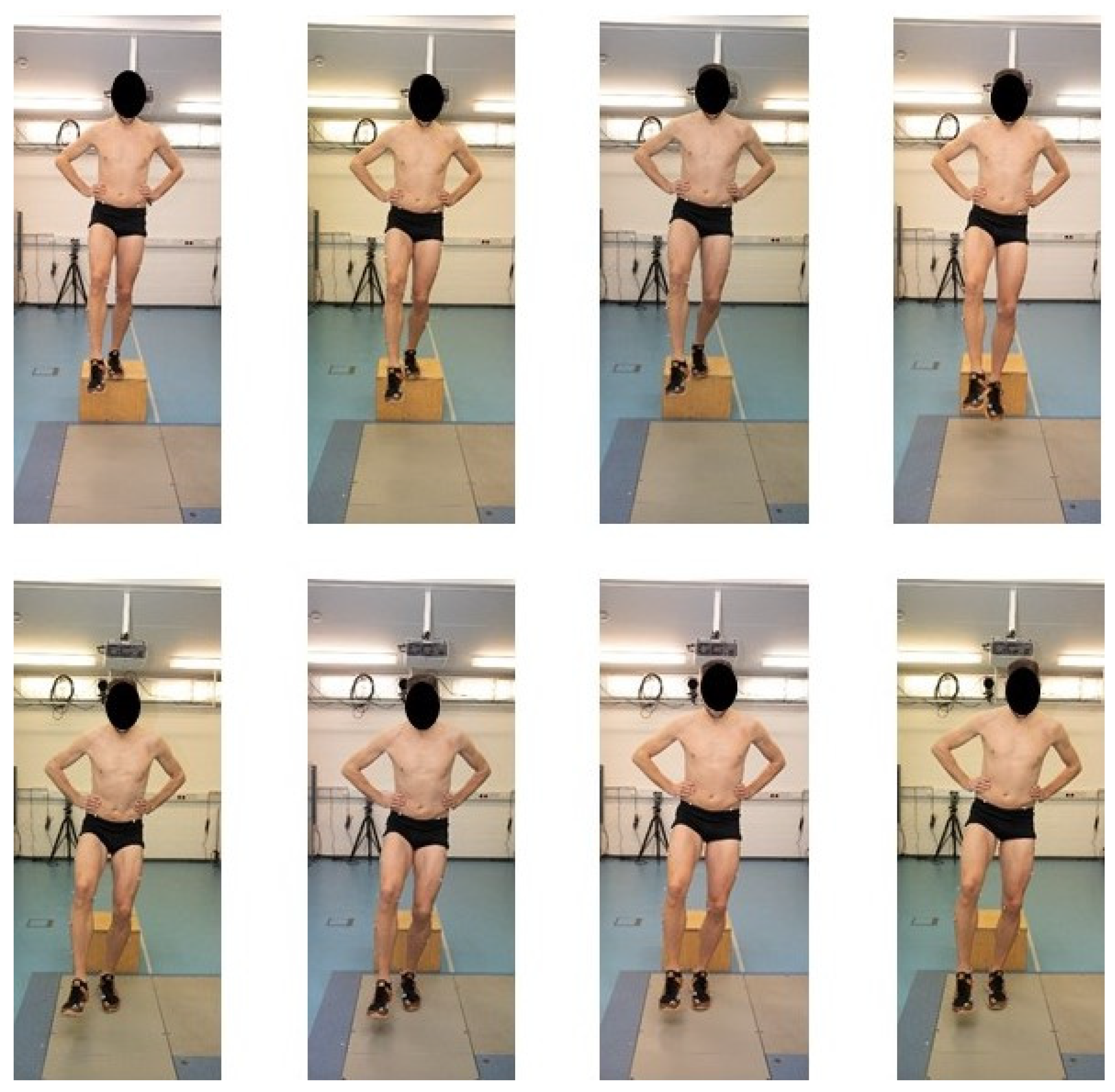
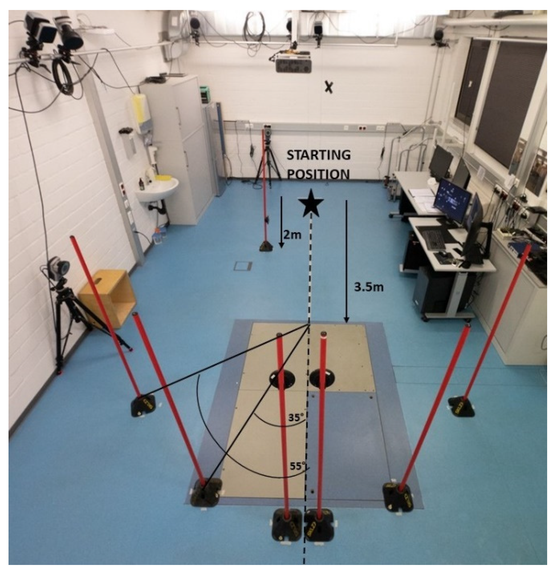
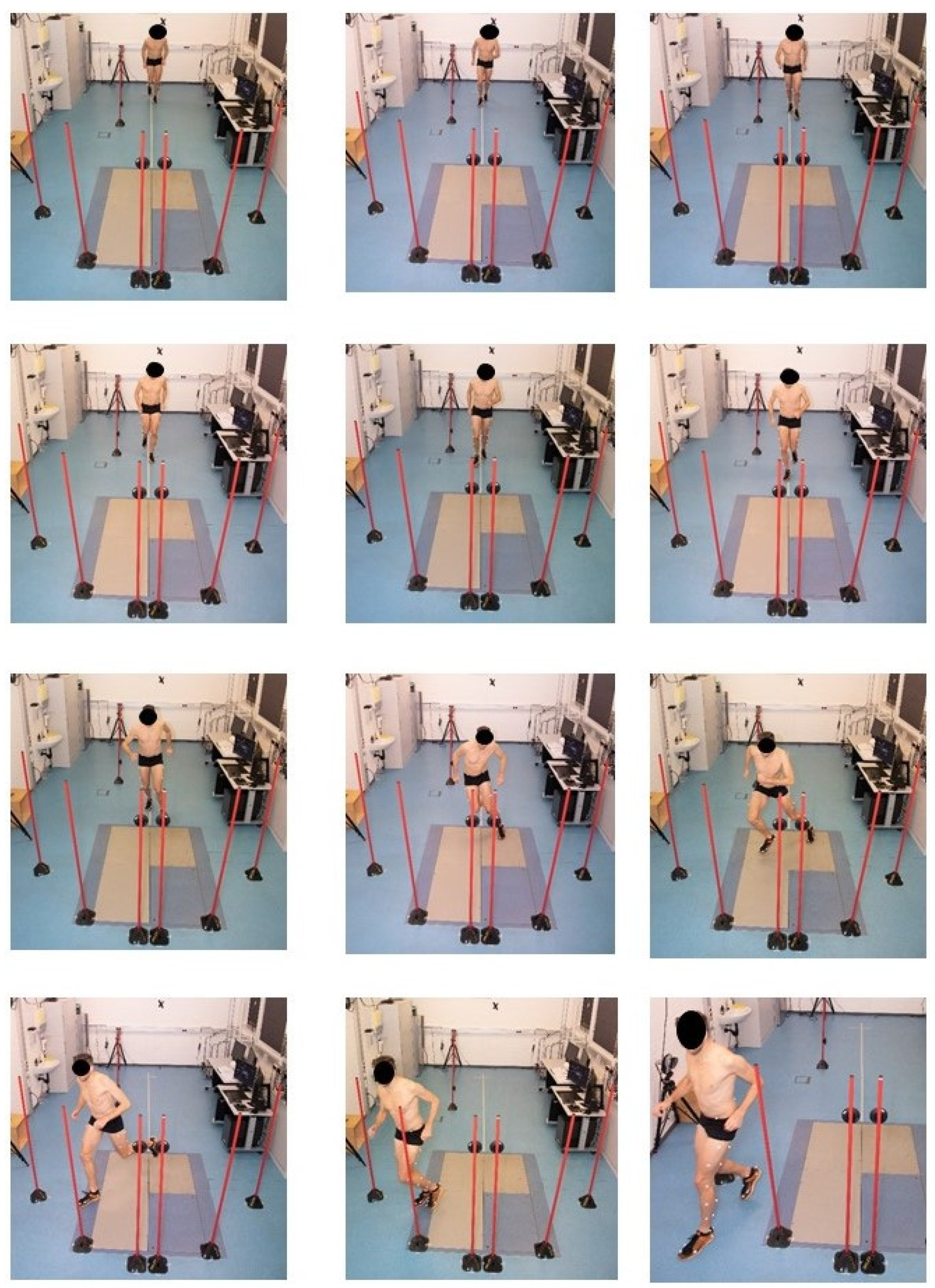
2.2. Experimental Protocol

All players performed a standardised warm-up, including 5 min of cycling on a bike ergometer, followed by movement preparation and plyometric exercises. After the warm-up, the players completed a single-leg drop landing (SLDL) task and an unanticipated side-step cutting (USSC) task with both legs. Adequate familiarisation with the tasks was facilitated in the form of two practice trials. Once the participants were comfortable with the tasks, they were asked to perform three successful trials for each leg. All landing and cutting tasks were performed on two force plates (AMTI Inc.®, 176 Waltham St, Watertown, MA, USA) measuring 0.9 m × 0.6 m each at a 1000 Hz sampling rate.
The SLDL task consisted of the participants starting on top of a box (30 cm in height), with their feet shoulder width apart and their hands fixed at the hip. The participants were instructed to shift their weight onto one leg and to position the other leg in front of the box. Starting from this position, they were asked to drop down (not jump) off the box to the force plate with the weight-bearing leg, adopt the landing for 2 s and not touch the ground with the opposite leg [44] (see Figure 1).
The USSC task was performed from a fixed starting distance of 3.5 m in front of the two force plates. Two poles with one light-powered LED sensor (Fitlight®, VISUS GmbH, Herrenberg, Germany) at a height of 1.5 m were placed directly behind the force plates. One pole with LED sensor at a height of 0.8 m was placed on the side 2 m after the start line and used as a light barrier. Poles placed 35° and 55° from the long axis of the runway were used to force the participants to cut at an angle of approximately 45° to the right or left side [45,46] (see Figure 2).
The participants stood in an athletic ready position and accelerated towards the force plates. Immediately after passing the light barrier, they saw a flashing light on either the left or right sensor, indicating the intended cutting direction. The athletes were instructed to perform the side-step cutting task as fast as possible and as close as possible to a match-play situation (see Figure 3). Three successful trials for each side, performed in random order, were selected for further analysis. A rest period of at least 30 s was provided between repetitions, and rests of at least 2 min were provided between tasks to reduce the effect of fatigue [47,48,49,50].

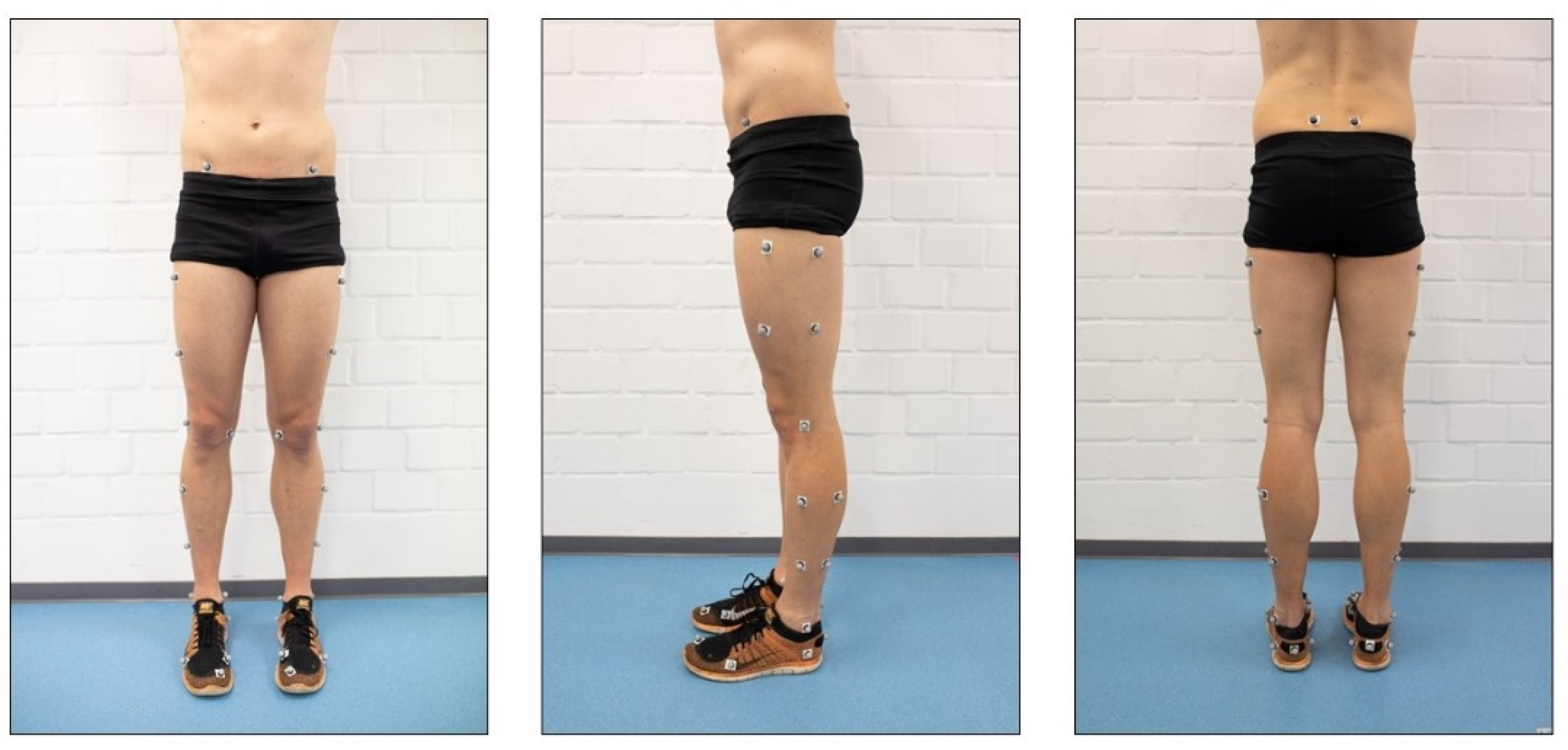
2.3. Kinematic and Kinetic Analysis

The participants’ lower-body motion was captured using a 3D motion capture system that consisted of 12 infrared cameras (120 Hz, Qualisys®, Göteborg, Sweden) and was time-synchronised to the force plates and a lower-body marker set of 40 markers [51] (see Figure 4).
Marker trajectory and force plate data were filtered with a fourth-order digital Butterworth filter with a cut-off frequency of 20 Hz to avoid impact-related artefacts in lower-extremity joint moments [52,53]. Lower-extremity joint angles and resultant internal joint moments were determined using a rigid body model of the lower extremities, including the forefoot, rearfoot, shank, thigh and pelvis segments [54,55]. Therefore, a customised MATLAB code (MathWorks Inc.®, Natick, MA, USA) was used to calculate kinematic and kinetic parameters during the ground-contact phase using a 3D inverse dynamics model. The ground-contact phases for the SLDL and USSC tasks were defined as the interval from foot strike on the force plate to take-off using a threshold of 20 N of the smoothed vertical GRF [46,49]. Hip and knee flexion–extension, abduction–adduction and internal–external rotation angles and ankle dorsiflexion–plantarflexion, eversion–inversion and internal–external rotation angles were calculated as kinematic variables. Kinetics implied hip and knee flexion–extension, abduction–adduction and internal–external rotation moments; ankle dorsiflexion–plantarflexion, eversion–inversion and internal–external rotation moments; and vertical GRF (vGRF). Joint moments were expressed as the external moment applied to the joint. Body mass was used to normalise all kinetic parameters. Joint torques were expressed in the anatomical coordinate systems of the respective proximal segments. Kinematic parameters were determined at two points during the stance phase: initial contact (IC) and peak (PEAK), whereas kinetic parameters were determined at PEAK only. IC was defined as the first instance of the ground-contact phase, whereas PEAK was defined as the peak value within the first 100 ms after IC [49]. This period was chosen because ACL injuries are likely to occur in this period [56]. The average value of both legs and three successful trials was calculated for each parameter and considered for further analysis for each task.

2.4. Statistical Analyses

Data are presented as means ± standard deviations. A decision tree was utilised to discriminate between injured and non-injured players. The tree was created using the classification and regression tree (CART) method [57]. In each iteration of the optimisation, the method chooses the cut in all available variables that maximises the reduction of the Gini impurity from the given observations to the subsequent partition into two groups. This process is repeated iteratively for all resulting partitions until the reduction is less than 0.01 or a partition contains fewer than 20 participants. This leads to a hierarchical tree-like structure of binary decisions, which is highly interpretable (e.g., via a dendrogram).
Once the decision tree was optimised, the association to injury was assessed using a 2 × 2 contingency table based on the decision tree’s classification and the true injury status of the players. Relative risk, sensitivity and specificity were calculated from the contingency table. Relative risk describes the increase in the probability of injury from players classified as injured to players classified as non-injured, whereas sensitivity and specificity served as performance measures for the decision tree [58,59,60]. Further information about the optimisation process of the decision tree and the calculation of relative risk, sensitivity and specificity has been described in detail elsewhere [43].

3. Results

3.1. Injury Epidemiology

Sixty-two elite youth soccer players (age: 17.2 ± 1.1 years; height: 179 ± 8 cm; weight: 70.4 ± 9.2 kg) were enrolled in the study. Because of technical problems, the data of six players could not be included in the analysis, which resulted in an attrition rate of 9.6%. Therefore, the data analysis was based on 56 players. There is no significant difference in age (p > 0.05), height (p < 0.05) and weight (p > 0.05) between injured and non-injured players. In this cohort, 23 non-contact injuries were registered over the 2018/2019 season, with 39% of the players (n = 22) having one or more non-contact injuries. Sixty-one percent of the players (n = 34) remained injury-free. The overall non-contact injury incidence was 1.2/1000 h of total exposure time (0.5 injuries per 1000 h of training and 3.9 injuries per 1000 h of competition). The most frequently injured body parts were the ankle (36%) and thigh muscles (hamstrings: 18%, quadriceps: 18%), followed by the adductors (16%) and the knee (12%). Sprains (48%) and strains (39%) were the most common injury types. Overuse injuries accounted for 13% of all non-contact injuries, whereas no fracture was counted.

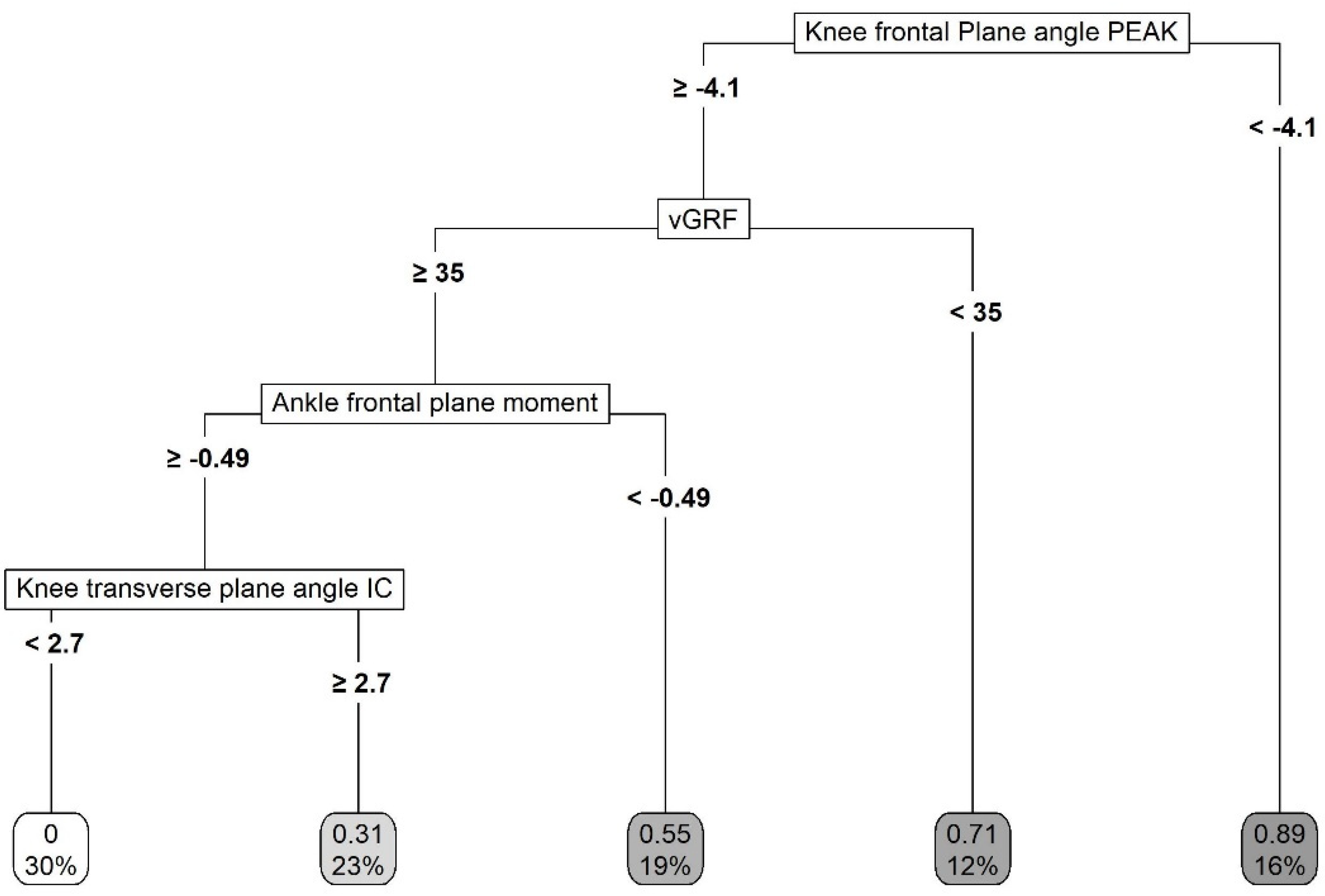
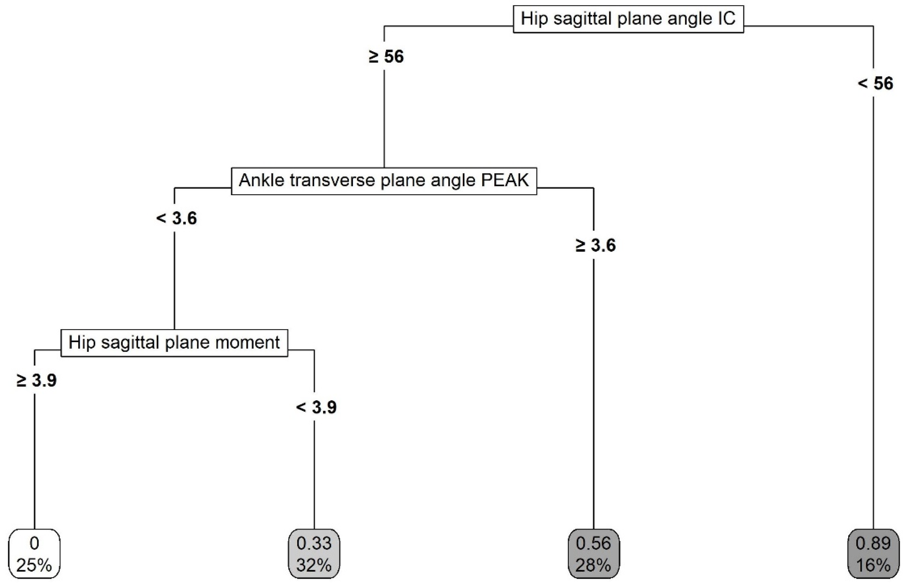
3.2. Multivariate Analysis

Table 1, Table 2 and Table 3 present the descriptive statistics for all kinematic and kinetic parameters obtained from the biomechanical injury risk screening.
The decision tree model indicated the knee frontal plane angle PEAK, peak vGRF, ankle frontal plane moment and knee transverse plane angle IC (in this hierarchical order) for the single-leg landing task (see dendrogram in Figure 5) as important biomechanical parameters to discriminate between injured and non-injured players. The hip sagittal plane angle IC, ankle transverse plane angle PEAK and the hip sagittal plane moment (in this hierarchical order) were indicated for the unanticipated cutting task (see dendrogram in Figure 6).
The classification of the decision tree model for the single-leg landing task showed a sensitivity of 0.70 and a specificity of 0.87. The relative risk was calculated as 3.5 (see Table 4).
The classification of the decision tree model for the unanticipated cutting task showed a sensitivity of 0.68 and a specificity of 0.82. The relative risk was calculated as 3.1 (see Table 5).

4. Discussion

Understanding the movements and forces distributed throughout the lower limbs during single-leg high-risk movements is required to understand potential biomechanical injury risk factors. Multivariate analysis using a decision tree model indicated the knee frontal plane angle PEAK, peak vGRF, ankle frontal plane moment and knee transverse plane angle IC for the single-leg landing task as important biomechanical parameters to discriminate between injured and non-injured players. The hip sagittal plane angle IC, ankle transverse plane angle PEAK and hip sagittal plane moment were indicated for the unanticipated cutting task. Players who were classified as injured according to the decision tree model for the single-leg landing task and for the unanticipated cutting task had a threefold increase in injury risk compared with those who were classified as non-injured. The properties of the decision tree model for the single-leg landing task indicated that the corresponding classification correctly identified 70% of injured players as being injured, whereas the classification correctly identified 68% of injured players as such for the unanticipated cutting task. In addition, the classification correctly identified 87% of uninjured players as such for the single-leg landing task and 82% for the unanticipated cutting task. Therefore, measuring ankle, knee and hip kinematics, as well as ankle and hip kinetics, during single-leg high-risk movements provides good indication of injury risk in elite youth soccer players.
To the best of our knowledge, the present study is the first to analyse kinematic and kinetic parameters of the ankle, knee and hip via 3D motion analysis as injury risk factors in elite youth soccer. There is currently a lack of evidence describing the kinematics and kinetics of the lower extremities during landing and cutting tasks for elite youth soccer players in general. In this respect, appropriate comparisons with previous studies are limited. Given the complexity of the decision tree results, we decided to discuss the cuts of the decision tree optimisation and to discuss only the paths that predict the highest probability of injury.

4.1. Single-Leg Landing Task

As a result of numerous studies with mostly small samples, knee abduction has become widely accepted as an undesirable movement pattern and injury risk factor [12,21,61]. The investigation of knee abduction during jump-landing tasks is valuable for identifying the non-contact injury risk for the knee joint and especially the ACL [12,13,14]. The results of our prospective injury risk factor identification process confirm this finding. The peak knee frontal plane angle was selected in the first cut of the decision tree optimisation and served as the root node. The related leaf node included 16% of the players with peak knee abduction angles greater than 4.1°, of whom 89% sustained an injury. At first, the importance of the knee abduction angle was surprising, as only three of the twenty-three injuries in this study affected the knee joint. In addition, only one injury could be described as traumatic (ACL rupture); the other two were due to overuse (patellofemoral pain). In general, a low number of knee injuries may be a consequence of successful injury prevention programmes in the last decade, especially those focused on reducing knee abduction (or dynamic valgus collapse; [62,63]). This notion is supported by the fact that Hewett, Myer, Ford, Heidt, Colosimo, McLean, van den Bogert, Paterno and Succop [12] found a 9° peak knee abduction angle in a landing task, whereas our data showed only a 0.9° peak knee abduction angle, which seems insufficient to damage knee structures [64]. However, we observed notably altered kinematics in the ACL-injured player compared with the non-injured players. Specifically, the ACL-injured player demonstrated a 15-fold higher peak knee abduction angle, and he was one of the players assigned to the related node. In contrast to our study, Krosshaug et al. [65] found no association between knee abduction kinematics and kinetics and the risk of ACL injury in their large cohort study. In their meta-analyses, Cronström et al. [66] and Romero-Franco, Ortego-Mate and Molina-Mula [64] did not find that the knee abduction angle at initial contact, peak knee abduction angle and peak knee abduction moment were injury risk factors. However, Krosshaug, Steffen, Kristianslund, Nilstad, Mok, Myklebust, Andersen, Holme, Engebretsen and Bahr [65], Cronström, Creaby and Ageberg [66], and Romero-Franco, Ortego-Mate and Molina-Mula [64] investigated only knee injuries, especially injuries to the ACL. Additionally, Krosshaug, Steffen, Kristianslund, Nilstad, Mok, Myklebust, Andersen, Holme, Engebretsen and Bahr [65] used a double-leg vertical drop jump. These methodological differences make comparison with the present study difficult, because ACL injuries have different underlying injury mechanisms than, for example, muscle injuries [67]. Further, double-leg actions may not fully represent athletes’ neuromuscular strategies during high-risk movements [25], and the dynamic multi-segmental malalignment patterns may become more apparent during single-leg screening tests because of the absence of the support of the contralateral leg and smaller base of support [26]. Interestingly, half of the ankle injuries in the present study occurred within the related leaf node. Therefore, knee abduction seems to be a critical kinematic variable for more than knee (and especially ACL) injuries. This interpretation is supported by a prospective study by Powers et al. [68], who found that weak hip abductor muscles predict ankle sprains in soccer players. Hip abductor weakness has also been related to increased knee frontal plane motion. Associations between hip abductor muscles and knee abduction were previously identified during a single-leg squat test and a landing task [69,70]. More research is needed on how the different elements (e.g., specific faulty movements or strength and postural control) of clinically oriented screening tools predict future lower-extremity injuries in elite youth soccer players.
The GRF is an important variable in biomechanical studies and presents an approximate measure of the external loading of the lower-extremity musculoskeletal system [71]. Peak vGRF was selected as the second splitting variable for the optimisation of the decision tree. One normally expects players who experience more severe loading to be at an increased risk of injury [72]. However, the optimisation of the decision tree showed that the risk of suffering a non-contact lower-extremity injury was higher for players with a lower vGRF. One potential explanation for the contrasting results could be that previous research on vGRF and injury risk focused on knee loading [27,73], whereas the knee was a rarely injured body part in the present study. Specifically, a stiff or low-flexion landing contributes to greater knee loading and results in greater vGRF compared with a soft or high-flexion landing [74]. A meta-analysis also showed no significant differences between the GRF of individuals with lower limb stress fractures and those in the control groups [75]. Therefore, we speculate that a soft landing strategy results in a greater relative contribution from the hip extensor to attenuate GRFs and that repetitive movements could exceed the tolerance of these muscles, which are frequently injured in soccer.
The ankle frontal plane moment was selected as the third splitting variable to optimise the decision tree. In principle, this classification showed that the risk of suffering a non-contact lower-extremity injury was higher for players with a higher ankle inversion moment. Koshino et al. [76] found that an internal rotated foot during a single-leg landing increased the ankle inversion moment, which we can confirm with our results, as injured players landed with a 9.6° internal rotated ankle. It has been frequently hypothesised that contacting the ground in an increased inversion position could result in an ankle sprain [77,78,79], which was the most frequently injured body part and injury type in the present study. However, to date, little is known about the biomechanical abnormalities of the ankle joint, especially in the frontal plane, in prospective injury risk studies.
Finally, the knee transverse plane angle at initial contact was selected as the fourth splitting variable to optimise the decision tree. In principle, this classification showed that the risk of suffering a non-contact lower-extremity injury was higher for players with a higher knee external rotation angle. This is in line with previous research investigating ACL injury risk and focusing on the ‘position of no return’ [80,81], which includes components of tibial external rotation and knee valgus [82]. By contrast, Everard et al. [83] and Dai et al. [84] emphasised the combined effects of less knee flexion and more significant knee internal rotation on an increased risk of knee injuries. However, all these studies investigated only knee injuries, especially those of the ACL, and therefore can only be compared with the present study to a limited extent. Overall, the role of knee transverse plane kinematics as injury risk factors in single-leg landings should be thoroughly investigated in future studies with larger sample sizes.

4.2. Unanticipated Cutting Task

During dynamic tasks, downward momentum of the body is stopped by flexing the joints of the lower limb after foot–ground contact to absorb the GRFs [85,86]. The need to attenuate and redirect forces in the sagittal plane is important, especially when changing direction [87,88]. The capability of appropriate joint flexion to absorb the generated kinetic energy and thus mitigate stress on passive structures, such as ligaments and tendons, has been discussed intensively [19,22,89]. This discussion is supported by a substantial body of evidence showing that sagittal plane factors contribute to acute and overuse injuries [12,90,91]. In line with these findings, the hip sagittal plane angle at initial contact was selected for the first cut in the present study to optimise the decision tree and serve as the root node. This showed that the risk of suffering a non-contact lower-extremity injury was higher for players with a lower hip flexion angle. The related leaf node included 16% of the players with a hip flexion angle of less than 59° at initial contact, 89% of whom sustained an injury. We assume that increased hip-joint stiffness reflects a compensating hip-dominant movement strategy during the USSC task, which has been identified in individuals in the chronic stages of lateral ankle sprains, a common injury in youth soccer [1,2]. A compensatory redistribution of the load within the limbs occurs in non-injured players to increase their reliance on the proximal joints and absorb the impact forces, thus protecting the ankle from injury [92]. The association between increased hip-joint stiffness and increased injury risk is supported by the selection of the hip sagittal plane moment as the third splitting variable for the optimisation of the decision tree. This showed that the risk of suffering a non-contact lower-extremity injury was higher for players with a lower hip flexion moment. This finding supports our conclusion that a high-risk posture in the USSC task for lower-extremity injuries includes hip-joint stiffness in the sagittal plane.
The peak ankle transverse plane angle was selected as the second splitting variable for the optimisation of the decision tree. This indicates that the risk of suffering a non-contact lower-extremity injury was higher for players with a greater ankle external rotation angle. The selection of the ankle transverse plane angle seems surprising, as researchers have demonstrated that the primary sagittal plane motion of the ankle is associated with injuries to the ankle, Achilles tendon and plantar-flexor muscles [22,93,94]. However, our results are supported by an overview of sub-optimal cutting techniques by Donelon, Dos’Santos, Pitchers, Brown and Jones [87]. An outwardly rotated foot has been shown to lead to increased susceptibility to eversion and pronation, which could also lead to knee abduction and tibial external rotation [87], which describes the ‘position of no return’ introduced by Ireland [80]. Overall, the role of ankle kinematics and kinetics as injury risk factors in single-leg high-risk movements should be investigated thoroughly in future studies that include larger sample sizes.

4.3. Practical Applications

In a targeted and successful injury prevention model, understanding why an injury occurs and the extent to which the occurrence of the injury is the result of a non-linear interaction between multiple risk factors is important [58,95,96]. Using the CART method [57], a decision tree was optimised, in which not only were risk factors identified using a multivariate procedure (as in previous research), but their interactions were also captured at the same time that practice-relevant cut-off values were determined, which is somewhat unique in injury prevention research. The resulting tree structure has a natural visualisation through its paths and nodes and thus a good data interpretability. Therefore, the use of the decision tree model and the identified cut-off values will allow practitioners and coaches to determine the injury risks of their players and enable customised injury prevention programmes to be provided as part of each player’s daily training schedule. It has been shown that injury prevention programmes have the potential to change faulty motion patterns in single-leg movement, which results in a decreased injury risk [97,98,99] and decreased injury rates [100,101]. This is supported by several reviews and meta-analyses on the effects of multicomponent exercise prevention programmes for effective injury risk reduction [102,103,104,105,106].

4.4. Limitations

A limitation of all laboratory studies is that one cannot conclude how the measured movement patterns relate to the biomechanics of real soccer-specific movements [49]. However, we added an unanticipated stimulus to the cutting task that may more closely simulate the kinematics and kinetics that the lower extremities experience during soccer-specific movements, as there is limited time for a player to make postural adjustments [30]. Accordingly, the unanticipated cutting protocol in the present study was not easy to standardise, as all athletes used their preferred cutting technique. However, the resulting variation in side-step cutting techniques likely reflects the variation in the cutting techniques used during active gameplay [30,49].
While biomechanical analyses of cutting have focused primarily on manoeuvres performed at lower degrees (e.g., 45°), research suggests that cutting to larger angles results in different lower-extremity biomechanics and higher knee loading in male athletes [107,108]. However, we had the players cut at an angle of approximately 45° to be consistent with the methods used in comparable studies (e.g., Pollard et al., 2007; Jeong et al., 2021).
It has been shown that jump and cutting speed affect lower-extremity biomechanics [109]. As we used a drop landing task, the jump height could not be used as a performance parameter.
Each injury was weighted equally, regardless of its occurrence during the season. As the physical performance level of the players has a tendency to deteriorate over the season and the overall level of fatigue may accumulate, the injury risk has to be evaluated during the time more closely preceding the injury event [110,111]. Therefore, a single preseason evaluation of players has limited value in predicting injury risk throughout the season because of the changing nature of the risk factor profile, and there is a need for multiple assessments throughout the season. Solely screening players in a non-fatigued state may not accurately identify those individuals whose movement patterns deteriorate towards the end of a match, affecting their relative risk of injury. Furthermore, changes in injury risk at different stages of the development of youth soccer players seem probable [112].
Knee joint angle measurements taken from skin-mounted markers are challenged by problems such as skin motion and soft-tissue artefacts. Consequently, in our calculations, we applied a method in which the marker coordinates of each trial were mathematically optimised to improve their compliance with rigid body assumptions [113]. Despite the well-known problems in calculating accurate joint kinematics, particularly for the transverse plane, the applied method is currently considered the gold standard approach for the non-invasive calculation of 3D joint kinematics.
Notwithstanding the many advantages of the decision tree (e.g., illustration of interactions and determination of cut-off scores), because of the underlying database, a sharp distinction between at-risk and non-at-risk players on the basis of the cut-off score must be treated with caution. Care should be taken when interpreting the present results, especially practice-relevant cut-off scores.
Furthermore, the primary purpose of the study was to investigate between-group differences. However, taking into account that soccer players are exposed to asymmetric musculoskeletal loading because of their dominant and non-dominant legs is important.
Taking all these limitations into account, the association between larger cutting angles and the risk of injury should be considered in further research. Additionally, the approach speed (m/s) in the unanticipated side-step cutting task should be determined and injury risk analyses of inter-subject differences should be conducted between injured and non-injured legs and between dominant and non-dominant legs. Finally, the number and distribution of injuries did not allow for in-depth analyses by subgroups, such as by type of injury, which could be beneficial for more detailed injury prevention programmes. Therefore, larger samples are required in future research. Finally, the occurrence of an injury represents a complex systemic reaction that includes not only biomechanical or neuromuscular risk factors but also psychological risk factors [95]. Post-traumatic stress disorder (PTSD) is categorised as a trauma disorder, and symptoms are a direct result of a severe injury [114]. However, the extent to which severe injuries in youth soccer players can lead to PTSD, which in turn influences future movement patterns during high-risk movements, is still not well-known.

5. Conclusions

This research suggests that assessing ankle, knee, and hip kinematics as well as ankle and hip kinetics during single-leg high-risk movements can provide a good indication of injury risk in elite youth soccer players. These preliminary results may have practical implications for future directions in injury risk screening and for planning and developing customised training programmes to counteract intrinsic injury risk factors. An enhanced understanding of the associated coupled lower-extremity joint kinematics and kinetics in all movement planes may help determine distinct movement strategies that can decrease lower-extremity injury risk in elite youth soccer players. However, the role of lower-extremity biomechanics as injury risk factors in single-leg high-risk movements should be thoroughly investigated in future studies with larger sample sizes.

Author Contributions

Conceptualization, M.K. and T.J.; methodology, M.K.; software, S.W. and K.N.; validation, M.S.; formal analysis, M.K., S.W., K.N. and M.S.; investigation, M.K.; resources, M.K.; data curation, S.W., K.N. and M.S.; writing—original draft preparation, M.K.; writing—review and editing, S.W., K.N. and T.J.; visualization, M.K.; supervision, M.K. and T.J.; project administration, M.K. All authors have read and agreed to the published version of the manuscript.

Funding

This research received no external funding.

Institutional Review Board Statement

The study was conducted in accordance with the Declaration of Helsinki and was approved by the Ethics Committee of TU Dortmund University (2018-07).

Informed Consent Statement

Informed consent was obtained from all participants involved in the study. Written informed consent was obtained from the participants to publish this paper.

Data Availability Statement

The data are not publicly available because the data owners are two elite soccer clubs in Germany which wanted to remain anonymous and did not give permission to make the original data publicly available. However, the authors can provide, upon reasonable request, transformed data that are processed in such a way that re-identifying the subjects involved in the study is not possible.

Acknowledgments

The authors would like to thank all the participants who volunteered to join in this study.

Conflicts of Interest

The authors declare no conflict of interest.

References

  1. Pfirrmann, D.; Herbst, M.; Ingelfinger, P.; Simon, P.; Tug, S. Analysis of injury incidences in male professional adult and elite youth soccer players: A systematic review. J. Athlet. Train. 2016, 51, 410–424. [Google Scholar] [CrossRef] [PubMed] [Green Version]
  2. Price, R.; Hawkins, R.; Hulse, M.; Hodson, A. The football association medical research programme: An audit of injuries in academy youth football. Br. J. Sports Med. 2004, 38, 466–471. [Google Scholar] [CrossRef] [PubMed] [Green Version]
  3. Renshaw, A.; Goodwin, P.C. Injury incidence in a premier league youth soccer academy using the consensus statement: A prospective cohort study. BMJ Open Sport Exerc. Med. 2016, 2, e000132. [Google Scholar] [CrossRef] [Green Version]
  4. Read, P.J.; Oliver, J.L.; De Ste Croix, M.B.; Myer, G.D.; Lloyd, R.S. Neuromuscular risk factors for knee and ankle ligament injuries in male youth soccer players. Sports Med. 2016, 46, 1059–1066. [Google Scholar] [CrossRef] [PubMed] [Green Version]
  5. Zazulak, B.T.; Ponce, P.L.; Straub, S.J.; Medvecky, M.J.; Avedisian, L.; Hewett, T.E. Gender comparison of hip muscle activity during single-leg landing. J. Orthop. Sports Phys. Ther. 2005, 35, 292–299. [Google Scholar] [CrossRef] [PubMed] [Green Version]
  6. Lehnert, M.; De Ste Croix, M.; Zaatar, A.; Hughes, J.; Varekova, R.; Lastovicka, O. Muscular and neuromuscular control following soccer-specific exercise in male youth: Changes in injury risk mechanisms. Scand. J. Med. Sci. Sports 2017, 27, 975–982. [Google Scholar] [CrossRef]
  7. Boden, B.P.; Sheehan, F.T.; Torg, J.S.; Hewett, T.E. Noncontact anterior cruciate ligament injuries: Mechanisms and risk factors. J. Am. Acad. Orthop. Surg. 2010, 18, 520–527. [Google Scholar] [CrossRef] [Green Version]
  8. Murphy, D.; Connolly, D.; Beynnon, B. Risk factors for lower extremity injury: A review of the literature. Br. J. Sports Med. 2003, 37, 13–29. [Google Scholar] [CrossRef] [Green Version]
  9. Whiting, W.C.; Zernicke, R.F. Biomechanics of Musculoskeletal Injury; Human Kinetics: Champaign, IL, USA, 2008. [Google Scholar]
  10. Ross, S.E.; Guskiewicz, K.M.; Yu, B. Single-leg jump-landing stabilization times in subjects with functionally unstable ankles. J. Athlet. Train. 2005, 40, 298–304. [Google Scholar]
  11. Hughes, G.; Watkins, J. A risk-factor model for anterior cruciate ligament injury. Sports Med. 2006, 36, 411–428. [Google Scholar] [CrossRef]
  12. Hewett, T.E.; Myer, G.D.; Ford, K.R.; Heidt, R.S., Jr.; Colosimo, A.J.; McLean, S.G.; van den Bogert, A.J.; Paterno, M.V.; Succop, P. Biomechanical measures of neuromuscular control and valgus loading of the knee predict anterior cruciate ligament injury risk in female athletes: A prospective study. Am. J. Sports Med. 2005, 33, 492–501. [Google Scholar] [CrossRef] [Green Version]
  13. Dingenen, B.; Malfait, B.; Nijs, S.; Peers, K.H.; Vereecken, S.; Verschueren, S.M.; Staes, F.F. Can two-dimensional video analysis during single-leg drop vertical jumps help identify non-contact knee injury risk? A one-year prospective study. Clin. Biomech. 2015, 30, 781–787. [Google Scholar] [CrossRef] [PubMed]
  14. Alentorn-Geli, E.; Mendiguchia, J.; Samuelsson, K.; Musahl, V.; Karlsson, J.; Cugat, R.; Myer, G.D. Prevention of anterior cruciate ligament injuries in sports. Part I: Systematic review of risk factors in male athletes. Knee Surg. Sports Traumatol. Arthrosc. 2014, 22, 3–15. [Google Scholar] [CrossRef]
  15. Fagenbaum, R.; Darling, W.G. Jump landing strategies in male and female college athletes and the implications of such strategies for anterior cruciate ligament injury. Am. J. Sports Med. 2003, 31, 233–240. [Google Scholar] [CrossRef] [PubMed] [Green Version]
  16. Van Mechelen, W.; Hlobil, H.; Kemper, H.C. Incidence, severity, aetiology and prevention of sports injuries. A review of concepts. Sports Med. 1992, 14, 82–99. [Google Scholar] [CrossRef] [PubMed]
  17. Bahr, R. Why screening tests to predict injury do not work—And probably never will…: A critical review. Br. J. Sports Med. 2016, 50, 776–780. [Google Scholar] [CrossRef] [Green Version]
  18. Raisanen, A.M.; Arkkila, H.; Vasankari, T.; Steffen, K.; Parkkari, J.; Kannus, P.; Forsman, H.; Pasanen, K. Investigation of knee control as a lower extremity injury risk factor: A prospective study in youth football. Scand. J. Med. Sci. Sports 2018, 28, 2084–2092. [Google Scholar] [CrossRef] [Green Version]
  19. Aerts, I.; Cumps, E.; Verhagen, E.; Verschueren, J.; Meeusen, R. A systematic review of different jump-landing variables in relation to injuries. J. Sports Med. Phys. Fit. 2013, 53, 509–519. [Google Scholar]
  20. Myer, G.D.; Ford, K.R.; Khoury, J.; Succop, P.; Hewett, T.E. Development and validation of a clinic-based prediction tool to identify female athletes at high risk for anterior cruciate ligament injury. Am. J. Sports Med. 2010, 38, 2025–2033. [Google Scholar] [CrossRef] [Green Version]
  21. Myer, G.D.; Ford, K.R.; Khoury, J.; Succop, P.; Hewett, T.E. Biomechanics laboratory-based prediction algorithm to identify female athletes with high knee loads that increase risk of acl injury. Br. J. Sports Med. 2011, 45, 245–252. [Google Scholar] [CrossRef] [Green Version]
  22. Boden, B.P.; Torg, J.S.; Knowles, S.B.; Hewett, T.E. Video analysis of anterior cruciate ligament injury abnormalities in hip and ankle kinematics. Am. J. Sports Med. 2009, 37, 252–259. [Google Scholar] [CrossRef] [PubMed]
  23. Stolen, T.; Chamari, K.; Castagna, C.; Wisloff, U. Physiology of soccer: An update. Sports Med. 2005, 35, 501–536. [Google Scholar] [CrossRef]
  24. Strudwick, A. Soccer Science; Human Kinetics: Champaign, IL, USA, 2016. [Google Scholar]
  25. Taylor, J.B.; Ford, K.R.; Nguyen, A.-D.; Shultz, S.J. Biomechanical comparison of single-and double-leg jump landings in the sagittal and frontal plane. Orthop. J. Sports Med. 2016, 4, 2325967116655158. [Google Scholar] [CrossRef] [PubMed] [Green Version]
  26. Dingenen, B.; Staes, F.F.; Janssens, L. A new method to analyze postural stability during a transition task from double-leg stance to single-leg stance. J. Biomech. 2013, 46, 2213–2219. [Google Scholar] [CrossRef] [PubMed]
  27. Yu, B.; Lin, C.-F.; Garrett, W.E. Lower extremity biomechanics during the landing of a stop-jump task. Clin. Biomech. 2006, 21, 297–305. [Google Scholar] [CrossRef]
  28. Bloomfield, J.; Polman, R.; O’Donoghue, P. Physical demands of different positions in fa premier league soccer. J. Sports Sci. Med. 2007, 6, 63. [Google Scholar]
  29. Almonroeder, T.G.; Garcia, E.; Kurt, M. The effects of anticipation on the mechanics of the knee during single-leg cutting tasks: A systematic review. Int. J. Sports Phys. Ther. 2015, 10, 918–928. [Google Scholar]
  30. Besier, T.F.; Lloyd, D.G.; Ackland, T.R.; Cochrane, J.L. Anticipatory effects on knee joint loading during running and cutting maneuvers. Med. Sci. Sports. Exerc. 2001, 33, 1176–1181. [Google Scholar] [CrossRef]
  31. Dempsey, A.R.; Lloyd, D.G.; Elliott, B.C.; Steele, J.R.; Munro, B.J. Changing sidestep cutting technique reduces knee valgus loading. Am. J. Sports Med. 2009, 37, 2194–2200. [Google Scholar] [CrossRef] [Green Version]
  32. Dempsey, A.R.; Lloyd, D.G.; Elliott, B.C.; Steele, J.R.; Munro, B.J.; Russo, K.A. The effect of technique change on knee loads during sidestep cutting. Med. Sci. Sports Exerc. 2007, 39, 1765–1773. [Google Scholar] [CrossRef] [Green Version]
  33. Chinnasee, C.; Weir, G.; Sasimontonkul, S.; Alderson, J.; Donnelly, C. A biomechanical comparison of single-leg landing and unplanned sidestepping. Int. J. Sports Med. 2018, 39, 636–645. [Google Scholar] [CrossRef] [PubMed]
  34. Arendt, E.; Dick, R. Knee injury patterns among men and women in collegiate basketball and soccer. Ncaa data and review of literature. Am. J. Sports Med. 1995, 23, 694–701. [Google Scholar] [CrossRef] [PubMed]
  35. Prodromos, C.C.; Han, Y.; Rogowski, J.; Joyce, B.; Shi, K. A meta-analysis of the incidence of anterior cruciate ligament tears as a function of gender, sport, and a knee injury–reduction regimen. Arthrosc. J. Arthrosc. Relat. Surg. 2007, 23, 1320–1325.e1326. [Google Scholar] [CrossRef] [PubMed]
  36. Waldén, M.; Hägglund, M.; Werner, J.; Ekstrand, J. The epidemiology of anterior cruciate ligament injury in football (soccer): A review of the literature from a gender-related perspective. Knee Surg. Sports Traumat. Arthrosc. 2011, 19, 3–10. [Google Scholar] [CrossRef] [PubMed]
  37. Shultz, S.J.; Schmitz, R.J.; Benjaminse, A.; Collins, M.; Ford, K.; Kulas, A.S. Acl research retreat vii: An update on anterior cruciate ligament injury risk factor identification, screening, and prevention. J. Athlet. Train. 2015, 50, 1076–1093. [Google Scholar] [CrossRef] [Green Version]
  38. Hrysomallis, C. Hip adductors’ strength, flexibility, and injury risk. J. Strength Cond. Res. 2009, 23, 1514–1517. [Google Scholar] [CrossRef]
  39. Wing, C.; Bishop, C. Hamstring strain injuries: Incidence, mechanisms, risk factors, and training recommendations. Strength Cond. J. 2020, 42, 40–57. [Google Scholar] [CrossRef]
  40. Fong, D.T.-P.; Hong, Y.; Chan, L.-K.; Yung, P.S.-H.; Chan, K.-M. A systematic review on ankle injury and ankle sprain in sports. Sports Med. 2007, 37, 73–94. [Google Scholar] [CrossRef] [Green Version]
  41. De Bleecker, C.; Vermeulen, S.; De Blaiser, C.; Willems, T.; De Ridder, R.; Roosen, P. Relationship between jump-landing kinematics and lower extremity overuse injuries in physically active populations: A systematic review and meta-analysis. Sports Med. 2020, 50, 1515–1532. [Google Scholar] [CrossRef]
  42. Fuller, C.W.; Ekstrand, J.; Junge, A.; Andersen, T.E.; Bahr, R.; Dvorak, J.; Hagglund, M.; McCrory, P.; Meeuwisse, W.H. Consensus statement on injury definitions and data collection procedures in studies of football (soccer) injuries. Br. J. Sports Med. 2006, 40, 193–201. [Google Scholar] [CrossRef]
  43. Kolodziej, M.; Nolte, K.; Schmidt, M.; Alt, T.; Jaitner, T. Identification of neuromuscular performance parameters as risk factors of non-contact injuries in male elite youth soccer players: A preliminary study on 62 players with 25 non-contact injuries. Front. Sports Act. Living 2021, 3, 615330. [Google Scholar] [CrossRef] [PubMed]
  44. Russell, K.A.; Palmieri, R.M.; Zinder, S.M.; Ingersoll, C.D. Sex differences in valgus knee angle during a single-leg drop jump. J. Athl. Train. 2006, 41, 166–171. [Google Scholar]
  45. Pollard, C.D.; Sigward, S.M.; Powers, C.M. Gender differences in hip joint kinematics and kinetics during side-step cutting maneuver. Clin. J. Sport Med. 2007, 17, 38–42. [Google Scholar] [CrossRef] [Green Version]
  46. Jeong, J.; Choi, D.-H.; Shin, C.S. Core strength training can alter neuromuscular and biomechanical risk factors for anterior cruciate ligament injury. Am. J. Sports Med. 2021, 49, 183–192. [Google Scholar] [CrossRef] [PubMed]
  47. Dix, C.; Arundale, A.; Silvers-Granelli, H.; Marmon, A.; Zarzycki, R.; Snyder-Mackler, L. Biomechanical measures during two sport-specific tasks differentiate between soccer players who go on to anterior cruciate ligament injury and those who do not: A prospective cohort analysis. Int. Sports Phys. Ther. 2020, 15, 928. [Google Scholar] [CrossRef]
  48. Thomas, C.; Dos’ Santos, T.; Comfort, P.; Jones, P.A. Male and female soccer players exhibit different knee joint mechanics during pre-planned change of direction. Sports Biomech. 2020, 1–14. [Google Scholar] [CrossRef] [PubMed]
  49. Kristianslund, E.; Krosshaug, T. Comparison of drop jumps and sport-specific sidestep cutting: Implications for anterior cruciate ligament injury risk screening. Am. J. Sports Med. 2013, 41, 684–688. [Google Scholar] [CrossRef]
  50. Kristianslund, E.; Faul, O.; Bahr, R.; Myklebust, G.; Krosshaug, T. Sidestep cutting technique and knee abduction loading: Implications for acl prevention exercises. Br. J. Sports Med. 2014, 48, 779–783. [Google Scholar] [CrossRef] [Green Version]
  51. Schmidt, M.; Nolte, K.; Terschluse, B.; Jaitner, T. Fatigue impairs kinematics but not kinetics of landing and cutting movements in elite youth female handball. ISBS Proc. Arch. 2020, 38, 696. [Google Scholar]
  52. Kristianslund, E.; Krosshaug, T.; Van den Bogert, A. Effect of low pass filtering on joint moments from inverse dynamics: Implications for injury prevention. J. Biomech. 2012, 45, 666–671. [Google Scholar] [CrossRef] [Green Version]
  53. Mai, P.; Willwacher, S. Effects of low-pass filter combinations on lower extremity joint moments in distance running. J. Biomech. 2019, 95, 109311. [Google Scholar] [CrossRef]
  54. Willwacher, S.; Kurz, M.; Menne, C.; Schrödter, E.; Brüggemann, G.-P. Biomechanical response to altered footwear longitudinal bending stiffness in the early acceleration phase of sprinting. Footwear Sci. 2016, 8, 99–108. [Google Scholar] [CrossRef]
  55. Willwacher, S.; Konig, M.; Braunstein, B.; Goldmann, J.P.; Bruggemann, G.P. The gearing function of running shoe longitudinal bending stiffness. Gait Posture 2014, 40, 386–390. [Google Scholar] [CrossRef] [PubMed]
  56. Koga, H.; Nakamae, A.; Shima, Y.; Iwasa, J.; Myklebust, G.; Engebretsen, L.; Bahr, R.; Krosshaug, T. Mechanisms for noncontact anterior cruciate ligament injuries: Knee joint kinematics in 10 injury situations from female team handball and basketball. Am. J. Sports Med. 2010, 38, 2218–2225. [Google Scholar] [CrossRef] [PubMed]
  57. Breiman, L.; Friedman, J.H.; Olshen, R.A.; Stone, C.J. Classification and regression trees. Belmont, ca: Wadsworth. Int. Group 1984, 432, 151–166. [Google Scholar]
  58. Ruddy, J.D.; Cormack, S.J.; Whiteley, R.; Williams, M.D.; Timmins, R.G.; Opar, D.A. Modeling the risk of team sport injuries: A narrative review of different statistical approaches. Front. Physiol. 2019, 10, 829. [Google Scholar] [CrossRef]
  59. Akobeng, A.K. Understanding diagnostic tests 1: Sensitivity, specificity and predictive values. Acta Paediatr. 2007, 96, 338–341. [Google Scholar] [CrossRef]
  60. Schmidt, C.O.; Kohlmann, T. When to use the odds ratio or the relative risk? Int. J. Public Health 2008, 53, 165. [Google Scholar] [CrossRef]
  61. Hewett, T.E.; Myer, G.D.; Ford, K.R.; Paterno, M.V.; Quatman, C.E. Mechanisms, prediction, and prevention of acl injuries: Cut risk with three sharpened and validated tools. J. Orthop. Res. 2016, 34, 1843–1855. [Google Scholar] [CrossRef] [Green Version]
  62. Nessler, T.; Denney, L.; Sampley, J. Acl injury prevention: What does research tell us? Curr. Rev. Musculoskelet. Med. 2017, 10, 281–288. [Google Scholar] [CrossRef] [Green Version]
  63. Silvers-Granelli, H.J.; Bizzini, M.; Arundale, A.; Mandelbaum, B.R.; Snyder-Mackler, L. Does the fifa 11+ injury prevention program reduce the incidence of acl injury in male soccer players? Clin. Orthop. Relat. Res. 2017, 475, 2447–2455. [Google Scholar] [CrossRef] [PubMed]
  64. Romero-Franco, N.; Ortego-Mate, M.d.C.; Molina-Mula, J. Knee kinematics during landing: Is it really a predictor of acute noncontact knee injuries in athletes? A systematic review and meta-analysis. Orthop. J. Sports Med. 2020, 8, 2325967120966952. [Google Scholar] [CrossRef] [PubMed]
  65. Krosshaug, T.; Steffen, K.; Kristianslund, E.; Nilstad, A.; Mok, K.M.; Myklebust, G.; Andersen, T.E.; Holme, I.; Engebretsen, L.; Bahr, R. The vertical drop jump is a poor screening test for acl injuries in female elite soccer and handball players: A prospective cohort study of 710 athletes. Am. J. Sports Med. 2016, 44, 874–883. [Google Scholar] [CrossRef] [PubMed] [Green Version]
  66. Cronström, A.; Creaby, M.W.; Ageberg, E. Do knee abduction kinematics and kinetics predict future anterior cruciate ligament injury risk? A systematic review and meta-analysis of prospective studies. BMC Musculoskelet. Disord. 2020, 21, 563. [Google Scholar] [CrossRef]
  67. Wong, P.; Hong, Y. Soccer injury in the lower extremities. Br. J. Sports Med. 2005, 39, 473–482. [Google Scholar] [CrossRef]
  68. Powers, C.M.; Ghoddosi, N.; Straub, R.K.; Khayambashi, K. Hip strength as a predictor of ankle sprains in male soccer players: A prospective study. J. Athlet. Train. 2017, 52, 1048–1055. [Google Scholar] [CrossRef] [Green Version]
  69. Claiborne, T.L.; Armstrong, C.W.; Gandhi, V.; Pincivero, D.M. Relationship between hip and knee strength and knee valgus during a single leg squat. J. Appl. Biomech. 2006, 22, 41–50. [Google Scholar] [CrossRef] [Green Version]
  70. De Marche Baldon, R.; Lobato, D.F.M.; Carvalho, L.P.; Santiago, P.R.P.; Benze, B.G.; Serrão, F.V. Relationship between eccentric hip torque and lower-limb kinematics: Gender differences. J. Appl. Biomech. 2011, 27, 223–232. [Google Scholar] [CrossRef] [Green Version]
  71. Zadpoor, A.A.; Nikooyan, A.A. The effects of lower-extremity muscle fatigue on the vertical ground reaction force: A meta-analysis. Proc. Inst. Mech. Eng. Part H J. Eng. Med. 2012, 226, 579–588. [Google Scholar] [CrossRef]
  72. Bates, N.A.; Ford, K.R.; Myer, G.D.; Hewett, T.E. Impact differences in ground reaction force and center of mass between the first and second landing phases of a drop vertical jump and their implications for injury risk assessment. J. Biomech. 2013, 46, 1237–1241. [Google Scholar] [CrossRef] [Green Version]
  73. Sell, T.C.; Ferris, C.M.; Abt, J.P.; Tsai, Y.S.; Myers, J.B.; Fu, F.H.; Lephart, S.M. Predictors of proximal tibia anterior shear force during a vertical stop-jump. J. Orthop. Res. 2007, 25, 1589–1597. [Google Scholar] [CrossRef] [PubMed]
  74. Devita, P.; Skelly, W.A. Effect of landing stiffness on joint kinetics and energetics in the lower extremity. Med. Sci. Sports Exerc. 1992, 24, 108–115. [Google Scholar] [CrossRef] [PubMed] [Green Version]
  75. Zadpoor, A.A.; Nikooyan, A.A. The relationship between lower-extremity stress fractures and the ground reaction force: A systematic review. Clin. Biomech. 2011, 26, 23–28. [Google Scholar] [CrossRef]
  76. Koshino, Y.; Ishida, T.; Yamanaka, M.; Samukawa, M.; Kobayashi, T.; Tohyama, H. Toe-in landing increases the ankle inversion angle and moment during single-leg landing: Implications in the prevention of lateral ankle sprains. J. Sport Rehab. 2017, 26, 530–535. [Google Scholar] [CrossRef] [PubMed]
  77. Wright, I.C.; Neptune, R.R.; van den Bogert, A.J.; Nigg, B.M. The influence of foot positioning on ankle sprains. J. Biomech. 2000, 33, 513–519. [Google Scholar] [CrossRef] [Green Version]
  78. Robbins, S.; Waked, E.; Rappel, R. Ankle taping improves proprioception before and after exercise in young men. Br. J. Sports Med. 1995, 29, 242–247. [Google Scholar] [CrossRef] [Green Version]
  79. Herb, C.C.; Grossman, K.; Feger, M.A.; Donovan, L.; Hertel, J. Lower extremity biomechanics during a drop-vertical jump in participants with or without chronic ankle instability. J. Athlet. Train. 2018, 53, 364–371. [Google Scholar] [CrossRef]
  80. Ireland, M.L. Anterior cruciate ligament injury in female athletes: Epidemiology. J. Athlet. Train. 1999, 34, 150. [Google Scholar]
  81. Ireland, M.L. The female acl: Why is it more prone to injury? Orthop. Clin. 2002, 33, 637–651. [Google Scholar] [CrossRef] [Green Version]
  82. Shultz, S.J.; Schmitz, R.J. Effects of transverse and frontal plane knee laxity on hip and knee neuromechanics during drop landings. Am. J. Sports Med. 2009, 37, 1821–1830. [Google Scholar] [CrossRef] [Green Version]
  83. Everard, E.; Lyons, M.; Harrison, A.J. An examination of the relationship between the functional movement screen, landing error scoring system, and 3d kinematic data during a drop jump task. J. Strength Cond. Res. 2019, 35, 3012–3020. [Google Scholar] [CrossRef] [PubMed]
  84. Dai, B.; Herman, D.; Liu, H.; Garrett, W.E.; Yu, B. Prevention of acl injury, part I: Injury characteristics, risk factors, and loading mechanism. Res. Sports Med. 2012, 20, 180–197. [Google Scholar] [CrossRef]
  85. Self, B.P.; Paine, D. Ankle biomechanics during four landing techniques. Med. Sci. Sports Exerc. 2001, 33, 1338–1344. [Google Scholar] [CrossRef] [PubMed]
  86. Zhang, S.-N.; Bates, B.T.; Dufek, J.S. Contributions of lower extremity joints to energy dissipation during landings. Med. Sci. Sports Exerc. 2000, 32, 812–819. [Google Scholar] [CrossRef]
  87. Donelon, T.A.; Dos’Santos, T.; Pitchers, G.; Brown, M.; Jones, P.A. Biomechanical determinants of knee joint loads associated with increased anterior cruciate ligament loading during cutting: A systematic review and technical framework. Sports Med.-Open 2020, 6, 53. [Google Scholar] [CrossRef] [PubMed]
  88. Kipp, K.; McLean, S.G.; Palmieri-Smith, R.M. Patterns of hip flexion motion predict frontal and transverse plane knee torques during a single-leg land-and-cut maneuver. Clin. Biomech. 2011, 26, 504–508. [Google Scholar] [CrossRef] [Green Version]
  89. Norcross, M.F.; Lewek, M.D.; Padua, D.A.; Shultz, S.J.; Weinhold, P.S.; Blackburn, J.T. Lower extremity energy absorption and biomechanics during landing, part i: Sagittal-plane energy absorption analyses. J. Athlet. Train. 2013, 48, 748–756. [Google Scholar] [CrossRef] [Green Version]
  90. Leppänen, M.; Pasanen, K.; Krosshaug, T.; Kannus, P.; Vasankari, T.; Kujala, U.M.; Bahr, R.; Perttunen, J.; Parkkari, J. Sagittal plane hip, knee, and ankle biomechanics and the risk of anterior cruciate ligament injury: A prospective study. Orthop. J. Sports Med. 2017, 5, 2325967117745487. [Google Scholar] [CrossRef] [Green Version]
  91. Leppänen, M.; Pasanen, K.; Kujala, U.M.; Vasankari, T.; Kannus, P.; Äyrämö, S.; Krosshaug, T.; Bahr, R.; Avela, J.; Perttunen, J. Stiff landings are associated with increased acl injury risk in young female basketball and floorball players. Am. J. Sports Med. 2017, 45, 386–393. [Google Scholar] [CrossRef]
  92. Simpson, J.D.; Stewart, E.M.; Turner, A.J.; Macias, D.M.; Chander, H.; Knight, A.C. Lower limb joint kinetics during a side-cutting task in participants with or without chronic ankle instability. J. Athlet. Train. 2020, 55, 169–175. [Google Scholar] [CrossRef] [Green Version]
  93. Bisseling, R.W.; Hof, A.L.; Bredeweg, S.W.; Zwerver, J.; Mulder, T. Are the take-off and landing phase dynamics of the volleyball spike jump related to patellar tendinopathy? Br. J. Sports Med. 2008, 42, 483–489. [Google Scholar] [CrossRef] [PubMed] [Green Version]
  94. Hadzic, V.; Sattler, T.; Topole, E.; Jarnovic, Z.; Burger, H.; Dervisevic, E. Risk factors for ankle sprain in volleyball players: A preliminary analysis. Isokinet. Exerc. Sci. 2009, 17, 155–160. [Google Scholar] [CrossRef]
  95. Bittencourt, N.F.N.; Meeuwisse, W.H.; Mendonça, L.D.; Nettel-Aguirre, A.; Ocarino, J.M.; Fonseca, S.T. Complex systems approach for sports injuries: Moving from risk factor identification to injury pattern recognition-narrative review and new concept. Br. J. Sports Med. 2016, 50, 1309–1314. [Google Scholar] [CrossRef] [Green Version]
  96. Edouard, P.; Ford, K.R. Great challenges toward sports injury prevention and rehabilitation. Front. Sports Act. Living 2020, 2, 80. [Google Scholar] [CrossRef]
  97. Pappas, E.; Nightingale, E.J.; Simic, M.; Ford, K.R.; Hewett, T.E.; Myer, G.D. Do exercises used in injury prevention programmes modify cutting task biomechanics? A systematic review with meta-analysis. Br. J. Sports Med. 2015, 49, 673–680. [Google Scholar] [CrossRef] [PubMed]
  98. Pollard, C.D.; Sigward, S.M.; Ota, S.; Langford, K.; Powers, C.M. The influence of in-season injury prevention training on lower-extremity kinematics during landing in female soccer players. Clin. J. Sport Med. 2006, 16, 223–227. [Google Scholar] [CrossRef]
  99. Myer, G.D.; Ford, K.R.; McLean, S.G.; Hewett, T.E. The effects of plyometric versus dynamic stabilization and balance training on lower extremity biomechanics. Am. J. Sports Med. 2006, 34, 445–455. [Google Scholar] [CrossRef]
  100. Mandelbaum, B.R.; Silvers, H.J.; Watanabe, D.S.; Knarr, J.F.; Thomas, S.D.; Griffin, L.Y.; Kirkendall, D.T.; Garrett, W., Jr. Effectiveness of a neuromuscular and proprioceptive training program in preventing anterior cruciate ligament injuries in female athletes: 2-year follow-up. Am. J. Sports Med. 2005, 33, 1003–1010. [Google Scholar] [CrossRef]
  101. Hewett, T.E.; Lindenfeld, T.N.; Riccobene, J.V.; Noyes, F.R. The effect of neuromuscular training on the incidence of knee injury in female athletes. Am. J. Sports Med. 1999, 27, 699–706. [Google Scholar] [CrossRef]
  102. Webster, K.E.; Hewett, T.E. Meta-analysis of meta-analyses of anterior cruciate ligament injury reduction training programs. J. Orthop. Res. 2018, 36, 2696–2708. [Google Scholar] [CrossRef] [PubMed]
  103. Al Attar, W.S.A.; Alshehri, M.A. A meta-analysis of meta-analyses of the effectiveness of fifa injury prevention programs in soccer. Scand. J. Med. Sci. Sports 2019, 29, 1846–1855. [Google Scholar] [CrossRef] [PubMed]
  104. Lopes, T.J.A.; Simic, M.; Myer, G.D.; Ford, K.R.; Hewett, T.E.; Pappas, E. The effects of injury prevention programs on the biomechanics of landing tasks: A systematic review with meta-analysis. Am. J. Sports Med. 2018, 46, 1492–1499. [Google Scholar] [CrossRef] [PubMed]
  105. Faude, O.; Rössler, R.; Petushek, E.J.; Roth, R.; Zahner, L.; Donath, L. Neuromuscular adaptations to multimodal injury prevention programs in youth sports: A systematic review with meta-analysis of randomized controlled trials. Front. Physiol. 2017, 8, 791. [Google Scholar] [CrossRef] [PubMed] [Green Version]
  106. Owoeye, O.B.; VanderWey, M.J.; Pike, I. Reducing injuries in soccer (football): An umbrella review of best evidence across the epidemiological framework for prevention. Sports Med.-Open 2020, 6, 46. [Google Scholar] [CrossRef]
  107. Besier, T.F.; Lloyd, D.G.; Cochrane, J.L.; Ackland, T.R. External loading of the knee joint during running and cutting maneuvers. Med. Sci. Sports Exerc. 2001, 33, 1168–1175. [Google Scholar] [CrossRef]
  108. Sigward, S.M.; Cesar, G.M.; Havens, K.L. Predictors of frontal plane knee moments during side-step cutting to 45 and 110 degrees in men and women: Implications for anterior cruciate ligament injury. Clin. J. Sport Med. 2015, 25, 529–534. [Google Scholar] [CrossRef] [Green Version]
  109. Dai, B.; Garrett, W.E.; Gross, M.T.; Padua, D.A.; Queen, R.M.; Yu, B. The effect of performance demands on lower extremity biomechanics during landing and cutting tasks. J. Sport Health Sci. 2019, 8, 228–234. [Google Scholar] [CrossRef] [Green Version]
  110. Meeuwisse, W.H.; Tyreman, H.; Hagel, B.; Emery, C. A dynamic model of etiology in sport injury: The recursive nature of risk and causation. Clin. J. Sport Med. 2007, 17, 215–219. [Google Scholar] [CrossRef]
  111. Emery, C.A.; Meeuwisse, W.H.; Hartmann, S.E. Evaluation of risk factors for injury in adolescent soccer: Implementation and validation of an injury surveillance system. Am. J. Sports Med. 2005, 33, 1882–1891. [Google Scholar] [CrossRef]
  112. Read, P.J.; Oliver, J.L.; De Ste Croix, M.; Myer, G.D.; Lloyd, R.S. A prospective investigation to evaluate risk factors for lower extremity injury risk in male youth soccer players. Scand. J. Med. Sci. Sports 2018, 28, 1244–1251. [Google Scholar] [CrossRef]
  113. Söderkvist, I.; Wedin, P.-Å. Determining the movements of the skeleton using well-configured markers. J. Biomech. 1993, 26, 1473–1477. [Google Scholar] [CrossRef]
  114. Lucke-Wold, B.; Nolan, R.; Nwafor, D.; Nguyen, L.; Cheyuo, C.; Turner, R.; Rosen, C.; Marsh, R. Post-traumatic stress disorder delineating the progression and underlying mechanisms following blast traumatic brain injury. J. Neurosci. Neuropharmacol. 2018, 4, 118. [Google Scholar] [PubMed]
Figure 1. Representation of the testing situation of the SLDL used in this study.
Biomechanics 02 00022 g001
Figure 2. Runway used for the USSC. Players started from a fixed starting position. Poles placed 35° and 55° from the long axis of the runway were used to force the players to cut at an angle of approximately 45° to the right or left side.
Biomechanics 02 00022 g002
Figure 3. Representation of the testing situation of the USSC used in this study.
Biomechanics 02 00022 g003
Figure 4. Lower-body marker set-up used for 3D motion analysis.
Biomechanics 02 00022 g004
Figure 5. Decision tree model for the SLDL to discriminate between injured and non-injured players. The number on the edge shows the optimal cut-off point ((°), Nm/kg or N/kg) to distinguish between injured and non-injured players. For each cut, the node with more non-injured players appears on the left and the node with more injured players appears on the right in the dendrogram. The lower number in the leaf node indicates the proportion of the sample, whereas the upper number indicates the proportion of the injured players in the node.
Biomechanics 02 00022 g005
Figure 6. Decision tree model for the USSC to discriminate between injured and non-injured players. The number on the edge shows the optimal cut-off point (either (°) or Nm/kg) to distinguish between injured and non-injured players. For each cut, the node with more non-injured players appears on the left and the node with more injured players appears on the right in the dendrogram. The lower number in the leaf node indicates the proportion of the sample, whereas the upper number indicates the proportion of the injured players in the node.
Biomechanics 02 00022 g006
Table 1. Descriptive statistics (mean ± standard deviation) for ankle, knee and hip kinematics during the SLDL of all players investigated according to their injury state (injured/non-injured).
Joint Kinematics at IC (°) and PEAK (°)All Players
(n = 56)
Injured Players
(n = 22)
Non-Injured Players
(n = 34)
Ankle
Plantarflexion(+)/Dorsiflexion(−) IC24.2 ± 5.324.2 ± 5.124.1 ± 5.5
Plantarflexion(+)/Dorsiflexion(−) PEAK26.1 ± 6.126.9 ± 6.425.6 ± 6.0
Eversion(+)/Inversion(−) IC−8.2 ± 3.8−8.2 ± 4.2−8.3 ± 3.5
Eversion(+)/Inversion(−) PEAK−9.4 ± 3.9−9.6 ± 4.2−9.2 ± 3.7
External Rotation(+)/Internal Rotation(−) IC−1.8 ± 3.9−1.5 ± 4.0−2.0 ± 3.9
External Rotation(+)/Internal Rotation(−) PEAK−3.2 ± 4.5−2.9 ± 4.6−3.4 ± 4.4
Knee
Flexion(+)/Extension(−) IC15.1 ± 5.015.0 ± 4.815.1 ± 5.2
Flexion(+)/Extension(−) PEAK52.3 ± 4.653.0 ± 4.351.8 ± 4.8
Adduction(+)/Abduction(−) IC2.0 ± 2.91.6 ± 3.02.2 ± 2.9
Adduction(+)/Abduction(− PEAK−0.9 ± 4.0−2.0 ± 4.6−0.1 ± 3.4
External Rotation(+)/Internal Rotation(−) IC2.0 ± 4.01.5 ± 4.22.4 ± 3.8
External Rotation(+)/Internal Rotation(−) PEAK−10.0 ± 4.4−9.4 ± 4.6−10.4 ± 4.3
Hip
Flexion(+)/Extension(−) IC30.0 ± 6.930.1 ± 7.530.0 ± 6.6
Flexion(+)/Extension(−) PEAK44.4 ± 8.644.4 ± 9.844.4 ± 7.9
Adduction(+)/Abduction(−) IC−9.6 ± 2.8−10.0 ± 2.7−9.4 ± 3.0
Adduction(+)/Abduction(−) PEAK0.3 ± 3.30.4 ± 3.50.2 ± 3.2
External Rotation(+)/Internal Rotation(−) IC4.8 ± 4.54.8 ± 5.64.8 ± 3.8
External Rotation(+)/Internal Rotation(−) PEAK−0.6 ± 4.2−1.1 ± 4.9−0.2 ± 3.7
SLDL, single-leg drop landing; IC, initial contact: first instance of ground-contact phase; PEAK, peak value: peak value within the first 100 ms after IC; (°), degrees.
Table 2. Descriptive statistics (mean ± standard deviation) for ankle, knee and hip kinematics during the USSC of all players investigated according to their injury state (injured/non-injured).
Joint Kinematics at IC (°) and PEAK (°)All Players
(n = 56)
Injured Players
(n = 22)
Non-Injured Players
(n = 34)
Ankle
Plantarflexion(+)/Dorsiflexion(−) IC−2.9 ± 9.2−1.7 ± 11.1−3.8 ± 7.7
Plantarflexion(+)/Dorsiflexion(−) PEAK3.3 ± 6.54.7 ± 6.92.4 ± 6.1
Eversion(+)/Inversion(−) IC−12.8 ± 5.8−13.0 ± 6.0−12.7 ± 5.7
Eversion(+)/Inversion(−) PEAK−22.0 ± 4.9−22.2 ± 5.3−21.9 ± 4.7
External Rotation(+)/Internal Rotation(−) IC3.1 ± 5.13.1 ± 5.93.1 ± 4.5
External Rotation(+)/Internal Rotation(−) PEAK−0.2 ± 5.20.9 ± 5.4−1.0 ± 5.0
Knee
Flexion(+)/Extension(−) IC42.8 ± 12.138.6 ± 12.345.6 ± 11.2
Flexion(+)/Extension(−) PEAK56.6 ± 5.455.5 ± 6.057.4 ± 4.8
Adduction(+)/Abduction(−) IC−1.1 ± 4.3−1.1 ± 4.0−1.1 ± 4.6
Adduction(+)/Abduction(−) PEAK−6.0 ± 4.7−6.2 ± 4.7−5.9 ± 4.8
External Rotation(+)/Internal Rotation(−) IC−7.1 ± 5.8−6.1 ± 6.4−7.8 ± 5.3
External Rotation(+)/Internal Rotation(−) PEAK−12.6 ± 5.4−11.5 ± 5.8−13.4 ± 5.0
Hip
Flexion(+)/Extension(−) IC66.7 ± 8.964.0 ± 10.768.5 ± 7.0
Flexion(+)/Extension(−) PEAK68.2 ± 8.665.6 ± 10.170.0 ± 7.0
Adduction(+)/Abduction(−) IC−7.1 ± 4.4−7.1 ± 4.5−7.2 ± 4.3
Adduction(+)/Abduction(−) PEAK−3.6 ± 4.6−4.0 ± 4.7−3.3 ± 4.5
External Rotation(+)/Internal Rotation(−) IC−2.6 ± 4.7−2.5 ± 5.9−2.7 ± 3.7
External Rotation(+)/Internal Rotation(−) PEAK6.1 ± 5.46.3 ± 7.16.0 ± 4.1
USSC, unanticipated side-step cutting; IC, initial contact: first instance of ground-contact phase; PEAK, peak value: peak value within the first 100 ms after IC; (°), degrees.
Table 3. Descriptive statistics (mean ± standard deviation) for ankle, knee and hip kinetics during the SLDL and the USSC of all players investigated according to their injury state (injured/non-injured).
PEAK Joint Moments (Nm/kg)All Players
(n = 56)
Injured Players
(n = 22)
Non-Injured Players
(n = 34)
Ankle
Plantarflexion(+)/Dorsiflexion(−) SLDL−0.1 ± 0.0−0.1 ± 0.0−0.1 ± 0.0
Plantarflexion(+)/Dorsiflexion(−) USSC0.1 ± 0.20.2 ± 0.20.1 ± 0.2
Eversion(+)/Inversion(−) SLDL−0.4 ± 0.1−0.4 ± 0.1−0.4 ± 0.1
Eversion(+)/Inversion(−) USSC−0.3 ± 0.1−0.3 ± 0.1−0.3 ± 0.1
External Rotation(+)/Internal Rotation(−) SLDL−0.4 ± 0.1−0.4 ± 0.1−0.4 ± 0.1
External Rotation(+)/Internal Rotation(−) USSC−0.1 ± 0.1−0.1 ± 0.1−0.1 ± 0.1
Knee
Flexion(+)/Extension(−) SLDL2.5 ± 0.42.5 ± 0.42.5 ± 0.4
Flexion(+)/Extension(−) USSC2.2 ± 0.52.3 ± 0.52.2 ± 0.5
Adduction(+)/Abduction(−) SLDL−0.7 ± 0.3−0.7 ± 0.3−0.7 ± 0.3
Adduction(+)/Abduction(−) USSC−0.9 ± 0.2−0.8 ± 0.2−0.9 ± 0.2
External Rotation(+)/Internal Rotation(−) SLDL−0.1 ± 0.1−0.1 ± 0.1−0.1 ± 0.0
External Rotation(+)/Internal Rotation(−) USSC−0.3 ± 0.2−0.3 ± 0.3−0.3 ± 0.2
Hip
Flexion(+)/Extension(−) SLDL3.6 ± 0.63.7 ± 0.63.6 ± 0.5
Flexion(+)/Extension(−) USSC3.7 ± 0.73.6 ± 0.73.8 ± 0.6
Adduction(+)/Abduction(−) SLDL1.0 ± 0.21.0 ± 0.21.0 ± 0.2
Adduction(+)/Abduction(−) USSC0.6 ± 0.30.6 ± 0.30.6 ± 0.3
External Rotation(+)/Internal Rotation(−) SLDL−0.6 ± 0.2−0.7 ± 0.3−0.6 ± 0.1
External Rotation(+)/Internal Rotation(−) USSC−0.6 ± 0.2−0.7 ± 0.3−0.6 ± 0.1
PEAK vGRF (N/kg)All Players
(n = 56)
Injured Players
(n = 22)
Non-Injured Players
(n = 34)
vGRF SLDL38.4 ± 3.938.1 ± 3.238.5 ± 4.3
vGRF USSC20.9 ± 2.520.8 ± 1.820.9 ± 2.8
USSC, unanticipated side-step cutting; SLDL, single-leg drop landing; vGRF, vertical ground reaction force; Nm, newton meter; N, newton; kg, kilogram; PEAK, peak value: peak value within the first 100 ms after IC.
Table 4. A 2 × 2 contingency table for the decision tree model for the SLDL. The frequency distribution of players that have been classified as injured or non-injured by the decision tree is displayed against the frequency distribution of players that did or did not suffer an injury.
Decision Tree Classification SLDL
InjuredNon-Injured
Injury StatusInjured19
True positive (TP)
4
False positive (FP)
Non-Injured8
False negative (FN)
26
True negative (TN)
Table 5. A 2 × 2 contingency table for the decision tree model for the USSC. The frequency distribution of players that have been classified as injured or non-injured by the decision tree is displayed against the frequency distribution of players that did or did not suffer an injury.
Decision Tree Classification USSC
InjuredNon-Injured
Injury StatusInjured17
True positive (TP)
6
False positive (FP)
Non-Injured8
False negative (FN)
26
True negative (TN)
Publisher’s Note: MDPI stays neutral with regard to jurisdictional claims in published maps and institutional affiliations.

