Communication

Relationship Between the Anteroposterior Acceleration of Lower Lumbar Spine and Pelvic Tilt Movements During Running

1
Department Judotherapy, Tokyo Ariake University of Medical and Health Sciences, 2-9-1 Ariake, Koto-ku, Tokyo 135-0063, Japan
2
Sayama Orthopedics and Internal Medicine, 1-19-19 Fujimi, Sayama-shi, Saitama 350-1306, Japan
*
Author to whom correspondence should be addressed.
Biomechanics 2024, 4(4), 765-772; https://doi.org/10.3390/biomechanics4040056
Submission received: 20 September 2024 / Revised: 12 November 2024 / Accepted: 14 November 2024 / Published: 2 December 2024
(This article belongs to the Collection Locomotion Biomechanics and Motor Control)

Abstract

:
Background/Objectives: Three-dimensional accelerometry data from the lower trunk during running is associated with intervertebral disc degeneration. The kinematic function known as the lumbo–pelvic–hip complex involves movements in the sagittal plane during running. If pelvic movement and acceleration in the anteroposterior direction are correlated, improving running mechanics may reduce the load on the intervertebral disc. This study investigated the relationship between the anteroposterior acceleration of the lower lumbar spine and pelvic tilt movements during running. Methods: Sixteen healthy male college students were enrolled and asked to run on a treadmill for 1 min at 16 km/h, and the acceleration data for their lower lumbar region and running motion in the sagittal plane were recorded. The pelvic tilt angle during running was calculated through two-dimensional motion analysis. Subsequently, a simple linear regression analysis was employed to clarify the relationship between the acceleration data of the lower lumbar region and the pelvic tilt angle during running. Results: The simple linear regression analysis indicated that the root mean squares of the anteroposterior acceleration of the lower lumbar spine were associated with the maximum pelvic tilt angle (r  =  0.32, p  =  0.003, adjusted R2 = 0.09) and its range (r  =  0.42, p  =  0.0001, adjusted R2 = 0.16). Conclusions: However, the adjusted R2 value was low, indicating that although the pelvic tilt angle during running may be related to acceleration in the anteroposterior direction, the effect is small.

1. Introduction

Several risk factors, including sports activities, are associated with intervertebral disc (IVD) degeneration [1,2]. Koyama et al. [3] reported that although the physical loads during sports activities significantly affect IVD degeneration, this is not always the case. A review showed that intradiscal pressures of 0.2–0.8 MPa are optimal for IVD [4]. Additionally, dynamic axial compressive loads within an optimal range induce an anabolic response in IVD [5]. Furthermore, activities such as walking and jogging cause loading in the “probably healthier” range of magnitude [5]. In contrast, short-distance running and sprinting likely result in loading patterns that exceed the “provably healthier” range of magnitude [5]. Another study using three-dimensional (3D) accelerometry data of the lower trunk showed that total physical activity levels are not related to positive IVD adaptations but rather to accelerations in a specific range [6]. Further, it reported that the IVD nucleus T2 time was most strongly associated with accelerations in the range of 0.44 and 0.59 g mean amplitude deviation. A high IVD nucleus T2 time indicates hydration and glycosaminoglycan levels. Ambulation at 2 m/s fell within this 0.44 and 0.59 g mean amplitude deviation range, whereas walking at 1.5 m/s or slower fell below this range, and running at 2.5 m/s or faster and jumping exceeded the range [6]. Moreover, Kawabata et al. [7] reported that vertical, anteroposterior, and medial-lateral acceleration amplitudes of the lower trunk and their root mean square (RMS) increase with running speed. However, the prevalence of IVD degeneration was not high among athlete-level runners (22.7%) [1]. This suggests that running speed is not the only factor influencing acceleration during running. It is speculated that by clarifying the factors that determine each acceleration during running, it may be possible to reduce the IVD load.
Vertical acceleration during running is related to the impact shock caused by foot contact [7]. Thus, decreasing the vertical acceleration reduces the IVD load during high-speed running. In the same way, a decrease in the acceleration in another direction may also reduce the IVD load.
Another study found that IVD degeneration mainly occurs in the lower discs (L4/L5 and L5/S1) [8], wherein the intradiscal pressure in segments L4/L5 exhibited the most pronounced changes during trunk flexion and extension. In particular, the pressure increased almost linearly to 1.08 MPa at a flexion of 36° [9], indicating that IVD may be affected by movement in the sagittal plane. The kinematic relationship between the hip, pelvis, and lumbar spine, known as the lumbo–pelvic–hip complex, during running involves movements in the sagittal plane. Timothy et al. [10] reported that the anteroposterior acceleration of the lower trunk is associated with the hip flexion–extension angle. Additionally, reduced active hip extension flexibility is correlated with a higher anterior pelvic tilt during running [11]. Subsequently, a higher anterior pelvic tilt was found to be associated with greater extension of the lumbar spine [12]. Hence, we speculated that a higher anterior pelvic tilt increases the acceleration in the anteroposterior direction. To test this hypothesis, this study investigated the relationship between anteroposterior acceleration of the lower lumbar spine and pelvic tilt movements during running. Although further studies are needed, confirming a relationship could allow for the reduction of load on the intervertebral disc by improving running form with consideration of the lumbo–pelvic–hip complex.

2. Materials and Methods

2.1. Subjects

Running at least twice a week in early adulthood is potentially associated with lumber IVD degeneration [13]. Conversely, the prevalence of IVD degeneration among athlete-level runners is relatively low (22.7%) [1]. There is currently no consensus on the prevalence of IVD degeneration in runners. Therefore, we believe that it is essential to first confirm the characteristics of non-runners. Sixteen recreationally active male participants (age = 20.5 ± 1.4 years; height = 172.0 ± 4.8 cm; body mass = 67.1 ± 6.9 kg; mean ± standard deviation) were recruited from within the university to participate in this study. Although the participants had sports experience (football, baseball, swimming, golf, judo, etc.), they did not specialize in long-distance running, The study protocol was approved by the Ethics Committee of Tokyo Ariake University of Medical and Health Sciences (approval no. 308). The participants were informed of the study purpose, potential risks, and protection of their rights before they provided written informed consent.

2.2. Procedures

The participants performed running trials on a treadmill (Star Trac 7731 ELITE; Star Trac, Vancouver, WA, USA), whose speed was gradually increased to 16 km/h, and they ran for 1 min at this speed. The participants were instructed to run in their usual running style wearing their usual running shoes. It has been shown that high-speed running, such as sprinting, induces a higher acceleration in the lower trunk [7]. After verifying the speed at which they could run for approximately 2 min through a pilot study, we selected 16 km/h for the experiments. As part of their warm-up and practice, the participants performed stretching exercises and ran in place to acclimate to the treadmill velocity set at 16 km/h.
As an indicator of running in the sagittal plane, we measured the left pelvic tilt during treadmill running through 2D analysis, similar to a previous study [14]. Prior to running on the treadmill, the participants were instructed to wear tight-fitting clothing, and markers were placed on the left anterior and posterior superior iliac spines of each participant by a qualified medical professional (Figure 1A). For the 2D analysis, the running motion was filmed using a digital camera (EX-FH25; CASIO, Tokyo, Japan) at 240 frames/s. The camera lens was placed at a height of 1.25 m and 5 m away from the treadmill, such that the left side of the participant was visible.

2.3. Data Analysis

The video data were processed using the Frame-DIAS VI image analysis program (DKH, Japan). The landmarks in the recorded images were manually digitized using the video data. Calibration was performed according to the 2D-4Points method implemented in kinematic analysis software (Frame-DIAS VI; Q’sfix, Tokyo, Japan), which employs the positions of calibration markers placed at four points (3 m wide and 2 m deep) to interpolate the position of each body landmark in 2D space. The treadmills were placed within the calibrated range.
The initial contact between the foot and toe-off was visually detected. A step was defined as the time from initial contact with the left foot to that with the right foot. The analysis comprised five steps at the running speed of 16 km/h. The pelvic tilt angle was defined as the angle between the horizontal line from the flat ground and that connecting the left anterior and posterior superior iliac spines [15] (Figure 1B). In this study, the anterior pelvic tilt angle was assigned a positive value. The maximum, minimum, average, and range (difference between the maximum and minimum pelvic tilt angles during each step) were measured at each step. Five steps of each participant were used for the analysis. The kinematic data were low-pass filtered using a fourth-order Butterworth filter (10 Hz) [16]. To evaluate the reliability of the 2D measurements obtained using this method, the step analysis for each participant was repeated ten times. The coefficient of variation for an average pelvic tilt angle of 10 times was below 3% [16].
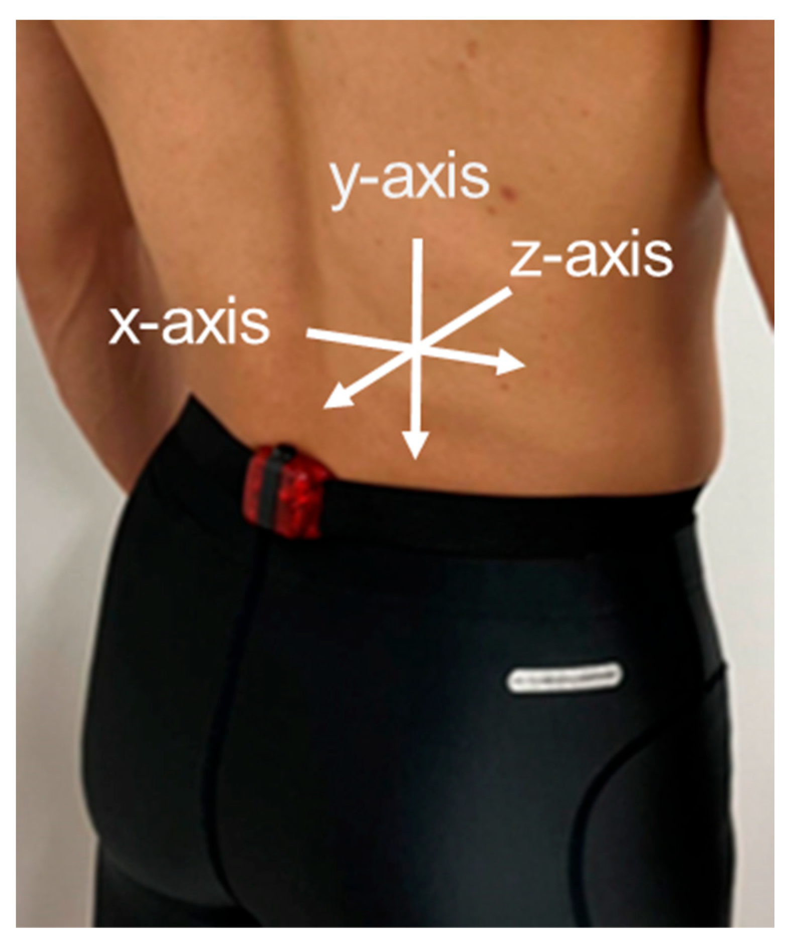
Additionally, a triaxial accelerometer (wGT3X-BT; ActiGraph, Pensacola, FL, USA) with a range of ±8.0 g was employed. The x-axis was oriented in the medial-lateral direction, the y-axis was oriented in the vertical direction, and the z-axis was oriented in the anteroposterior direction. Positive X, Y, and Z values represented the right, downward, and backward accelerations, respectively. The accelerometer was attached between the L4 and L5 spinous processes using a belt by medically qualified staff (Figure 2). Raw acceleration data were collected at 100 Hz during the treadmill running task, transferred to analysis software (OT BioLab; OT Bioelettronica, Torino, Italy), and filtered at a cut-off frequency of 10 Hz [17]. The stride duration from the acceleration data was determined as two consecutive peak-to-peak durations of vertical acceleration [7]. Because the accelerometer also recorded time data, accelerometer data at the same step as that recorded by the digital camera were extracted by recording the start time of the step. The acceleration data for the five steps were analyzed during the same period as the 2D analysis, and those along the z-axis oriented in the anteroposterior direction were used for the analysis. The RMS of the z-axis acceleration (AP-RMS) was calculated for each step.

2.4. Statistical Analysis

A simple linear regression analysis was used to investigate the relationship between the AP-RMS and the pelvic tilt angle, which were the dependent and predictor variables (maximum, minimum, range, and average), respectively. All statistical analyses were performed using the IBM SPSS Statistics 23 software for Windows (SPSS IBM; Japan Inc., Tokyo, Japan) with a significance level of p < 0.05.

3. Results

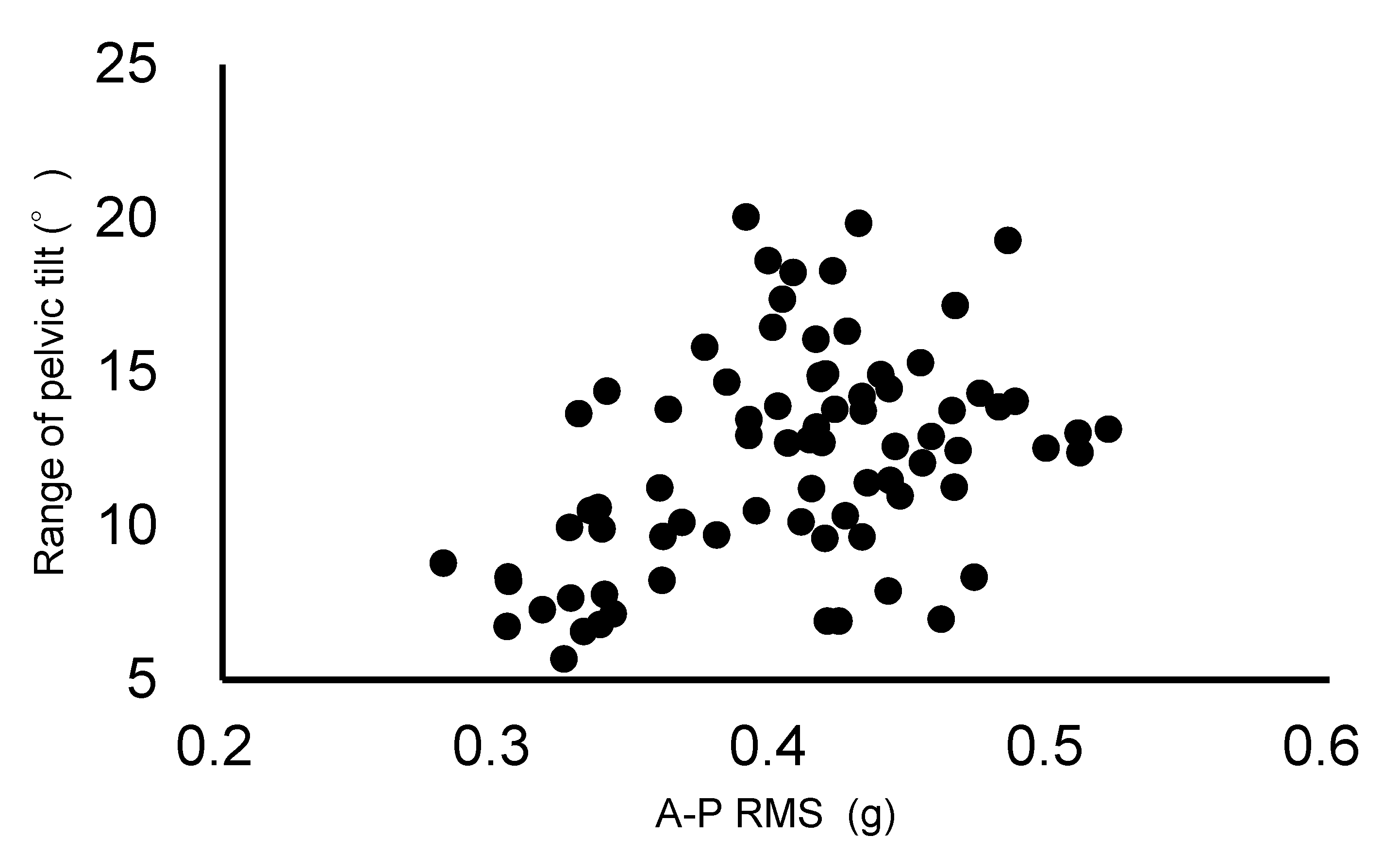
The result of the AP-RMS was 0.41 ± 0.06 g. The maximum, minimum, average, and range of the pelvic tilt angle were 24.8 ± 4.2°, 12.5 ± 4.6°, 19.0 ± 4.1°, and 12.2 ± 3.5°, respectively. The simple linear regression analysis indicated that the AP-RMS was associated with the maximum pelvic tilt angle (r  =  0.32, p  =  0.003) and its range (r  =  0.42, p  =  0.0001). The regression models were as follows: AP-RMS = 0.299 + 0.004 × maximum pelvic tilt angle (adjusted R2 = 0.09) (Figure 3) and AP-RMS = 0.324 + 0.007 × range of pelvic tilt angle (adjusted R2 = 0.16) (Figure 4). However, no relationship was observed between the AP-RMS and the minimum or average pelvic tilt angle. Data of Figure 3 and Figure 4 can be confirmed at Supplementary Materials.

4. Discussion

A previous study [7] reported that running produced a significantly greater excursion than walking in the total lumbar range of motion. Additionally, the 3D trunk acceleration increased with the running speed [7]. Therefore, it is presumed that the range of motion and acceleration of the lumbar spine are related to running. The intradiscal pressure in the L4/L5 segments exhibited the most pronounced change during trunk flexion and extension [9]. Hence, it is assumed that the higher the flexion range and lumbar spine extension, the higher the load on the lumbar spine. Thus, reducing the acceleration in the anteroposterior direction may prevent IVD degeneration. Although we did not investigate the lumbar range of motion during running, we speculate that a higher AP-RMS indicates higher excursion in the lumbar spine.
This study aimed to investigate the relationship between the anteroposterior acceleration of the lower lumbar spine and pelvic tilt movements during running. The results revealed a weak relationship between the AP-RMS and the maximum pelvic tilt angle (r  =  0.32, p  =  0.003) and between the AP-RMS and the range of the pelvic tilt angle (r  =  0.42, p  =  0.0001), suggesting that the larger the maximum or range of the pelvic tilt angle, the higher the AP-RMS. Regarding the lumbo–pelvic–hip complex, a higher anterior pelvic tilt is associated with greater extension of the lumbar spine during running [12]. Crosbie et al. [18] reported that the movement patterns in the lumbar segment complement those in the pelvis. Although the pelvis rotates into a negative pelvic tilt (posterior pelvic tilt) at the heel strike, it causes maximum lumbar spine flexion. Thus, pelvic movements affect the lower lumbar region. Therefore, a higher pelvic tilt range may increase the range of motion in the lower lumbar spine during running.
However, the adjusted R2 in the relationship between the AP-RMS and the maximum pelvic tilt angle (adjusted R2 = 0.09) or the pelvic tilt angle range was low (adjusted R2 = 0.16) in this study. The anteroposterior acceleration of the lower lumbar region during running does not only elucidate the pelvic tilt angle, and other factors should be considered. Additionally, the regression constant obtained through simple linear regression analysis was larger than the regression coefficient in this study. The constant may be related to other factors, such as gait, which should also be considered. A stepwise regression showed that the maximum hip flexion angle, hip angle at mid-stance, hip angle at toe-off, foot forward, and vertical displacement of the center of gravity from mid-stance to toe-off were related to AP-RMS in the lower lumbar region during running [10]. These variables may be among the factors that contributed to the high regression constant obtained in this study. Therefore, it is suggested that the anteroposterior acceleration of the lower lumbar region is slightly influenced by the pelvic tilt angle.
Finally, this study had several limitations. As the running form was analyzed in 2D, it was difficult to evaluate the pelvic rotation and all gait cycles. Therefore, the analysis was limited to the period from the initial contact of the left and right feet. Although further studies using 3D analysis are required, it is assumed that they will yield similar results on the opposite side because running is a symmetrical movement. Additionally, this study only employed treadmill running. Therefore, it is unclear whether similar results will be obtained for ground running, and further studies are required to evaluate this. Furthermore, the participants included in this study were not long-distance runners. Therefore, further studies are required with long-distance runners; however, the results of this study may provide important data for comparisons with those of long-distance runners.

5. Conclusions

This study aimed to investigate the relationship between the anteroposterior acceleration of the lower lumbar spine and pelvic tilt movements during running. We speculated that a higher anterior pelvic tilt may increase the acceleration in the anteroposterior direction. The results suggested that a large pelvic tilt range during running may lead to significant acceleration in the anteroposterior direction. However, the pelvic tilt angle has a small effect on the AP-RMS.

Supplementary Materials

The following supporting information can be downloaded at https://www.mdpi.com/article/10.3390/biomechanics4040056/s1: Data S1: Correlations between anteroposterior acceleration and maximum pelvic tilt angle (data for Figure 3); Data S2: Correlations between anteroposterior acceleration and the range of pelvic tilt angle (data for Figure 4).

Author Contributions

Conceptualization, Y.K. and K.K.; methodology, Y.K., K.K. and T.K.; formal analysis, Y.K.; investigation, Y.K. and T.K.; resources, Y.K., K.K. and T.K.; data curation, Y.K.; writing—original draft preparation, Y.K.; writing—review and editing, K.K.; visualization, Y.K.; supervision, Y.K.; project administration, Y.K.; funding acquisition, Y.K. All authors have read and agreed to the published version of the manuscript.

Funding

This work was supported by the TAU Research Fund from Tokyo Ariake University of Medical and Health Sciences.

Institutional Review Board Statement

The study was conducted in accordance with the Declaration of Helsinki and approved by the Ethics Committee of Tokyo Ariake University of Medical and Health Sciences (Approval no. 308; 14 July 2020).

Informed Consent Statement

Written informed consent was obtained from all participants involved in the study.

Data Availability Statement

The data presented in this study are available in the insert article and Supplementary Materials here.

Acknowledgments

The authors would like to thank all members of the research team and all the participants who took part in the study.

Conflicts of Interest

The authors declare no conflicts of interest.

References

  1. Min, S.K.; Nakazato, K.; Yamamoto, Y.; Gushiken, K.; Fujimoto, H.; Fujishiro, H.; Kobayakawa, Y.; Hiranuma, K. Cartilage intermediate layer protein gene is associated with lumbar disc degeneration in male, but not female, collegiate athletes. Am. J. Sports Med. 2010, 38, 2552–2557. [Google Scholar] [CrossRef] [PubMed]
  2. Hangai, M.; Kaneoka, K.; Hinotsu, S.; Shimizu, K.; Okubo, Y.; Miyakawa, S.; Mukai, N.; Sakane, M.; Ochiai, N. Lumbar intervertebral disk degeneration in athletes. Am. J. Sports Med. 2009, 37, 149–155. [Google Scholar] [CrossRef] [PubMed]
  3. Koyama, K.; Nakazato, K.; Hiranuma, K. Etiology and nature of intervertebral disc degeneration and its correlation with low back pain. J. Phys. Fit. Sports Med. 2015, 4, 63–72. [Google Scholar] [CrossRef]
  4. Chan, S.C.; Ferguson, S.J.; Gantenbein-Ritter, B. The effects of dynamic loading on the intervertebral disc. Eur. Spine J. 2011, 20, 1796–1812. [Google Scholar] [CrossRef] [PubMed]
  5. Belavy, D.L.; Albracht, K.; Bruggemann, G.P.; Vergroesen, P.P.; Van Dieen, J.H. Can Exercise Positively Influence the Intervertebral Disc? Sports Med. 2016, 46, 473–485. [Google Scholar] [CrossRef] [PubMed]
  6. Belavy, D.L.; Quittner, M.J.; Ridgers, N.; Ling, Y.; Connell, D.; Rantalainen, T. Running exercise strengthens the intervertebral disc. Sci. Rep. 2017, 7, 45975. [Google Scholar] [CrossRef] [PubMed]
  7. Kawabata, M.; Goto, K.; Fukusaki, C.; Sasaki, K.; Hihara, E.; Mizushina, T.; Ishii, N. Acceleration patterns in the lower and upper trunk during running. J. Sports Sci. 2013, 31, 1841–1853. [Google Scholar] [CrossRef] [PubMed]
  8. Takatalo, J.; Karppinen, J.; Niinimaki, J.; Taimela, S.; Nayha, S.; Jarvelin, M.R.; Kyllonen, E.; Tervonen, O. Prevalence of degenerative imaging findings in lumbar magnetic resonance imaging among young adults. Spine 2009, 34, 1716–1721. [Google Scholar] [CrossRef]
  9. Wilke, H.; Neef, P.; Hinz, B.; Seidel, H.; Claes, L. Intradiscal pressure together with anthropometric data—A data set for the validation of models. Clin. Biomech. 2001, 16 (Suppl. S1), S111–S126. [Google Scholar] [CrossRef]
  10. Lindsay, T.R.; Yaggie, J.A.; McGregor, S.J. Contributions of lower extremity kinematics to trunk accelerations during moderate treadmill running. J. Neuroeng. Rehabil. 2014, 11, 162. [Google Scholar] [CrossRef]
  11. Franz, J.R.; Paylo, K.W.; Dicharry, J.; Riley, P.O.; Kerrigan, D.C. Changes in the coordination of hip and pelvis kinematics with mode of locomotion. Gait Posture 2009, 29, 494–498. [Google Scholar] [CrossRef]
  12. Schache, A.G.; Blanch, P.; Rath, D.; Wrigley, T.; Bennell, K. Three-dimensional angular kinematics of the lumbar spine and pelvis during running. Hum. Mov. Sci. 2002, 21, 273–293. [Google Scholar] [CrossRef] [PubMed]
  13. Takatalo, J.; Karppinen, J.; Nayha, S.; Taimela, S.; Niinimaki, J.; Blanco, S.R.; Tammelin, T.; Auvinen, J.; Tervonen, O. Association between adolescent sport activities and lumbar disk degeneration among young adults. Scand. J. Med. Sci. Sports 2017, 27, 1993–2001. [Google Scholar] [CrossRef] [PubMed]
  14. Sekine, C.; Matsunaga, N.; Kaneoka, K. Changes in Lumbopelvic Motion and Trunk Muscle Activity during 2000 m Rowing Ergometer Trial. Int. J. Sport Health Sci. 2021, 19, 47–57. [Google Scholar] [CrossRef]
  15. Schache, A.G.; Blanch, P.D.; Murphy, A.T. Relation of anterior pelvic tilt during running to clinical and kinematic measures of hip extension. Br. J. Sports Med. 2000, 34, 279–283. [Google Scholar] [CrossRef] [PubMed]
  16. Miyamoto, A.; Takeshita, T.; Yanagiya, T. Differences in sprinting performance and kinematics between preadolescent boys who are fore/mid and rear foot strikers. PLoS ONE 2018, 13, e0205906. [Google Scholar] [CrossRef] [PubMed]
  17. Wundersitz, D.W.; Gastin, P.B.; Richter, C.; Robertson, S.J.; Netto, K.J. Validity of a trunk-mounted accelerometer to assess peak accelerations during walking, jogging and running. Eur. J. Sport Sci. 2015, 15, 382–390. [Google Scholar] [CrossRef] [PubMed]
  18. Crosbie, J.; Vachalathiti, R.; Smith, R. Patterns of spinal motion during walking. Gait Posture 1997, 5, 6–12. [Google Scholar] [CrossRef]
Figure 1. (A) Participants with the markers. Marker was attached to the anterior the left anterior and posterior superior iliac spines. (B) A participant during running trials on the treadmill. The pelvic tilt angle was defined as the angle between the horizontal line from the flat ground and that connecting the left anterior and posterior superior iliac spines. The anterior pelvic tilt angle was assigned a positive value.
Biomechanics 04 00056 g001
Figure 2. The accelerometer was attached between the L4 and L5 spinous processes using a belt. The x-axis was oriented in the medial-lateral direction, the y-axis was oriented in the vertical direction, and the z-axis was oriented in the anteroposterior direction.
Biomechanics 04 00056 g002
Figure 3. Correlations between anteroposterior acceleration (0.41 ± 0.06 g) and maximum pelvic tilt angle (24.8 ± 4.2°) for the regression model of AP-RMS = 0.299 + 0.004 × maximum pelvic tilt angle (adjusted R2 = 0.09).
Biomechanics 04 00056 g003
Figure 4. Correlations between anteroposterior acceleration (0.41 ± 0.06 g) and the range of pelvic tilt angle (12.2 ± 3.5°) for the regression model of AP-RMS = 0.324 + 0.007 × range of the pelvic tilt angle (adjusted R2 = 0.16).
Biomechanics 04 00056 g004
Disclaimer/Publisher’s Note: The statements, opinions and data contained in all publications are solely those of the individual author(s) and contributor(s) and not of MDPI and/or the editor(s). MDPI and/or the editor(s) disclaim responsibility for any injury to people or property resulting from any ideas, methods, instructions or products referred to in the content.