Review

iBTA-Induced Biotube® Blood Vessels: 2020 Update

1
Osaka Laboratory, Biotube Co., Ltd., Osaka 565-0842, Japan
2
Department of Cardiovascular Surgery, Yokohama General Hospital, Kanagawa 225-0025, Japan
3
Pre-Clinical Research Center, Institute of Development, Aging and Cancer, Tohoku University, Miyagi 980-8575, Japan
4
Department of Cardiovascular Surgery, Oita University Hospital, Oita 879-5593, Japan
5
Department of Mechanical Engineering, Faculty of Engineering Science, Kansai University, Osaka 564-8680, Japan
6
Department of Nephrology, Kameda Medical Center, Chiba 296-8602, Japan
7
Institute of Frontier Science and Technology, Okayama University of Science, Okayama 700-0005, Japan
8
Advanced Medical Engineering Research Center, Asahikawa Medical University, Hokkaido 078-8510, Japan
*
Author to whom correspondence should be addressed.
Kidney Dial. 2021, 1(1), 3-13; https://doi.org/10.3390/kidneydial1010002
Submission received: 12 April 2021 / Revised: 18 May 2021 / Accepted: 24 May 2021 / Published: 17 June 2021
(This article belongs to the Special Issue Dialysis Access—A New Era)

Abstract

:
Blood access is a lifeline for dialysis patients. However, serious problems such as stenosis or obstruction of access blood vessels, which are life-threatening conditions in daily clinical practice, still remain. One of the most promising candidates for solving these problems may be Biotube blood vessels. More than 20 years have passed since the development of in-body tissue architecture (iBTA), a technology for preparing tissues for autologous implantation in patients. The tissues obtained by iBTA do not elicit immunological rejection, which is one of the ultimate goals of regenerative medical engineering; however, their practical applications were quite challenging. The seemingly unorthodox iBTA concepts that do not follow the current pre-established medical system may not be readily accepted in general medicine. In contrast, there are many diseases that cannot be adequately addressed even with the latest and most advanced medical technology. However, iBTA may be able to save patients with serious diseases. It is natural that the development of high-risk medical devices that do not fit the corporate logic would be avoided. In order to actively treat such largely unattached diseases, we started Biotube Co., Ltd. with an aim to contribute to society. Biotubes induced by iBTA are collagenous tubular tissues prepared in the patient’s body for autologous implantation. The application of Biotubes as tissues for vascular implantation has been studied for many years. Biotubes may have excellent potential as small-diameter artificial blood vessels, one of the most difficult to clinically achieve. Their possibility is currently being confirmed in preclinical tests. Biotubes may save hundreds of thousands of patients worldwide annually from amputation. In addition, we aim to eliminate the recuring access vascular problems in millions of dialysis patients. This study provides an update on the current development status and future possibilities of Biotubes and their preparation molds, Biotube Makers.

1. Introduction

What are the characteristics of an ideal artificial blood vessel? It is essential for a blood vessel to maintain blood flow without thrombus formation, stenosis or rupture. Additionally, special attributes will be required depending on the application purpose and the implant site. Artificial vessels for dialysis access are mainly used for the preparation of subcutaneously fixed superficial arteries and arteriovenous shunts to initiate dialysis. For arteries, high strength to withstand arterial pressure is required. In contrast, because the arteriovenous shunt is a low-pressure system, pressure resistance is not necessary. However, because its blood flow does not exist in a normal living body, it may require special functions other than those required for normal blood vessels in the living body. It is imperative that dialysis access prostheses withstand repeated punctures for dialysis.
Currently, various types of artificial blood vessels are commercially available for dialysis. If strong artificial materials are used, the strength of the vessels is guaranteed. However, there is a limit to maintaining the access function because the puncture is retained. In addition to allowing blood to flow, artificial blood vessels for dialysis are required to have a higher degree of durability than artificial blood vessels for other purposes. Furthermore, because artificial blood vessels are manufactured using artificial materials, there is an inherent risk of infection. Westerners have a large physique and thick arms, so vein tends to run deeply. Therefore, they are often physically more difficult to puncture, and the use of artificial blood vessels in Europe and the United States for preparing dialysis access is higher than in Japan. However, globally, there is a reluctance to use artificial materials to avoid infection. In Japan, autologous blood vessels are used first-line for preparing dialysis shunts, but autologous blood vessels may be insufficient depending on the flow or caliber of the veins. In addition, a shortage of access blood vessels is becoming a challenge due to the extension of dialysis life owing to advances in technology. Therefore, an artificial blood vessel close to an autologous blood vessel with self-repairing ability and resistance to infection is ideal.
One viable candidate is Biotube. Biotube is a general term for tubular tissues for autologous implantation that can be produced in the patient’s body. Its application is not limited to blood vessels and can potentially be applied to any tubular structures in the body such as lymphatic vessels, ureters, trachea [1,2,3], esophagus [4], and intestines. Furthermore, when the Biotube is cut open, it becomes a membrane or plate-shaped tissues, known as Biosheet. Animal experiments have confirmed their potential application as substitutes or repair materials for various defects in the cornea [5], diaphragm [6], urinary bladder [7], peritoneum [8,9], myocardium, valve membrane [10,11], dura mater, etc. The technique for preparing tissues for implantation in the body is called in-body tissue architecture technology (iBTA), but it is not a special technique. It is based on the encapsulation reaction—a type of biological defense reaction that occurs when a foreign substance is implanted in the body.
When iBTA was initially developed ~20 years ago, tubular tissues were formed on the surface of the implants from a simple encapsulation reaction by embedding plastic cylinders as a foreign substance [12,13,14,15,16,17]. For many years, we have been investigating whether the obtained tissues can be used as a substitute blood vessel. The tissues obtained were generally extremely thin and uneven in thickness, but they withstood arterial pressure sufficiently as vascular tissues with a small diameter of about several mm. Collagen was exposed on the luminal surface of Biotubes but, fortunately, that they had high patency without thrombus formation for an extended time [18,19]. Moreover, it was confirmed that such sufficiently strong tubular tissue can be formed even in the human body [20]. However, since the encapsulation-based tissues are very thin, the suitability for suture is poor, and it was thought that their general dissemination would be difficult. The encapsulation-based tissue preparation for implantation has long been proposed by other research groups worldwide [21]. However, its application beyond small animal experiments has been challenging.
Recently, Biotube-like tubular connective tissues have been developed that thicken and harden the tissue walls by overexpressing the encapsulation reaction. A Dutch group has developed a special material that can enhances the foreign body reaction [22,23]. Until ~10 years ago, we were also developing ways to make embedded types functional: (1) biochemical stimulation methods such as drug application or release [24,25,26], (2) light stimulation methods by incorporating LED irradiators in molds [27], or (3) a mechanical stimulation method by dynamic diameter expansion using repeated balloon dilation, or by minute inner diameter fluctuation using vibrating elements [28]. All of these stimulation methods were able to exert a special effect for promoting tissue formation under certain conditions. However, it is very difficult to stabilize their optimum conditions because they were highly dependent on the individual or physical condition of the animals, and they were often accompanied by inhibition of tissue formation due to excessive inflammation.
A breakthrough or turning point was the invention of a mold that was assembled by covering a conventional cylindrical rod with a pipe [29,30,31]. Instead of the previous method for preparing tissues on the outer surface of a conventional foreign substance, it was a completely new idea of preparing tissues inside the mold with a gap inside. By adjusting the size or gap of the space in the mold, it has become possible to freely prepare tubular tissues as Biotubes with a desired diameter and thickness [10,32]. The previous tissue formation was body-dependent, but it has become possible to control the shape and dimensions of the tissues using designed molds. By making the wall thickness of Biotube above a certain level, the applicability of its implantation could be significantly improved. Biotube Co., Ltd., [33] which the authors founded, is intended for social implementation of Biotubes using molds.
In this study, we describe the development of Biotube Maker for preparing Biotube with a diameter <4 mm, which is designated as a medical device in the SAKIGAKE program [34] of the Ministry of Health, Labour and Welfare in 2019, for bypassing the lower limb artery in patients with chronic limb-threatening ischemia. We will provide the latest information regarding Biotubes in 2020, including the progress of preclinical tests currently underway with the support of the Hashiwatashi project in Japan Agency for Medical Research and Development (AMED).

2. Development Process of the Molds for Biotube Preparation (Biotube Makers)

2.1. Development of Original Straight Mold

Since its conception, the first developed mold as Biotube Maker has been a straight columnar type (original mold in Figure 1a), which is assembled by covering the outside of a plastic core rod with a stainless steel pipe with a predetermined gap space [30,35]. The pipe contains many thin slits. When the mold is implanted subcutaneously, skin fibroblasts migrate into the mold through slits. The cells produce collagen fibers and, in over a month, the space in the mold is completely occupied with collagen-based tissue. A tubular Biotube was obtained by removing all parts from the harvested mold. The outer diameter of the core rod corresponded to the inner diameter of the formed Biotube, and the inner diameter of the cover pipe was the outer diameter of Biotube. Therefore, the gap between the rod and the cover was the wall thickness of Biotube.
The minimum diameter of the previously prepared Biotube was 0.6 mm, which was the smallest artificial blood vessel worldwide [18,19]. In principle, if the rod diameter is made smaller, a small-diameter Biotube can be produced. Similarly, if the rod material is made larger, a large-diameter Biotube can be easily obtained. Currently, large and thick Biotubes can be produced up to approximately 3 cm in diameter and 2 mm in thickness. Interestingly, a large or thick Biotube could be formed in approximately a month. A Biotube that is created immediately after being harvested from a living body is flexible and soft, but if temporarily dehydrated by immersing in alcohol for a short time, it can maintain its tubular shape even after immersing it in physiological saline. In addition, it can be stored in alcohol for a year even at room temperature. Almost all previous implantation experiments in animals were performed using Biotubes preserved in alcohol.

2.2. First-in-Man (FIM) Study of Straight Mold

Biotubes obtained from the straight mold have already been clinically applied to three dialysis patients as a First-in-Man (FIM) study [35]. Blood vessels on the venous side of the arteriovenous shunt are prone to stenosis. Three patients had repeated venous stenosis for over a year and underwent painful balloon dilation. Therefore, at Tenri Hospital, the patients underwent bypass surgery using Biotubes. Two molds were implanted subcutaneously in the patient’s abdomen, and two months later, all molds produced Biotubes with a length of 7 cm and a diameter of 6 mm. One patient maintained patency for more than 2 years and did not require postoperative balloon dilatation.

2.3. Development of Plastic Spiral Biotube Maker for Long Length Biotube

A long Biotube is required for use as an artificial blood vessel for a dialysis shunt. However, in a straight mold, there is a limit to the length that can be embedded in the body. Therefore, we developed a spiral-shaped Biotube Maker (Figure 1). The Maker was manufactured by injection molding of plastic. Since the total length of the formed Biotube can be adjusted by the number of turns of the spiral, a 25-cm-long Biotube could be obtained from a mini-sized Biotube Maker with two turns (1G mini in Figure 1a). By increasing the number of turns of the Maker spiral by one more (1G), a Biotube with a length of 50 cm was obtained. The 50-cm-long Biotube was introduced as the longest tissue engineering artificial blood vessel worldwide at the time of development [36]. The first-generation spiral Biotube Maker (1G) had the same large slit-shaped pores similar to that of the straight mold. Large pores were designed to facilitate the entry of subcutaneous fibroblasts into the mold. Since tissue formation occurs on the inner and outer surfaces of the mold, the Biotube structure formed inside the Maker and the capsule structure formed on the outer periphery of the Maker are strongly connected at a large pore. Therefore, it was often difficult to harvest the mold from the new tissues formed around the mold. Occasionally many surrounding tissues were attached to the harvested mold. Removing excess adherent tissues from the Maker surface was cumbersome.
The mold was improved to reduce the pore area as much as possible while maintaining the opening ratio. In the second generation Biotube Maker (2G), the pore area was reduced to a 2 mm × 2 mm square, and in the third generation Maker (3G), it was further reduced to half the area (2 mm × 1 mm). However, the strength of the tissues connecting the inner and outer surfaces of the mold at the pore was quite strong, and it is still difficult to peel off the outer peripheral structure even in a small pore. In contrast, due to the technical problem of plastic processing, there is a limit to the precise processing of minute pores with high density. Moreover, since the plastic material has low strength, the thickness of the plastic plate has to be increased to prevent structural deformation.

2.4. Development of Stainless Steel Spiral Biotube Maker

The previous Makers were made of plastic, so they lacked precision machining. Therefore, in 2020, we decided to change the material of the spiral Biotube Maker from plastic to stainless steel. A press method was adopted to process the stainless steel plate into a spiral shape. First, a thin stainless steel plate was etched to create many fine round pores. By hydraulically pressing the plate, spiral irregularities were formed. Finally, a spiral stainless steel plate was produced by cutting it into a disk shape. A stainless steel spiral Maker was assembled by sandwiching a separately prepared plastic rod between two spiral stainless steel plates as outer shell parts (4G in Figure 1a). Compared with the first generation (1G), the area and thickness of the opening hole of the 4G Maker could be reduced by about 1/100 and about 1/3, respectively. At present, the mold is thinned by flattening the cross-sectional shape of the rod material from a circle to an ellipse (5G) (Figure 1b,c).
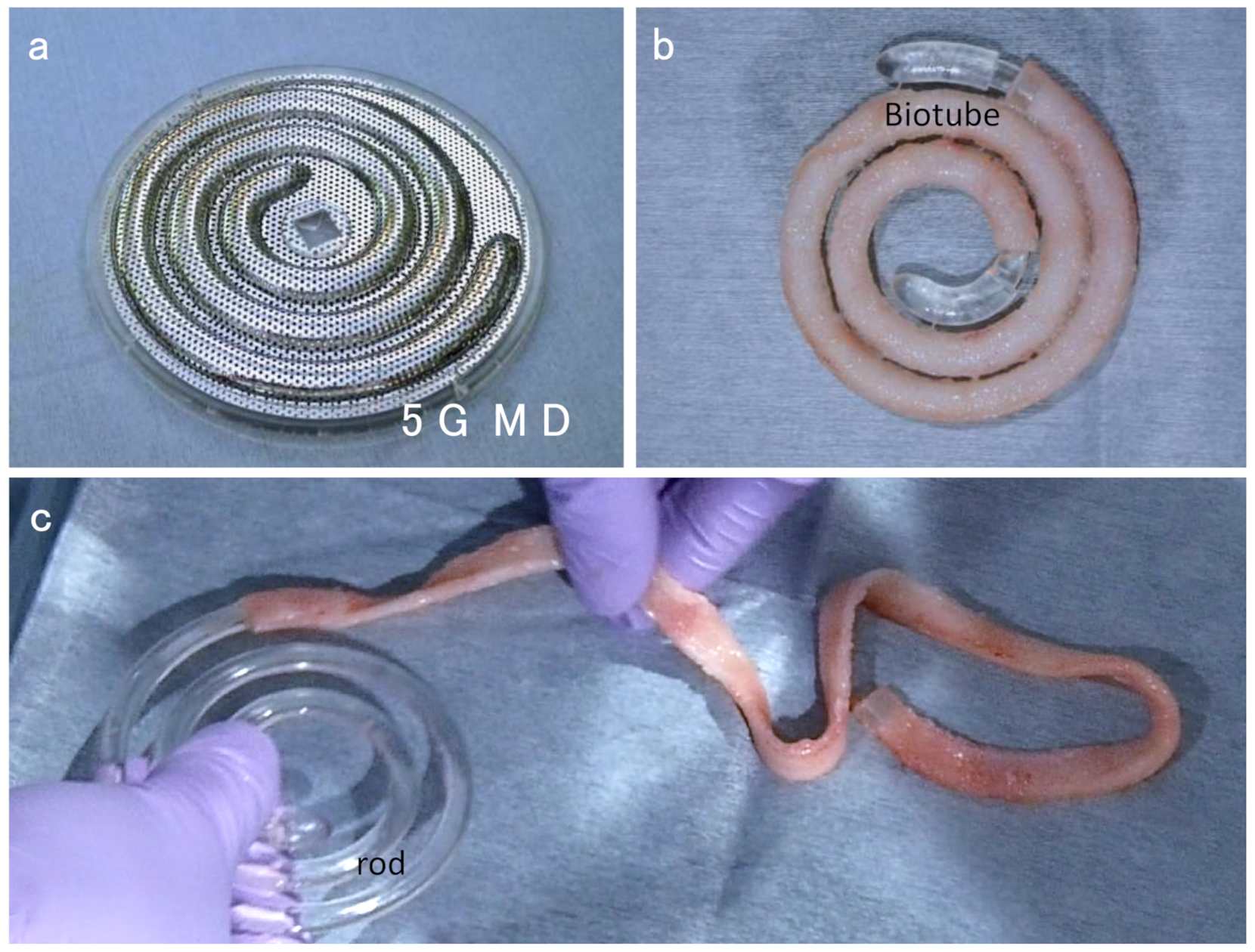
In contrast, we are also developing a Maker for medium-diameter Biotubes with an inner diameter of 5 mm for access in dialysis (5G MD in Figure 2a). The basic structure was the same as the Maker for the lower limbs. The size of the Maker was 9 cm, which is the same as for the lower limbs (5G). By implanting under the skin for 1 month, Biotubes for dialysis were formed on the surface of the core rod inside the Maker (Figure 2b). The removal of Biotube from the rod was smooth with almost no resistance (Figure 2c). The length of the resulting Biotube was about 10 cm shorter than that for the lower limbs.

3. Progress of Preclinical Tests

3.1. Biotube Formation Test

In the 4th SAKIGAKE program of the Ministry of Health, Labour and Welfare of Japan in 2019, the mold, Biotube Maker for in-body preparation of Biotubes, was designated as a medical device. The Maker was the third implantable medical device specified in the program. The purpose of the SAKIGAKE program is to promptly provide the world’s most advanced medical devices to Japanese patients. The Biotubes are intended for patients with chronic limb-threatening ischemia. Chronic limb-threatening ischemia (CLTI) is the final stage of peripheral arterial disease. It is a disease associated with a high morbidity rate, leading to serious mortality and loss of limbs and resulting in poor quality of life. There are no small-diameter conduits of less than 4 mm based on artificial materials such as expanded polytetrafluoroethylene and polyethylene terephthalate (Dacron) for surgical bypass of CLTI to infrapopliteal targets. In contrast, the patency rate in endovascular interventions is inadequate. High quality of autologous venous conduits is effective in bypass surgeries. However, no veins are available when spent on previous procedures such as coronary or peripheral bypass.
These Biotubes provide a substitute blood vessel with a diameter of <4 mm for revascularization by lower limb artery bypass. Using the stainless steel 5G Maker, which was successfully developed in mid-2020, preclinical clinical tests have begun. In vivo performance tests are conducted using goats at Oita University and Tohoku University. All animal experiments were performed in accordance with the Guide for the Care and Use of Laboratory Animals, published by the United States National Institutes of Health (NIH Publication No. 85-23, received 1996). All animal experiments were approved by the Tohoku University Ethics Committee (No. 2019AcA-041) and the Oita University Ethics comittee (No. 182201).
As one of the in vivo performance tests, we investigated whether the 5G Makers could form Biotubes with the designed dimension. The method was as follows [36,37]. An incision was made in the skin on the back of the goats, and the skin was peeled off using a special device like a “Shamoji” to make a subcutaneous pocket (Figure 3a). After checking the size of the pocket with a disk-shaped plate (Figure 3b), and the Maker was embedded in the pocket (Figure 3c). After closing the wound, normal breeding was performed for follow-up (Figure 3d). One month after the Makers were embedded, they could be removed from the same incision position as they were embedded (Figure 3e). Since the opening hole of the stainless steel Maker is extremely small, the adhesion between the surrounding tissue under the skin and the Maker was weak, and the Maker could be easily removed from the subcutaneous tissue by hand. The connective tissue adhering to the outer surface of the Maker could be easily removed with a cotton gauze. When the two stainless steel plates were opened, the formation of Biotubes as designed was observed inside the Maker (Figure 3f). The Biotubes created had an inner diameter of 4 mm, and one end was tapered to an inner diameter of 3 mm. The length was over 50 cm and the wall thickness was 0.85 mm. The entire outer surface of the Biotubes had small protrusions but was relatively smooth overall (Figure 3g). In contrast, the previous Biotubes obtained from 3G Makers had large protrusions on their outer surface. However, the inner surface of all Biotubes was very smooth regardless of the type of Maker used (Figure 3h).

3.2. Biotube Implantation Test

The Biotubes obtained using the 5G spiral Maker were straightened by immediate immersion in a 70% alcohol solution. When water was poured into the lumen of the Biotubes, no leakage, tears or defects were observed (Figure 4a). Biotubes withstood an internal pressure of over 200 mmHg. The tensile strength of the samples obtained by cutting the Biotubes into a ring shape (width 5 mm) was ca. 7 to 10 N when measured in the operating room with a portable tensile tester (Stency, AcroEdge, Osaka, Japan). Therefore, the converted pressure resistance was extremely high to be ca. 4500 to 6500 mmHg. When the Biotubes were stored in an alcohol solution, there was almost no change in their strength even after 1 month.
The carotid arteries of goats were dissected and after intravenous injection of heparin sodium (200 IU/kg), the carotid artery was cross-clamped. One edge of the Biotube was anastomosed to the proximal site of the carotid artery in a side-to-end manner using 7-0 polypropylene sutures via the continuous manner using the parachute method (Figure 4b). The resistance to needle sticks was slightly higher than that of native blood vessels. There was no bleeding from the needle hole. No cutting occurred during suturing. Since the shape of Biotubes was stable and their lumen was maintained as compared with native blood vessel, there was almost no stress in suturing. Even if there was bleeding from the anastomotic site, hemostasis could be easily performed by adding one or two needles. Biotubes under arterial pressure were easily bent and had excellent flexibility (Figure 4c). Biotubes did not kink even after being almost completely bent (Figure 4d). The other end of the Biotube was similarly anastomosed to the distal site of the carotid artery. The carotid artery was ligated between the proximal and distal anastomosis sites. Biotubes were buried under the skin (Figure 4e). The implantation distance of the Biotube was set to 15 to 20 cm. After implantation, animals received antiplatelet drug (clopidogrel 75 mg/head, PO, SID) for one month.
In a preliminary acute phase study using Biotubes from 3G Makers [37], complete patency was successful in all six cases with an implantation period of 1 month. Histological observation revealed the progression of vascular tissue remodeling, including the formation of an endothelial cell layer on the lumen surface. The implantation test in the chronic phase is ongoing using Biotubes from 5G Makers and, at present, the observation period of up to 4 months has elapsed, and there is no abnormal vascular deformation such as stenosis or dilation with maintained patency. Since the structure of Biotube is expected to approach that of native blood vessels with the implantation period, it is expected that favorable chronic phase results will be obtained. In the future, we would like to investigate the possibility of applying Biotube as a blood vessel for dialysis access, to determine whether it can be repeatedly punctured and whether it is resistant to infection, using a 5 mm diameter Biotube prepared from a 5G MC Maker.

4. Future Plan

Human acellular vessels from Humacyte Co., (Durham, NC, USA) are probably the most advanced in the field of regenerative blood vessels [38,39]. Tubular tissue is prepared by culturing human vascular smooth muscle cells collected from corpses on biodegradable scaffolds and decellularized to finally obtain a collagen-containing tube containing without artificial substance. It can be said that Biotube-like implants are artificially manufactured. It takes more than half a year to manufacture it, but once it is manufactured, it can be used as off-the-shelf products for emergency use. Many good clinical results have already been reported. Two clinical trials are undergoing. One is a phase 2 trial in thoracic or limb vascular replacement or reconstruction. Additionally, the other is pilot study in above-knee femoral popliteal bypass implantation. Biotubes, on the other hand, could only be applied to a few dialysis patients in a single facility. However, Biotubes provide an absolute sense of security that is obtained from one’s own tissues in one’s body and the overwhelming economic efficiency that does not require manufacturing equipment.
In addition to the in vivo tests introduced in this paper, physical and chemical tests and biological safety tests are also conducted in parallel as preclinical tests for the stainless steel Makers (5G) used for making Biotubes for bypass grafts in CLTI. All preclinical tests are scheduled to be completed in 2021 and physician-initiated clinical tests are scheduled to begin in 2022. We aim for regulatory approval of Biotube in 2025; therefore, further accelerated development is progressing. The practical application of Biotubes for lower limb artery bypass, which has been designated as a pioneer application, is a top priority. Furthermore, application to other fields including dialysis will be conducted in parallel as far as possible.

Author Contributions

Conceptualization, Y.N.; methodology, T.U., A.Y., K.M., M.M., R.I., T.T. (Takeshi Terazawa) and T.O.; software, T.T. (Tsutomu Tajikawa); validation, R.H. and Y.S.; writing—original draft preparation, Y.N.; writing—review and editing, Y.N., S.M. and M.O.; supervision, Y.N.; project administration, T.Y. and S.M.; funding acquisition, Y.N. and S.M. All authors have read and agreed to the published version of the manuscript.

Funding

This research was supported by Biotube Co., Ltd. and grants from AMED (Japan Agency for Medical Research and Development).

Institutional Review Board Statement

All animal experiments were approved by the Tohoku University Ethics Committee (No. 2019AcA-041) and the Oita University Ethics Committee (No. 182201). The clinical research process was approved by the ethics committee of the National Cerebral and Cardiovascular Center (No. M27-084) and of Tenri Hospital (No. 677).

Informed Consent Statement

Informed consent was obtained from all subjects involved in the study.

Data Availability Statement

The data are not publicly available due to their containing information that could compromise the privacy of research participants.

Acknowledgments

We thank Yumiko Nakashima of Oita University and all staff of Narita Animal Science (NAS) Laboratory Co., Ltd. for their great help in the management of goats breeding. We also thank Satoki Kadota and Saya Yamawaki of Clinical Research, Innovation and Education Center, Tohoku University Hospital (CRIETO) for their kind support in the progress of the AMED project.

Conflicts of Interest

Y.N., R.H. and T.O. are employees and stock-holders of Biotube Co., Ltd. The other authors declare no conflict of interest. The funders had no role in the design of the study; in the collection, analyses, or interpretation of data; in the writing of the manuscript, or in the decision to publish the results.

References

  1. Komura, M.; Komura, H.; Satake, R.; Suzuki, K.; Yonekawa, H.; Ikebukuro, K.; Komuro, H.; Hoshi, K.; Takato, T.; Moriwaki, T.; et al. Fabrication of an anatomy-mimicking BIO-AIR-TUBE with engineered cartilage. Regen. Ther. 2019, 11, 176–181. [Google Scholar] [CrossRef]
  2. Umeda, S.; Nakayama, Y.; Terazawa, T.; Iwai, R.; Hiwatashi, S.; Nakahata, K.; Takama, Y.; Okuyama, H. Long-term outcomes of patch tracheoplasty using collagenous tissue membranes (biosheets) produced by in-body tissue architecture in a beagle model. Surg. Today 2019, 49, 958–964. [Google Scholar] [CrossRef]
  3. Hiwatashi, S.; Nakayama, Y.; Umeda, S.; Takama, Y.; Terazawa, T.; Okuyama, H. Tracheal Replacement Using an In-Body Tissue-Engineered Collagenous Tube “BIOTUBE” with a Biodegradable Stent in a Beagle Model: A Preliminary Report on a New Technique. Eur. J. Pediatr. Surg. 2019, 29, 90–96. [Google Scholar] [PubMed]
  4. Okuyama, H.; Umeda, S.; Takama, Y.; Terasawa, T.; Nakayama, Y. Patch esophagoplasty using an in-body-tissue-engineered collagenous connective tissue membrane. J. Pediatr. Surg. 2018, 53, 223–226. [Google Scholar] [CrossRef]
  5. Takiyama, N.; Mizuno, T.; Iwai, R.; Uechi, M.; Nakayama, Y. In-body tissue-engineered collagenous connective tissue membranes (BIOSHEETs) for potential corneal stromal substitution. J. Tissue Eng. Regen. Med. 2016, 10, E518–E526. [Google Scholar] [CrossRef] [PubMed]
  6. Suzuki, K.; Komura, M.; Terawaki, K.; Kodaka, T.; Gohara, T.; Komura, H.; Nakayama, Y. Engineering and repair of diaphragm using biosheet (a collagenous connective tissue membrane) in rabbits. J. Pediatr. Surg. 2018, 53, 330–334. [Google Scholar] [CrossRef] [PubMed]
  7. Iimori, Y.; Iwai, R.; Nagatani, K.; Inoue, Y.; Funayama-Iwai, M.; Okamoto, M.; Nakata, M.; Mie, K.; Nishida, H.; Nakayama, Y.; et al. Urinary bladder reconstruction using autologous collagenous connective tissue membrane “Biosheet®” induced by in-body tissue architecture: A pilot study. Regen. Ther. 2020, 15, 274–280. [Google Scholar] [CrossRef]
  8. Terazawa, T.; Furukoshi, M.; Nakayama, Y. One-year follow-up study of iBTA-induced allogenic biosheet for repair of abdominal wall defects in a beagle model: A pilot study. Hernia 2019, 23, 149–155. [Google Scholar] [CrossRef] [PubMed]
  9. Nakayama, Y.; Oshima, N.; Tatsumi, E.; Ichii, O.; Nishimura, T. iBTA-induced bovine Biosheet for repair of abdominal wall defects in a beagle model: Proof of concept. Hernia 2018, 22, 1033–1039. [Google Scholar] [CrossRef]
  10. Terazawa, T.; Kawashima, T.; Umeno, T.; Wada, T.; Ozaki, S.; Miyamoto, S.; Nakayama, Y. Mechanical characterization of an in-body tissue-engineered autologous collagenous sheet for application as an aortic valve reconstruction material. J. Biomech. 2020, 99, 109528. [Google Scholar] [CrossRef]
  11. Kawashima, T.; Umeno, T.; Terazawa, T.; Wada, T.; Shuto, T.; Nishida, H.; Anai, H.; Nakayama, Y.; Miyamoto, S. Aortic valve neocuspidization with in-body tissue-engineered autologous membranes: Preliminary results in a long-term goat model. Interact. Cardiovasc. Thorac. Surg. 2021, ivab015. [Google Scholar] [CrossRef] [PubMed]
  12. Nakayama, Y.; Ishibashi-Ueda, H.; Takamizawa, K. In vivo tissue-engineered small- caliber arterial graft prosthesis consisting of autologous tissue (biotube). Cell Transplant. 2004, 13, 439–449. [Google Scholar] [CrossRef] [PubMed] [Green Version]
  13. Watanabe, T.; Kanda, K.; Ishibashi-Ueda, H.; Yaku, H.; Nakayama, Y. Development of biotube vascular grafts incorporating cuffs for easy implantation. J. Artif. Organs 2007, 10, 10–15. [Google Scholar] [CrossRef] [PubMed]
  14. Sakai, O.; Kanda, K.; Ishibashi-Ueda, H.; Takamizawa, K.; Ametani, A.; Yaku, H.; Nakayama, Y. Development of the wing-attached rod for acceleration of “Biotube” vascular grafts fabrication in vivo. J. Biomed. Mater. Res. Part B Appl. Biomater. 2007, 83, 240–247. [Google Scholar] [CrossRef] [PubMed]
  15. Watanabe, T.; Kanda, K.; Ishibashi-Ueda, H.; Yaku, H.; Nakayama, Y. Autologous small-caliber “biotube” vascular grafts with argatroban loading: A histomorphological examination after implantation to rabbits. J. Biomed. Mater. Res. Part B Appl. Biomater. 2010, 92, 236–242. [Google Scholar] [CrossRef] [PubMed]
  16. Watanabe, T.; Kanda, K.; Yamanami, M.; Ishibashi-Ueda, H.; Yaku, H.; Nakayama, Y. Long-term animal implantation study of biotube-autologous small-caliber vascular graft fabricated by in-body tissue architecture. J. Biomed. Mater. Res. Part B Appl. Biomater. 2011, 98, 120–126. [Google Scholar] [CrossRef]
  17. Yamanami, M.; Ishibashi-Ueda, H.; Yamamoto, A.; Iida, H.; Watanabe, T.; Kanda, K.; Yaku, H.; Nakayama, Y. Implantation study of small-caliber “biotube” vascular grafts in a rat model. J. Artif. Organs 2013, 16, 59–65. [Google Scholar] [CrossRef]
  18. Ishii, D.; Enmi, J.I.; Iwai, R.; Kurisu, K.; Tatsumi, E.; Nakayama, Y. One year Rat Study of iBTA-induced “Microbiotube” Microvascular Grafts with an Ultra-Small Diameter of 0.6 mm. Eur. J. Vasc. Endovasc. Surg. 2018, 55, 882–887. [Google Scholar] [CrossRef] [Green Version]
  19. Ishii, D.; Enmi, J.; Moriwaki, T.; Ishibashi-Ueda, H.; Kobayashi, M.; Iwana, S.; Iida, H.; Satow, T.; Takahashi, J.C.; Kurisu, K.; et al. Development of in vivo tissue-engineered microvascular grafts with an ultra small diameter of 0.6 mm (MicroBiotubes): Acute phase evaluation by optical coherence tomography and magnetic resonance angiography. J. Artif. Organs 2016, 19, 262–269. [Google Scholar] [CrossRef] [PubMed]
  20. Nakayama, Y.; Kaneko, Y.; Takewa, Y.; Okumura, N. Mechanical properties of human autologous tubular connective tissues (human biotubes) obtained from patients undergoing peritoneal dialysis. J. Biomed. Mater. Res. Part B Appl. Biomater. 2016, 104, 1431–1437. [Google Scholar] [CrossRef] [PubMed]
  21. Campbell, J.H.; Efendy, J.L.; Campbell, G.R. Novel vascular graft grown within recipient’s own peritoneal cavity. Circ. Res. 1999, 85, 1173–1178. [Google Scholar] [CrossRef] [PubMed] [Green Version]
  22. Geelhoed, W.J.; Moroni, L.; Rotmans, J.I. Utilizing the Foreign Body Response to Grow Tissue Engineered Blood Vessels in Vivo. J. Cardiovasc. Transl. Res. 2017, 10, 167–179. [Google Scholar] [CrossRef] [PubMed] [Green Version]
  23. Geelhoed, W.J.; van der Bogt, K.E.A.; Rothuizen, T.C.; Damanik, F.F.R.; Hamming, J.F.; Mota, C.D.; van Agen, M.S.; de Boer, H.C.; Restrepo, M.T.; Hinz, B.; et al. A novel method for engineering autologous non-thrombogenic in situ tissue-engineered blood vessels for arteriovenous grafting. Biomaterials 2020, 229, 119577. [Google Scholar] [CrossRef] [PubMed]
  24. Sakai, O.; Kanda, K.; Takamizawa, K.; Sato, T.; Yaku, H.; Nakayama, Y. Faster and stronger vascular “Biotube” graft fabrication in vivo using a novel nicotine-containing mold. J. Biomed. Mater. Res. Part B Appl. Biomater. 2009, 90, 412–420. [Google Scholar] [CrossRef]
  25. Nakayama, Y.; Tsujinaka, T. Acceleration of robust “biotube” vascular graft fabrication by in-body tissue architecture technology using a novel eosin Y-releasing mold. J. Biomed. Mater. Res. Part B Appl. Biomater. 2014, 102, 231–238. [Google Scholar] [CrossRef] [PubMed]
  26. Iwai, R.; Tsujinaka, T.; Nakayama, Y. Preparation of Biotubes with vascular cells component by in vivo incubation using adipose-derived stromal cell-exuding multi-microporous molds. J. Artif. Organs 2015, 18, 322–329. [Google Scholar] [CrossRef]
  27. Oie, T.; Yamanami, M.; Ishibashi-Ueda, H.; Kanda, K.; Yaku, H.; Nakayama, Y. In-body optical stimulation formed connective tissue vascular grafts, “biotubes,” with many capillaries and elastic fibers. J. Artif. Organs 2010, 13, 235–240. [Google Scholar] [CrossRef]
  28. Huang, H.; Zhou, Y.M.; Ishibashi-Ueda, H.; Takamizawa, K.; Ando, J.; Kanda, K.; Yaku, H.; Nakayama, Y. In vitro maturation of “biotube” vascular grafts induced by a 2-day pulsatile flow loading. J. Biomed. Mater. Res. Part B Appl. Biomater. 2009, 91, 320–328. [Google Scholar] [CrossRef]
  29. Furukoshi, M.; Moriwaki, T.; Nakayama, Y. Development of an in vivo tissue-engineered vascular graft with designed wall thickness (biotube type C) based on a novel caged mold. J. Artif. Organs 2016, 19, 54–61. [Google Scholar] [CrossRef]
  30. Nakayama, Y.; Furukoshi, M.; Tatsumi, E. Shape memory of in-body tissue-engineered Biotube® vascular grafts and the preliminary evaluation in animal implantation experiments. J. Cardiovasc. Surg. 2020, 61, 208–213. [Google Scholar] [CrossRef]
  31. Furukoshi, M.; Tatsumi, E.; Nakayama, Y. Application of in-body tissue architecture-induced Biotube vascular grafts for vascular access: Proof of concept in a beagle dog model. J. Vasc. Access 2020, 21, 314–321. [Google Scholar] [CrossRef]
  32. Terazawa, T.; Nishimura, T.; Mitani, T.; Ichii, O.; Ikeda, T.; Kosenda, K.; Tatsumi, E.; Nakayama, Y. Wall thickness control in biotubes prepared using type-C mold. J. Artif. Organs 2018, 21, 387–391. [Google Scholar] [CrossRef]
  33. Introduction of Biotube®. Available online: http://www.medical-biotube.com. (In Japanese).
  34. Sakigake Designation Scheme. Available online: https://www.mhlw.go.jp/file/05-Shingikai-11121000-Iyakushokuhinkyoku-Soumuka/0000123357.pdf (accessed on 24 June 2014). (In Japanese)
  35. Nakayama, Y.; Kaneko, Y.; Okumura, N.; Terazawa, T. Initial 3-year results of first human use of an in-body tissue-engineered autologous “Biotube” vascular graft for hemodialysis. J. Vasc. Access 2020, 21, 110–115. [Google Scholar] [CrossRef] [PubMed]
  36. Nakayama, Y.; Furukoshi, M.; Terazawa, T.; Iwai, R. Development of long in vivo tissue-engineered “Biotube” vascular grafts. Biomaterials 2018, 185, 232–239. [Google Scholar] [CrossRef] [PubMed]
  37. Higashita, R.; Nakayama, Y.; Shiraishi, Y.; Iwai, R.; Inoue, Y.; Yamada, A.; Terazawa, T.; Tajikawa, T.; Miyazaki, M.; Ohara, M.; et al. Acute phase evaluation of small-diameter long iBTA-induced vascular graft “Biotube” in a goat model. unpublished in preparation.
  38. Niklason, L.E.; Lawson, J.H. Bioengineered human blood vessels. Science 2020, 9, 6513. [Google Scholar] [CrossRef]
  39. Gutowski, P.; Gage, S.M.; Guziewicz, M.; Ilzecki, M.; Kazimierczak, A.; Kirkton, R.D.; Niklason, L.E.; Pilgrim, A.; Prichard, H.L.; Przywara, S.; et al. Arterial reconstruction with human bioengineered acellular blood vessels in patients with peripheral arterial disease. J. Vasc. Surg. 2020, 72, 1247–1258. [Google Scholar] [CrossRef]
Figure 1. (a) Photo of a series of molds, Biotube Makers, for Biotube preparation with stainless steel pipe (original), plastic spiral case (1G to 3G) and stainless steel cover plates (4G and 5G) with a core rod inside all the molds as a scaffold for Biotube formation. The original and 1G molds have straight slit pores for tissue migration into the molds. The 2G and 3G molds have square pores. 4G and 5G molds have round pores. Illustration of the cross-section of 3G with a round-shaped rod (b) and 5G with an ellipse-shaped rod (c). The red parts are cover plates and the white ones are core rods.
Kidneydial 01 00002 g001
Figure 2. (a) Photo of stainless steel spiral Biotube Maker for the preparation of Biotube with minimum diameter (5 mm) for use in blood access in dialysis (5G MD). (b) Biotube formed around the core rod in the 5G MD Maker. (c) Biotube removing from the core rod.
Kidneydial 01 00002 g002
Figure 3. (a) Peeling the skin using a Shamoji-like device. (b) Checking the pocket size using a disk-shape plate. (c) Subcutaneous embedding of the stainless steel spiral Biotube Maker (5G) into goats. (d) Follow-up after embedding. (e) Maker harvesting from goats after 1 month of embedding. (f) Observation of Biotube preparation in the Maker. (g) Comparison of surface structure of two kinds of Biotubes obtained from 3G or 5G Maker. (h) Luminal surface of Biotube obtained from 3G Maker.
Kidneydial 01 00002 g003
Figure 4. (a) Test for leakage, tear or defect by water injection into the lumen of Biotube before implantation. (b) End-to-side anastomosis of Biotube to the carotid artery of goat. (c) Flexibility test of Biotube by wrapping it around an 1-mL syringe. (d) Kink test of Biotube by its complete bending. (e) Biotube implantation into carotid artery of goat through subcutaneous tunnel.
Kidneydial 01 00002 g004
Publisher’s Note: MDPI stays neutral with regard to jurisdictional claims in published maps and institutional affiliations.