Case Report

An Unusual Cause of Acute Kidney Injury in Pregnancy: Beware of HELLP Look-Alikes

by
Oralia Alejandra Orozco-Guillen
1,*,†,
Virgilia Soto-Abram
2,†,
Bernardo Moguel-Gonzalez
3,
Magdalena Madero
3,‡ and
Giorgina Barbara Piccoli
4,‡
1
Department of Nephrology, Instituto Nacional de Perinatología, Isidro Espinosa de los Reyes, Mexico City 11000, Mexico
2
Department of Pathology, National Institute of Cardiology “Ignacio Chávez”, Mexico City 11000, Mexico
3
Department of Nephrology, National Institute of Cardiology “Ignacio Chávez”, Mexico City 11000, Mexico
4
Department of Nephrologie, Centre Hospitalier Le Mans, 72037 Le Mans, France
*
Author to whom correspondence should be addressed.
These authors contributed equally to this work.
Kidney Dial. 2022, 2(4), 588-594; https://doi.org/10.3390/kidneydial2040053
Submission received: 17 September 2022 / Revised: 10 October 2022 / Accepted: 25 October 2022 / Published: 10 November 2022
(This article belongs to the Collection Teaching Cases in Nephrology, Dialysis and Transplantation)

Abstract

:
The differential diagnosis between new occurrence or revelation of chronic kidney diseases in pregnancy and hypertensive disorders of pregnancy is not easy, and the presence of a hypertensive disorder superimposed on a glomerular disease is even more challenging, as this case exemplifies. A 29-year-old woman was referred with HELLP (hemolysis, elevated liver enzymes, low platelets) syndrome at the end of her pregnancy. Immediately after cesarean delivery, she developed anuria, unexplained by blood loss or hypotension, and in the absence of known nephrotoxic drugs. While the laboratory features of HELLP rapidly resolved, AKI persisted, and the finding of high-level proteinuria was the hint leading to diagnosis of a glomerular disease (focal segmental glomerulosclerosis, FSGS), later proven by kidney biopsy. This case, reporting on the rare association between HELLP and FSGS, offers the opportunity to discuss the role of proteinuria, hypertension, and in the differential diagnosis of pregnancy-related acute kidney injury (pAKI).

1. Background

The HELLP (hemolysis, elevated liver enzymes, and low platelets) syndrome is a rare but potential serious cause of pregnancy-related acute kidney injury (AKI); in most of the cases it is an indication for immediate delivery, as the full-blown syndrome is associated with high maternal and fetal mortality and morbidity [1].
In this context, AKI is a relatively frequent complication, whose frequency is however very variably reported, with a wide range, from 7.7% to about 60% [2,3,4].
While the full-blown disease is conspicuous, many women initially present with nonspecific or vague symptoms, mimicking different medical or surgical disorders, including gastroenteritis, hepatitis, pyelonephritis, appendicitis, gallbladder disease, acute fatty liver of pregnancy, while the presence of hemolysis requires a differential diagnosis with other potentially stormy conditions, including idiopathic thrombocytopenic purpura and hemolytic uremic syndrome [5].
Conversely, it is well-known that the presence of a glomerular disease, even when in clinical remission, is associated with an increased risk for development of preeclampsia (PE) or HELLP syndrome [2,6].
The link between focal segmental glomerulosclerosis (FSGS), one of the most common causes of proteinuria and nephrotic syndrome in women in childbearing age, and PE is particularly intriguing, and has long been known [7,8,9,10].
FSGS is a relatively frequent cause of nephrotic syndrome in pregnancy, and as such, it may first appear or may flare up, becoming clinically manifest in pregnancy. Also, as reported in a previous series of our group, pregnancy may modulate its clinical presentation [11], and FSGS presenting during pregnancy may be easily misdiagnosed as PE [12]. In addition to cases with a stormy presentation, typical of “primary”, and particularly collapsing FSGS, the hyperfiltration stress has been identified as a potential risk factor for the progression of all other forms FSGS, and FSGS lesions have been described in kidney biopsies of pre-eclamptic women, in particular in series from Japan [4,9,13,14,15].
The present case, reporting on a rare association between FSGS and HELLP syndrome, offers the opportunity to discuss the clinical overlap between FSGS and hypertensive disorders of pregnancy, suggesting hints for early diagnosis and timely care.

2. The Case

A 29-year-old Hispanic woman was referred during her first pregnancy to a tertiary center in Mexico City with a suspicion of HELLP syndrome.
Her medical history was unremarkable. She worked as an architect and reported no surgery or allergy. We asked about all kind of pills, including antibiotics, herbs, homeopathic drugs, and dietary supplements, and she denied all of them. At hospital arrival, she complained of epigastric pain. Physical examination was remarkable for leg edema and moderate hypertension (140/100 mmHg). At referral, according to the last menses, gestation was estimated as at 33.6 gestational weeks, but the height of the uterine fundus was consistent with 38 weeks’ gestation.
Oligohydramnios was identified, suggesting that pregnancy was probably near term, and fetal weight was estimated at about 2500 g (at the 4th centile according to Hadlock).
Initial laboratory findings revealed a platelet count of 48,000/mm3, with acute kidney injury (AKI). Blood urea nitrogen was 30.8 mg/dL, serum creatinine 3.3 mg/dL; urate 8.5 mg/dL; liver enzymes were elevated (alanine aminotransferase, 750 U/L; aspartate aminotransferase, 1265 U/L; and lactate dehydrogenase, 978 U/L); and prothrombin time, partial thromboplastin time, fibrinogen, and fibrinogen degradation products were normal. A peripheral blood smear was also performed, finding a few schistocytes. A diagnosis of HELLP syndrome was made. At ultrasounds, the kidneys were normal in size and form, without hydronephrosis or kidney stones.
Blood pressure was controlled with oral nifedipine and alpha-methyldopa. Due to the high maternal fetal risks and to the advanced gestation period, a cesarean section was performed; during the intervention, blood loss was estimated at 400 mL; platelet concentrates from apheresis were transfused. We reviewed the clinical charts, including obstetrical, anesthesiology, and nursing records, and found no report of even transitory hypotension.
A healthy female baby weighting 2345 g and measuring 45 cm of height was delivered, with an Apgar score of 9/9 at 1 and 5 min; Capurro score was consistent with a gestational age of 39.5 weeks.
The patient suffered one seizure and developed oligoanuria in the 12 h after delivery. She received fluids and loop diuretic at high doses; in the absence of diuretic response and due to persistent anuria, hemodialysis was started 48 h after delivery.
Her liver enzymes, platelet count, and schistocytes on peripheral smear normalized within 2 days after delivery. Antinuclear antibodies and dsDNA were absent, anticardiolipin IgG, IgM were negative, C3 and C4 levels were normal.
One week after delivery, urine output gradually increased; however, serum creatinine remained elevated, at 6.3 mg/dL, and, in this context, urinalysis revealed intense proteinuria (>300 mg/dL); proteinuria resulted of 11,390 g/24 h, mainly composed by albumin. The urinary sediment was blunt and hematuria was absent.
Since the picture was not in keeping with the effect of HELLP or of partial recovery after acute tubular necrosis, on the account of the very high level of proteinuria and on its selective pattern, we hypothesized the presence of idiopathic nephrotic syndrome, and, in the wait for a kidney biopsy, she started treatment with 3 pulses of methylprednisolone, 250 mg per day, followed by 30 mg of oral prednisone (0.5 g/kg/day).
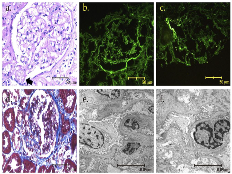
Three weeks after delivery, a kidney biopsy was performed. Focal segmental glomerulosclerosis (tip lesion) was diagnosed, in addition to acute tubular injury and active tubulointerstitial nephritis (Figure 1 and Figure 2).
We continued with prednisone 30 mg per day for 4 weeks and started tapering thereafter, on the account of a rapid clinical response. Eight weeks after delivery serum creatinine was 0.9 mg/dL, and proteinuria was 0.486 mg/24 h.(Table 1)

3. Discussion

Every year, as many as 500,000 mothers lose their lives due to pregnancy-related complications and 99% of these deaths occur in low- and middle-income countries [16]. These maternal deaths are most often linked with complications associated with preeclampsia, eclampsia, and HELLP syndrome [17].
The incidence of HELLP syndrome is estimated at 0.17–0.85% in all pregnancies and maternal mortality in HELLP syndrome ranges from 1% to 24% [1,16,17]. The relationship with AKI is well-known; back in 1993, Sibai described 33 cases of HELLP syndrome with AKI, with average creatinine clearance of 7 mL/min [1]. More recently, a systematic review published by Liu in 2020, including 11 cohort studies, with 355 cases of HELLP syndrome, found a strong association with AKI (Odds Ratio, OR 4.87), fetal mortality (OR 1.56), and maternal death (OR 3.70) [4]. Acute tubular necrosis was the most common finding in persistent AKI in HELLP syndrome, which can explain the reversibility in most patients even in those who experienced required dialysis. Thus, the finding of acute tubular necrosis in our case was expected [3].
However, our patient presented some clues for the differential diagnosis between “primary” HELLP and HELLP superimposed on a glomerular disease [2]. Indeed, while HELLP syndrome may be associated with AKI, this often occurs in more severe HELLP, while in our case, the main biochemical alterations resolved rapidly, and we observed dissociation between rapid healing of the biochemical alterations (elevated liver enzymes, low platelets, and signs of hemolysis) and the persistence of anuria. Furthermore, in keeping with a diagnosis different from HELLP syndrome, the kidney biopsy did not show fibrin thrombi in glomerular capillaries or in arterioles thus excluding a thrombotic microangiopathy severe enough to cause anuria. Since, at least in cases associated with placental ischemia, a derangement in the ratio angiogenic–antiangiogenic drugs is observed, we would like to underline the importance of more extensively using these important biomarkers, also for facilitation the differential diagnosis between hypertensive disorders of pregnancy and CKD.
Secondly, our patient did not experience shock or severe hypotension in the occasion of the cesarean section; her blood loss was moderate, and she had not been treated by tranexanic acid, thus excluding the three main causes of acute tubular necrosis or, even, acute cortical necrosis [3,18].
In such cases, a renal magnetic resonance may be of help in identifying cortical necrosis; however, our patient resumed some diuresis before we could schedule this imaging test, and since the finding of a very high level of proteinuria was not typical of the recovery phase from acute tubular necrosis and, even less, of partial cortical necrosis, this strongly oriented towards a primary kidney disease.
The presence of a peritubular infiltrate rich in eosinophils evokes the differential diagnosis of interstitial nephritis, which may have played a role both in the clinical manifestations of the disease and in the good response to steroid treatment. While it is not possible to formally exclude a role for an added interstitial damage, in the majority of cases, this is associated with the use of drugs, herbs, homeopathy, and food supplements. However, our patient denied use of drugs and over-the-counter toxins. The association between glomerular lesions and interstitial infiltrates is common, incompletely described, and not necessarily associated with classic interstitial disease, as it has been described for example in diabetic nephrology. The sensitivity of eosinophils to steroids may be one of the reasons for the rapid response to treatment [19].
As a third diagnostic hint, the selective proteinuria and the normal immunology pattern was suggestive for the presence of an “idiopathic nephrotic syndrome,” an umbrella term still occasionally used to encompass minimal change disease and focal segmental glomerulosclerosis [7,14]. The differential diagnosis necessitates a renal biopsy, whose interest in this context is not only diagnostic but also prognostic, as it allows assessing extension and type of the glomerulosclerotic lesions; presence of tubular necrosis; presence and extension of associated lesions including microangiopathic lesions (even if the rapid disappearance of schistocytes and the normalization of blood pressure did not suggest such an involvement); and presence and extension of glomerular endotheliosis [12].
A fourth diagnostic hint may result from urinary sediment analysis: while microscopic hematuria is so frequent in pregnancy to lose its diagnostic value, its absence is strongly against the presence of prolipherative lesions (including rapidly progressive glomerulonephritis), once more pointing towards the presence of either minimal change disease or focal segmental glomerulosclerosis [2,12,13].
The availability of the now-classic biomarkers of PE (sflt-1 and PlGF) could have further supported the differential diagnosis between hypertensive disorders of pregnancy and glomerulonephritis, since their behavior is clearly different in the “pure forms”, even if intermediate pictures are often present in PE or HELLP superimposed on kidney diseases [10,19,20,21,22]. However, the availability of these precious diagnostic aids is not widespread, and, in any case, their availability would have not changed the decision to perform the caesarean section.
Considering the high probability of minimal change disease in FSGS, steroid treatment was empirically started and, even if, due to the recent surgery, on the account of the fragility of the patient, we started with steroid doses lower than those usually employed. The rapid response to the treatment indirectly supported this diagnosis, which was indeed confirmed at the kidney biopsy. This response is also in keeping with previous findings of good and rapid response to treatment in the case of diseases whose renal prognosis is grim, as has been described by our group and others, and may further stress the advantages of timely treatment of glomerular diseases disclosed in pregnancy [11,23].
This case, reporting on the rare association between HELLP and FSGS, is a reminder to systematically consider kidney diseases in the differential diagnosis with all types of hypertensive disorders of pregnancy and is also a call for establishing close collaboration between obstetricians and nephrologists to safely manage these vulnerable women [6].
In addition, this case emphasizes the importance of pregnancy for diagnosing kidney disease and the need for further follow-up, to avoid the shift from AKI to chronic kidney disease.

Author Contributions

Conceptualization, writing—draft preparation: G.B.P. and O.A.O.-G.; performed Kidney biopsy: B.M.-G.; Nephropatology analisis: V.S.-A.; review and editing: G.B.P. and M.M. All authors have read and agreed to the published version of the manuscript.

Funding

This research received no external funding.

Informed Consent Statement

The patient provided signed informed consent for the anonymous publication of her data in the present report.

Acknowledgments

To Susan Finnel for her careful language revision.

Conflicts of Interest

The authors declare no conflict of interest.

References

  1. Sibai, B.M.; Ramadan, M.K.; Usta, I.; Salama, M.; Mercer, B.M.; Friedman, S.A. Maternal morbidity and mortality in 442 pregnancies with hemolysis, elevated liver enzymes, and low platelets (HELLP syndrome). Am. J. Obs. Gynecol. 1993, 169, 1000–1006. [Google Scholar] [CrossRef]
  2. Kandukurti, K.; Sun, J.; Venuto, R. Multiple pathologies in the kidney biopsy of a recently pregnant woman. Case Rep. Nephrol. Urol. 2013, 3, 9–15. [Google Scholar] [CrossRef] [PubMed]
  3. Ye, W.; Shu, H.; Wen, Y.; Ye, W.; Li, H.; Qin, Y.; Chen, L.; Li, X. Renal histopathology of prolonged acute kidney injury in HELLP syndrome: A case series and literature review. Int. Urol. Nephrol. 2019, 51, 987–994. [Google Scholar] [CrossRef] [PubMed]
  4. Liu, Q.; Ling, G.J.; Zhang, S.Q.; Zhai, W.Q.; Chen, Y.J. Effect of HELLP syndrome on acute kidney injury in pregnancy and pregnancy outcomes: A systematic review and meta-analysis. BMC Pregnancy Childbirth 2020, 20, 657. [Google Scholar] [CrossRef] [PubMed]
  5. Fakhouri, F.; Scully, M.; Provôt, F.; Blasco, M.; Coppo, P.; Noris, M.; Paizis, K.; Kavanagh, D.; Pène, F.; Quezada, S.; et al. Management of thrombotic microangiopathy in pregnancy and postpartum: Report from an international working group. Blood 2020, 136, 2103–2117. [Google Scholar] [CrossRef]
  6. Saei Ghare Naz, M.; Rahmati, M.; Azizi, F.; Ramezani Tehrani, F. Risk of chronic kidney disease in women with a history of preterm delivery: Tehran Lipid and Glucose Study. J. Nephrol. 2021, 34, 1621–1629. [Google Scholar] [CrossRef]
  7. Kountouris, E.; Clark, K.; Kay, P.; Roberts, N.; Bramham, K.; Kametas, N.A. Postnatal assessment for renal dysfunction in women with hypertensive disorders of pregnancy : A prospective observational study. J. Nephrol. 2021, 34, 1641–1649. [Google Scholar] [CrossRef]
  8. Cabiddu, G.; Mannucci, C.; Fois, A.; Maxia, S.; Chatrenet, A.; Osadolor, S.; Kimani, E.; Torreggiani, M.; Attini, R.; Masturzo, B.; et al. Preeclampsia is a valuable opportunity to diagnose chronic kidney disease: A multicentre study. Nephrol. Dial. Transplant. 2021, 2, gfab225. [Google Scholar]
  9. Gaber, L.W.; Spargo, B.H. Pregnancy-induced nephropathy: The significance of focal segmental glomerulosclerosis. Am. J. Kidney Dis. 1987, 9, 317–323. [Google Scholar] [CrossRef]
  10. Molina-Pérez, C.J.; Nolasco-Leaños, A.G.; Carrillo-Juárez, R.I.; Leaños-Miranda, A. Clinical usefulness of angiogenic factors in women with chronic kidney disease and suspected superimposed preeclampsia. J. Nephrol. 2022, 35, 1699–1708. [Google Scholar] [CrossRef]
  11. Orozco Guillén, O.A.; Velazquez Silva, R.I.; Gonzalez, B.M.; Becerra Gamba, T.; Gutiérrez Marín, A.; Paredes, N.R.; Cardona Pérez, J.A.; Soto Abraham, V.; Piccoli, G.B.; Madero, M. Collapsing Lesions and Focal Segmental Glomerulosclerosis in Pregnancy: A Report of 3 Cases. Am. J. Kidney Dis. 2019, 74, 837–843. [Google Scholar] [CrossRef] [PubMed]
  12. Smyth, A.; Wall, C.A. Nephrotic syndrome due to focal segmental glomerulosclerosis occurring in early pregnancy. Obs. Med. 2011, 4, 80–82. [Google Scholar] [CrossRef] [PubMed] [Green Version]
  13. Kopp, J.B.; Rosenberg, A.Z. One Actor, Many Roles: Histopathologies Associated With APOL1 Genetic Variants. Adv. Anat. Pathol. 2019, 26, 215–219. [Google Scholar] [CrossRef] [PubMed]
  14. De Castro, I.; Easterling, T.R.; Bansal, N.; Jefferson, J.A. Nephrotic syndrome in pregnancy poses risks with both maternal and fetal complications. Kidney Int. 2017, 91, 1464–1472. [Google Scholar] [CrossRef]
  15. Cabiddu, G.; Longhitano, E.; Cataldo, E.; Lepori, N.; Chatrenet, A.; Torreggiani, M.; Attini, R.; Masturzo, B.; Rossini, M.; Versino, E.; et al. History of Preeclampsia in Patients Undergoing a Kidney Biopsy: A Biphasic, Multiple-Hit Pathogenic Hypothesis. Kidney Int. Rep. 2021, 7, 547–557. [Google Scholar] [CrossRef]
  16. World Health Organization. The World Health Report 2005: Make Every Mother and Child Count; World Health Organization: Geneva, Switzerland, 2005. [Google Scholar]
  17. Weinstein, L. Syndrome of hemolysis, elevated liver enzymes, and low platelet count: A severe consequence of hypertension in pregnancy. Am. J. Obstet. Gynecol. 1982, 142, 159–167. [Google Scholar] [CrossRef]
  18. Jamme, M.; Rafat, C.; Gaugain, S.; Chousterman, B.G. Postpartum renal cortical necrosis. Kidney Int. 2018, 94, 1240. [Google Scholar] [CrossRef]
  19. Dai, D.F.; Sasaki, K.; Lin, M.Y.; Smith, K.D.; Nicosia, R.F.; Alpers, C.E.; Najafian, B. Interstitial eosinophilic aggregates in diabetic nephropathy: Allergy or not? Nephrol Dial. Transplant. 2015, 30, 1370–1376. [Google Scholar] [CrossRef] [Green Version]
  20. Rolfo, A.; Attini, R.; Tavassoli, E.; Neve, F.V.; Nigra, M.; Cicilano, M.; Nuzzo, A.M.; Giuffrida, D.; Biolcati, M.; Nichelatti, M.; et al. Is It Possible to Differentiate Chronic Kidney Disease and Preeclampsia by means of New and Old Biomarkers? A Prospective Study. Dis. Markers 2015, 2015, 127083. [Google Scholar] [CrossRef] [Green Version]
  21. Wiles, K.; Bramham, K.; Seed, P.T.; Kurlak, L.O.; Mistry, H.D.; Nelson-Piercy, C.; Lightstone, L.; Chappell, L.C. Diagnostic Indicators of Superimposed Preeclampsia in Women With CKD. Kidney Int. Rep. 2019, 4, 842–853. [Google Scholar] [CrossRef] [Green Version]
  22. Bramham, K.; Seed, P.T.; Lightstone, L.; Nelson-Piercy, C.; Gill, C.; Webster, P.; Poston, L.; Chappell, L.C. Diagnostic and predictive biomarkers for pre-eclampsia in patients with established hypertension and chronic kidney disease. Kidney Int. 2016, 89, 874–885. [Google Scholar] [CrossRef] [PubMed]
  23. Kwiatkowski, S.; Kwiatkowska, E.; Rzepka, R.; Kurkiewicz, V.; Mikołajek-Bedner, W.; Torbè, A. Development of a focal segmental glomerulosclerosis after pregnancy complicated by preeclampsia: Case report and review of literature. J. Matern. Fetal Neonatal Med. 2016, 29, 1566–1569. [Google Scholar] [CrossRef] [PubMed]
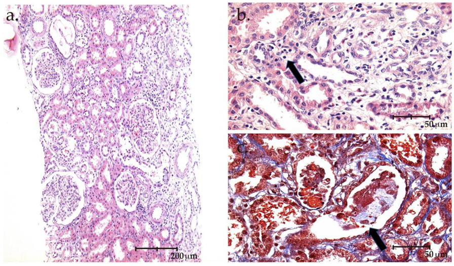
Figure 1. (a) 10× photomicrography stained with H&E showing the renal biopsy sample at low magnification, displaying interstitial edema and multiple foci of inflammatory infiltrate. (b) 40× photomicrography stained with H&E in which the arrows indicate that there are abundant eosinophils in the interstitial inflammatory infiltrate. (c) 40× photomicrograph stained with Masson’s trichrome, the arrows show the intratubular cellular and heme casts.
Kidneydial 02 00053 g001
Figure 2. (a,d) 40× photomicrographs stained with H&E and Masson’s trichrome respectively; the arrows show the lesions of the focal and segmental sclerosis, affecting the tubular pole, with endothelial edema. (b,c) 40× photomicrograph of the direct immunofluorescence study, which was negative for all the immunoreactants. (e,f) Electron photomicrograph 800× magnification, showing diffuse obliteration of the podocyte processes.
Kidneydial 02 00053 g002
Table 1. Biochemical data from referral to delivery and after pregnancy.
At ReferralPuerperium3 Weeks after Delivery, Kidney Biopsy8 Weeks after Delivery
S Cr (mg/dL)3.36.33.30.9
BUN (mg/dL)30.854.767.812.1
e-GFR (mL/min)--1989
Albumin (g/dL)4.554.13.74
Total proteins7.57.277.3
Proteinuria (g/24 h)--11.390.486
Haemoglobin (g/dL)14.611.211.412.9
Platelets (mm3)48,000142397396
LDH (UI/L)978432242260
AST (UI/L)7503634625
ALT (UI/L)12652735143
Total bilirubin(mg/dL)3.20.60.30.4
Potassium (mEq/L)4.75.74.24
HCO3 (mEq/L)1410.419.225
Immunologic markers: C3 96.5 mg/dL (90–180 mg/dL), C4 12.20 mg/dL (10–40 mg/dL), ANA negative
B2 glycoprotein (IgG) 3.9 CU, b2 Glycoprotein (IgM) 1.4 CU, anticardiolipin (IgM) 1.4 CU, anti-SM antibodies >3.3 CU, DNA double-strain antibodies >9.8 UI/ml
Thrombotic microangiopathic screening: IgG antibodies ADAMS-13(J) 1.83 U/mL negative (negative <12); positive >15
Adams-13 activity (j)   0.75   (0.4–1.3 U/mL)
Adams-13 activity      75% (40–130%)
Haptoglobin 27.1            (30–200 mg/dL)
Viral markers: Hepatitis B, C, HIV negative
Legend: sCr: serum creatinine, BUN: blood urea nitrogen; e-GFR: estimated glomerular filtration rate; w: week; HIV: human immunodeficiency virus; ANA: anti-nuclear antibodies; AST: aspartate aminotransferase; ALT: alanine aminotransferase; LDH: lactate dehydrogenase; HCO3: sodium bicarbonate, CU: control unit.
Publisher’s Note: MDPI stays neutral with regard to jurisdictional claims in published maps and institutional affiliations.