Review

A Review of High-Intensity Focused Ultrasound

by
Ben Turner
1 and
David Cranston
2,*
1
Bristol Medical School, Bristol University, Bristol BS8 1UD, UK
2
Nuffield Department of Surgery, University of Oxford, Oxford OX3 7LE, UK
*
Author to whom correspondence should be addressed.
Int. J. Transl. Med. 2024, 4(1), 197-207; https://doi.org/10.3390/ijtm4010011
Submission received: 30 January 2024 / Revised: 7 March 2024 / Accepted: 8 March 2024 / Published: 12 March 2024

Abstract

:
For 80 years, high-intensity focused ultrasound (HIFU) has been the subject of interest in medical research. It is a non-invasive procedure that causes the death of cells in a very select area through one of two mechanisms, either heat or cavitation. While diagnostic ultrasound is well known in the medical profession and ultrasound is also used in physiotherapy, high-intensity focused ultrasound is less known but is becoming increasingly important as a non-invasive tool that can be used in many ways, including in the treatment of several cancers as well as benign uterine fibroids. Other interesting developments are underway, including its use in the treatment through an intact skull of essential tremors and the tremor associated with Parkinson’s disease, and in a modified form, it is used to target drug delivery to the brain due to its potential opening of the blood–brain barrier. The depth of penetration of HIFU is variable depending on the type of transducer used and the distance from it. Clinical trials of abdominal malignancies and benign uterine fibroids are reviewed in this article along with potential side effects of the procedure. Over the past two decades, the technology has improved considerably, and the clinical indications have broadened. The current limitations of the technology are also discussed, along with the potential advances in the field that may be made over the next decade.

1. Introduction

High-intensity focused ultrasound (HIFU) is a therapeutic technology based on ultrasound which has been studied by of a number of research laboratories and translational clinical applications over the last 85 years. Diagnostic ultrasound beams have frequencies in the scale of 2–16 MHz, with intensities up to several hundreds of mW/cm2 being used in B-mode scanning, whereas HIFU treatments designed to cause cell death will use frequencies in the range from 300 kHz to several MHz, with intensities at the target that may be in excess of 1500 watts/cm2. The focused waves can be targeted on precise volumes in tissue, ref. [1], ablating the targeted area. The final temperature achieved in the focus depends on the acoustic absorption coefficient. Since acoustic attenuation arises from both absorption and scatter, tissues with low attenuation coefficients will heat up less than those for which attenuation is high.
Histotripsy is another form of high energy that is mediated by cavitation rather than heat to emulsify tissue and is now being used in studies to treat renal and prostate cancers [2].
Since HIFU is a non-invasive technique, it is an attractive alternative to traditional surgical techniques. Before the point of focus, the emitted waves are insufficiently powerful to damage normal tissue and it is only when the waves meet at the focal point that the energy contained within them is high enough to cause tissue necrosis [3].
HIFU causes necrosis in two ways. Firstly, it generates high temperatures of over 80 °C, which is considerably higher than the 56 °C where an exposure of more than one second causes cell death [3]. These high temperatures are localised by keeping exposure times short, and this reduces the risk of damage to surrounding tissues. The cooling effect of perfusion, which limits the reproducibility of other forms of hyperthermia treatment, can be minimised by keeping exposure times below three seconds [4]. Secondly, HIFU causes tissue destruction through cavitation. Acoustic cavitation is complex and difficult to control, but the resulting mechanical stresses and thermal injury cause cell death. Histotripsy by contrast leads to mechanical tissue emulsification without a thermal effect.
Ultrasound causes vibration of the tissues, subjecting them to alternating rarefaction and compression. During the former, gas forms bubbles as it is drawn out of solution. These oscillate in size and may collapse, causing mechanical stresses and generating temperatures of up to 5000 °C in the microenvironment. The effects of heating the tissues are more predictable than cavitation and also more repeatable [5], and this makes tissue heating the favoured mode of action in most clinical therapies. However, it is possible that the mechanical disruption caused by cavitation may boost the development of antitumor antibodies, which in turn could have an advantageous effect on distant metastases [6].
Minimally invasive treatment techniques like HIFU have many potential benefits over traditional surgery. Some are obvious: the absence of an incision reduces infection risk, often resulting in a more rapid recovery, and the lack of a scar can be cosmetically valuable. HIFU may also have additional advantages not common to other minimally invasive techniques. For example, one downside of radiotherapy is its immunosuppressive effect [7]. HIFU does not lead to immunosuppression, and there is some evidence to suggest that HIFU may be able to boost the antitumoral immune response [8]. Unlike radiotherapy, HIFU has no maximum dose limit, meaning repeat administrations are possible to ensure satisfactory outcomes [3].
High-intensity focused ultrasound is generated under water and passed through a degassed water-filled coupling balloon into the body, usually without damage to the overlying structures, into the focal region, which may be as far as 15 cm from the source. HIFU does not propagate well through air. This means that gas-containing structures within the body such as the lungs or bowel are not suitable targets [9], although it may be possible to treat areas in the lung if one lung is flooded [10]. Furthermore, these structures can be at risk when HIFU is delivered to other structures in close proximity. Another limitation is the time it takes to deliver an effective HIFU treatment. Some sessions of HIFU on larger tumours can take many hours. Due to the precise nature of HIFU, the patient must remain still during the treatment, and so general anaesthetics may be required [9]. However, other treatments, such as for uterine fibroids, can be undertaken with sedation.

2. History of HIFU

The biological effects of high-energy ultrasound waves were first reported by Wood and Loomis in 1927 [11] and further early research was conducted by Lynn et al. in 1942 [12].
The clinical story of HIFU begins in the 1950s, where it was trialled for the treatment of Parkinson’s disease and other neurological conditions by brothers William and Frank Fry [13,14,15]. However, these treatments involved the opening of the skull via a craniotomy, and this highly invasive requirement inevitably meant that this use of HIFU was not further pursued [1].
The technology was explored again in the 1990s, with variable success. China has been one of the leaders in clinical HIFU use in recent times. In the past 20 years, thousands of patients have been treated in China and more widely in Asia for conditions such as uterine fibroids and benign prostate hypertrophy [16].
Professor Gail ter Haar and her colleagues in Surrey pioneered HIFU research in the UK, and in 2002, a dedicated HIFU unit was founded by Professor David Cranston in Oxford. This unit has carried out several trials on a variety of different clinical applications of HIFU [17].

3. The Technology

Today, there are many HIFU devices produced for various indications for both extracorporeal (Figure 1) and intracavitary applications [18], the latter in particular for use in prostate cancer, such the Ablatherm® (EDAP-Technomed, Lyon, France) and the Sonablate®500 (Focus Surgery, Indianapolis, IN, USA) systems. Historically, HIFU treatments have been guided either by magnetic resonance imaging (MRI) or ultrasound imaging, although it has now become possible to fuse these two modalities.
Ultrasound-guided HIFU combines the diagnostic and therapeutic forms of ultrasound and, to all intents and purposes, allows for real-time imaging with a diagnostic transducer placed in close proximity to the treatment transducer. In ultrasound-guided HIFU, successful ablation is seen as grey scale changes and bright echoes in the targeted area, resulting from bubbles that are produced as a result of water boiling within the tissues.
Ultrasound-guided HIFU is cheaper, smaller, and more widely available than the alternative MRI-guided HIFU, where, apart from being more expensive, the imaging is not performed in ‘real time’; however, the pictures produced by MRI are three-dimensional and clearer to interpret than those produced by ultrasound-guided HIFU, and in addition to this, MRI is able to produce temperature maps of the targeted areas.

4. Applications of HIFU

4.1. Kidney

There is a limited amount of research on renal tissue, although some experimental studies have shown the destruction of both malignant and benign renal lesions [19], but in the early studies, there were technical problems with skin burns and the amount of tissue ablation was variable. Clinical treatment of renal tumours with HIFU has been the subject of very few clinical trials; however, a study in 2010 by the Oxford HIFU team investigated 17 patients with renal tumours [20]. The patients had an average tumour size of 2.5 cm and were all treated under a general anaesthetic and then spent one night in hospital after their treatments. The machine used was an ultrasound-guided HIFU machine (JC Chongqing Haifu, Chongqing, China). The patients were then followed up with an MRI scan after 12 days, and then every 6 months for a mean duration of 36 months. Of the 17 original patients, 15 had the full HIFU treatment—two of the procedures had to be stopped due to bowel obstruction. Of those 15, 7 showed tumour ablation at the 12-day follow-up. After the 36 months, the overall results showed that two thirds of the patients experienced some tumour ablation, with a mean reduction in tumour volume of 30%.
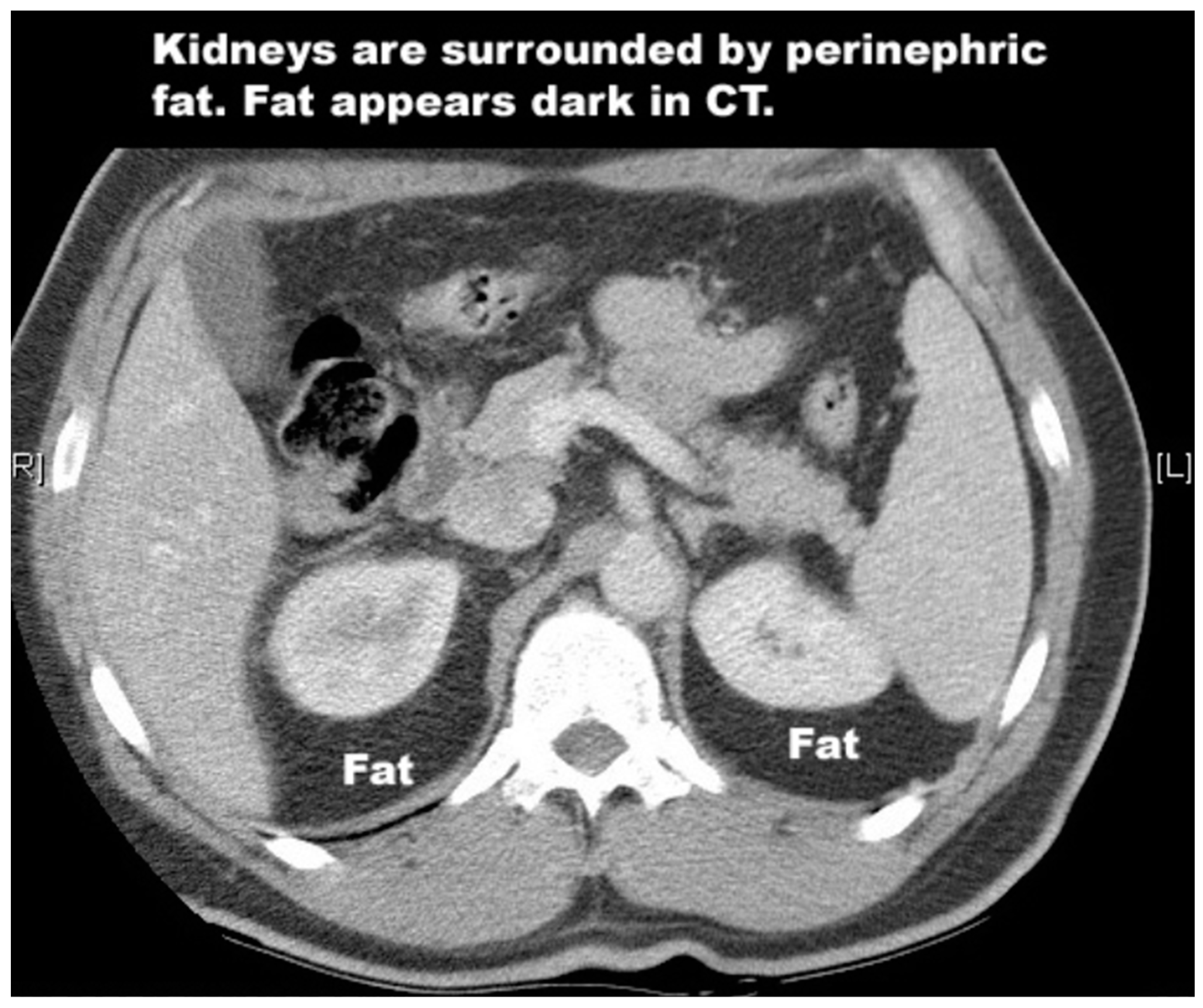
Renal tumours are a potentially challenging target for HIFU given the dense layers of perinephric fat that surround the kidneys (Figure 2). These layers absorb some of the energy of the HIFU beam. A 2020 study investigated this issue to determine the percentage drop in HIFU energy compared to the depth of the perinephric fat [21]. The reduction was significant: from 58% of the output energy level at 2 cm deep, to 26% at 5 cm. To compensate for this, higher output energies are required, but this increases the risk of surrounding tissue damage. However, transplanted kidneys have had their perinephric fat removed, and so transplant patients are perhaps better candidates for HIFU treatment. This was demonstrated by the treatment of a tumour in a renal transplant in two patients in Oxford who were treated with HIFU. The first treatment was unsuccessful due to technical issues. The second patient [22] received two treatments of HIFU over 4 months. On both occasions, the patient experienced no pain and was discharged within 24 h. Post-treatment analysis of the tumour showed a 90% reduction in tumour size.
A laparoscopic HIFU system has also been used for the ablations of small kidney tumours. The device is called the Sonatherm and is manufactured be Misonix Inc, an American company. The first treatment on a human was performed in 2006 in Oxford. Phase 1 studies were performed in Oxford (12 patients) and Vienna (10 patients) with encouraging results [23]. However, while laparoscopic HIFU was feasible and safe, and good ablation seen with low morbidity, laparoscopic HIFU remains an invasive procedure, meaning it loses one of the major benefits of non-invasive extracorporeal HIFU, and due to financial constraints, and the fact that this remains an invasive procedure, no further studies were performed.

4.2. Prostate

The technique for using HIFU to target the prostate involves inserting a small probe into the rectum which then emits the ultrasound waves. For benign prostatic disease, HIFU has been trialled and compared with transurethral resection of the prostate (TURP); however, HIFU has not yet been demonstrated to be superior [24].
The first reports that HIFU could be used to treat prostate carcinoma were published in 1995 by Madersbacher et al. [25], and since that time, there have been many more studies using the Ablatherm® or the Sonablate®500 systems. Both are examples of ultrasound-guided HIFU where the ultrasound imager and treatment transducer are combined in the endorectal probe.
In the context of prostate cancer, HIFU may have many advantages over the traditional methods of surgical radical prostatectomy and radiotherapy. In 2019, the UK National Prostate Cancer Audit published the results of a study involving patient-reported outcomes from over 25,000 men who had received the traditional prostate cancer treatment methods [26]. This audit highlighted the significant risk of side effects and complications from traditional treatments for prostate cancer. One in ten men experienced ‘severe urinary complications’ following surgery or ‘severe bowel complications’ following radiotherapy. These results demonstrate the significant negative impact of established treatments for prostate cancer. Could HIFU offer equally effective treatment with fewer side effects?
The French Urological Association undertook a six-year [27] study (2009–2015) for the treatment of primary prostate cancer in 111 patients with low or medium risk disease, finding a 95% absence of clinically significant cancer, and a treatment-free survival rate of 89% at two years [28]. These results show the potential efficacy of HIFU as a prostate cancer treatment. It is important to note though that two years is a short follow-up in the context of evaluating the effectiveness of radical treatments for prostate cancer. A follow-up duration of 10–15 years may be required to effectively compare treatments [29].
Medium-term outcomes were reported by Dickinson et al. [30] in 569 men who received HIFU to the whole of the prostate for localised prostate cancer. In total, 754 interventions were carried out as the study protocol allowed for further treatment sessions where necessary. They found that this could be undertaken as a day case procedure with few side effects. The incontinence rates of those needing pads was 12%. Erectile dysfunction was 61% but this was similar to other whole-gland treatments. One patient developed a recto-urethral fistula—though this is a potentially devastating complication. These results demonstrate the possible advantages that HIFU has over other prostate cancer treatment options in terms of its low side-effect and complication rates.
In 2019, Stabile et al. [31] carried out focal treatment in 1032 men, 80% of whom had medium or high-risk cancer. The treatment was carried out using the Sonablate 500 device. They found that at 96 months post HIFU, 81% of the patients had avoided any further radical treatment. Furthermore, the survival rate at 96 months was 97%. As a result of this, they felt that focal HIFU for prostatic carcinoma was a feasible therapeutic strategy, with good survival rates and a reduction in the re-treatment rates at 96 months.
The first study concerning HIFU treatment of prostate cancer in the United States of America has also recently been published [32]. The report was retrospective but showed similarly positive results to the Stabile et al. study, with a sample size of 100 men. HIFU was shown to be effective (91% of the men avoided further radical treatment within the first 2 years after treatment), and all 100 of the patients maintained full urinary continence after the treatment.
HFIU has also been tested as a method of salvage treatment for recurrent prostate cancer after previous radical treatment. Houstiou et al. evaluated salvage HIFU for recurrent prostate cancer after previous brachytherapy and radiotherapy treatment [33]. They treated 50 patients over 12 years between 2003 and 2015. They found encouraging results, both in terms of oncological and complication outcomes. Progression-free survival was 45%, and overall survival was 93%. The HIFU was delivered with specific post-brachytherapy and post-radiotherapy parameters.
Crouzet et al. carried out a retrospective study on 418 patients and examined the outcomes of their salvage treatments from 1995 to 2009 [34]. The patients had all received salvage HIFU for locally recurrent prostate cancer following external beam radiotherapy. The 7-year cancer-specific survival rate was over 80%; however, they initially reported worse morbidity results than the Houstiou et al. study. Complication rates of incontinence, outflow obstruction, and recto-urethral fistula were all higher in the period 1995–2002. In 2002, however, treatment-specific parameters were introduced for the salvage HIFU. These, in a similar way to the Houstiou et al. study, accounted for the tissue changes that previous radiotherapy induced, such as decreased vascularisation of the prostate gland, and per-prostatic tissue. These alterations in HIFU treatment improved complication rates post-2002, resulting in the recto-urethral fistula rate decreasing from 9% to 0.6%.

4.3. Liver

A 2005 study conducted in Oxford examined the safety and feasibility of HIFU for the treatment of both liver and kidney tumours in a Western population [35]. A total of 30 patients were recruited, with 22 having liver tumours. Each patient received a single HIFU session under general anaesthetic. Of the 30 patients treated, 27 were able to have their response to treatment evaluated, primarily through an MRI scan at 12 days post-HIFU. Evidence of tumour ablation was seen in 25 patients (93%). The accuracy of the treatment was also evaluated. This involved observing the zones of ablation to see if they fell within the tumour area or impacted the surrounding tissue. Accuracy was said to be ‘good’ if the ablation only effected the tumour, and ‘poor’ if it was outside the tumour. Accuracy was assessed as ‘good’ in 21 patients. The patients were also monitored for side effects post-treatment. The only clinically relevant symptom reported was ‘discomfort’ around the site of treatment, which was reported as ‘mild’ in severity in 80% of cases. Skin toxicity was seen in eight cases (27%). In seven of these, toxicity consisted of very small (pinhead) blisters or tracks that were not clinically relevant and resolved without any need for treatment.
A more recent study by Yang, T et al. also investigated the treatment of liver tumours [36]. This phase 1 clinical trial aimed to evaluate HIFU as a treatment for colorectal liver metastases (CRLM), specifically metastases that were deemed ‘difficult’ and unsuitable for resection or radiological ablation. A total of 13 patients between the ages of 20 and 80 were recruited. Ultrasound-guided HIFU was used to ablate the liver tumours.
The surgical method of hepatectomy can achieve a 5-year 50% survival rate for CRLM [37,38]. However, up to 80% of patients diagnosed with CRLM have an unresectable tumour at the time of diagnosis. The median survival time for patients diagnosed at this stage is less than a year [39]. The results of this study were positive. Firstly, adverse events were minimal, with the most commonly reported being pain and fatigue. The median follow-up to the treatment was 25 months. The 2-year overall survival was 77.8%, and the median overall survival time was 25 months. This indicates that HIFU is safe and able to achieve good results in patients with difficult-to-treat CRLM. The authors concluded that this treatment should be considered for patients who were considered unsuitable for other treatment options.

4.4. Uterine Fibroids

In females, HIFU can be used to treat uterine fibroids, which occur in between 20% and 25% of women of childbearing age [40].
A study from 20 centres in China in collaboration with the University of Oxford under the IDEAL framework of a prospective collaborative cohort study [27] looked at 2411 Chinese women with symptomatic fibroids to investigate the clinical results of HIFU in treating uterine fibroids, comparing it with hysterectomy or myomectomy. They looked at hospital stay, complications, return to normal activities, and quality of life at baseline, and six and twelve months, and whether further treatment was necessary. In total, 1353 women received HIFU, 472 hysterectomy, and 586 myomectomy. Quality of life improved more rapidly after HIFU than after surgery, and the authors concluded that HIFU caused substantially less morbidity than surgery, with similar longer-term quality of life outcomes.
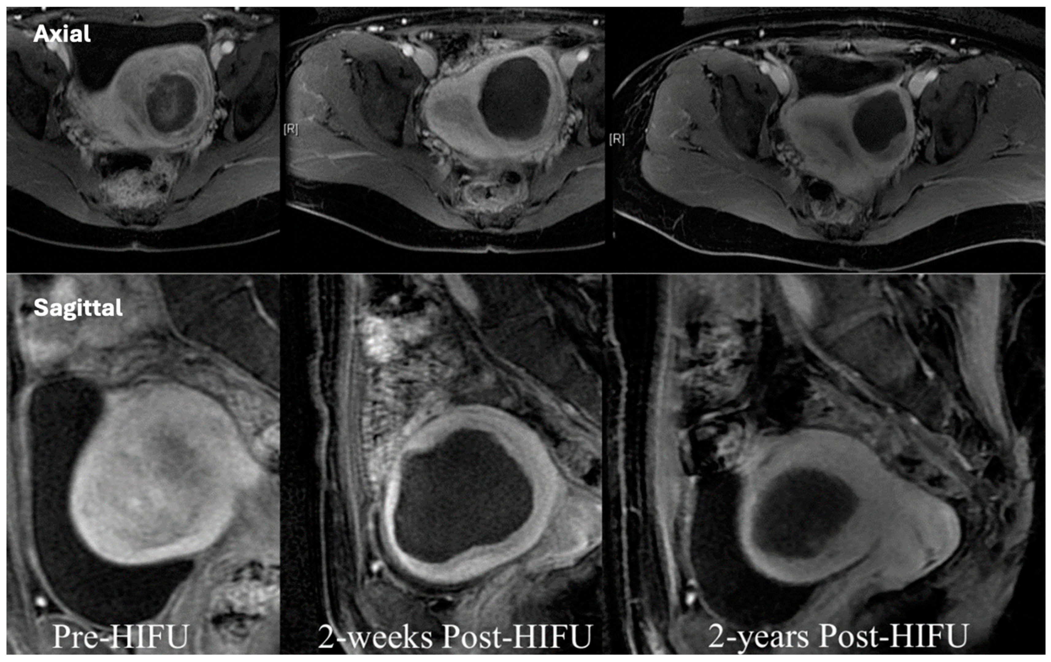
Oxford University Hospitals conducted the first study of HIFU treatment for this condition on an NHS population [41]. Out of 22 patients referred, 12 were treated with a single session of ultrasound-guided HIFU ablation, targeting a total of 14 fibroids. These patients were then monitored for a two-year period. While no serious adverse events were recorded, there was an isolated case of a second-degree skin burn seen in a patient with a surgical scar from a previous caesarean section. Significant improvements were noted in mean symptom severity scores, dropping from 56.5 ± 29.1 (SD) at baseline to 45.0 ± 35.4 (p < 0.05) at one year, and 40.6 ± 32.7 (p < 0.01) at two years post-treatment. In summary, the study suggests that HIFU ablation led to a substantial improvement in symptoms over a 2-year time period. The occurrence of adverse events was generally low, with the exception of a single case of a second-degree skin burn in a patient with a specific medical history. Figure 3 shows the dark avascular areas post ablation.
A systematic review investigating the outcomes of HIFU in patients with uterine artery embolisation and traditional surgeries for treating symptomatic uterine fibroids was carried out by Yan et al. [42]. This showed that although HIFU has a relatively high rate of re-intervention, the pregnancy rate may be higher, with little influence on ovarian function, thus making it an good option for treating fibroids that are symptomatic in young women who wish to plan for a future pregnancy.

4.5. Chordomas

Chordomas are malignant tumours that arise from remnants of the notochord [43] and form in areas such as the sacrum [44]. Surgical resection is the treatment of choice for sacral chordomas, but this is not always possible due to their location and proximity to essential structures. Radiotherapy is an alternative to surgery; however, recurrence is very common [45], and the long-term prognosis is poor [46]. HIFU has great potential in this area. In a 2017 clinical trial at Churchill Hospital, Oxford, four patients with sacral chordomas were successfully treated [47]. Three of the four patients were treated under general anaesthetic, and the other was given only sedation (allowing verbal feedback to be given throughout the treatment). Only three of the patients were able to be followed up as one lived abroad. All three saw a reduction in tumour volume over time, and tumour necrosis was demonstrated in two patients. The side effects were minimal, with the most prominent symptom being mild discomfort.

4.6. Pancreas and Breast Cancer

Pancreatic cancers generally have a poor prognosis. Surgery gives the best chance of being cured, but typically, only 15–20% of patients are suitable for surgery [48]. In Oxford, a multi-centre clinical trial is being carried out to investigate HIFU for patients with pancreatic cancer. Patients with pancreatic cancer, who are unsuitable for surgery, and a lack of metastases are being recruited. Patients are initially treated with chemotherapy for three months, and then receive HIFU. They are then followed up for two years. Multiple patients so far have been successfully treated with no significant side effects. This is therefore a promising direction for HIFU treatment.
Breast cancer is another area where trials are ongoing for HIFU therapy [49]. Breast tissue is an ideal location for HIFU beams [50] as it sits superficially and so does not have vulnerable or gas-filled structures nearby. HIFU is an established treatment for breast cancer in China [1], and a trial in Oxford has been approved.

4.7. Ultrasound-Mediated Drug Delivery

Ultrasound waves can be used to facilitate the delivery of drugs and genes into living cells. This occurs by sonoporation, whereby ultrasound induces plasma membrane perforation. The authors of a 2022 review of the technology [50] concluded that ‘This drug delivery approach, when coupled with concurrent advances in ultrasound imaging, has potential to become an effective therapeutic paradigm’.
The TARDOX trial was an Oxford-based phase 1 clinical trial involving 10 patients with non-ablatable and non-resectable primary and secondary liver tumours [51]. Patients were given a single infusion of doxorubicin which was encapsulated in a thermosensitive carrier (a liposome). The tumours were then targeted with HIFU, and the heat generated by the HIFU resulted in targeted drug release. Biopsies were taken pre- and post-HIFU to assess the impact. The mean change in intratumoural concentrations of the drug was a 3.7-fold increase post-HIFU. Reported side effects included neutropenia in five patients, although this was only transient. Whilst preliminary, this study raises exciting possibilities for future HIFU applications.

4.8. Possible Immunological Benefits of HIFU

HIFU may have positive immunological impacts with regards to cancer-specific immunity. Murine studies suggest that this might arise from destroyed tumour cells that remain in situ and which can act as sources of antigens that provoke a tumour-specific immune response [52]. Another study involving rats showed that this phenomenon may be mediated in part via heat shock proteins (HSPs) [53]. Cancer cells express these proteins during times of stress, such as when targeted with HIFU therapy. These proteins can then stimulate cytotoxic T-cell activity. Whilst more research needs to be conducted in this area, the fact that HIFU may potentially benefit a patients’ immune response puts it into stark contrast with other non-surgical cancer treatments. Both chemotherapy and radiotherapy, for example, have the opposite effect.

5. Discussion

Since the early days of HIFU research and treatment, many developments have taken place and increasing numbers of clinicians and pre-clinical research workers around the world are using and developing the technology. Like any medical technology, it has potential side effects, and as has been discussed above, there are areas of the body which are more difficult to access, partly due to overlying ribs, and partly, as in the case of kidneys, due to the surrounding perinephric fat. In terms of depth penetration, it is possible to target 14 or 15 cm into the body on the JC 200 device, and due to time reversal technology, it is now possible to treat through an intact skull essential tremors and the tremor associated with Parkinson’s disease. Currently, treatment may take several hours for a large tumour, and this requires the patient to be still either under general anaesthesia or sedation. Respiratory movement needs to be controlled when targeting abdominal organs, and in due course, improvements in the technology will link treatment with respiration, which will help to overcome these problems. With the ability to fuse ultrasound and MRI images, better planning and targeting of treatment will be available, and where HIFU is appropriate, it is likely to be much cheaper than surgery and does not need the use of an operating theatre and staff requirements are less. Unlike radiotherapy, treatment can be repeated, and as further clinical trials are undertaken, there will be an increased understanding of the role of HIFU not only as a solitary treatment but also where it sits in combination of other types of therapy, especially for malignant tumours.
While HIFU is considered to be non-invasive, it is not without its side effects. The most common ones are superficial skin burns and occasional deep skin burns, especially if treatment is undertaken either through previous scars or in areas that have previously been treated by radiotherapy. In terms of prostate cancer, fistulas from the urethra into the rectum have been reported and there is a potential for perforation of the bowel if the ultrasound beam penetrates the bowel. Pain is usually short-lived, and the majority of patients can be treated either as a day case or with minimal hospital stay.

6. Conclusions

There is little doubt that HIFU is becoming an increasingly important non-invasive addition to the medical armamentarium. It is now an accepted treatment for some patients with localised prostate cancer and, in many parts of the world, a definitive treatment for uterine fibroids. Other areas will continue to progress as the technology improves. Image fusion, faster treatments, and new phased array transducers will increase the speed and accuracy of treatment, and the problems caused by the ribs and breathing will be overcome with motion compensation. This in turn will help to facilitate the successful treatment of kidneys and other abdominal tumours where movement is an issue. In due course, there will be newer machines for treating prostate cancer and other abdominal organs which will further minimise the side effects of radical treatment. Other areas of targeted drug delivery, including within the brain, will improve, and with an increased understanding of HIFU in combination with other treatments, the technology will become increasingly important in the next decade.

Author Contributions

B.T. and D.C. had an equal contribution to the conceptualization, writing, review, and editing of this paper. All authors have read and agreed to the published version of the manuscript.

Funding

This research received no external funding.

Conflicts of Interest

DC is an Adjunct Professor at Chongqing Medical University China.

References

  1. Kennedy, J.E.; ter Haar, G.R.; Cranston, D. High intensity focused ultrasound: Surgery of the future? Br. J. Radiol. 2003, 76, 590–599. [Google Scholar] [CrossRef]
  2. Xu, Z.; Hall, T.L.; Vlaisavljevich, E.; Lee, F.T. Histotripsy: The first noninvasive, non-ionizing, non-thermal ablation technique based on ultrasound. Int. J. Hyperth. 2021, 38, 561–575. [Google Scholar] [CrossRef]
  3. Kennedy, J.E. High-intensity focused ultrasound in the treatment of solid tumours. Nat. Rev. Cancer 2005, 5, 321–327. [Google Scholar] [CrossRef]
  4. Chen, L.; ter Haar, G.R.; Hill, C.R.; Dworkin, M.; Carnochan, P.; Young, H.; Bensted, J.F.M. Effect of blood perfusion on the ablation of liver parenchyma with high-intensity focused ultrasound. Phys. Med. Biol. 1993, 38, 1661–1673. [Google Scholar] [CrossRef]
  5. Hill, C.R.; Rivens, I.; Vaughan, M.G.; ter Haar, G. Lesion development in focused ultrasound surgery: A general model. Ultrasound Med. Biol. 1994, 20, 259–269. [Google Scholar] [CrossRef]
  6. Kepp, O.; Marabelle, A.; Zitvogel, L. Oncolysis without viruses—Inducing systemic anticancer immune responses with local therapies. Nat. Rev. Clin. Oncol. 2020, 17, 49–64. [Google Scholar] [CrossRef]
  7. Rückert, M.; Flohr, A.-S.; Hecht, M.; Gaipl, U.S. Radiotherapy and the immune system: More than just immune suppression. Stem Cells 2021, 39, 1155–1165. [Google Scholar] [CrossRef]
  8. Kennedy, J.; Wu, F.; ter Haar, G.; Gleeson, F.; Phillips, R.; Middleton, M.; Cranston, D. High-intensity focused ultrasound for the treatment of liver tumours. Ultrasonics 2004, 42, 931–935. [Google Scholar] [CrossRef] [PubMed]
  9. Izadifar, Z.; Izadifar, Z.; Chapman, D.; Babyn, P. An introduction to high intensity focused ultrasound: Systematic review on principles, devices, and clinical applications. J. Clin. Med. 2020, 9, 460. [Google Scholar] [CrossRef] [PubMed]
  10. Wolfram, F.; Lesser, T.G. A simulation study of the HIFU ablation process on lung tumours, showing consequences of atypical acoustic properties in flooded lung. Z. Med. Phys. 2019, 29, 49–58. [Google Scholar] [CrossRef] [PubMed]
  11. Wood, R.; Loomis, A.L. The physical and biological effects of high-frequency sound-waves of great intensity. Lond. Edinb. Dublin Philos. Mag. J. Sci. 1927, 4, 417–436. [Google Scholar] [CrossRef]
  12. Lynn, J.G.; Zwemer, R.L.; Chick, A.J.; Miller, A.E. A new method for the generation and use of focused ultrasound in experimental biology. J. Gen. Physiol. 1942, 26, 179–193. [Google Scholar] [CrossRef]
  13. Fry, W.J.; Barnard, J.W.; Fry, F.J.; Krumins, R.F.; Brennan, J.F. Ultrasonic lesions in the mammalian central nervous system. Science 1955, 122, 517–518. [Google Scholar] [CrossRef]
  14. Fry, F.J. Precision high intensity focusing ultrasonic machines for surgery. Am. J. Phys. Med. Rehabil. 1958, 37, 152–156. [Google Scholar] [CrossRef]
  15. Burov, A.K. High-intensity ultrasonic vibrations for action on animal and human malignant tumours. Dokl Akad. Nauk SSSR 1956, 106, 239–241. [Google Scholar]
  16. Lee, K.W. The Asian perspective on Hifu. Int. J. Hyperth. 2021, 38, 5–8. [Google Scholar] [CrossRef]
  17. ter Haar, G.; Sinnett, D.; Rivens, I. High intensity focused ultrasound-a surgical technique for the treatment of discrete liver tumours. Phys. Med. Biol. 1989, 34, 1743–1750. [Google Scholar] [CrossRef] [PubMed]
  18. Bachu, V.S.; Kedda, J.; Suk, I.; Green, J.J.; Tyler, B. High-intensity focused ultrasound: A review of mechanisms and clinical applications. Ann. Biomed. Eng. 2021, 49, 1975–1991. [Google Scholar] [CrossRef]
  19. Koerhrman, K. High intensity focused ultrasound for non-invasive tissue ablation in the kidney, prostate and uterus. J. Urol. 2000, 163, 698. [Google Scholar]
  20. Ritchie, R.W.; Leslie, T.; Phillips, R.; Wu, F.; Illing, R.; Ter Haar, G.; Protheroe, A.; Cranston, D. Extracorporeal high intensity focused ultrasound for renal tumours: A 3-year follow-up. BJU Int. 2010, 106, 1004–1009. [Google Scholar] [CrossRef] [PubMed]
  21. Mancini, M.; Righetto, M.; Baggio, G. Gender-related approach to kidney cancer management: Moving forward. Int. J. Mol. Sci. 2020, 21, 3378. [Google Scholar] [CrossRef] [PubMed]
  22. Chakera, A.; Leslie, T.; Roberts, I.; O’Callaghan, C.A.; Cranston, D. A lucky fall? Case report. Transplant. Proc. 2010, 42, 3883–3886. [Google Scholar] [CrossRef] [PubMed]
  23. Klingler, H.C.; Susani, M.; Seip, R.; Mauermann, J.; Sanghvi, N.; Marberger, M.J. A novel approach to energy ablative therapy of small renal tumours: Laparoscopic high-intensity focused ultrasound. Eur. Urol. 2008, 53, 810–816. [Google Scholar] [CrossRef]
  24. Nakamura, K. Clinical application of high-intensity focused ultrasound for benign prostatic hyperplasia. In Treatment of Benign Prostatic Hyperplasia; Springer: Tokyo, Japan, 2000; pp. 123–132. [Google Scholar] [CrossRef]
  25. Madersbacher, S.; Pedevilla, M.; Vingers, L.; Susani, M.; Marberger, M. Effect of High-Intensity Focused Ultrasound on human prostate cancer in vivo. Cancer Res. 1995, 55, 3346–3351. [Google Scholar]
  26. National Prostate Cancer Audit. NPCA Annual Report 2019. 2020. Available online: https://www.npca.org.uk/reports/npca-annual-report-2019/ (accessed on 25 January 2024).
  27. McCulloch, P.; Altman, D.G.; Campbell, W.B.; Flum, D.R.; Glasziou, P.; Marshall, J.C.; Nicholl, J. No surgical intervention without evaluation. The IDEAL recommendations. Lancet 2009, 374, 1105–1112. [Google Scholar] [CrossRef]
  28. Rischmann, P.; Gelet, A.; Riche, B.; Villers, A.; Pasticier, G.; Bondil, P.; Jung, J.L.; Bugel, H.; Petit, J.; Toledano, H.; et al. Focal High Intensity Focused Ultrasound of Unilateral Localized Prostate Cancer: A Prospective Multicentric Hemiablation Study of 111 Patients. Eur. Urol. 2017, 71, 267–273. [Google Scholar] [CrossRef]
  29. Cooperberg, M.R. RE: Fifteen-year outcomes after monitoring, surgery, or radiotherapy for prostate cancer. Eur. Urol. 2023, 84, 435–436. [Google Scholar] [CrossRef]
  30. Dickinson, L.; Arya, M.; Afzal, N.; Cathcart, P.; Charman, S.C.; Cornaby, A.; Hindley, R.G.; Lewi, H.; McCartan, N.; Moore, C.M.; et al. Medium-term Outcomes After Whole-gland High-intensity Focused Ultrasound for the Treatment of Nonmetastatic Prostate Cancer from a Multicentre Registry Cohort. Eur. Urol. 2016, 70, 668–674. [Google Scholar] [CrossRef]
  31. Stabile, A.; Orczyk, C.; Hosking-Jervis, F.; Giganti, F.; Arya, M.; Hindley, R.G.; Dickinson, L.; Allen, L.; Punwani, S.; Jameson, C.; et al. Medium-term Oncological Outcomes in a Large Cohort of Men Treated with Either Focal or Hemi-Ablation Using High-Intensity Focused Ultrasonography for Primary Localized Prostate Cancer. BJU Int. 2019, 124, 431–440. [Google Scholar] [CrossRef] [PubMed]
  32. Abreu, A.L.; Peretsman, S.; Iwata, A.; Shakir, A.; Iwata, T.; Brooks, J.; Tafuri, A.; Ashrafi, A.; Park, D.; Cacciamani, G.E.; et al. High Intensity Focused Ultrasound Hemi-gland Ablation for Prostate Cancer: Initial Outcomes of a United States Series. J. Urol. 2020, 204, 741–747. [Google Scholar] [CrossRef] [PubMed]
  33. Hostiou, T.; Gelet, A.; Chapelon, J.Y.; Rouviere, O.; Mege-Lechevalier, F.; Lafon, C.; Tonoli-Catez, H.; Badet, L.; Crouzet, S. Salvage High-Intensity Focused Ultrasound for Locally Recurrent Prostate Cancer After Low-Dose-Rate Brachytherapy: Oncological and Functional Outcomes. BJU Int. 2019, 124, 746–757. [Google Scholar] [CrossRef] [PubMed]
  34. Crouzet, S.; Blana, A.; Murat, F.J.; Pasticier, G.; Brown, S.C.; Conti, G.N.; Ganzer, R.; Chapet, O.; Gelet, A.; Chaussy, C.G.; et al. Salvage High-Intensity Focused Ultrasound (Hifu) For Locally Recurrent Prostate Cancer After Failed Radiation Therapy: Multi-Institutional Analysis Of 418 Patients. BJU Int. 2017, 119, 896–904. [Google Scholar] [CrossRef]
  35. Illing, R.O.; Kennedy, J.E.; Wu, F.; Ter Haar, G.R.; Protheroe, A.S.; Friend, P.J.; Gleeson, F.V.; Cranston, D.W.; Phillips, R.R.; Middleton, M.R. The safety and feasibility of extracorporeal high-intensity focused ultrasound (HIFU) for the treatment of liver and kidney tumours in a Western population. Br. J. Cancer 2005, 93, 890–895. [Google Scholar] [CrossRef] [PubMed]
  36. Yang, T. HIFU for the treatment of difficult colorectal liver metastases with unsuitable indications for resection and radiofrequency ablation: A phase I clinical trial. J. Clin. Oncol. 2020, 38 (Suppl. 15), e16715. [Google Scholar] [CrossRef]
  37. Rees, M.; Tekkis, P.P.; Welsh, F.K.; O’rourke, T.; John, T.G. Evaluation of long-term survival after hepatic resection for metastatic colorectal cancer: A multifactorial model of 929 patients. Ann. Surg. 2008, 247, 125–135. [Google Scholar] [CrossRef] [PubMed]
  38. Pawlik, T.M.; Scoggins, C.R.; Zorzi, D.; Abdalla, E.K.; Andres, A.; Eng, C.; Vauthey, J.N. Efect of surgical margin status on survival and site of recurrence after hepatic resection for colorectal metastases. Ann. Surg. 2005, 241, 715–722. [Google Scholar] [CrossRef] [PubMed]
  39. Nordlinger, B.; Sorbye, H.; Glimelius, B.; Poston, G.J.; Schlag, P.M.; Rougier, P.; Gruenberger, T. Perioperative FOLFOX4 chemotherapy and surgery versus surgery alone for resectable liver metastases from colorectal cancer (EORTC 40983): Long-term results of a randomised, controlled, phase 3 trial. Lancet Oncol. 2013, 14, 1208–1215. [Google Scholar] [CrossRef]
  40. Chen, J.; Li, Y.; Wang, Z.; McCulloch, P.; Hu, L.; Chen, W.; Liu, G.; Li, J.; Lang, J. Evaluation of high-intensity focused ultrasound ablation for uterine fibroids: An ideal prospective exploration study. BJOG Int. J. Obstet. Gynaecol. 2017, 125, 354–364. [Google Scholar] [CrossRef]
  41. Lyon, P.C.; Rai, V.; Price, N.; Shah, A.; Wu, F.; Cranston, D. Ultrasound-guided high intensity focused ultrasound ablation for symptomatic uterine fibroids: Preliminary clinical experience. Ultraschall Med.-Eur. J. Ultrasound 2019, 41, 550–556. [Google Scholar] [CrossRef]
  42. Yan, L.; Huang, H.; Lin, J.; Yu, R. High-intensity focused ultrasound treatment for symptomatic uterine fibroids: A systematic review and meta-analysis. Int. J. Hyperth. 2022, 39, 230–238. [Google Scholar] [CrossRef]
  43. Yakkioui, Y.; van Overbeeke, J.J.; Santegoeds, R.; van Engeland, M.; Temel, Y. Chordoma: The entity. Biochim. Biophys. Acta 2014, 1846, 655–669. [Google Scholar] [CrossRef]
  44. Chetan, M.R.; Lyon, P.C.; Wu, F.; Phillips, R.; Cranston, D.; Gillies, M.J.; Bojanic, S. Role of diffusion-weighted imaging in monitoring treatment response following high-intensity focused ultrasound ablation of recurrent sacral chordoma. Radiol. Case Rep. 2019, 14, 1197–1201. [Google Scholar] [CrossRef] [PubMed]
  45. Yang, Y.; Niu, X.; Li, Y.; Liu, W.; Xu, H. Recurrence and survival factors analysis of 171 cases of sacral chordoma in a single institute. Eur. Spine J. 2017, 26, 1910–1916. [Google Scholar] [CrossRef] [PubMed]
  46. Radaelli, S.; Stacchiotti, S.; Ruggieri, P.; Donati, D.; Casali, P.; Palmerini, E.; Collini, P.; Gambarotti, M.; Porcu, L.; Boriana, S.; et al. Sacral chordoma: Long-term outcome of a large series of patients surgically treated at two reference centres. Spine. 2016, 41, 1049–1057. [Google Scholar] [CrossRef] [PubMed]
  47. Gillies, M.J.; Lyon, P.C.; Wu, F.; Leslie, T.; Chung, D.Y.; Gleeson, F.; Cranston, D.; Bojanic, S. High-intensity focused ultrasonic ablation of sacral chordoma is feasible: A series of four cases and details of a national clinical trial. Br. J. Neurosurg. 2016, 31, 446–451. [Google Scholar] [CrossRef] [PubMed]
  48. Prachee, I.; Wu, F.; Cranston, D. Oxford’s clinical experience in the development of high intensity focused ultrasound therapy. Int. J. Hyperth. 2021, 38, 81–88. [Google Scholar] [CrossRef] [PubMed]
  49. Maloney, E.; Hwang, J.H. Emerging HIFU applications in cancer therapy. Int. J. Hyperth. 2015, 31, 302–309. [Google Scholar] [CrossRef] [PubMed]
  50. Tu, J.; Yu, A.C. Ultrasound-mediated drug delivery: Sonoporation Mechanisms, biophysics, and critical factors. BME Front. 2022, 2022, 9807347. [Google Scholar] [CrossRef] [PubMed]
  51. Lyon, P.C.; Gray, M.D.; Mannaris, C.; Folkes, L.K.; Stratford, M.; Campo, L.; Chung, D.Y.F.; Scott, S.; Anderson, M.; Goldin, R.; et al. Safety and feasibility of ultrasound-triggered targeted drug delivery of doxorubicin from thermosensitive liposomes in liver tumours (TARDOX): A single-centre, open-label, phase 1 trial. Lancet Oncol. 2018, 19, 1027–1039. [Google Scholar] [CrossRef]
  52. den Brok, M.H.; Sutmuller, R.P.M.; van der Voort, R.; Bennink, E.J.; Figdor, C.G.; Ruers, T.J.M.; Adema, G.J. In situ tumor ablation creates an antigen source for the generation of antitumor immunity. Cancer Res. 2004, 64, 4024–4029. [Google Scholar] [CrossRef]
  53. Schueller, G.; Kettenbach, J.; Sedivy, R.; Bergmeister, H.; Stift, A.; Fried, J.; Gnant, M.; Lammer, J. Expression of heat shock proteins in human hepatocellular carcinoma after radiofrequency ablation in an animal model. Oncol. Rep. 2004, 12, 495–499. [Google Scholar] [CrossRef] [PubMed]
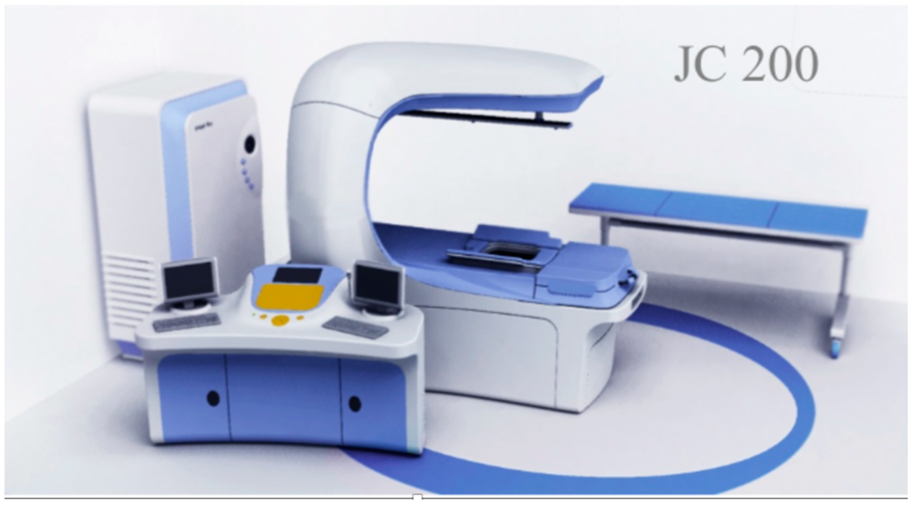
Figure 1. The JC200 ultrasound machine. Chongqing Haifu Company, Chongqing, China.
Ijtm 04 00011 g001
Figure 2. CT scan showing perinephric fat.
Ijtm 04 00011 g002
Figure 3. The treatment of uterine fibroids with HIFU (MRI).
Ijtm 04 00011 g003
Disclaimer/Publisher’s Note: The statements, opinions and data contained in all publications are solely those of the individual author(s) and contributor(s) and not of MDPI and/or the editor(s). MDPI and/or the editor(s) disclaim responsibility for any injury to people or property resulting from any ideas, methods, instructions or products referred to in the content.