Article

Administration of Polyphenol-Rich Sugarcane Extract Alleviates Deficits Induced by Amyloid-Beta1–42 (Aβ1–42) in Transgenic C. elegans

1
Department of Microbiology, Anatomy, Physiology & Pharmacology, School of Agriculture, Biomedicine and Environment, La Trobe University, Melbourne, VIC 3086, Australia
2
TPM Bioactive Division, The Product Makers Pty. Ltd., Melbourne, VIC 3173, Australia
3
Department of Food Science and Technology, Faculty of Applied Sciences, University of Sri Jayewardenepura, Gangodawila, Colombo 10250, Sri Lanka
*
Author to whom correspondence should be addressed.
J. Ageing Longev. 2025, 5(2), 12; https://doi.org/10.3390/jal5020012
Submission received: 17 December 2024 / Revised: 24 March 2025 / Accepted: 25 March 2025 / Published: 2 April 2025

Abstract

:
Polyphenol-Rich Sugarcane Extract (PRSE), derived from Saccharum officinarum, demonstrates significant neuroprotective effects against amyloid-beta (Aβ1–42)-induced deficits associated with Alzheimer’s disease (AD). This study utilized transgenic C. elegans expressing Aβ1–42 to investigate PRSE’s impact on lifespan, sensory behavior, learning, memory, and amyloid fibril accumulation. Supplementation with 5 mg/mL of PRSE extended the mean lifespan of Aβ1–42 worms by 11% (17.78 ± 0.36 days) and reduced amyloid fibril levels by 34% in aged worms compared to untreated worms. PRSE also improved sensory behavior, with a 27% increase in naïve chemotaxis at day 8. Memory deficits were mitigated, with PRSE-treated worms showing 21% and 30% reductions in short-term associative memory loss after 1 h intervals on days 8 and 12, respectively. These improvements can be associated with the polyphenolic compounds in PRSE, which aid in reducing amyloid aggregation. The findings highlight PRSE’s potential as a dietary supplement to address AD-related symptoms and pathologies. Further studies are needed to understand its mechanisms and confirm its effectiveness in mammals, supporting its potential use as a natural preventative supplement for Alzheimer’s and related neurodegenerative diseases.

1. Introduction

A progressive neurodegenerative disorder that leads to cognitive decline and is the leading cause of dementia is Alzheimer’s disease (AD). It is marked by memory loss, cognitive decline, and behavioral changes that worsen over time. A key feature of AD is the buildup of amyloid-beta (Aβ) peptides, particularly Aβ1–42, which clump together to form toxic fibrils. These fibrils interfere with communication between neurons, disrupt the balance of cellular proteins, and trigger widespread damage, including oxidative stress and mitochondrial dysfunction, ultimately leading to the death of neurons and the progression of cognitive impairments [1,2].
Mitochondrial dysfunction is a major factor in the early stages of AD. Aβ peptides accumulate inside mitochondria, where they interfere with energy production, increase harmful reactive oxygen species (ROS), and disrupt calcium levels. This cascade of damage weakens neurons, making them more vulnerable to degeneration and worsening AD symptoms [3,4]. These mitochondrial problems also contribute to the buildup of more Aβ, creating a vicious cycle of damage. Additionally, they activate processes like inflammation and disrupt synapses, the connections between neurons, further advancing the disease [5].
Oxidative stress also plays a critical role in AD. The accumulation of Aβ leads to an overproduction of ROS, which damage lipids, proteins, and DNA in neurons. This type of damage is especially severe in brain regions like the hippocampus and cortex, which are responsible for memory and thinking and are among the first areas affected in AD [6,7]. Together, mitochondrial dysfunction and oxidative stress set the stage for the progression of AD, creating a destructive cycle that drives neuronal damage and cognitive decline. Breaking this cycle is a key focus for potential therapies aimed at slowing the disease and preserving brain health.
Lifestyle and diet play a critical role in brain health, with numerous studies suggesting that a healthy diet rich in bioactive compounds can delay the onset or progression of neurodegenerative diseases, including AD. Polyphenols, abundant in fruits, vegetables, and plant-based products, have garnered significant attention due to their potent antioxidative, anti-inflammatory, and amyloid-inhibitory properties. Epidemiological studies have shown that higher dietary intake of polyphenols is associated with reduced cognitive decline and a lower risk of developing dementia [8,9].
Polyphenols are diverse compounds that target key pathological mechanisms of AD. They inhibit amyloid-beta aggregation, promote proteostasis by enhancing autophagy and proteasomal activity, and combat oxidative stress by scavenging reactive oxygen species. For instance, cocoa polyphenols significantly decreased amyloid-beta fibril accumulation in C. elegans models, improving memory retention and extending lifespan [10].
Polyphenol-Rich Sugarcane Extract (PRSE), derived from Saccharum officinarum, is a natural source of potent bioactive compounds such as apigenin, luteolin, and chlorogenic acid. These compounds have been shown to scavenge free radicals, inhibit amyloid-beta fibril formation, and enhance proteostasis. Preliminary studies using C. elegans models have demonstrated that PRSE improves lifespan and mitigates cognitive decline [11,12].
Previous research indicates that PRSE may promote cellular health by mitigating oxidative stress and preventing amyloid aggregation, potentially contributing to its lifespan-extending properties. The optimal PRSE concentration for lifespan extension was identified as 5 mg/mL, which increased the lifespan of N2 worms by 18.12%. Notably, this lifespan extension was observed only when PRSE was introduced early in life, with no significant effects seen when administered later. PRSE at 5 mg/mL had no impact on the lifespan of DAF-16 and DAF-2 mutants, reinforcing the idea that aging is regulated by the IIS pathway. Despite this, PRSE enhanced thermotolerance, particularly in young and middle-aged worms. These results suggest that early and sustained PRSE intake may improve heat stress resistance and extend lifespan, likely through the insulin/IGF-1 signaling pathway in C. elegans [11].
The neuroprotective effects of PRSE may also involve the modulation of key proteolytic enzymes such as neprilysin and insulin-degrading enzyme, which are responsible for amyloid-beta clearance [13].
Given the increasing evidence linking dietary polyphenols to neuroprotection, this study investigates the therapeutic potential of PRSE in addressing Aβ1–42-induced deficits in lifespan, sensory behavior, learning, memory, and amyloid fibril accumulation using a transgenic C. elegans model. The findings aim to provide insights into the role of polyphenol-rich diets as a promising intervention for mitigating the progression of AD and other neurodegenerative conditions.

2. Materials and Methods

2.1. Polyphenol-Rich Sugarcane Extract and Treatment

A patented Polyphenol-Rich Sugarcane Extract (PRSE), derived from the Saccharum officinarum plant and developed by the Product Makers in Keysborough, Victoria, Australia, was employed in this study. The product’s specifications, manufacturing process, key polyphenols, and antioxidant properties have been previously documented [11,12,14]. To ensure consistency with prior research, a single sample of PRSE was used, with a concentration of 5 mg/mL identified as the most effective within the tested range (1–5 mg/mL). This concentration showed significant benefits, including enhanced longevity, improved heat stress resistance, and amelioration of chemosensation, learning, and short-term memory loss. Its efficacy was further evaluated through experiments conducted on transgenic strains of C. elegans designed to model deficits associated with amyloid-beta1–42 (Aβ1–42).
Concentrated E. coli OP50 was prepared by suspending 1 g of E. coli pellet in 16 mL of M9 buffer. This bacterial suspension was applied to NGM (Nematode Growth Medium) plates and incubated overnight at 20 °C to create a bacterial lawn. A stock solution of PRSE, 5 mg/mL of PRSE suspension, was then applied to the bacterial lawn at a ratio of 2:1 (PRSE suspension to E. coli). Specifically, 400 µL of PRSE suspension was combined with 200 µL of E. coli for small plates, while 800 µL of PRSE suspension was mixed with 400 µL of E. coli for medium plates. This resulted in a final PRSE concentration of 0.1 mg/mL based on the previous study [11,12].
PRSE treatment was initiated at the first larval stage (L1) in all experiments, conducted in triplicate, and maintained at 20 °C. This study utilized PRSE to assess its impact on several key age-associated factors, including lifespan, chemosensation, short-term associative memory, and amyloid-beta (Aβ) fibril accumulation quantified through Thioflavin-T (ThT) staining.

2.2. Culture Conditions for Escherichia coli OP50 and Maintenance of C. elegans

The transgenic C. elegans strains [GRU101 gnals1 (myo-2p::YFP) and GRU102 gnals2 (myo-2p::YFP + unc-119p::Aβ1–42)] were sourced from the Caenorhabditis Genetics Center (Minneapolis, MN, USA). Concentrated Escherichia coli OP50 was prepared by resuspending 1 g of cultured E. coli OP50 pellet in 12 mL of M9 buffer. To synchronize the development of L1-stage transgenic nematodes, gravid adults were treated with a solution containing 1 mL of bleach and 0.5 mL of 5N NaOH [12]. The extracted eggs were incubated in 3 mL of M9 buffer for 48 h to achieve age synchronization. All transgenic nematodes were cultured on NGM plates seeded with E. coli OP50 and maintained at a constant temperature of 20 °C throughout their lifespan.

2.3. Lifespan Assay

L1-stage worms were synchronized and cultured on 60 × 10 mm NGM plates seeded with E. coli OP50 in triplicate. The lifespan assay was performed without the addition of 5′-fluorodeoxyuridine (FUdR) to avoid its potential longevity-extending effects. To maintain synchronized populations, worms were transferred to fresh NGM plates daily during their reproductive phase. After day 10, transfers were reduced to every three days [11].
Survival was monitored daily by touch provocation with a platinum loop to confirm viability. Lifespan was tracked from day 1 (L1 stage) until all worms had died, excluding individuals that left the plates. The mean, median, and maximum lifespans were calculated, and survival curves were generated to illustrate the results [11].

2.4. Chemotaxis Behavior, Learning, and Short-Term Associative Memory

This study utilized chemotaxis, learning, and short-term associative memory assays as previously described by Heydarian et al. [12] and modified from the protocols described by Munasinghe et al. [10], Kauffman et al. [15], and Margie et al. [16]. These assays were conducted to assess the effects of PRSE supplementation on age-synchronized C. elegans at days 4, 8, and 12, representing the young, middle-aged, and old stages of adulthood, respectively. Naïve chemotaxis was performed to assess the worms’ innate response to butanone (a chemoattractant), while learning and memory assays examined their ability to associate butanone with food (Figure 1).
The chemotaxis index (CI) was calculated using the following formula:
CI = ( T 1 + T 2 ) ( C 1 + C 2 ) ( Total   number   of   scored   worms )
T1 and T2 represent the number of worms paralyzed in the butanone quadrants, and C1 and C2 represent those in the ethanol quadrants. Worms within the origin circle (1 cm diameter) were excluded from scoring. For learning, positive butanone conditioning, as described by Kauffman et al., was employed. Worms were exposed to butanone and food simultaneously, and their learning index (LI) was determined using the following formula:
LI = Chemotaxis   index t Chemotaxis   index naïve
In this formula, the learning index (LI) represents the difference between the chemotaxis index measured at specific time points and the baseline (naïve) chemotaxis index, providing a measure of associative learning in the worms.
Short-term associative memory (SAML), adapted from Heydarian et al. [12], was assessed at 1 and 2 h post-conditioning to evaluate the retention of the learned association. Memory loss at a given time (t) was calculated as follows:
Short term   associative   memory   loss   index t = Learning   index 0 Learning   index t
The SAML indext represents the calculated memory loss score at a specific time point, indicating the degree to which the learned association has deteriorated since the initial measurement. The Learning index0 refers to the baseline score immediately after conditioning, reflecting the worm’s initial memory performance. The Learning indext is measured at a later time point (1 h and 2 h post-conditioning) and indicates the worm’s retention of the learned association over time. The assays were conducted on days four, eight, and twelve of worm development, with PRSE-treated worms compared to untreated controls. The experiment was performed in triplicate, using approximately 100–250 worms per assay, to ensure robust and reproducible results. These modifications allowed for a comprehensive evaluation of PRSE’s effects on chemotaxis, associative learning, and memory functions in C. elegans (Figure 1).

2.5. Quantitative Detection of Aβ Fibrils Using Thioflavin-T Staining

Quantitative staining of amyloid-beta (Aβ) fibrils with Thioflavin-T (ThT) was carried out following a modified protocol based on Xin et al. [17] and Munasinghe et al. [10]. Worms were collected from plates on days four, eight and twelve by washing with 1 mL of M9 buffer and transferring them into Eppendorf tubes. The worms were pelleted by centrifugation at 14,000 rpm for 2 min, and the supernatant was discarded. To eliminate residual food, the pellets were washed twice more with M9 buffer.
The washed worm pellets were resuspended in 500 µL of M9 buffer, snap-frozen in liquid nitrogen, and stored at −80 °C for later analysis. Before staining, the samples were thawed and homogenized using a Precellys®24 Homogenizer (Bertin Technologies, Paris, France) with a Precellys® lysing kit containing 0.5 mm glass beads. The homogenization was performed at 6400 rpm for two cycles of 10 s each (totaling 40 s) in 500 µL of M9 buffer. After homogenization, the samples were centrifuged at 14,000 rpm for 2 min, and the supernatant containing soluble protein was collected.
The protein concentration in each sample was quantified using the bicinchoninic acid (BCA) assay (Pierce™ BCA Protein Assay Kit, Thermo Fisher Scientific™, Rockford, IL, USA). Equal amounts of protein from each sample were used for triplicate analyses. For each replicate, 10 µL of M9 buffer and 2 µL of 1 mM ThT (Sigma-Aldrich, St. Louis, MI, USA) were added to reach a final reaction volume of 100 µL. Fluorescence intensity was measured with a CLARIOstar multi-mode plate reader (BMG LABTECH, Ortenberg, Germany) at 440 nm excitation and 482 nm emission. The fluorescence readings were averaged from three independent experiments to ensure reliability and consistency of the results.

2.6. Statistical Analysis

All statistical analyses were conducted using IBM SPSS® Statistics software (version 29.0.2.0), while figures and visual representations were created using GraphPad Prism (version 9.1.0). Data were expressed as mean ± standard error of the mean (SEM), unless otherwise stated. For survival analysis, Kaplan–Meier curves were used, and p values for survival function comparisons between treated and untreated groups were calculated using the log-rank (Mantel–Cox) test. A p value of <0.05 was considered statistically significant. Maximum lifespan, defined as the average lifespan of the 10 longest-lived worms, was analyzed using one-way ANOVA followed by Tukey’s post hoc test to determine group differences.
To assess differences in chemotaxis, learning, and memory loss indices, a General Linear Model (GLM) with a multivariate test was applied. This model analyzed the effects of two fixed factors: day (representing worm age) and treatment (PRSE vs. no PRSE). The GLM multivariate test accounted for potential interactions and correlations between dependent variables, providing a robust statistical framework. Differences in amyloid-beta (Aβ) levels between groups were analyzed using a General Linear Model univariate test, to determine statistical significance.

3. Results

3.1. PRSE Supplementation Restores Lifespan in Pan-Neuronal Aβ1–42 Expressing C. elegans

Under laboratory conditions at 20 °C, GRU101 untreated worms exhibited a mean lifespan of 17.60 ± 0.33 days, while GRU102 untreated worms, expressing pan-neuronal Aβ1–42, had a significantly reduced mean lifespan of 15.95 ± 0.43 days (p < 0.05, Figure 2A, Table 1). The median lifespan of GRU101 untreated worms was 18.00 ± 0.41 days, whereas GRU102 untreated worms had a median lifespan of 17.00 ± 0.60 days, a difference that was not statistically significant (Figure 2A, Table 1).
Supplementation with PRSE at a dose of 5 mg significantly improved the lifespan of GRU102 worms. The mean lifespan of GRU102 worms increased by 11% to 17.78 ± 0.36 days, compared to untreated GRU102 (p < 0.05, Figure 2B, Table 1). The median lifespan also improved significantly to 19.00 ± 0.52 days, representing an increase of 12%, compared to untreated GRU102 (p < 0.05, Figure 2B, Table 1). Furthermore, PRSE significantly enhanced the maximum lifespan in GRU102 worm strains. GRU102 worms supplemented with PRSE reached a maximum lifespan of 21.60 ± 0.93 days, showing significant increases compared to untreated GRU102 (p < 0.05, Figure 2B, Table 1).

3.2. PRSE Supplement Intervention Improved Chemosensation, Learning, and Memory Loss of C. elegans

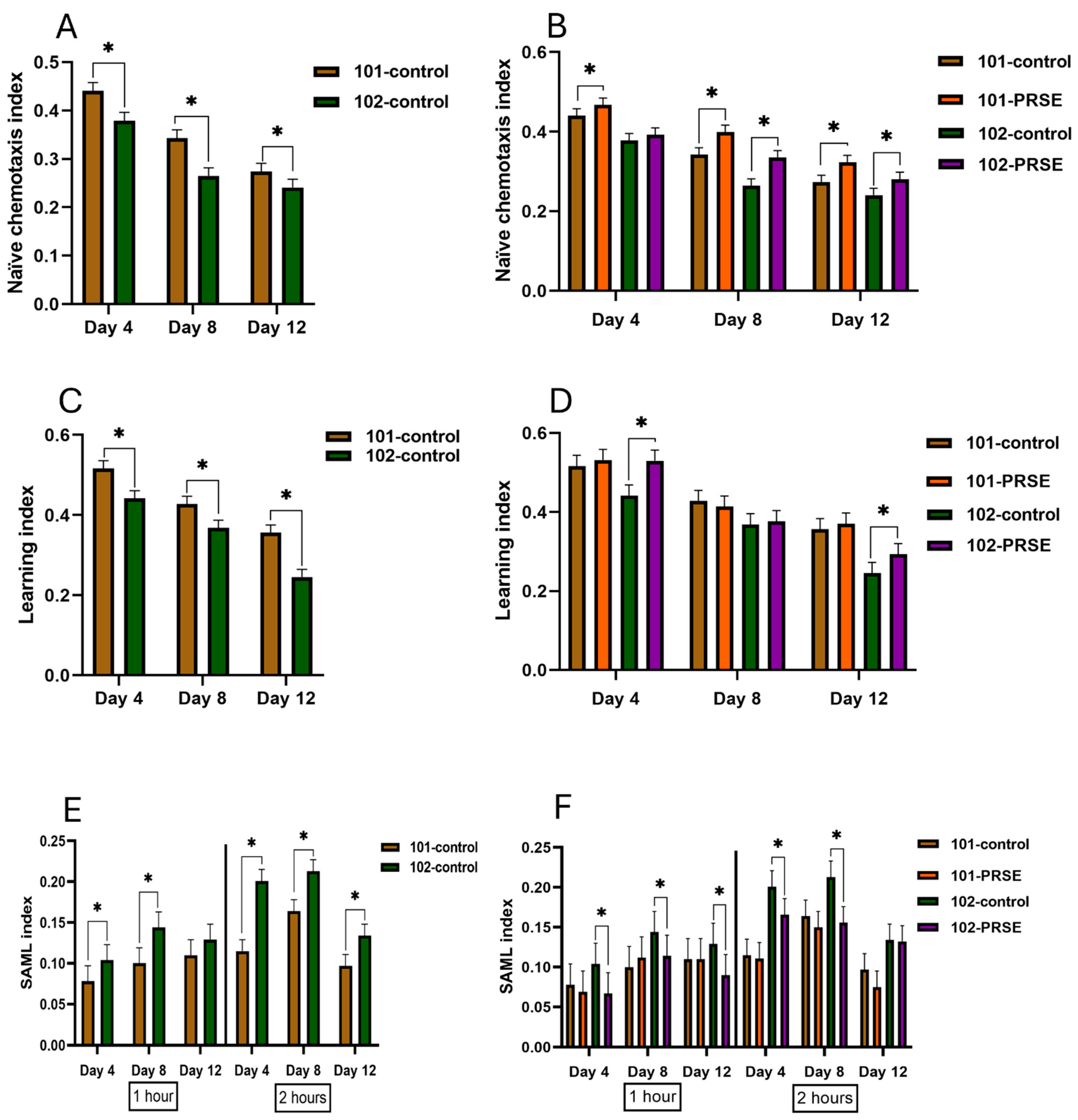
As worms showed a progressive reduction in the naïve chemotaxis index with age, significant differences were observed at later stages (Figure 3A,B). There was no significant difference in the naïve chemotaxis index between untreated GRU102 and PRSE treated GRU102 worms at day four (young stage) (Figure 3A,B). However, by day eight (middle age) and day twelve (old age), PRSE treated GRU102 worms showed significantly improved naïve chemotaxis compared to GRU102 untreated worms (p < 0.05, Figure 2B). PRSE supplementation improved the naïve chemotaxis index in GRU101 worms across all stages (days four, eight and twelve), with significant increases of 6%, 17%, and 18%, respectively, compared to their untreated controls (p < 0.05, Figure 3B). In GRU102 worms, PRSE supplementation did not result in a statistically significant improvement in naïve chemotaxis at day four compared to untreated GRU102 worms. However, at day eight and day twelve, PRSE significantly increased the naïve chemotaxis index of GRU102 worms by 27% and 17%, respectively, compared to untreated GRU102 worms (p < 0.05, Figure 3B).
For the learning index, GRU102 worms consistently showed a significant learning decrease compared to GRU101 worms across all stages (Figure 3C). PRSE supplementation significantly improved the learning index in GRU102 worms at day four and day twelve (p < 0.05, Figure 3D). However, no significant improvement was observed at day eight for PRSE treated GRU102 worms compared to untreated GRU102.
GRU102-control worms showed significantly higher short-term associative memory loss (SAML) than GRU101-control worms at both 1 h and 2 h time points at all stages (p < 0.05, Figure 3E). PRSE supplementation significantly reduced memory loss in GRU102 worms across all stages after 1 h (p < 0.05, Figure 3F). However, PRSE reduced memory loss significantly in GRU102 worms at day four and day eight after 2 h (p < 0.05, Figure 3F). At day eight, PRSE-treated GRU102 worms showed 21% and 27% memory loss reduction, respectively, after 1 h and 2 h compared to untreated GRU102 worms (p < 0.05, Figure 3F). At day twelve, PRSE-treated GRU102 worms showed 30% memory loss reduction after 1 h compared to untreated GRU102 worms (p < 0.05, Figure 3F).

3.3. PRSE Supplementation Decreased Aβ Fibril Accumulation in Pan-Neuronal Aβ1–42-Expressing Worms During the Young, Middle, and Old-Aged Stages

GRU102 (Aβ1–42 expressing) worms showed a significantly higher fluorescence intensity compared to GRU101 (control) worms when stained with Thioflavin-T dye at day four (young stage), day eight (middle age), and day twelve (old age) (p < 0.05, Figure 4A). In GRU102 worms supplemented with PRSE, Aβ fibril levels were significantly reduced at day four, day eight, and day twelve compared to their untreated counterparts (p < 0.05, Figure 4B).

4. Discussion

This study utilized the transgenic C. elegans strain GRU102, which pan-neuronally expresses amyloid-beta1–42 (Aβ1–42), to investigate the neurotoxic effects associated with Alzheimer’s disease (AD), particularly short-term memory deficits. Long-term supplementation with Polyphenol-Rich Sugarcane Extract (PRSE) from the early stage of life significantly improved these deficits, reducing amyloid deposition and improving lifespan, chemosensation, learning, and memory-related behaviors. These findings emphasize the potential of PRSE as a neuroprotective agent capable of addressing key pathologies in AD. The impact of 5 mg/mL PRSE on longevity, chemosensation, learning, and memory loss was previously evaluated using the wild-type C. elegans strain N2, while daf-2 and daf-16 mutant strains were employed to elucidate the specific longevity pathways affected by PRSE [11,12]. Polyphenol-Rich Sugarcane Extract (PRSE), with a total polyphenol content of 221 mg/g Gallic Acid Equivalency (GAE), contains a diverse array of bioactive phenolic compounds, including apigenin, luteolin, tricin, diosmin, syringic acid, and chlorogenic acid [14,18]. These polyphenols are well-known for their potent antioxidant activities, as demonstrated in previous studies on sugarcane-derived phenolic compounds [19]. Antioxidant properties of these compounds allow them to neutralize reactive oxygen species (ROS) and mitigate oxidative damage, a key factor in aging and neurodegenerative diseases [20,21]. Additionally, specific compounds like luteolin and apigenin have shown anti-inflammatory and neuroprotective effects, potentially modulating pathways associated with longevity and cognitive function, such as the insulin/IGF-1 signaling and autophagy pathways [22]. Syringic acid and chlorogenic acid further contribute to PRSE’s health-promoting effects through their roles in reducing inflammation and supporting proteostasis. This comprehensive polyphenolic profile underscores PRSE’s potential as a dietary intervention for enhancing antioxidant defenses and addressing degenerative conditions, aligning with its demonstrated efficacy in experimental models. The limited bioavailability of natural polyphenols, including those in PRSE, is compensated for by their microbial metabolism, which produces smaller, more bioactive metabolites that may improve their bioefficacy [23]. This makes PRSE a promising dietary intervention for age-associated conditions and neurodegenerative diseases, offering multi-faceted benefits through its rich polyphenolic profile. By combining potent antioxidative and anti-inflammatory actions with the capacity to influence critical longevity pathways, PRSE presents itself as a valuable agent in promoting lifespan and mitigating degenerative conditions.
Apigenin and luteolin, key flavonoids found in various plant sources, exhibit significant neuroprotective properties and potential to improve cognitive and behavioral functions. Apigenin has been shown to reduce inflammation, oxidative stress, and apoptosis, while enhancing neurogenesis and neurotransmitter regulation, particularly through pathways like ERK/CREB/BDNF in neurodegenerative models [24,25]. Similarly, luteolin, a flavone with anti-inflammatory and antioxidant properties, has demonstrated efficacy in suppressing neuroinflammatory responses and mitigating oxidative stress via the activation of PI3K/Akt and Nrf2 pathways, making it a promising nutraceutical candidate for conditions like Alzheimer’s and Parkinson’s diseases [26,27]. Additionally, luteolin’s ability to regulate diverse signaling pathways, including JAK2/STAT and MAPK, further highlights its neuroprotective potential [28,29]. These findings underscore the therapeutic relevance of apigenin and luteolin in enhancing neuronal health and addressing neurodegenerative diseases. Further exploration of their combined effects with other bioactive phenolics, such as syringic acid and chlorogenic acid, could open up new avenues for holistic approaches to neuroprotection.
In this study, worms were distinguished into three age groups—young (day four), middle-aged (day eight), and old (day twelve)—to evaluate the impact of Polyphenol-Rich Sugarcane Extract (PRSE) supplementation on various biological and cognitive functions at distinct life stages.
Lifespan analysis revealed that GRU102-control worms, which express Aβ1–42, displayed a significant reduction in both mean and maximum lifespan compared to GRU101-control. PRSE supplementation restored the lifespan of GRU102 worms to levels comparable to GRU101-control. These results indicate that PRSE mitigates the adverse effects of Aβ1–42 toxicity on longevity, potentially through its antioxidant activity and inhibition of amyloid aggregation. This aligns with previous studies indicating the antioxidative potential of polyphenol-rich compounds in improving lifespan and reducing proteotoxic stress [11]. These results are consistent with earlier studies highlighting the benefits of polyphenol-rich compounds in extending lifespan and alleviating proteotoxic stress. Compounds like quercetin and sugarcane extract have been shown to improve lifespan and resilience to stress in model organisms by supporting protein homeostasis and influencing key pathways such as DAF-16 and insulin/IGF-1 signaling [11,30]. This evidence underscores the antioxidant and anti-amyloid properties of polyphenol-rich substances like PRSE in combating age-related protein damage and supporting healthier aging.
Chemosensory behavior, measured by naïve chemotaxis to butanone, was significantly worse in GRU102-control worms at all ages compared to GRU101-control. This reflects the detrimental effects of Aβ1–42 toxicity on sensory neurons. PRSE supplementation significantly improved chemotaxis in GRU102 worms, particularly at middle (day eight) and old (day twelve) ages. These findings suggest that PRSE has neuroprotective properties capable of enhancing sensory function, potentially by reducing amyloid-induced damage to neuronal circuits and also related to Amphid Wing Cell (AWC) neuronal functioning, which was improved by PRSE in wild-type N2 worms [12]. The reduced chemotaxis to butanone observed in GRU102 worms highlights the damaging effects of Aβ1–42 toxicity on sensory neurons, leading to a significant decline in chemosensory behavior. Remarkably, PRSE supplementation improved chemotaxis in these worms, particularly at middle (day eight) and old (day twelve) ages, suggesting that PRSE offers neuroprotective benefits. These benefits likely arise from its ability to counteract amyloid-induced neuronal damage and enhance sensory function. This observation is supported by research showing that polyphenols can inhibit amyloid-beta aggregation, which is a major factor in its neurotoxicity. Polyphenols, such as luteolin, have been found to bind directly to amyloid-beta, preventing its aggregation and protecting neurons from its harmful effects [31]. Similarly, certain polyphenols can directly interfere with the formation of toxic amyloid-beta aggregates, reducing their neurotoxic impact [32]. The improvement in chemotaxis could also be linked to enhanced functionality of AWC neurons, which are essential for navigating chemical gradients in C. elegans. Studies suggest that polyphenols can improve neuronal resilience and modulate signaling pathways, helping restore normal sensory function in circuits affected by amyloid toxicity [33]. In conclusion, the ability of PRSE to reverse chemosensory deficits in GRU102 worms highlights the neuroprotective potential of polyphenol-rich extracts. This points to dietary polyphenols as a promising approach for addressing sensory and neurological impairments associated with conditions like neurodegenerative diseases.
The improvement in learning ability observed in GRU102 worms supplemented with PRSE highlights its potential to support brain function and counteract the effects of Aβ1–42 toxicity. GRU102 worms exhibited the most significant learning deficits during middle age (day eight), a period of heightened vulnerability to amyloid toxicity. The absence of a PRSE effect on the learning index in day 8 animals may be explained by age-related changes in neuronal plasticity and resilience in C. elegans. By day 8 (mid-adulthood), C. elegans exhibits a notable decline in cognitive capacity, synaptic function, and cellular stress resistance, which could diminish the neuroprotective potential of PRSE. Previous studies, including our earlier work with wild-type worms [12], indicated that the beneficial effects of PRSE on sensory and cognitive behavior are most prominent in early life stages (day 4), aligning with other polyphenol interventions showing stronger impacts when administered before midlife decline sets in [10]. This age-dependent effect may arise from the progressive accumulation of Aβ toxicity, oxidative damage, and proteostasis disruption by day 8, potentially overwhelming PRSE’s antioxidant and anti-amyloidogenic capacity [34,35]. Furthermore, studies on age-dependent memory decline in C. elegans suggest that synaptic plasticity and associative learning circuits become less responsive to interventions with age, possibly due to impaired insulin/IGF-1 signaling and transcriptional deregulation [36,37]. Additionally, it is possible that PRSE’s bioactive compounds primarily act as preventive agents rather than rescuing age-progressed deficits. Future studies could examine whether initiating PRSE supplementation earlier or combining it with interventions targeting age-associated transcriptional and metabolic decline would sustain its cognitive benefits into later stages. Additionally, Kauffman et al. [38] demonstrated that C. elegans learning and memory decline follows a non-linear pattern, with middle-aged worms (comparable to day 8 in this study) experiencing a distinct phase of cognitive vulnerability. This period is marked by a sharp decline in learning ability due to dysregulation in insulin/IGF-1 signaling, which compromises neuronal plasticity and stress resistance. Notably, Kauffman et al. [38] also found that dietary restriction, despite improving cognition in early and late life stages, was ineffective in middle age, suggesting an intrinsic resistance to interventions during this phase. Thus, PRSE’s lack of effect on day 8 worms may reflect this biologically vulnerable period, where heightened oxidative stress and impaired proteostasis limit responsiveness to treatment. However, PRSE supplementation improved their learning index, particularly at days four and twelve, demonstrating its ability to enhance cognitive resilience and protect against amyloid-induced damage. This aligns with extensive research on the cognitive benefits of polyphenol-rich compounds. Extra virgin olive oil, rich in polyphenols, improved learning and memory in aging SAMP8 mice by reducing brain oxidative damage and increasing protective antioxidants like glutathione [39]. Polyphenols also work by preventing the formation of toxic amyloid-beta aggregates. Grape seed polyphenolic extract, for example, reduced memory-impairing amyloid-beta oligomers in Alzheimer’s mouse models and significantly improved cognitive performance [40]. Resveratrol, another well-known polyphenol, reversed learning and memory deficits in amyloid-beta-infused mice by modulating the cAMP-CREB-BDNF signaling pathway, reducing inflammation, and protecting neurons from cell death [41]. Overall, the ability of PRSE to improve learning in GRU102 worms aligns with the well-documented benefits of polyphenols in supporting brain health. By reducing oxidative stress, preventing amyloid aggregation, and enhancing neuronal plasticity, PRSE offers a promising natural strategy for mitigating cognitive impairments associated with neurodegenerative conditions like Alzheimer’s disease.
Short-term memory, assessed using the short-term associative memory loss index (SAML index) over one- and two-hour intervals, revealed significant memory deficits in GRU102 worms across all age groups compared to GRU101-control. PRSE supplementation significantly improved memory retention in GRU102 worms, particularly at young (day four) and old (day twelve) ages. These results highlight PRSE’s ability to mitigate memory deficits associated with Aβ1–42 toxicity, demonstrating its potential as a dietary intervention for cognitive impairments. These findings align with research showing the cognitive benefits of polyphenols. As an example, resveratrol has been found to reduce oxidative stress and enhance the production of memory-related proteins in hippocampal neurons affected by amyloid-beta, supporting its role in protecting memory function [42]. Similar improvements have been observed with grape seed polyphenolic extract (GSPE), which inhibits the formation of toxic amyloid-beta aggregates, reducing memory deficits in animal models [40]. Polyphenols like EGCG from green tea have also demonstrated an ability to reduce oxidative damage, improve synaptic plasticity, and mitigate amyloid-beta-induced cognitive dysfunction [43]. These studies and the results in GRU102 worms support PRSE’s potential to preserve short-term memory by reducing amyloid-beta toxicity, enhancing neuronal health, and protecting against oxidative stress. These findings highlight PRSE as a promising natural approach to address memory loss associated with neurodegenerative diseases.
While this study demonstrated that PRSE improved lifespan, sensory behavior, and cognitive function in both the control strain for pan-neuronal amyloid beta1–42-expressing strain GRU102 (GRU101) and Aβ1–42-expressing (GRU102) worms, it is important to emphasize that a key disease-specific effect was observed in GRU102 worms: the significant reduction in Aβ fibril accumulation. This effect was not observed in GRU101 worms, highlighting PRSE’s potential anti-amyloidogenic properties. The reduction in fibril burden aligns with existing evidence on polyphenols such as apigenin, luteolin, and resveratrol, which have been shown to inhibit Aβ aggregation, reduce oxidative stress, and enhance proteostasis [31,32]. Therefore, although PRSE exerts general health-promoting benefits across different strains, its specific capacity to mitigate Aβ1–42 toxicity through fibril reduction highlights its relevance as a potential dietary intervention against Alzheimer’s disease pathology. Future research can explore whether PRSE’s effects involve modulation of Aβ clearance pathways to further elucidate its mechanism of action. As the locomotory function in the current study was not assessed, it is important to note that Aβ1–42-expressing C. elegans models often display early neuronal dysfunction and behavioral impairments before overt locomotory deficits or muscle degeneration become prominent [34,44]. Furthermore, locomotory impairments in these models often emerge later and are commonly associated with severe muscle Aβ aggregation, particularly in models that express Aβ in muscle cells [45]. In this study, behavioral assays were conducted from early adulthood (day 4), a stage at which significant muscle degeneration is unlikely to be the primary cause of functional decline. However, we acknowledge the importance of assessing locomotory function and muscle integrity to further disentangle PRSE’s potential effects on neuromuscular health. Future studies will include locomotory assays, muscle structure analysis, and neuronal integrity evaluations to better elucidate the relationship between PRSE’s neuroprotective and muscle-preserving effects in Aβ1–42-expressing C. elegans models.
Amyloid-beta (Aβ) fibril accumulation is a key feature of Alzheimer’s disease (AD) and plays a major role in its progression. In GRU102 worms, Thioflavin-T staining revealed a significant age-related increase in Aβ1–42 fibril levels, highlighting the harmful aggregation of misfolded proteins. The fluorescence intensity background in Amyloid-beta fibril quantification is likely due to non-specific binding and autofluorescence from intestinal lipofuscin and gut granules, both well-known sources of interference in C. elegans fluorescence imaging [46], as well as the inherent limitations of ThT binding specificity, which can be influenced by dye concentration, electrostatic interactions, and fibril morphology [47,48]. Despite this background, the fluorescence intensity was consistently and significantly higher in GRU102 worms, indicating specific Aβ fibril accumulation. While the experiment was conducted in a partially blinded manner, we acknowledge the need for fully double-blind evaluations in future work. Additionally, future experiments could incorporate complementary aggregation detection methods, such as Congo Red staining or hyperspectral imaging, to enhance specificity [49].
However, supplementation with PRSE dramatically reduced fibril levels at days four, eight, and twelve, indicating its potential to block amyloid fibrillogenesis. This effect is likely due to PRSE’s rich polyphenolic content, which enhances proteostasis and reduces the formation of toxic protein aggregates. These findings are supported by previous studies on the effects of polyphenols. For example, Munasinghe et al. [10] showed that cocoa polyphenols reduced amyloid-beta fibril accumulation in C. elegans models expressing Aβ1–42. In addition to lowering fibril levels, long-term cocoa supplementation reversed related behavioral issues, including memory loss and shortened lifespan, underscoring the neuroprotective benefits of polyphenols [10]. Similarly, Fong et al. [34] investigated a novel C. elegans model of AD with neuronal Aβ1–42 expression. This model exhibited age-dependent behavioral and metabolic impairments alongside amyloid aggregation and mitochondrial dysfunction. Polyphenol interventions improved proteostasis, enhanced mitochondrial health, and reduced amyloid toxicity, demonstrating the wide-ranging protective effects of these compounds [34]. PRSE’s ability to reduce amyloid fibril accumulation aligns with these findings and supports the broader evidence on dietary polyphenols. Compounds such as catechins and epicatechins inhibit amyloid-beta aggregation by binding to the peptide, preventing fibril formation. Additionally, these polyphenols promote autophagy and proteasomal activity, which are essential for maintaining proteostasis and protecting neurons from damage. In summary, PRSE’s effectiveness in reducing amyloid-beta fibril accumulation highlights its potential as a therapeutic option for neurodegenerative diseases like AD. By leveraging the antioxidative and proteostasis-enhancing properties of its polyphenolic content, PRSE not only prevents amyloid aggregation but also supports overall neuronal health, offering a promising approach to mitigating age-related cognitive decline and neurodegeneration.

5. Conclusions

This study highlights the promising potential of Polyphenol-Rich Sugarcane Extract (PRSE) as a natural supplement to combat the neurotoxic effects of amyloid-beta1–42 (Aβ1–42), a key factor in Alzheimer’s disease (AD). PRSE supplementation significantly improved lifespan, sensory behavior, learning, and memory while reducing amyloid fibril accumulation in C. elegans. These effects are likely mediated by the extract’s polyphenolic compounds, which support cellular health by improving proteostasis, reducing oxidative stress, and preventing amyloid aggregation. While these findings are encouraging, further research is essential to elucidate the underlying mechanisms and evaluate the extract’s efficacy in mammalian models. These steps will help establish PRSE as a potential preventative supplement for AD and related neurodegenerative conditions.

Author Contributions

Conceptualization: D.H. and M.J.; methodology: D.H., M.J. and M.M.; formal analysis: D.H. and M.J.; resources: D.H. and M.F.; writing—original draft: D.H.; writing—review and editing: D.H., M.F., M.J., M.M. and J.T.; supervision: J.T.; project administration: D.H.; funding acquisition: D.H. All authors have read and agreed to the published version of the manuscript.

Funding

This study was supported by the La Trobe University and Product Makers PTY Ltd. Postgraduate Research Scholarship (LTUPRS) and the La Trobe University Full-Fee Research Scholarship (LTUFFRS). The authors declare that this study received partial funding from the Product Makers PTY Ltd. via an industry-based PhD scholarship.

Institutional Review Board Statement

Not applicable.

Informed Consent Statement

Not applicable.

Data Availability Statement

Data will be provided upon request to the corresponding author.

Conflicts of Interest

Author Matthew Flavel was employed by the company the Product Makers PTY Ltd. The remaining authors declare that this research was conducted in the absence of any commercial or financial relationships that could be construed as potential conflicts of interest. The funder had the following involvement with this study: supplying their product and partially funding the La Trobe Postgraduate Research Scholarship (LTUPRS). The funder was not involved in the study design, collection, analysis, interpretation of data, the writing of this article, or the decision to submit it for publication.

References

  1. Butterfield, D.A.; Drake, J.; Pocernich, C.; Castegna, A. Evidence of Oxidative Damage in Alzheimer’s Disease Brain: Central Role for Amyloid β-Peptide. Trends Mol. Med. 2001, 7, 548–554. [Google Scholar] [CrossRef]
  2. Reddy, P.H.; Beal, M.F. Amyloid Beta, Mitochondrial Dysfunction and Synaptic Damage: Implications for Cognitive Decline in Aging and Alzheimer’s Disease. Trends Mol. Med. 2008, 14, 45–53. [Google Scholar] [CrossRef]
  3. Perez Ortiz, J.M.; Swerdlow, R.H. Mitochondrial Dysfunction in Alzheimer’s Disease: Role in Pathogenesis and Novel Therapeutic Opportunities. Br. J. Pharmacol. 2019, 176, 3489–3507. [Google Scholar] [CrossRef]
  4. Monsalvo-Maraver, L.Á.; Maya-López, M.; Rangel-López, E.; Túnez, I.; Tinkov, A.A.; Skalny, A.; Ferrer, B.; Aschner, M.; Santamaría, A. Amyloid Beta Peptide-Mediated Alterations in Mitochondrial Dynamicsand Its Implications for Alzheimer’s Disease. CNS Neurol. Disord. Drug Targets 2023, 22, 1039–1056. [Google Scholar] [CrossRef]
  5. Bhatia, V.; Sharma, S. Role of Mitochondrial Dysfunction, Oxidative Stress and Autophagy in Progression of Alzheimer’s Disease. J. Neurol. Sci. 2021, 421, 117253. [Google Scholar] [CrossRef]
  6. Varadarajan, S.; Yatin, S.; Aksenova, M.; Butterfield, D.A. Review: Alzheimer’s Amyloid β-Peptide-Associated Free Radical Oxidative Stress and Neurotoxicity. J. Struct. Biol. 2000, 130, 184–208. [Google Scholar] [CrossRef]
  7. Babaei Abraki, S.; Chavoshi-Nezhad, S. Mitochondrial Defects and Oxidative Stress in Alzheimer Disease. Neurosci. J. Shefaye Khatam 2014, 2, 85–94. [Google Scholar] [CrossRef]
  8. Scarmeas, N.; Stern, Y.; Tang, M.; Mayeux, R.; Luchsinger, J.A. Mediterranean Diet and Risk for Alzheimer’s Disease. Ann. Neurol. 2006, 59, 912–921. [Google Scholar] [CrossRef]
  9. Singh, B.; Parsaik, A.K.; Mielke, M.M.; Erwin, P.J.; Knopman, D.S.; Petersen, R.C.; Roberts, R.O. Association of Mediterranean Diet with Mild Cognitive Impairment and Alzheimer’s Disease: A Systematic Review and Meta-Analysis. J. Alzheimer’s Dis. 2014, 39, 271–282. [Google Scholar] [CrossRef]
  10. Munasinghe, M.; Almotayri, A.; Kolivas, D.; Thomas, J.; Heydarian, D.; Jois, M. Cocoa Supplementation Reduces Amyloid-Beta1–42 (Aβ1–42) Induced Deficits in a Transgenic C. elegans. Nutr. Healthy Aging 2021, 6, 117–130. [Google Scholar] [CrossRef]
  11. Heydarian, D.; Flavel, M.; Munasinghe, M.; Almotayri, A.; Jois, M.; Thomas, J. Early Exposure to Polyphenol-Rich Sugarcane Extract (PRSE) Mitigates Aging While Enhancing Thermotolerance in C. elegans. J. Ageing Longev. 2024, 4, 15–27. [Google Scholar] [CrossRef]
  12. Heydarian, D.; Flavel, M.; Munasinghe, M.; Jois, M.; Thomas, J. Improving Cognitive and Chemosensory Function in Caenorhabditis Elegans Through Polyphenol-Rich Sugarcane Extract. Stresses 2024, 4, 816–826. [Google Scholar] [CrossRef]
  13. Malito, E.; Hulse, R.E.; Tang, W.-J. Amyloid β-Degrading Cryptidases: Insulin Degrading Enzyme, Presequence Peptidase, and Neprilysin. Cell. Mol. Life Sci. 2008, 65, 2574–2585. [Google Scholar] [CrossRef]
  14. Deseo, M.A.; Elkins, A.; Rochfort, S.; Kitchen, B. Antioxidant Activity and Polyphenol Composition of Sugarcane Molasses Extract. Food Chem. 2020, 314, 126180. [Google Scholar] [CrossRef]
  15. Kauffman, A.; Parsons, L.; Stein, G.; Wills, A.; Kaletsky, R.; Murphy, C. C. elegans Positive Butanone Learning, Short-Term, and Long-Term Associative Memory Assays. J. Vis. Exp. 2011, 49, 2490. [Google Scholar] [CrossRef]
  16. Margie, O.; Palmer, C.; Chin-Sang, I. C. elegans Chemotaxis Assay. J. Vis. Exp. 2013, 74, 50069. [Google Scholar] [CrossRef]
  17. Xin, L.; Yamujala, R.; Wang, Y.; Wang, H.; Wu, W.-H.; Lawton, M.A.; Long, C.; Di, R. Acetylcholineestarase-Inhibiting Alkaloids from Lycoris Radiata Delay Paralysis of Amyloid Beta-Expressing Transgenic C. elegans CL4176. PLoS ONE 2013, 8, e63874. [Google Scholar] [CrossRef]
  18. Ji, J.; Yang, X.; Flavel, M.; Shields, Z.P.-I.; Kitchen, B. Antioxidant and Anti-Diabetic Functions of a Polyphenol-Rich Sugarcane Extract. J. Am. Coll. Nutr. 2019, 38, 670–680. [Google Scholar] [CrossRef]
  19. Hewawansa, U.H.A.J.; Houghton, M.J.; Barber, E.; Costa, R.J.S.; Kitchen, B.; Williamson, G. Flavonoids and Phenolic Acids from Sugarcane: Distribution in the Plant, Changes during Processing, and Potential Benefits to Industry and Health. Compr. Rev. Food Sci. Food Saf. 2024, 23, e13307. [Google Scholar] [CrossRef]
  20. Singla, R.K.; Dubey, A.K.; Garg, A.; Sharma, R.K.; Fiorino, M.; Ameen, S.M.; Haddad, M.A.; Al-Hiary, M. Natural Polyphenols: Chemical Classification, Definition of Classes, Subcategories, and Structures. J. AOAC Int. 2019, 102, 1397–1400. [Google Scholar] [CrossRef]
  21. Liu, X.; Le Bourvellec, C.; Guyot, S.; Renard, C.M.G.C. Reactivity of Flavanols: Their Fate in Physical Food Processing and Recent Advances in Their Analysis by Depolymerization. Compr. Rev. Food Sci. Food Saf. 2021, 20, 4841–4880. [Google Scholar] [CrossRef]
  22. Di Lorenzo, C.; Colombo, F.; Biella, S.; Stockley, C.; Restani, P. Polyphenols and Human Health: The Role of Bioavailability. Nutrients 2021, 13, 273. [Google Scholar] [CrossRef]
  23. Manach, C.; Williamson, G.; Morand, C.; Scalbert, A.; Rémésy, C. Bioavailability and Bioefficacy of Polyphenols in Humans. I. Review of 97 Bioavailability Studies. Am. J. Clin. Nutr. 2005, 81, 230S–242S. [Google Scholar] [CrossRef]
  24. Olasehinde, T.A.; Olaokun, O.O. The Beneficial Role of Apigenin against Cognitive and Neurobehavioural Dysfunction: A Systematic Review of Preclinical Investigations. Biomedicines 2024, 12, 178. [Google Scholar] [CrossRef]
  25. Balez, R.; Steiner, N.; Engel, M.; Muñoz, S.S.; Lum, J.S.; Wu, Y.; Wang, D.; Vallotton, P.; Sachdev, P.; O’Connor, M.; et al. Neuroprotective Effects of Apigenin against Inflammation, Neuronal Excitability and Apoptosis in an Induced Pluripotent Stem Cell Model of Alzheimer’s Disease. Sci. Rep. 2016, 6, 31450. [Google Scholar] [CrossRef]
  26. Nabavi, S.F.; Braidy, N.; Gortzi, O.; Sobarzo-Sanchez, E.; Daglia, M.; Skalicka-Woźniak, K.; Nabavi, S.M. Luteolin as an Anti-Inflammatory and Neuroprotective Agent: A Brief Review. Brain Res. Bull. 2015, 119, 1–11. [Google Scholar] [CrossRef]
  27. Kempuraj, D.; Thangavel, R.; Kempuraj, D.D.; Ahmed, M.E.; Selvakumar, G.P.; Raikwar, S.P.; Zaheer, S.A.; Iyer, S.S.; Govindarajan, R.; Chandrasekaran, P.N.; et al. Neuroprotective Effects of Flavone Luteolin in Neuroinflammation and Neurotrauma. BioFactors 2021, 47, 190–197. [Google Scholar] [CrossRef]
  28. Singh, N.K.; Bhushan, B.; Singh, P.; Sahu, K.K. Therapeutic Expedition of Luteolin against Brain-Related Disorders:An Updated Review. Comb. Chem. High Throughput Screen. 2024, 28, 371–391. [Google Scholar] [CrossRef]
  29. Goyal, A.; Solanki, K.; Verma, A. Luteolin: Nature’s Promising Warrior against Alzheimer’s and Parkinson’s Disease. J. Biochem. Mol. Toxicol. 2024, 38, e23619. [Google Scholar] [CrossRef]
  30. Dilberger, B.; Passon, M.; Asseburg, H.; Silaidos, C.V.; Schmitt, F.; Schmiedl, T.; Schieber, A.; Eckert, G.P. Polyphenols and Metabolites Enhance Survival in Rodents and Nematodes—Impact of Mitochondria. Nutrients 2019, 11, 1886. [Google Scholar] [CrossRef]
  31. Das, S.; Stark, L.; Musgrave, I.F.; Pukala, T.; Smid, S.D. Bioactive Polyphenol Interactions with β Amyloid: A Comparison of Binding Modelling, Effects on Fibril and Aggregate Formation and Neuroprotective Capacity. Food Funct. 2016, 7, 1138–1146. [Google Scholar] [CrossRef]
  32. Baruah, P.; Moorthy, H.; Ramesh, M.; Padhi, D.; Govindaraju, T. A Natural Polyphenol Activates and Enhances GPX4 to Mitigate Amyloid-β Induced Ferroptosis in Alzheimer’s Disease. Chem. Sci. 2023, 14, 9427–9438. [Google Scholar] [CrossRef]
  33. Vauzour, D.; Vafeiadou, K.; Rodriguez-Mateos, A.; Rendeiro, C.; Spencer, J.P.E. The Neuroprotective Potential of Flavonoids: A Multiplicity of Effects. Genes Nutr. 2008, 3, 115–126. [Google Scholar] [CrossRef]
  34. Fong, S.; Teo, E.; Ng, L.F.; Chen, C.-B.; Lakshmanan, L.N.; Tsoi, S.Y.; Moore, P.K.; Inoue, T.; Halliwell, B.; Gruber, J. Energy Crisis Precedes Global Metabolic Failure in a Novel Caenorhabditis Elegans Alzheimer Disease Model. Sci. Rep. 2016, 6, 33781. [Google Scholar] [CrossRef]
  35. Kang, N.; Luan, Y.; Jiang, Y.; Cheng, W.; Liu, Y.; Su, Z.; Liu, Y.; Tan, P. Neuroprotective Effects of Oligosaccharides in Rehmanniae Radix on Transgenic Caenorhabditis Elegans Models for Alzheimer’s Disease. Front. Pharmacol. 2022, 13, 878631. [Google Scholar] [CrossRef]
  36. Weng, Y.; Zhou, S.; Morillo, K.; Kaletsky, R.; Lin, S.; Murphy, C.T. The Neuron-Specific IIS/FOXO Transcriptome in Aged Animals Reveals Regulatory Mechanisms of Cognitive Aging. eLife 2024, 13, RP95621. [Google Scholar] [CrossRef]
  37. Mastrandreas, P.; Arnold, A.; Boglari, C.; De Quervain, D.J.-F.; Stetak, A.; Papassotiropoulos, A. (−)-Gossypol Inhibition of Musashi-Mediated Forgetting Improves Memory and Age-Dependent Memory Decline in Caenorhabditis Elegans. Mol. Neurobiol. 2023, 60, 820–835. [Google Scholar] [CrossRef]
  38. Kauffman, A.L.; Ashraf, J.M.; Corces-Zimmerman, M.R.; Landis, J.N.; Murphy, C.T. Insulin Signaling and Dietary Restriction Differentially Influence the Decline of Learning and Memory with Age. PLoS Biol. 2010, 8, e1000372. [Google Scholar] [CrossRef]
  39. Farr, S.A.; Price, T.O.; Dominguez, L.J.; Motisi, A.; Saiano, F.; Niehoff, M.L.; Morley, J.E.; Banks, W.A.; Ercal, N.; Barbagallo, M. Extra Virgin Olive Oil Improves Learning and Memory in SAMP8 Mice. J. Alzheimer’s Dis. 2012, 28, 81–92. [Google Scholar] [CrossRef]
  40. Liu, P.; Kemper, L.J.; Wang, J.; Zahs, K.R.; Ashe, K.H.; Pasinetti, G.M. Grape Seed Polyphenolic Extract Specifically Decreases Aβ*56 in the Brains of Tg2576 Mice. J. Alzheimer’s Dis. 2011, 26, 657–666. [Google Scholar] [CrossRef]
  41. Wang, G.; Chen, L.; Pan, X.; Chen, J.; Wang, L.; Wang, W.; Cheng, R.; Wu, F.; Feng, X.; Yu, Y.; et al. The Effect of Resveratrol on Beta Amyloid-Induced Memory Impairment Involves Inhibition of Phosphodiesterase-4 Related Signaling. Oncotarget 2016, 7, 17380–17392. [Google Scholar] [CrossRef] [PubMed]
  42. Rege, S.; Geetha, T.; Broderick, T.; Babu, J. Resveratrol Protects β Amyloid-Induced Oxidative Damage and Memory Associated Proteins in H19-7 Hippocampal Neuronal Cells. Curr. Alzheimer Res. 2015, 12, 147–156. [Google Scholar] [CrossRef]
  43. Choi, Y.-T.; Jung, C.-H.; Lee, S.-R.; Bae, J.-H.; Baek, W.-K.; Suh, M.-H.; Park, J.; Park, C.-W.; Suh, S.-I. The Green Tea Polyphenol (−)-Epigallocatechin Gallate Attenuates β-Amyloid-Induced Neurotoxicity in Cultured Hippocampal Neurons. Life Sci. 2001, 70, 603–614. [Google Scholar] [CrossRef]
  44. Xue, W.; Lei, Z.; Liu, B.; Guo, H.; Yan, W.; Jin, Y.N.; Yu, Y.V. Olfactory Dysfunction as an Early Pathogenic Indicator in C. elegans Models of Alzheimer’s and Polyglutamine Diseases. Front. Aging Neurosci. 2024, 16, 1462238. [Google Scholar] [CrossRef]
  45. Machino, K.; Link, C.D.; Wang, S.; Murakami, H.; Murakami, S. A Semi-Automated Motion-Tracking Analysis of Locomotion Speed in the C. elegans Transgenics Overexpressing Beta-Amyloid in Neurons. Front. Genet. 2014, 5, 202. [Google Scholar] [CrossRef]
  46. Coburn, C.; Gems, D. The Mysterious Case of the C. elegans Gut Granule: Death Fluorescence, Anthranilic Acid and the Kynurenine Pathway. Front. Genet. 2013, 4, 151. [Google Scholar] [CrossRef]
  47. Arad, E.; Green, H.; Jelinek, R.; Rapaport, H. Revisiting Thioflavin T (ThT) Fluorescence as a Marker of Protein Fibrillation—The Prominent Role of Electrostatic Interactions. J. Colloid Interface Sci. 2020, 573, 87–95. [Google Scholar] [CrossRef]
  48. Komatsu, H.; Meurice, C.; Grasso, G.; Lippert, L.G.; Goldman, Y.E.; Axelsen, P.H. Variable Binding of Thioflavin T by Amyloid Fibrils. Biophys. J. 2017, 112, 198a. [Google Scholar] [CrossRef]
  49. Oba, R.; Ujike, N.; Ono, Y.; Okano, T.; Murakami, T. Label-Free Autofluorescence and Hyperspectral Imaging of Cerebral Amyloid-β Lesions in Aged Squirrel Monkeys. J. Vet. Diagn. Investig. 2024, 36, 41–45. [Google Scholar] [CrossRef]
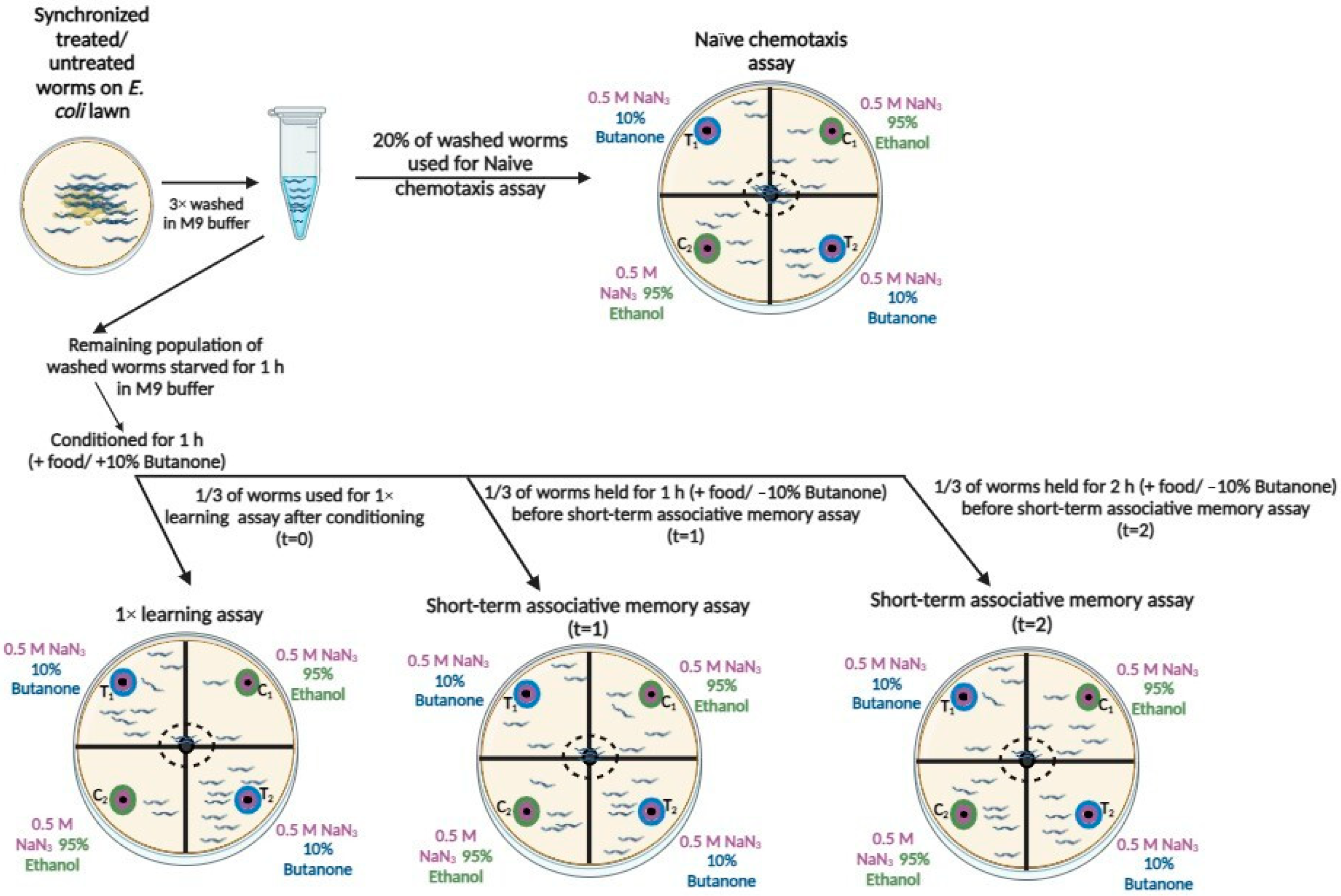
Figure 1. The flow of the naïve chemotaxis, 1× learning, and short-term associative memory assays, adapted from Heydarian et al. [12]. Synchronized worms (treated or untreated) were washed from an E. coli lawn using M9 buffer. A subset of 100–250 worms was used for the naïve chemotaxis assay, where worms were placed on the assay plate and exposed to either 10% butanone or 95% ethanol. Worms were paralyzed in respective quadrants, and their distribution was recorded after one hour for naïve chemotaxis analysis. The remaining worms were starved for one hour in M9 buffer, transferred to NGM plates seeded with E. coli, and conditioned for one hour with 2 µL of 10% butanone applied to the plate’s lid. Post-conditioning, the worms were divided into three equal groups: the first group underwent the 1× learning assay immediately (t = 0), while the second and third groups were transferred to E. coli-seeded NGM plates for one hour (t = 1) and two hours (t = 2) before short-term associative memory assays, respectively. For the short-term assays, worms were washed and placed on assay plates where they were exposed to butanone or ethanol for one hour. At the end of each assay, the number of paralyzed worms in each quadrant was recorded for analysis. Data were collected across naïve chemotaxis, 1× learning, and short-term memory assays to evaluate the worms’ behavioral responses.
Jal 05 00012 g001
Figure 2. Survival curves for untreated and PRSE-supplemented GRU101 and GRU102 (Aβ1–42-expressing) worms. Experiments performed in triplicate (n = 93 for GRU101-control, n = 86 for gru102-control, n = 82 for GRU101-PRSE, n = 95 for GRU102-PRSE). (A) GRU102-control worms showed a significantly shorter mean lifespan compared to GRU101-control worms. (B) In GRU102 worms, PRSE also significantly extended the median lifespan by 12% compared to controls. Additionally, PRSE extended the maximum lifespan of GRU102 worms compared to their respective controls. Experiments were conducted in triplicate with ~100 worms per group. Statistical significance was assessed using the log-rank (Mantel–Cox) test.
Jal 05 00012 g002
Figure 3. Naïve chemotaxis index, learning index, and short-term associative memory loss index (SAML index) in GRU101 (control) and PRSE-treated GRU102 (Aβ1–42 expressing) worms at days four, eight, and twelve. (A) GRU102-control worms showed a significantly lower naïve chemotaxis index compared to GRU101-control worms at day four, day eight, and day twelve (* p < 0.05). (B) PRSE supplementation significantly increased the naïve chemotaxis index in GRU101 worms at all stages (days four, eight, and twelve) compared to their untreated control group (* p < 0.05). PRSE also restored the naïve chemotaxis index in GRU102 worms closer to the levels of GRU102-control worms at day four. PRSE supplementation significantly increased the naïve chemotaxis index in GRU101 worms at all stages (days four, eight, and twelve) compared to their untreated control group (* p < 0.05). PRSE also restored the naïve chemotaxis index in GRU102 worms closer to the levels of GRU102-control worms at day four. At days eight and twelve, PRSE significantly improved the naive chemotaxis index in GRU102 worms compared to their untreated control group (* p < 0.05). (C) GRU101 (control) worms consistently exhibited a higher learning index compared to GRU102 (control) worms across all stages (days four, eight, and twelve), with significant differences observed (* p < 0.05). (D) PRSE supplementation significantly improved the learning index in GRU102 worms at the young and old stages (day four and day twelve, * p < 0.05) compared to their own untreated control group. For GRU101 worms, PRSE supplementation did not significantly affect the learning index at any stage compared to their own untreated control group. (E) GRU102 (control) worms showed significantly higher memory loss compared to GRU101 (control) worms at both 1 h and 2 h across all stages (days four, eight, and twelve) (* p < 0.05). (F) PRSE supplementation significantly reduced short-term associative memory loss in GRU102 worms at the 1 h time point across all stages (days four, eight, and twelve) compared to their untreated control group (* p < 0.05). PRSE supplementation significantly reduced short-term associative memory loss in GRU102 worms at 2 h (at days four and eight) compared to their untreated control group (* p < 0.05). No significant differences in memory loss were observed in GRU101 worms with or without PRSE supplementation. Three independent experiments were performed with approximately 100–250 worms per group. Values are expressed as mean ± SEM, and statistical significance was determined using the multivariate General Linear Model (* p < 0.05).
Jal 05 00012 g003
Figure 4. Quantification of Aβ fibril levels in GRU101 (control) and GRU102 (Aβ1–42 expressing) worms with and without PRSE treatment at days four, eight, and twelve. (A) Untreated GRU102 worms exhibited significantly higher ThT fluorescence compared to untreated GRU101 worms at all stages (days four, eight, and twelve) (* p < 0.05). PRSE treatment significantly reduced ThT fluorescence in GRU102 worms at days four, eight, and twelve compared to their untreated control groups (* p < 0.05). (B) Adjusted graph accounting for GRU101 background fluorescence, depicting that PRSE treatment reduced Aβ fibril levels in GRU102 worms at days four, eight, and twelve (* p < 0.05) compared to untreated GRU102 worms.
Jal 05 00012 g004
Table 1. The effect of PRSE on the lifespan of GRU101 and GRU102 worms.
TreatmentMean Lifespan
(Days)
% Extension Compared to ControlMedian Lifespan
(Days)
% Extension Compared to ControlMaximum Lifespan
(Days)
101-control17.60 ± 0.33 18.00 ± 0.41 20.97 ± 0.20
102-control15.95 ± 0.43 a 17.00 ± 0.60 19.90 ± 0.23 a
101-PRSE18.18 ± 0.35 18.00 ± 0.50 21.37 ± 0.18
102-PRSE17.78 ± 0.36 b1119.00 ± 0.52 ab6 a, 12 b21.60 ± 0.93 b
The comparisons between treatments and their respective control groups were statistically significant, as denoted by a compared to GRU101-control and b compared to GRU102-control.
Disclaimer/Publisher’s Note: The statements, opinions and data contained in all publications are solely those of the individual author(s) and contributor(s) and not of MDPI and/or the editor(s). MDPI and/or the editor(s) disclaim responsibility for any injury to people or property resulting from any ideas, methods, instructions or products referred to in the content.