Article

Blood Bacterial Microbiota of the American Bison (Bison bison) in Northern Mexico: A Reference for Health and Conservation

by
Juan Carlos Ontiveros-Chacón
1,
Cristina García-De La Peña
1,*,
Joel Domínguez-Viveros
2,
Guadalupe Nelson Aguilar-Palma
2,
Verónica Ávila-Rodríguez
1,
Josué Raymundo Estrada-Arellano
1,
Quetzaly Karmy Siller-Rodríguez
1,
Luis Manuel Valenzuela-Núñez
1,
Jesús Vásquez-Arroyo
3,
Juan Carlos Herrera-Salazar
1,
Annely Zamudio-López
1 and
Judith Correa-Gómez
1
1
Conservation Medicine Laboratory, Biological Sciences Faculty, Juarez University of the State of Durango, Gomez Palacio 35010, Mexico
2
Faculty of Zootechnics and Ecology, Autonomous University of Chihuahua, Chihuahua 31453, Mexico
3
Faculty of Chemical Sciences, Juarez University of the State of Durango, Gomez Palacio 35010, Mexico
*
Author to whom correspondence should be addressed.
Ruminants 2025, 5(1), 10; https://doi.org/10.3390/ruminants5010010
Submission received: 25 January 2025 / Revised: 26 February 2025 / Accepted: 27 February 2025 / Published: 4 March 2025

Simple Summary

Bacteria play vital roles in animal welfare, and some bacteria are even present in the blood of healthy individuals, reflecting overall microbial ecology. In this study, the blood microbiota of endangered American bison in a Mexican biosphere reserve was analyzed to better understand their health and support conservation efforts. The blood bacterial communities of 12 juvenile and 12 adult bison were compared using advanced DNA sequencing techniques. Juveniles exhibited less diverse microbiota (33 phyla, 333 families, and 704 genera) than adults (49 phyla, 583 families, and 1439 genera), with significant differences in composition and abundance. Notably, Mycoplasma wenyonii was more prevalent in juveniles. Many bacteria found in bison blood, including Firmicutes and Proteobacteria, are typically abundant in their digestive systems, suggesting common translocation into the bloodstream. These findings reveal age-related microbiota differences and potential links to health. Understanding the blood microbiota offers insights into disease risk, environmental influences, and overall well-being, thus supporting the development of conservation strategies to preserve vulnerable species such as the American bison.

Abstract

The emerging field of study of blood microbiota reveals the presence of bacteria in the blood of healthy animals. In endangered species such as the American bison (Bison bison), the analysis of this microbiota is crucial for conservation, as changes in these communities or the development of pathogens may affect their health and compromise herd viability. Here, we analyzed and compared the bacterial blood microbiota of healthy adult and juvenile bison in Mexico (Janos, Chihuahua), identifying those bacterial taxa with potential pathogenicity for these individuals. Blood samples were collected from 12 juvenile and 12 adult bison. The V3–V4 region of the 16S rRNA gene was amplified, and next-generation sequencing was subsequently performed on the Illumina NovaSeq platform. The bacterial taxa observed in the blood of these individuals (Firmicutes, Proteobacteria, Bacteroidota, Actinobacteria, Fusobacteriaceae, Lachnospiraceae, Oscillospiraceae, and Ruminococcaceae) have been previously reported to be abundant in the rumen and feces of bison. The most notable difference was observed for Mycoplasma wenyonii, which was significantly enriched in juveniles compared with adults. New sequencing technologies can be practically applied to improve the management and conservation of vulnerable species such as the American bison.

1. Introduction

Bacteria form diverse and complex communities that inhabit animal species, and constitute their microbiota [1]. Several studies have revealed important ecological and evolutionary insights, revealing a significant connection between the microbiota and animal health [2]. In particular, the study of bacterial diversity in endangered or vulnerable species has become highly relevant in conservation because the microbiota plays crucial roles in health, environmental adaptation, and species survival [3,4]. Changes in the microbiota, such as those caused by captivity, reintroductions, translocations, or climate change, can negatively impact host health and the ability to adapt [5,6]. These findings highlight the importance of integrating the study of bacterial diversity into management and conservation strategies for at-risk species to optimize individual health and promote the sustainability of their populations.
The study of bacteria in the bloodstream, known as the blood microbiota/microbiome, is an emerging area of research in microbiology and medicine. Blood was once considered a sterile environment under normal conditions; however, recent studies have identified the presence of bacterial communities in the blood of healthy individuals, raising questions about their origin, physiological role, and relationship to health [7,8]. Next-generation sequencing (NGS) has enabled the characterization of the blood bacterial microbiota in healthy humans [9,10] as well as in other animal species such as cats, mice, and dogs [11,12,13]. Various studies suggest that bacteria circulating in the blood of vertebrates may be translocated from other body parts, such as the gut, mouth, or skin, through the processes of intestinal permeability, microlesions in epithelial barriers, or acquisition via arthropod vector transmission [9,14,15,16,17,18]. Because blood connects all body organs, its microbiota may reflect to some extent the bacteria that inhabit various body compartments and those acquired through arthropod vectors [19,20,21,22]. In the context of vulnerable animals, studying their blood microbiota could provide a better understanding of their microbial ecology as well as the bacterial species with potential pathogenicity circulating in their populations, which under certain circumstances may cause disease in individuals.
The American bison (Bison bison) is the largest terrestrial mammal in North America and is adapted to grassland ecosystems. This ruminant contributes significantly to the ecology of the Great Plains, influencing the plants and animals with which it interacts [23]. Furthermore, it was fundamental to the culture and economy of indigenous people, who used it as a source of food, shelter, and fuel [24]. Before European colonization, it is estimated that there were between 30 and 60 million bison in North America [25]. However, between 1830 and 1880, the population drastically declined to approximately 1000 individuals due to overhunting, habitat fragmentation, disease, and strategic use to weaken indigenous communities who were dependent on this species [26,27,28]. The International Union for Conservation of Nature (IUCN) Red List places the American bison in the Critically Depleted category owing to its absence from much of its original distribution. Most of the current herds within this range are the result of reintroductions, providing them with a significant conservation legacy [29]. In Mexico, bison once inhabited the arid plains of the Chihuahuan Desert in Sonora, Chihuahua, Coahuila, Nuevo León, and Durango [30]. To date, only 512 individuals remain in two conservation herds within Mexico: Rancho El Uno in the Janos Biosphere Reserve, Chihuahua (395 individuals, Robles-Félix, personal communication) and the Maderas del Carmen Protected Natural Area, Coahuila (117 individuals, Delgadillo-Villalobos, personal communication). The Mexican government has listed the American bison as endangered since 2010 [31] because of its small population and high risk of extinction [32,33,34].
Some studies have provided records on the ruminal, gastrointestinal, and fecal microbiota of the American bison [35,36,37,38,39]; however, no information has yet been generated regarding its blood microbiota. These data will enhance the understanding of the microbial ecology of these ruminants and serve as a microbiological reference for herds currently protected in other regions. Therefore, the present study analyzed and compared the bacterial blood microbiota of healthy adults and juvenile bison in a biosphere reserve in northwestern Mexico. Additionally, bacterial species with potential pathogenicity for these individuals were identified.

2. Materials and Methods

2.1. Study Area

The study was conducted at Rancho El Uno, which is located in the Janos Biosphere Reserve, Chihuahua, Mexico (30°50′15.87″ N, 108°25′39.29″ W; Figure 1). This site is located in the northwestern part of the state of Chihuahua, adjacent to Sonora and New Mexico, USA. The average annual temperature is 16 °C, and the average annual precipitation is 300 mm. The dominant vegetation type is open medium grassland [40].

2.2. Blood Sample Collection

In November 2023, blood samples were collected from 12 juvenile bison (less than 1 year old) and 12 adult bison (1 to 10 years old). Individuals were randomly selected from the herd. The animals were immobilized in a livestock press adapted for bison. Aseptic procedures were performed at the puncture site (coccygeal vein) using povidone–iodine (PoviCare™, AdvaCare Pharma USA, Cheyenne, WY, USA). Blood samples were then extracted using 3 mL syringes and 20 G needles [41]. For each animal, 1 mL of blood was collected and placed into a lysis Bashing Bead tube containing 750 μL of lysing/stabilizing solution (Zymo Research, Irvine, CA, USA). Each tube was processed in a Terralyzer (Zymo Research, Irvine, CA, USA) cell disruptor for 30 s to macerate the sample and preserve the DNA.

2.3. Laboratory Work

DNA was extracted from the blood samples of the bison using a commercial kit (Zymobiomics DNA MiniPrep Kit from Zymo Research, Irvine, CA, USA). This process was carried out in a UV laminar flow hood following all sterility protocols. Samples were processed at Novogene Corporation, Inc. (Davis, CA, USA), and the V3–V4 region of the 16S rRNA gene was amplified via the primers 341F (CCTAYGGGRBGCASCAG) and 806R (GGACTACNNGGTATCTAAT). PCR was carried out using 15 µL of Phusion® High-Fidelity PCR Master Mix (New England Biolabs, Ipswich, MA, USA), 2 µM of forward and reverse primers, and approximately 10 ng of template DNA. The thermal program consisted of initial denaturation at 98 °C for 1 min, followed by 30 cycles of denaturation at 98 °C for 10 s, alignment at 50 °C for 30 s, and elongation at 72 °C for 30 s. An equal volume of 1X loading buffer (containing SYBRTM green, Thermo Scientific, Waltham, MA, USA) and PCR products was mixed for electrophoresis on a 2% agarose gel for detection. The PCR products were mixed in equal proportions. The mixed PCR products were then purified using a Qiagen gel extraction kit (QiagenTM, Hilden, Germany). Sequencing libraries were generated using a PCR-free TruSeq® DNA sample preparation kit (Illumina, San Diego, CA, USA) following the manufacturer’s instructions, with index codes added. Library quality was assessed using a Qubit® 2.0 fluorometer (Thermo Scientific, Waltham, MA, USA) and an Agilent Bioanalyzer 2100 (Agilent, Santa Clara, CA, USA) system. Finally, the library was sequenced on an Illumina NovaSeq platform, generating paired-end reads of 250 bp.

2.4. Bioinformatic Analysis

The bioinformatic analysis was performed using Quantitative Insights into Microbial Ecology (QIIME2) on the Linux Ubuntu platform [42]. The DADA2 algorithm (divisive amplicon denoising algorithm) [43] was employed to remove low-quality sequences, filter chimeric sequences, and generate amplicon sequence variants (ASVs) [44]. The Greengenes2 database [45] was used for taxonomy assignment. ASVs at the species level were confirmed on the National Center for Biotechnology Information (NCBI) database using the Basic Local Alignment Search Tool (BLAST) tool. To accept an ASV as a valid species, three conditions had to be met: (1) the identity percentage must be greater than 98.0%, and this number must not be equal to that of other related taxa; (2) the E value must be 0.0 or less; and (3) the query cover (alignment coverage) must be 100%. If any of these three conditions were not met, the taxon remained at the genus level. Reaching the species level (latinized name) was considered important because accurate taxonomic identification of the bacteria circulating in bison blood is key for understanding their function, preventing disease, and improving the health and well-being of these animals. The relative abundance of the dominant ASVs at the phylum, family, and species levels for juvenile and adult bison was graphed using heatmaps in the Morpheus program (Broad Institute), and Venn diagrams (Venny 2.1.0) were used to observe the number of ASVs at these three levels that were unique or shared between age classes. To statistically compare the bacterial microbiota between the age classes of B. bison, a rarefaction process was performed to unify the number of sequences for all samples. With this new absolute abundance file, four alpha diversity metrics were obtained (observed features, Shannon index, Pielou evenness, and Faith’s phylogenetic diversity index), and a Mann–Whitney test (p < 0.05) was applied to each one to test for significant differences between the age classes. Significant alpha diversity metrics were visualized in boxplots using GraphPad Prism ver. 8.0.2. Four beta diversity metrics were subsequently obtained: (1) Jaccard [46], a qualitative dissimilarity metric based on the presence/absence of species between communities that varies between 0 (communities have no species in common) and 1 (communities are identical in terms of species present); (2) Bray–Curtis [47], a quantitative dissimilarity metric based on the abundance of each species in the communities, varying between 0 (the two communities are identical in species composition and abundance) and 1 (the two communities do not share any species); and (3) unweighted UniFrac and (4) weighted UniFrac. Both UniFrac [48] metrics generate a dissimilarity value that varies between 0 and 1: 0 when communities are completely identical in terms of phylogenetic composition (unweighted) or composition and abundance (weighted) and 1 when communities do not share a phylogeny and all lineages are exclusive to a community. Permutational multivariate analysis of variance (PERMANOVA) tests were applied (p < 0.05) to beta diversity metrics across ages. Significant beta diversity metrics were visualized in principal coordinate analysis (PCoA) graphs generated by Emperor [49]. To determine the bacterial taxa causing dissimilarity between ages, similarity percentage analysis (SIMPER) [50] was applied at the phylum, family, and species levels using PAST 5.1 (University of Oslo, Norway), then, Mann–Whitney tests (p < 0.05) were performed between ages for the taxa whose contribution to dissimilarity was greater than 0.1%. Finally, significantly different bacterial taxa were represented in heatmaps using the CLUSTVIS program [51].

3. Results

The average number of reads obtained for juvenile bison was 193,631.3, whereas for adult bison, it was 182,494.8. The average number of nonchimeric sequences for juveniles was 147,468.8 (75.36%), and for adults, it was 148,193.6 (81.07%) (Table S1). The total number of ASVs for both age classes was 13,008 (3083 for juveniles and 10,379 for adults). The ASV richness of juvenile bison included 33 phyla, 69 classes, 195 orders, 333 families, 704 genera, and 890 species (109 achieved a latinized name), whereas for adult bison, it included 49 phyla, 99 classes, 293 orders, 583 families, 1439 genera, and 2112 species (351 achieved a latinized name) (Table S2).
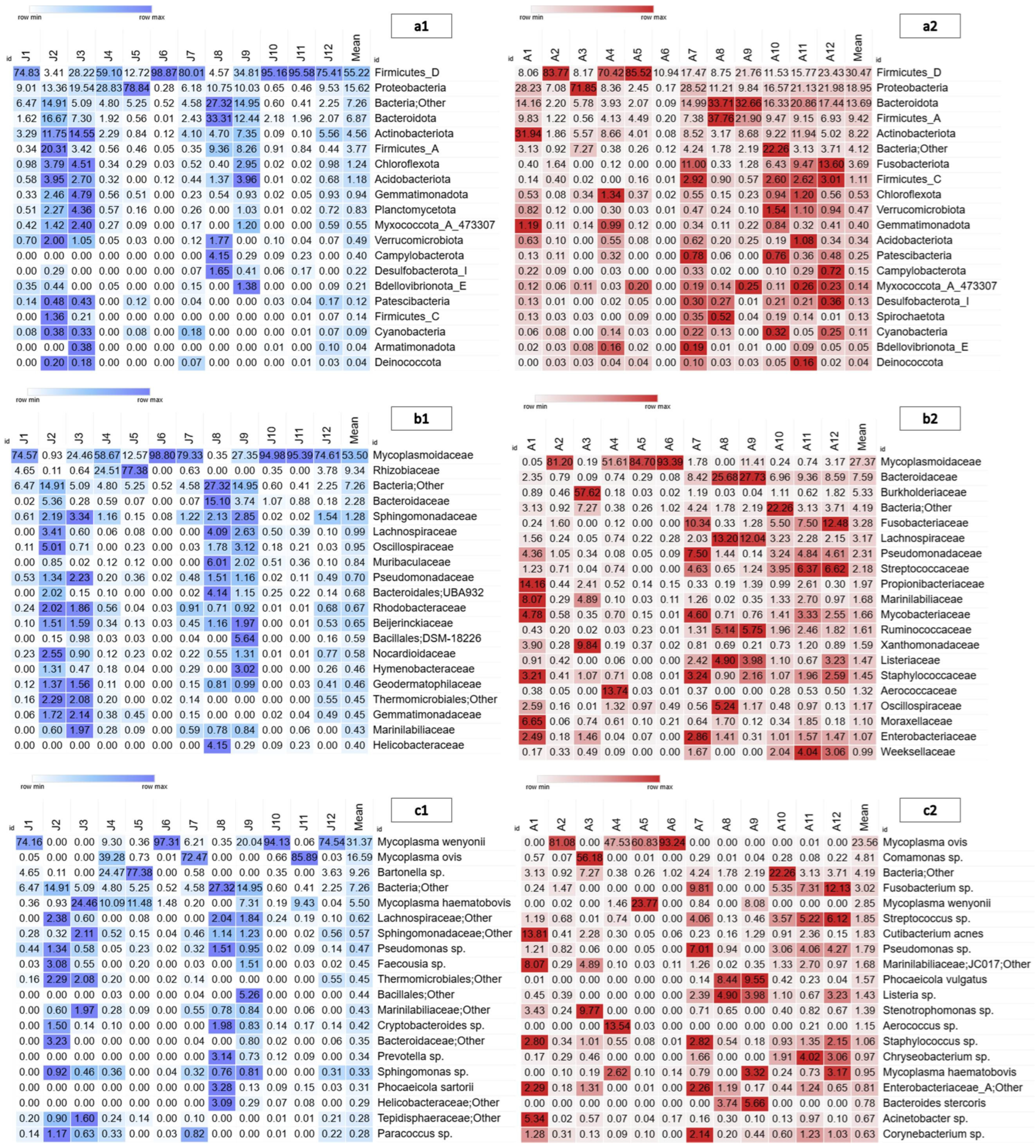
For both age groups, the phyla Firmicutes_D and Proteobacteria were the most abundant (juveniles: mean = 55.22%, 15.62%; adults: mean = 30.47%, 18.95%, Figure 2(a1,a2)). The most abundant family in juvenile bison was Mycoplasmoidaceae (mean = 53.50%), followed by Rhizobiaceae (mean = 9.34%). In adult bison, the most abundant family was Mycoplasmoidaceae (mean = 27.37%), followed by Bacteroidaceae (mean = 7.59%) and Burkholderiaceae (mean = 5.33%; Figure 2(b1,b2)). The most abundant genus in juvenile bison was Mycoplasma (mean = 47.9%), followed by Bartonella (mean = 9.2%). In adult bison, Mycoplasma (mean = 26.4%) was also the most abundant genus, followed by Comamonas (mean = 5.1%). The most abundant species in juveniles was Mycoplasma wenyonii (mean = 31.3%), followed by M. ovis (mean = 16.5%), whereas in adults, M. ovis (mean = 23.5%) was the most abundant species (Figure 2(c1,c2)).
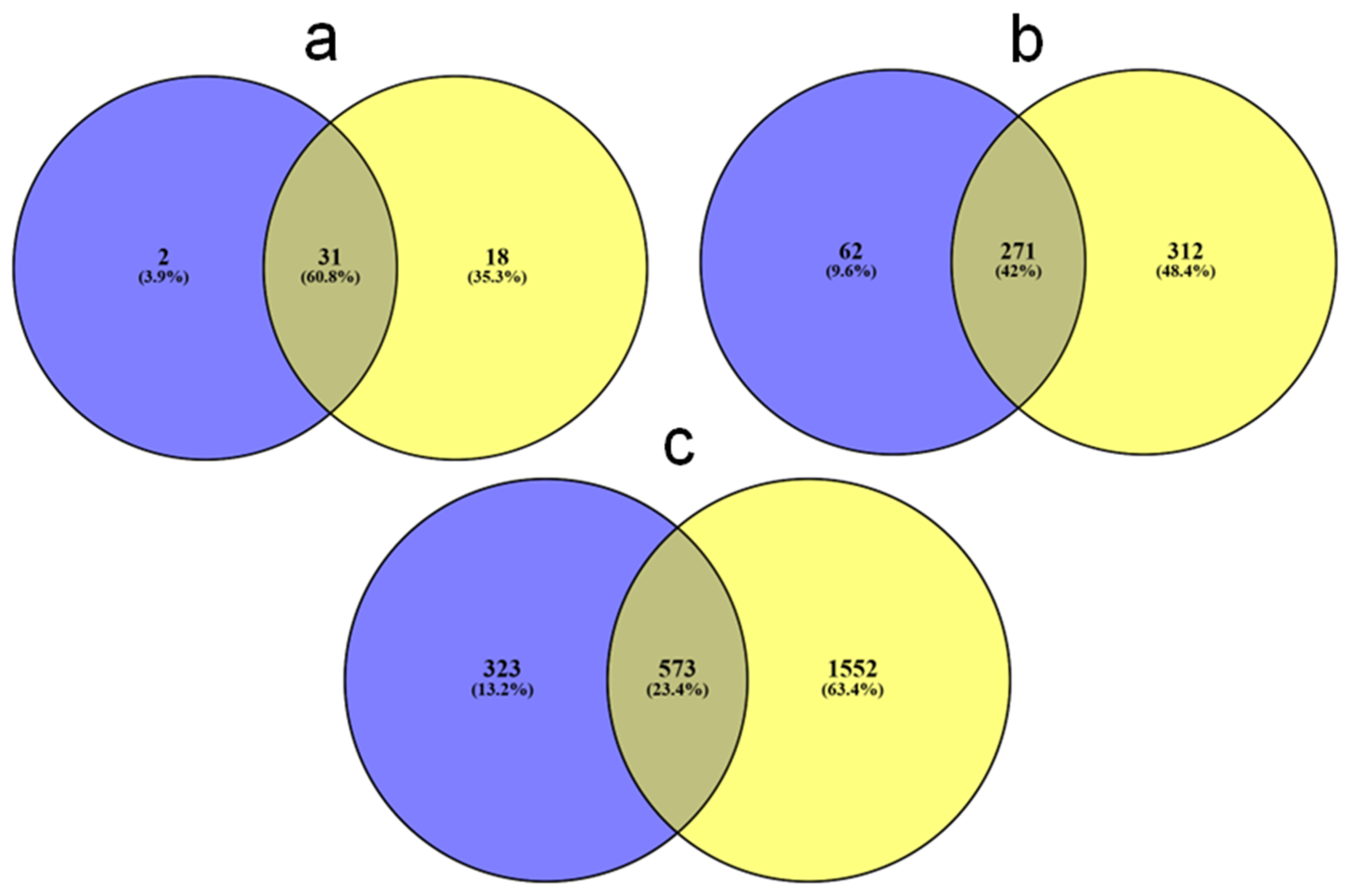
Juveniles possessed two phyla that were absent in adults (Deferribacterota and Myxococcota A437813), whereas adults possessed 18 phyla (Fusobacteriota, Zixibacteria, Calditrichota, Methylomirabilota, UBA10199, Fibrobacterota, SAR324, Firmicutes B370541, Eisenbacteria, Desulfobacterota G459544, WOR-3, TA06, Krumholzibacteriota, Dependentiae, Synergistota, Thermosulfidibacterota, Thermotogota, and Riflebacteria) that were not observed in juveniles. The two age classes shared 31 phyla (60.8%) (Figure 3a). At the family level, juvenile bison possessed 62 taxa not present in adults, whereas adults possessed 312 families that were not observed in juveniles, while 271 families (42%) were found in both age classes (Figure 3b). Juvenile bison possessed 323 species not reported in adults, whereas adults possessed 1552 species not present in juveniles, while 573 species (23.4%) were found in both juvenile and adult bison (Figure 3c).
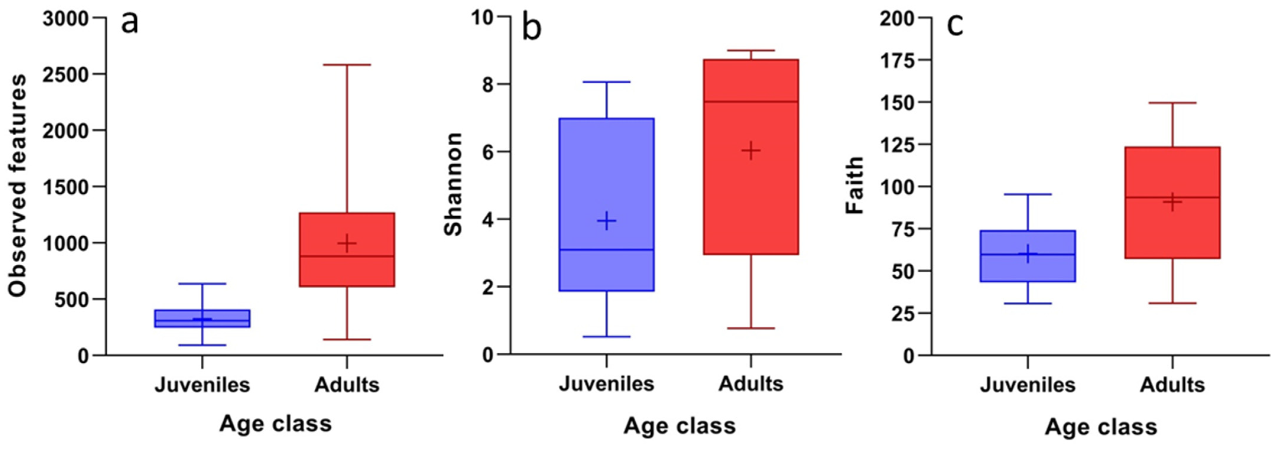
Differences were observed in three of the four alpha diversity metrics, with significantly higher values for adults (A) than for juveniles (J): number of ASVs (U = 14, p = 0.0009; Jmean = 324.6, Amean = 997), Shannon index (U = 37, p = 0.046; Jmean = 3.88, Amean = 6.01), and Faith’s phylogenetic diversity (U = 34, p = 0.030; Jmean = 60.22, Amean = 91.68) (Figure 4). Pielou evenness was not significantly different across age classes (U = 56, p = 0.370; Jmean = 0.46, Amean = 0.60).
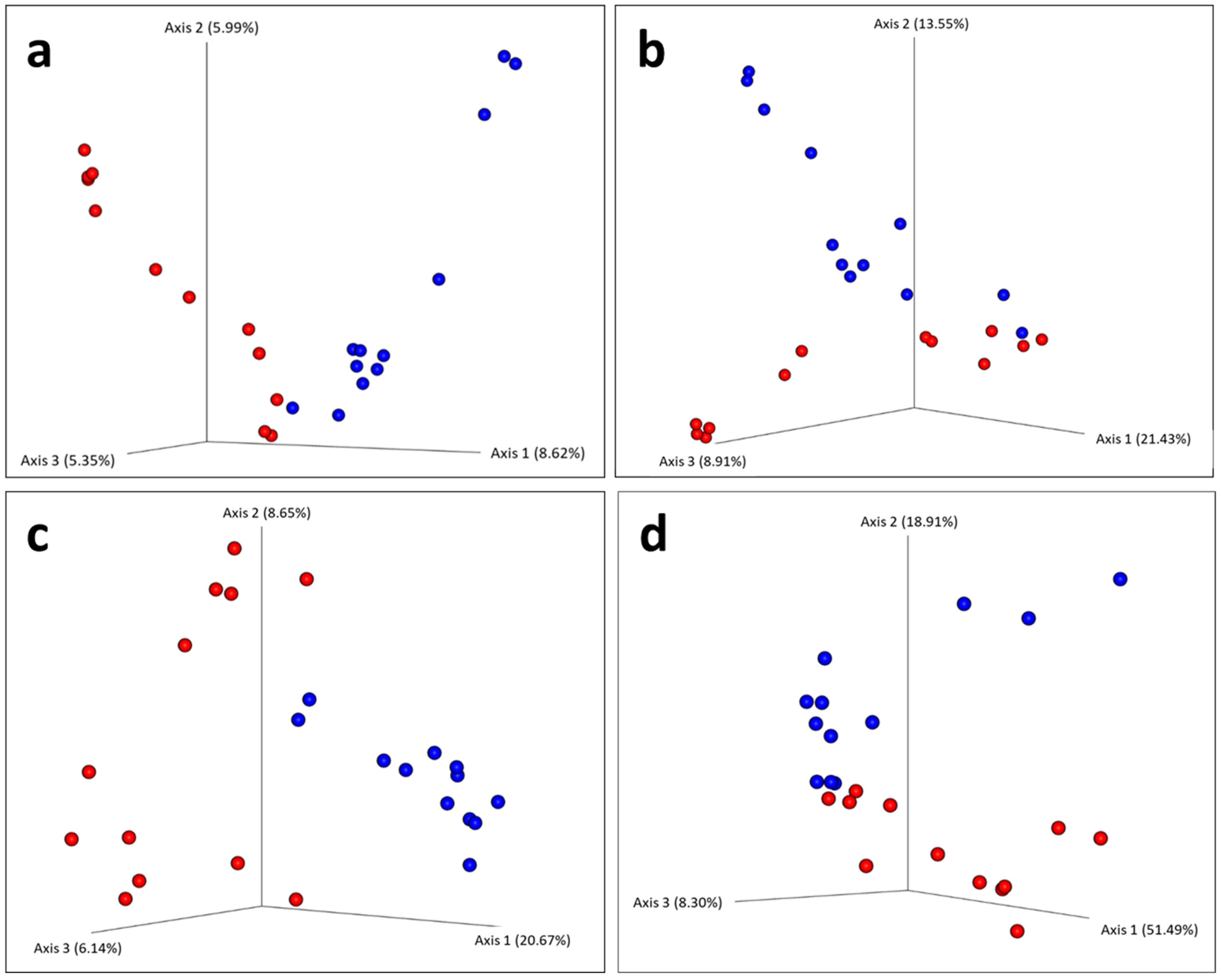
With respect to beta diversity, all four indices showed significant differences: the Jaccard index PERMANOVA pseudo-F = 1.97, p = 0.001; Bray–Curtis PERMANOVA pseudo-F = 2.30. p = 0.007; unweighted UniFrac PERMANOVA pseudo-F = 5.56, p = 0.001; and weighted UniFrac PERMANOVA pseudo-F = 4.36, p = 0.008. The principal coordinate analysis (PCoA) plots show the separation of the two age classes for the four beta diversity indices (Figure 5a–d).
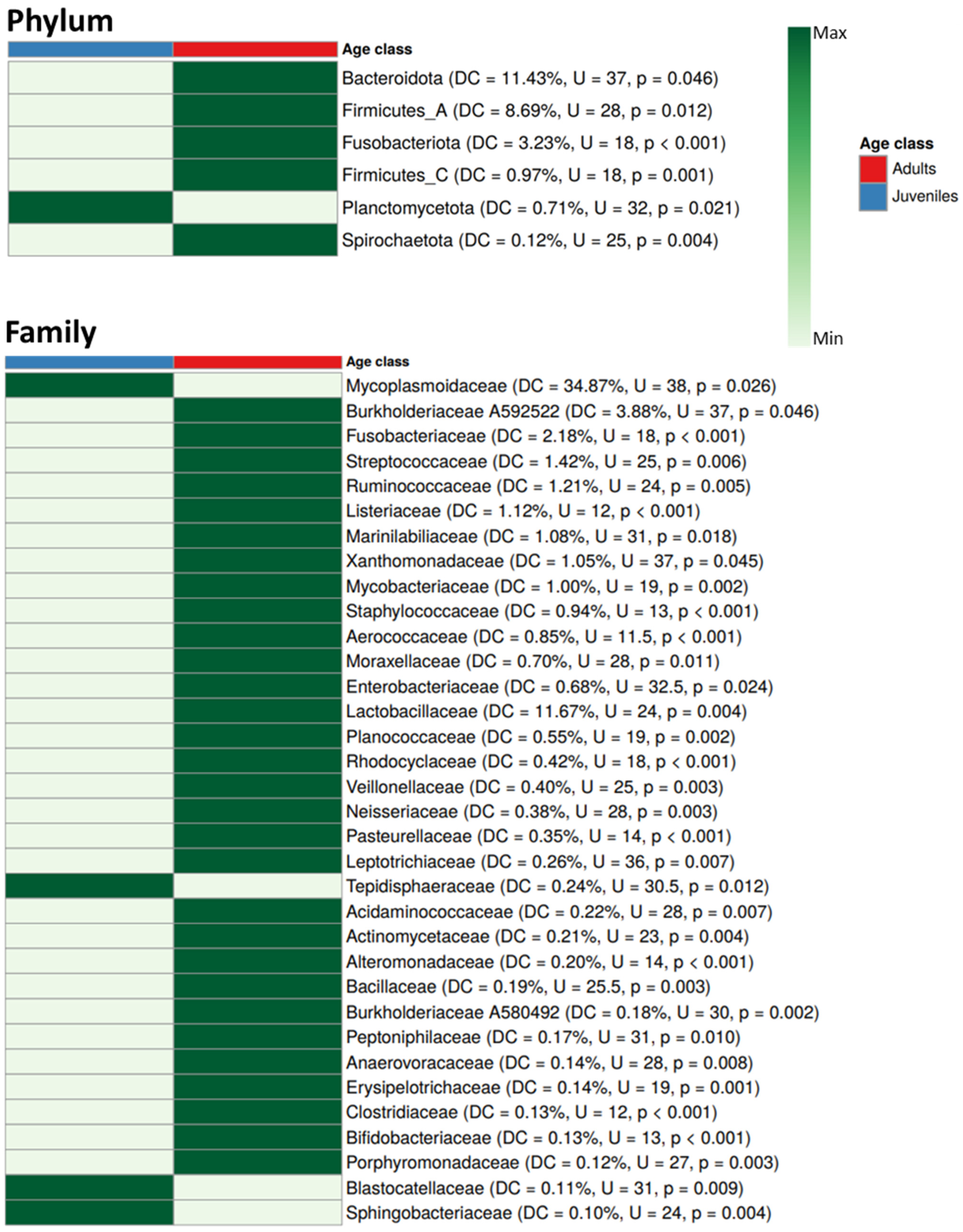
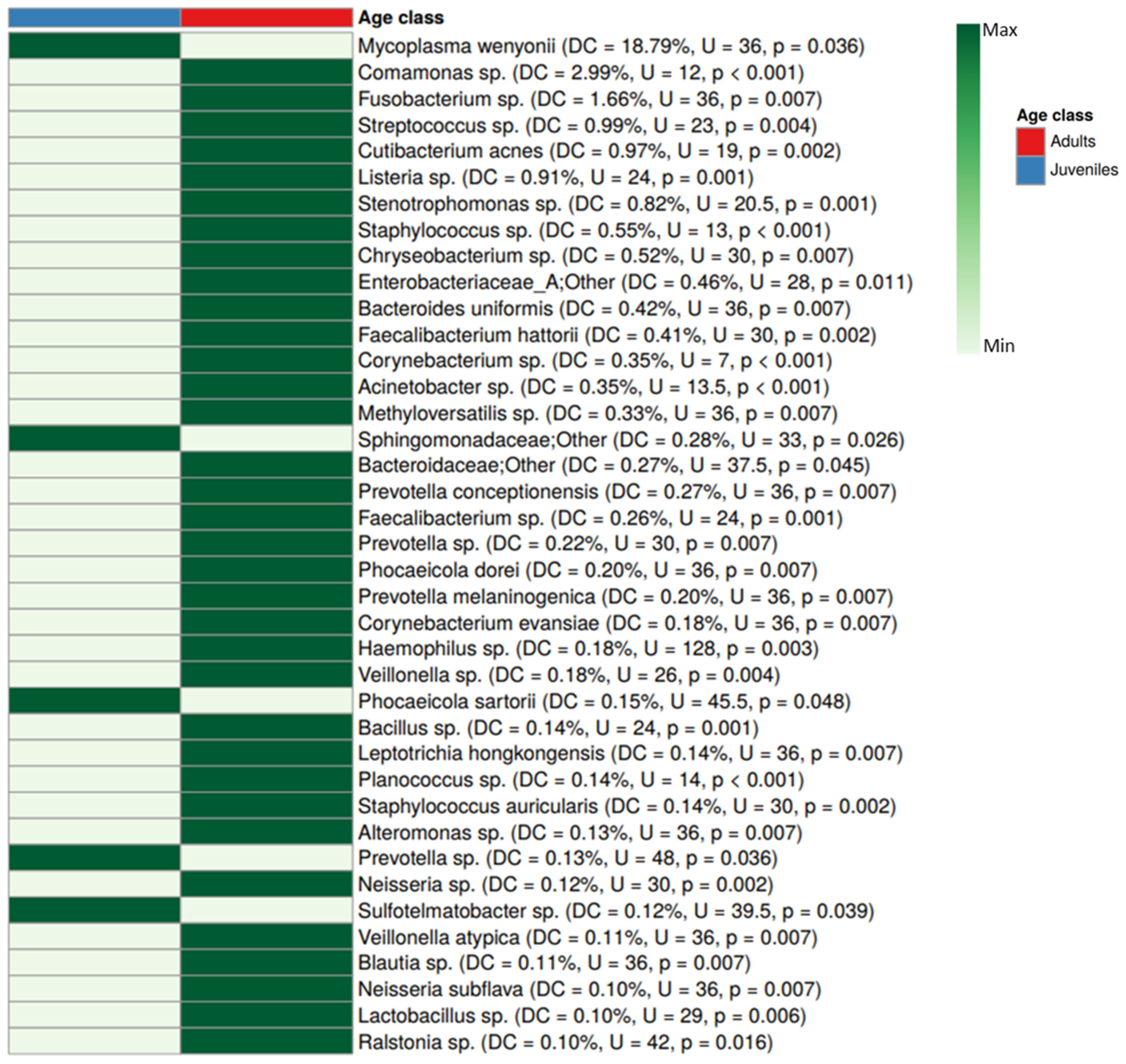
Based on the SIMPER analysis, the overall dissimilarity at the phylum level between juveniles and adults was 55.13%. Six phyla showed significant differences between age groups, with adults exhibiting five enriched phyla and juveniles showing only one, the Planctomycetota (Figure 6). At the family level, the overall dissimilarity was 72.67%, with 34 taxa differing (30 enriched in adults and four enriched in juveniles: Mycoplasmoidaceae, Tepidisphaeraceae, Blastocatellaceae, and Sphingobacteriaceae) (Figure 6). At the species level, the overall dissimilarity was 86.95%. Significant differences were observed in 39 taxa, five of which were more abundant in juveniles (Mycoplasma wenyonii, Sphingomonadaceae—other, Phocaeicola sartorii, Prevotella sp., and Sulfotelmatobacter sp.) and 34 in adults (Figure 7).

4. Discussion

The blood bacterial microbiota is an emerging field of research currently under development in various vertebrate species, including humans, that primarily utilizes next-generation sequencing techniques [7,20,52]. For the American bison, there are few studies on the bacterial microbiota, and those have focused primarily on the digestive system. Our study documents for the first time the blood microbiota of bison, revealing a high number of bacterial taxa circulating in the bloodstream of healthy juveniles and adults.

4.1. Ecological Context of the American Bison Blood Microbiota

In general, the bacterial phyla observed in the blood of the bison in this study (mainly Firmicutes, Proteobacteria, Bacteroidota, and Actinobacterota) have been recorded as the most abundant taxa in the rumen and feces of bison in Canada and the USA. Nguyen et al. [38] reported Firmicutes (51%), Bacteroidota (30%), Fibrobacterota (6%), Spirochaetota (5%), and Pseudomonadota (2%) in the rumens of healthy bison from Canada. Similarly, Weese et al. [35] described the fecal microbiota of juvenile and adult wood bison (Bison bison athabascae) in a national park in Canada and identified 21 bacterial phyla, with Firmicutes, Proteobacteria, and Actinobacteria being the most abundant. These findings are similar to those reported by Bergmann et al. [37], who analyzed the fecal microbiota of healthy bison in Kansas, USA, reporting Firmicutes as the most abundant phylum (53%), followed by Bacteroidota (33%). This information is consistent with that of Fresno-Rueda et al. [39], who analyzed the ruminal and fecal microbiota of healthy bison transitioning from pasture to a free-choice grain diet and reported Firmicutes and Bacteroidota as the most abundant phyla. The high abundance of these and other phyla in the rumen and feces has also been documented in other ruminants such as cows [53,54], deer [54,55], yaks [56], water buffalo [57], goats [58], and sheep [54,59]. Therefore, it is likely that a large portion of the bacterial phyla found in the blood of Janos bison originates from the rumen and feces, and through the process of translocation, these bacteria reach the bloodstream [7,9]. However, less abundant or rare phyla may originate from other compartments of the body. According to Jeon et al. [60], samples of the blood, feces, vagina, and uterus from Holstein dairy cows share a significant abundance of core bacterial genera. Furthermore, these authors indicated that bacteria originating from the gut (Bacteroides, Fusobacterium, and Porphyromonas) may be translocated to the uterus via the bloodstream. Similarly, Scarsella et al. [52] discussed the similarities among bacteria in the feces, blood, and milk of dairy cows, where Firmicutes, Bacteroidetes, and Actinobacteria were present at all three sites, although in variable proportions. This evidence suggests that the blood serves as a means of transport and bacterial connection between different organs in cows, a process that most likely occurs in other vertebrates, including bison.
The most abundant family in both age classes of bison was Mycoplasmoidaceae. In juveniles, Mycoplasma wenyonii was the most abundant species, followed by M. ovis and, in a lower proportion, M. haematobovis (formerly M. haemobos [61]). In adults, M. ovis was the most abundant species, with lower proportions of M. wenyonii and M. haematobovis. The genus Mycoplasma comprises small pleomorphic bacteria that lack a cell wall, are widely distributed in nature, and are classified as hemotropic (parasites of vertebrate erythrocytes and blood-feeding arthropods) or nonhemotropic (parasites of epithelial cells in the respiratory, urinary, and genital tracts) [62,63,64]. The three Mycoplasma species detected in the blood of Janos bison belong to the hemotropic group. Both M. wenyonii and M. haemobos have been reported in the blood of domestic ruminants (cattle, goats, sheep, and water buffalo) in countries such as Japan, Brazil, China, Cuba, and France [65,66,67,68,69]. In the USA, Schambow et al. [70] reported that more than 70% of sampled cows and more than 5% of cows in all herds tested positive for M. wenyonii or M. haemobos. In Mexico, Martínez-Ocampo et al. [71] and Quiroz-Castañeda et al. [72] documented “Candidatus Mycoplasma haemobos” and M. wenyonii, respectively, in sick cattle from the state of Chihuahua (the same state where the Janos Reserve is located). Additionally, Jaimes-Martínez et al. [73] reported the presence of M. haemobos in cattle blood across several states in Mexico, including Morelos, Durango, Veracruz, Jalisco, and Querétaro. M. ovis is a globally distributed bacterium that has been reported in the blood of sheep, deer, goats, and reindeer [74,75,76,77,78]. Horizontal transmission appears to be the most common way that ruminants acquire these Mycoplasma species. According to a review compiled by Arendt et al. [78], the following hematophagous arthropods have been implicated as vectors for Mycoplasma species: for M. wenyonii (e.g., Haematopinus eurysternus, Stomoxys calcitrans, Tabanus megalops, Rhipicephalus microplus, Haemaphysalis bispinosa, Ixodes ricinus, and mosquitoes), for M. haemobos (e.g., R. microplus, Haemaphysalis longicornis), for M. wenyonii and M. hemobos together (e.g., Haematobia irritans, S. calcitrans, Tabanus bovinus, T. bromius), and for M. ovis (e.g., Aedes camptorhynchus, Culex annulirostris, Stomoxys spp., Bovicula ovis, R. microplus). However, vertical transmission (transplacental or intrauterine) of M. wenyonii has also been documented in neonatal cattle born to mothers carrying this bacterium [79,80]. To determine how Janos bison acquire these Mycoplasma species, analyses of the microbiota between mothers and neonates as well as studies on the diversity and microbiota of the hematophagous vectors interacting with this herd are necessary. Other Mycoplasma species previously detected in bison include M. bovis, M. bovirhinis, M. bovoculi, M. arginini, and M. dispar [81].
The genus Bartonella (family Rhizobiaceae) was also relatively abundant in juvenile bison. These facultative intracellular bacteria are widely distributed, and their vectors in livestock include lice, ticks, and biting flies [82]. The remaining bacterial taxa detected in juveniles and adults, as shown in Figure 2, mainly correspond to bacteria associated with the rumen and intestines of ruminants (e.g., Ruminococcaceae, Sphingomonadaceae, Fusobacteriaceae, Lachnospiraceae, and Oscillospiraceae), and some are also commonly found in the environment (e.g., Comamonas sp.) [83,84,85]. These bacteria are probably also translocated into the bloodstream of bison. However, further studies are needed to definitively determine the origin of all bacteria detected in these blood samples.
Notably, other bacteria not typically associated with the digestive tract of ruminants were detected in bison blood, although in minimal proportions (Table S2). For example, the genus Rickettsia was detected in one juvenile bison and one adult bison, whereas Coxiella was identified in two adults. These bacterial taxa are potentially transmitted by ticks [86,87]. Beristain-Ruiz et al. [88] reported the presence of Rickettsia rickettsii in three individuals from this same Janos herd using conventional PCR and suggested some species of wildlife ticks as potential vectors (Rhipicephalus sanguineus s.l., Dermacentor parumapertus, D. albipictus, Ornithodoros sp., and Ixodes sp.). Although ticks or mites have not yet been collected from these bison, the detection of bacteria in their blood originating from ticks or other hematophagous arthropod bites suggests that it would be valuable to identify the ectoparasites hosted by this ruminant species as well as the associated microbiota hosted by the ectoparasites.

4.2. Differences in Blood Microbiota Between Age Classes

The present study revealed significant differences in the blood microbiota of juvenile and adult bison. Initially, the number of ASVs recorded at the species level in adults (2110) was more than double that recorded in juveniles (890). The Venn diagrams (Figure 3) illustrate the increase in the number of taxa at the phylum, family, and species levels in adult bison compared with juveniles. Adults exhibited significantly higher values for observed features, Shannon index, and Faith’s phylogenetic diversity index, indicating that the bacterial microbiota in the blood of this age class is more diverse compared with that of juveniles. Additionally, all four beta diversity metrics used in the present study showed significant dissimilarity, supporting strong evidence for differences in blood bacteria between juveniles and adults. The most notable difference was observed in the family Mycoplasmoidaceae (DC = 34.87%) and the species Mycoplasma wenyonii (DC = 18.79%), with significant enrichment in juveniles compared with those of adults. This result may suggest that the transmission route of M. wenyonii in this herd is primarily vertical and that this taxon is most abundant in juveniles during this life stage because their immune system takes time to regulate its presence. In contrast, adults showed a low mean abundance of M. wenyonii (2.85%). Another, albeit less likely, explanation is that juveniles are more susceptible to the hematophagous ectoparasites that vector this bacterium, increasing its transmission. These questions could be addressed through future studies on ectoparasitism in these bison. For other taxa that exhibited significant differences in abundance between age classes, their contributions to dissimilarity were low (phylum: <11.5%, family: <4%, species: <3%; Figure 6 and Figure 7). However, given that most bacteria in the blood of Janos bison likely translocate from the rumen and intestine into the bloodstream, it is reasonable to explain these age-related differences in terms of the maturation of the digestive microbiota to support nutrition. The Janos bison graze freely over an area of more than 18,500 hectares, allowing access to a wide variety of plant species in the open medium grassland. Therefore, their ruminal microbiota is expected to be highly diverse. Compared with juveniles, adult bison were significantly enriched in five phyla, 30 families, and 34 species. This may indicate that adults possess a more mature, diverse, and complex ruminal–intestinal microbiota that is reflected in their blood, whereas juveniles are still in the developmental stage. This observation aligns with findings by Liang et al. [89], who argued that the ruminal and intestinal microbiota of ruminants is significantly influenced by age, which impacts the health and metabolic processes of these animals. Indeed, several studies have documented significant differences in the ruminal microbiota between young and adult cattle [90,91,92,93], water buffalo [57], sheep [94], and goats [95]. Young livestock have a relatively diverse microbiota that becomes more complex with maturity, which is crucial for efficient digestion and overall health [96]. As animals mature, their microbiota composition changes, with increases in fiber-digesting bacteria that increase the nutritional capacity of the host. Based on the results obtained in Janos, this same transition may occur in bison, as reflected by the bacteria translocated into their blood. However, to conclusively confirm this transition in the ruminal microbiota of juvenile and adult bison, additional studies in this herd are necessary.

4.3. Health Context and Conservation of the American Bison Based on Its Blood Microbiota

The IUCN Bison Specialist Group recognizes disease as a significant concern for bison conservation, as pathogens can hinder population recovery by reducing individual survival and/or reproduction [97]. According to Mackintosh et al. [98], various bacterial diseases have been reported in farmed bison, including brucellosis (Brucella), bovine tuberculosis (Mycobacterium), anaplasmosis (Anaplasma), clostridial diseases (Clostridium), Johne’s disease (Mycobacterium), yersiniosis (Yersinia), leptospirosis (Leptospira), pasteurellosis (Pasteurella), anthrax (Bacillus), salmonellosis (Salmonella), and colibacillosis (Escherichia). Fortunately, in the present study, only the genera Mycobacterium, Clostridium, and Escherichia were detected, and all were present at very low levels.
With respect to the genus Mycoplasma, M. bovis has been documented to cause infections and mortality in bison in the United States [99,100], and this bacterium is known to be transmitted directly between individuals. Janardhan et al. [100] argued that although the origin of M. bovis transmission could not be confirmed, it was likely transmitted from cattle adjacent to the bison herd. In the present study, M. bovis was not detected in blood samples. However, maintaining health monitoring and strict preventative measures to prevent contact between this bison herd and cattle remains crucial. The Mycoplasma species identified in the Janos bison included M. wenyonii, M. ovis, and M. haematobovis (all abundant in juveniles, with M. ovis also present in adults). According to Khoza et al. [22], the majority of cattle infected with M. wenyonii remain clinically asymptomatic unless concurrent diseases cause immunosuppression. Some studies have reported M. wenyonii infections in cattle [101,102]. According to Strugnell and McAuliffe [103], clinical signs include hemolytic anemia, pyrexia, prefemoral lymphadenopathy, weight loss, infertility, a rough hair coat, decreased milk production, and scrotal, teat, and hind limb edema. Meli et al. [104] detected M. haemobos and M. wenyonii in sick cattle during a fatal outbreak of anaplasmosis in Switzerland and suggested that coinfection with these species could increase their pathogenicity in ruminants. Although no infections from these bacteria have been reported in the Janos bison herd or other populations, their presence and abundance (particularly in juveniles) should be a point of attention for veterinarians and reserve managers. Because the bison analyzed in this study appeared to be healthy, it is likely that Mycoplasma species coexist in immune equilibrium with their hosts. However, it is important to note that exposure to stressors could trigger opportunistic pathogen infections. For example, stress events such as transportation, entry into feedlots, social stress due to mixing with unfamiliar animals, and temperature-related stress have been associated with increased nasal shedding of M. bovis in cattle [105]. The information generated in the present study for Janos bison represents a baseline for monitoring possible infections caused by bacteria circulating in their bloodstream if these animals are immunosuppressed. A comprehensive management strategy should take this information into account, along with behavioral monitoring to measure possible animal stress. However, disease prevention is always the best option for the successful conservation of vulnerable species, which implies the permanent maintenance of a high level of animal welfare.

5. Conclusions

The information generated in this study represents the first reference on the blood bacterial composition of healthy juvenile and adult bison living freely in an ecologically appropriate habitat. Because blood connects to all body organs, bacteria found in this bloodstream may provide a general estimate of the diversity of an individual’s overall microbiota (native, environmentally acquired, and potential pathogens) and by extension their health status. However, this is only a starting point, as the true challenge lies in long-term population and individual monitoring and timely veterinary care if needed. New high-throughput sequencing techniques are invaluable tools for addressing previously unresolved questions about bacteria. However, it is desirable to apply these techniques in practical ways, as demonstrated in this study, to improve the management and conservation of the American bison.

Supplementary Materials

The following supporting information can be downloaded at https://www.mdpi.com/article/10.3390/ruminants5010010/s1, Table S1: V3–V4 16S rRNA amplicon sequencing results for each blood sample (numbers of reads before and after quality filtering for each sample) of juvenile and adult bison from Janos, Chihuahua, Mexico; Table S2: Absolute and relative abundance of the bacterial taxa in the blood of juvenile (J) and adult (A) bison (Bison bison) from Janos, Chihuahua, Mexico.

Author Contributions

Conceptualization, J.C.O.-C. and C.G.-D.L.P.; methodology, J.D.-V. and G.N.A.-P.; validation, C.G.-D.L.P.; formal analysis, J.C.O.-C. and V.Á.-R.; investigation, J.R.E.-A.; data curation, Q.K.S.-R. and L.M.V.-N.; writing—original draft preparation, J.C.O.-C.; writing—review and editing, C.G.-D.L.P.; visualization, A.Z.-L. and J.C.-G.; supervision, J.V.-A.; project administration, J.C.H.-S.; funding acquisition, J.C.O.-C. and C.G.-D.L.P. All authors have read and agreed to the published version of the manuscript.

Funding

This research was funded by SECIHTI (Secretaría de Ciencia, Humanidades, Tecnología e Innovación) through a doctoral scholarship awarded to Juan Carlos Ontiveros-Chacón (number 376796).

Institutional Review Board Statement

All the methods and activities of this study were in strict accordance with accepted guidelines for the ethical use, care, and welfare of animals in research at the international level and were approved by the Biological Sciences Faculty UJED ethics committee on 18 September 2023 (code: 0012).

Informed Consent Statement

Not applicable.

Data Availability Statement

The raw data supporting the conclusions of this article are available at the National Center for Biotechnology Information (NCBI; PRJNA675923).

Acknowledgments

To SECIHTI (Secretaría de Ciencia, Humanidades, Tecnología e Innovación) for the doctoral scholarship awarded to Juan Carlos Ontiveros-Chacón; to the team at Rancho El Uno and Cuenca los Ojos; and to the colleagues at the Conservation Medicine Laboratory for their support in this study.

Conflicts of Interest

The authors declare no conflicts of interest.

Abbreviations

The following abbreviations are used in this manuscript:
DNAdeoxyribonucleic acid
rRNAribosomal ribonucleic acid
NGSnext-generation sequencing
QIIME2Quantitative Insights into Microbial Ecology
ASVsamplicon sequence variants
BLASTBasic Local Alignment Search Tool
PCoAprincipal coordinate analysis
PERMANOVApermutational multivariate analysis of variance
SIMPERsimilarity percentage analysis

References

  1. Colston, T.J.; Jackson, C.R. Microbiome evolution along divergent branches of the vertebrate tree of life: What is known and unknown. Mol. Ecol. 2016, 25, 3776–3800. [Google Scholar] [CrossRef] [PubMed]
  2. Costa, M.C.; Arroyo, L.G.; Allen-Vercoe, E.; Stämpfli, H.R.; Kim, P.T.; Sturgeon, A.; Weese, J.S. Comparison of the fecal microbiota of healthy horses and horses with colitis by high throughput sequencing of the V3–V5 region of the 16S rRNA gene. PLoS ONE 2012, 7, e41484. [Google Scholar] [CrossRef] [PubMed]
  3. Santoro, A.; Zhao, J.; Wu, L.; Carru, C.; Biagi, E.; Franceschi, C. Microbiomes other than the gut: Inflammaging and age-related diseases. Semin. Immunopathol. 2020, 42, 589–605. [Google Scholar] [CrossRef] [PubMed]
  4. García-Dela Peña, C.; Garduño-Niño, E.; Vaca-Paniagua, F.; Díaz-Velásquez, C.; Barrows, C.W.; Gomez-Gil, B.; Valenzuela-Núñez, L.M. Comparison of the fecal bacterial microbiota composition between wild and captive Bolson tortoises (Gopherus flavomarginatus). Herpetol. Conserv. Biol. 2019, 14, 587–600. [Google Scholar]
  5. Zhu, L.; Wang, J.; Bahrndorff, S. Editorial: The wildlife gut microbiome and its implication for conservation biology. Front. Microbiol. 2021, 12, 697499. [Google Scholar] [CrossRef]
  6. Diaz, J.; Reese, A.T. Possibilities and limits for using the gut microbiome to improve captive animal health. Anim. Microbiome 2021, 3, 89. [Google Scholar] [CrossRef]
  7. Castillo, D.J.; Rifkin, R.F.; Cowan, D.A.; Potgieter, M. The healthy human blood microbiome: Fact or fiction? Front. Cell. Infect. Microbiol. 2019, 9, 148. [Google Scholar] [CrossRef]
  8. Cheng, H.S.; Tan, S.P.; Wong, D.M.K.; Koo, W.L.Y.; Wong, S.H.; Tan, N.S. The blood microbiome and health: Current evidence, controversies, and challenges. Int. J. Mol. Sci. 2023, 24, 5633. [Google Scholar] [CrossRef]
  9. Païssé, S.; Valle, C.; Servant, F.; Courtney, M.; Burcelin, R.; Amar, J.; Lelouvier, B. Comprehensive description of blood microbiome from healthy donors assessed by 16S targeted metagenomic sequencing. Transfusion 2016, 56, 1138–1147. [Google Scholar] [CrossRef]
  10. Tsafarova, B.; Hodzhev, Y.; Yordanov, G.; Tolchkov, V.; Kalfin, R.; Panaiotov, S. Morphology of blood microbiota in healthy individuals assessed by light and electron microscopy. Front. Cell. Infect. Microbiol. 2023, 12, 1091341. [Google Scholar] [CrossRef]
  11. Vientós-Plotts, A.I.; Ericsson, A.C.; Rindt, H.; Grobman, M.E.; Graham, A.; Bishop, K.; Cohn, L.A.; Reinero, C.R. Dynamic changes of the respiratory microbiota and its relationship to fecal and blood microbiota in healthy young cats. PLoS ONE 2017, 12, e0173818. [Google Scholar] [CrossRef] [PubMed]
  12. Wang, J.; Lang, T.; Shen, J.; Dai, J.; Tian, L.; Wang, X. Core gut bacteria analysis of healthy mice. Front. Microbiol. 2019, 10, 887. [Google Scholar] [CrossRef] [PubMed]
  13. Rojas, C.A.; Park, B.; Scarsella, E.; Jospin, G.; Entrolezo, Z.; Jarett, J.K.; Martin, A.; Ganz, H.H. Species-level characterization of the core microbiome in healthy dogs using full-length 16S rRNA gene sequencing. Front. Veter-Sci. 2024, 11, 1405470. [Google Scholar] [CrossRef]
  14. Berg, R.D. Bacterial translocation from the gastrointestinal tract. Adv. Exp. Med. Biol. 1999, 473, 11–30. [Google Scholar] [CrossRef] [PubMed]
  15. Blekhman, R.; Goodrich, J.K.; Huang, K.; Sun, Q.; Bukowski, R.; Bell, J.T.; Spector, T.D.; Keinan, A.; Ley, R.E.; Gevers, D.; et al. Host genetic variation impacts microbiome composition across human body sites. Genome Biol. 2015, 16, 191. [Google Scholar] [CrossRef]
  16. Lloyd-Price, J.; Abu-Ali, G.; Huttenhower, C. The healthy human microbiome. Genome Med. 2016, 8, 51. [Google Scholar] [CrossRef]
  17. Nagpal, R.; Yadav, H. Bacterial translocation from the gut to the distant organs: An overview. Ann. Nutr. Metab. 2017, 71 (Suppl. S1), 11–16. [Google Scholar] [CrossRef] [PubMed]
  18. Laroche, M.; Raoult, D.; Parola, P. Insects and the transmission of bacterial agents. Microbiol. Spectr. 2018, 6, MTBP0017-2016. [Google Scholar] [CrossRef]
  19. Whittle, E.; Leonard, M.O.; Harrison, R.; Gant, T.W.; Tonge, D.P. Multi-method characterization of the human circulating microbiome. Front. Microbiol. 2018, 9, 3266. [Google Scholar] [CrossRef]
  20. Scarsella, E.; Sandri, M.; Monego, S.D.; Licastro, D.; Stefanon, B. Blood microbiome: A new marker of gut microbial population in dogs? Vet. Sci. 2020, 7, 198. [Google Scholar] [CrossRef]
  21. Yuan, X.; Yang, X.; Xu, Z.; Li, J.; Sun, C.; Chen, R.; Wei, H.; Chen, L.; Du, H.; Li, G.; et al. The profile of blood microbiome in new-onset type 1 diabetes children. iScience 2024, 27, 110252. [Google Scholar] [CrossRef]
  22. Khoza, B.L.; Byaruhanga, C.; Makgabo, S.M.; Nyangiwe, N.; Mnisi, T.; Nxumalo, S.; Oosthuizen, M.C.; Mnisi, Z.T.H. Tick distribution and comparative analysis of bovine blood microbiome in two provinces of South Africa using 16S rRNA PacBio sequencing approach. Front. Trop. Dis. 2024, 5, 1399364. [Google Scholar] [CrossRef]
  23. Knapp, A.K.; Briggs, J.M.; Collins, S.L.; Hartnett, D.C.; Johnson, L.C.; Towne, E.G. The keystone role of Bison in North American Tallgrass Prairie. BioScience 1999, 49, 39–50. [Google Scholar] [CrossRef]
  24. SEMARNAT. Programa de Acción Para la Conservación de la Especie Bisonte (Bison bison); SEMARNAT/CONANP: Ciudad de México, Mexico, 2018. [Google Scholar]
  25. Shaw, J.H. How many bison originally populated western rangelands? Rangelands 1995, 17, 148–150. [Google Scholar]
  26. Potter, B.A.; Gerlach, S.C.; Gates, C.C.; Boyd, D.P.; Oetelaar, G.A.; Shaw, J.S. History of bison in North America. In American bison: Status Survey and Conservation Guidelines; International Union for Conservation of Nature: Gland, Switzerland, 2010; pp. 5–12. [Google Scholar]
  27. Redford, K.H.; Fearn, E. Ecological Future of Bison in North America: A Report from a Multi-Stakeholder, Transboundary Meeting; Wildlife Conservation Society: Bronx, NY, USA, 2007; pp. 2–4. [Google Scholar]
  28. Sanderson, E.W.; Redford, K.H.; Weber, B.; Aune, K.; Baldes, D.; Berger, J.; Carter, D.; Derr, J.; Dobrott, S.; Fearn, E.; et al. The ecological future of the North American bison: Conceiving long-term, large-scale conservation of wildlife. Conserv. Biol. 2008, 22, 252–266. [Google Scholar] [CrossRef]
  29. Rogers, L.R.; Ranglack, D.H.; Plumb, G. Bison bison (Green Status Assessment). The IUCN Red List of Threatened Species 2022, e.T2815A281520242. 2022. Available online: https://www.iucnredlist.org (accessed on 10 January 2025).
  30. List, R.; Ceballos, G.; Curtin, C.; Gogan, P.J.P.; Pacheco, J.; Truett, J. Historic distribution and challenges to bison recovery in the Northern Chihuahuan Desert. Conserv. Biol. 2007, 21, 1487–1494. [Google Scholar] [CrossRef]
  31. SEMARNAT. Norma Oficial Mexicana NOM-059-SEMARNAT-2010, Protección y Manejo Ambiental de Especies Nativas de México de Flora y Fauna Silvestres, Categoría de Riesgo y Especificaciones Para su Inclusión, Exclusión o Cambio (NOM-059-SEMARNAT-2010). Secretaría de Medio Ambiente y Recursos Naturales. 2010. Available online: https://www.gob.mx/profepa/documentos/norma-oficial-mexicana-nom-059-semarnat-2010 (accessed on 20 January 2025).
  32. Solari, S.; Baker, R.J. Mammal Species of the World: A Taxonomic and Geographic Reference; Wilson, D.E., Reeder, D.M., Eds.; Johns Hopkins University Press: Baltimore, MD, USA, 2005; Volume 1. [Google Scholar]
  33. Gates, C.; Ellison, K.; Gates, C.C. Numerical and geographic status. In American Bison: Status Survey and Conservation Guidelines; International Union for Conservation of Nature: Gland, Switzerland, 2010; pp. 55–62. [Google Scholar]
  34. Vázquez-Mantecón, M.C. El Bisonte de América. Historia, Polémica, Leyenda; Universidad Nacional Autónoma de México: Ciudad de México, Mexico, 2013. [Google Scholar]
  35. Weese, J.S.; Shury, T.; Jelinski, M.D. The fecal microbiota of semi-free-ranging wood bison (Bison bison athabascae). BMC Vet. Res. 2014, 10, 120. [Google Scholar] [CrossRef] [PubMed]
  36. Bergmann, G.T. Microbial community composition along the digestive tract in forage- and grain-fed bison. BMC Vet. Res. 2017, 13, 253. [Google Scholar] [CrossRef]
  37. Bergmann, G.T.; Craine, J.M.; Robeson, M.S., II; Fierer, N. Seasonal shifts in diet and gut microbiota of the American bison (Bison bison). PLoS ONE 2015, 10, e0142409. [Google Scholar] [CrossRef] [PubMed]
  38. Nguyen, T.T.M.; Badhan, A.K.; Reid, I.D.; Ribeiro, G.; Gruninger, R.; Tsang, A.; Guan, L.L.; McAllister, T. Comparative analysis of functional diversity of rumen microbiome in bison and beef heifers. Appl. Environ. Microbiol. 2023, 89, e0132023. [Google Scholar] [CrossRef] [PubMed]
  39. Fresno-Rueda, A.F.; Griffith, J.E.; Kruse, C.; St-Pierre, B. Effects of grain-based diets on the rumen and fecal bacterial communities of the North American bison (Bison bison). Front. Microbiol. 2023, 14, 1163423. [Google Scholar] [CrossRef]
  40. List, R.; Pacheco, J.; Ponce, E.; Sierra-Corona, R.; Ceballos, G.; Sierra, R. The Janos Biosphere Reserve, Northern Mexico. Int. J. Wilderness 2010, 16, 35–41. [Google Scholar]
  41. Zambrano, L.J.; Díaz, S. Comité de Bioética. Guía Para la Correcta Toma de Sangre en Bovinos: A Partir de la Vena Coccígea y la Vena Yugular Externa; Universidad Nacional de Colombia: Bogotá, Colombia, 2014; pp. 1–3. [Google Scholar]
  42. Bolyen, E.; Rideout, J.R.; Dillon, M.R.; Bokulich, N.A.; Abnet, C.C.; Al-Ghalith, G.A.; Alexander, H.; Alm, E.J.; Arumugam, M.; Asnicar, F.; et al. Reproducible, interactive, scalable and extensible microbiome data science using QIIME 2. Nat. Biotechnol. 2019, 37, 852–857. [Google Scholar] [CrossRef]
  43. Callahan, B.J.; Mcmurdie, P.J.; Rosen, M.J.; Han, A.W.; Johnson, A.J.A.; Holmes, S.P. DADA2: High-resolution sample inference from Illumina amplicon data. Nat. Methods 2016, 13, 581–583. [Google Scholar] [CrossRef] [PubMed]
  44. Callahan, B.J.; McMurdie, P.J.; Holmes, S.P. Exact sequence variants should replace operational taxonomic units in marker-gene data analysis. ISME J. 2017, 11, 2639–2643. [Google Scholar] [CrossRef] [PubMed]
  45. McDonald, D.; Jiang, Y.; Balaban, M.; Cantrell, K.; Zhu, Q.; Gonzalez, A.; Morton, J.T.; Nicolaou, G.; Parks, D.H.; Karst, S.M.; et al. Greengenes2 unifies microbial data in a single reference tree. Nat. Biotechnol. 2023, 42, 715–718. [Google Scholar] [CrossRef] [PubMed]
  46. Jaccard, P. The distribution of the flora in the alpine zone. New Phytol. 1912, 11, 37–50. [Google Scholar] [CrossRef]
  47. Bray, J.R.; Curtis, J.T. An ordination of the upland forest communities of Southern Wisconsin. Ecol. Monogr. 1957, 27, 325–349. [Google Scholar] [CrossRef]
  48. Lozupone, C.; Knight, R. UniFrac: A New Phylogenetic Method for Comparing Microbial Communities. Appl. Environ. Microbiol. 2005, 71, 8228–8235. [Google Scholar] [CrossRef]
  49. Vázquez-Baeza, Y.; Pirrung, M.; Gonzalez, A.; Knight, R. EMPeror: A tool for visualizing high-throughput microbial community data. GigaScience. 2013, 2, 16. [Google Scholar] [CrossRef]
  50. Khomich, M.; Måge, I.; Rud, I.; Berget, I. Analysing microbiome intervention design studies: Comparison of alternative multivariate statistical methods. PLoS ONE 2021, 16, e0259973. [Google Scholar] [CrossRef]
  51. Metsalu, T.; Vilo, J. Clustvis: A web tool for visualizing clustering of multivariate data using Principal Component Analysis and heatmap. Nucleic Acids Res. 2015, 43, W566–W570. [Google Scholar] [CrossRef]
  52. Scarsella, E.; Zecconi, A.; Cintio, M.; Stefanon, B. Characterization of microbiome on feces, blood and milk in dairy cows with different milk leucocyte patterns. Animals. 2021, 11, 1463. [Google Scholar] [CrossRef] [PubMed]
  53. Rudi, K.; Moen, B.; Sekelja, M.; Frisli, T.; Lee, M.R. An eight-year investigation of bovine livestock fecal microbiota. Vet. Microbiol. 2012, 160, 369–377. [Google Scholar] [CrossRef]
  54. Glendinning, L.; Genç, B.; Wallace, R.J.; Watson, M. Metagenomic analysis of the cow, sheep, reindeer and red deer rumen. Sci. Rep. 2021, 11, 1990. [Google Scholar] [CrossRef] [PubMed]
  55. Gruninger, R.J.; Sensen, C.W.; McAllister, T.A.; Forster, R.J. Diversity of rumen bacteria in Canadian cervids. PLoS ONE 2014, 9, e89682. [Google Scholar] [CrossRef] [PubMed]
  56. Guo, W.; Li, Y.; Wang, L.; Wang, J.; Xu, Q.; Yan, T.; Xue, B. Evaluation of composition and individual variability of rumen microbiota in yaks by 16S rRNA high-throughput sequencing technology. Anaerobe 2015, 34, 74–79. [Google Scholar] [CrossRef] [PubMed]
  57. Koringa, P.G.; Thakkar, J.R.; Pandit, R.J.; Hinsu, A.T.; Parekh, M.J.; Shah, R.K.; Jakhesara, S.J.; Joshi, C.G. Metagenomic characterisation of ruminal bacterial diversity in buffaloes from birth to adulthood using 16S rRNA gene amplicon sequencing. Funct. Integr. Genom. 2019, 19, 237–247. [Google Scholar] [CrossRef]
  58. Giger-Reverdin, S.; Domange, C.; Broudiscou, L.P.; Sauvant, D.; Berthelot, V. Rumen function in goats, an example of adaptive capacity. J. Dairy Res. 2020, 87, 45–51. [Google Scholar] [CrossRef] [PubMed]
  59. Rabee, A.E.; Kewan, K.Z.; Sabra, E.A.; El Shaer, H.M.; Lamara, M. Rumen bacterial community profile and fermentation in Barki sheep fed olive cake and date palm byproducts. PeerJ 2021, 9, e12447. [Google Scholar] [CrossRef]
  60. Jeon, S.J.; Cunha, F.; Vieira-Neto, A.; Bicalho, R.C.; Lima, S.; Bicalho, M.L.; Galvão, K.N. Blood as a route of transmission of uterine pathogens from the gut to the uterus in cows. Microbiome 2017, 5, 109. [Google Scholar] [CrossRef]
  61. Oren, A. A plea for linguistic accuracy–also for Candidatus taxa. Int. J. Syst. Evol. Microbiol. 2017, 67, 1085–1094. [Google Scholar] [CrossRef]
  62. Razin, S.; Hayflick, L. Highlights of mycoplasma research—An historical perspective. Biologicals 2010, 38, 183–190. [Google Scholar] [CrossRef] [PubMed]
  63. Brown, D.R.; May, M.; Bradbury, J.M.; Balish, M.F.; Calcutt, M.J.; Glass, J.I.; Tasker, S.; Messick, J.B.; Johansson, K.E.; Neimark, H. Mycoplasma. In Bergey’s Manual of Systematics of Archaea and Bacteria; Whitman, W.B., Kämpfer, P., Trujillo, M., Chun, J., DeVos, P., Hedlund, B., Dedysh, S., Eds.; Springer Science and Business Media: Berlin/Heidelberg, Germany, 2015; pp. 1–78. [Google Scholar]
  64. Paul, B.T.; Jesse, F.F.A.; Chung, E.L.T.; Che-Amat, A.; Lila, M.A.M.; Hashi, H.A.; Norsidin, M.J. Review of clinical aspects, epidemiology and diagnosis of haemotropic Mycoplasma ovis in small ruminants: Current status and future perspectives in tropics focusing on Malaysia. Trop. Anim. Health Prod. 2020, 52, 2829–2844. [Google Scholar] [CrossRef] [PubMed]
  65. Tagawa, M.; Matsumoto, K.; Inokuma, H. Molecular detection of Mycoplasma wenyonii and ‘Candidatus Mycoplasma haemobos’ in cattle in Hokkaido, Japan. Vet. Microbiol. 2008, 132, 177–180. [Google Scholar] [CrossRef] [PubMed]
  66. Girotto, A.; Zangirolamo, A.F.; Bogado, A.L.G.; Souza, A.S.L.E.; da Silva, G.C.F.; Garcia, J.L.; Vilas Boas, L.A.; Biondo, A.W.; Vidotto, O. Molecular detection and occurrence of “Candidatus Mycoplasma haemobos” in dairy cattle of Southern Brazil. Rev. Bras. Parasitol. Vet. 2012, 21, 342–344. [Google Scholar] [CrossRef]
  67. Shi, H.; Hu, Y.; Leng, C.; Shi, H.; Jiao, Z.; Chen, X.; Peng, Y.; Yang, H.; Kan, Y.; Yao, L. Molecular investigation of “Candidatus Mycoplasma haemobos” in goats and sheep in central China. Transbound. Emerg. Dis. 2019, 66, 22–27. [Google Scholar] [CrossRef] [PubMed]
  68. Díaz-Sánchez, A.A.; Corona-González, B.; Meli, M.L.; Álvarez, D.O.; Cañizares, E.V.; Rodríguez, O.F.; Rivero, E.L.; Hofmann-Lehmann, R. First molecular evidence of bovine hemoplasma species (Mycoplasma spp.) in water buffalo and dairy cattle herds in Cuba. Parasites Vectors 2019, 12, 78. [Google Scholar] [CrossRef] [PubMed]
  69. Nouvel, L.X.; Hygonenq, M.-C.; Catays, G.; Martinelli, E.; Le Page, P.; Collin, É.; Inokuma, H.; Schelcher, F.; Citti, C.; Maillard, R. First detection of Mycoplasma wenyonii in France: Identification, evaluation of the clinical impact and development of a new specific detection assay. Comp. Immunol. Microbiol. Infect. Dis. 2019, 63, 148–153. [Google Scholar] [CrossRef] [PubMed]
  70. Schambow, R.; Poulsen, K.; Bolin, S.; Krahn, D.; Norby, B.; Sockett, D.; Ruegg, P. Apparent prevalence of Mycoplasma wenyonii, “Candidatus Mycoplasma haemobos”, and bovine leukemia virus in Wisconsin and Michigan dairy cattle herds. JDS Commun. 2021, 2, 61–66. [Google Scholar] [CrossRef]
  71. Martínez-Ocampo, F.; Rodríguez-Camarillo, S.D.; Amaro-Estrada, I.; Quiroz-Castañeda, R.E. Draft Genome Sequence of “Candidatus Mycoplasma haemobos”, a Hemotropic Mycoplasma Identified in Cattle in Mexico. Genome Announc. 2016, 4, e00656-16. [Google Scholar] [CrossRef]
  72. Quiroz-Castañeda, R.E.; Martínez-Ocampo, F.; Dantán-González, E. Draft genome sequence of Mycoplasma wenyonii, a second hemotropic Mycoplasma species identified in Mexican bovine cattle. Microbiol. Resour. Announc. 2018, 7, 10–1128. [Google Scholar] [CrossRef]
  73. Jaimes-Martínez, C.A.; Quiroz-Castañeda, R.E.; Preciado-de la Torre, J.F.; Amaro-Estrada, I. Detección molecular del hemoplasma Candidatus Mycoplasma haemobos en ganado bovino de México. Acta Agrícola y Pecuaria. 2018, 4, 99–107. [Google Scholar] [CrossRef]
  74. Aguirre, D.H.; Thompson, C.; Neumann, R.D.; Salatin, A.O.; Gaido, A.B.; Torioni de Echaide, S. Brote de micoplasmosis clínica por Mycoplasma ovis en ovinos de Salta, Argentina. Diagnóstico clínico, microbiológico y molecular. Rev. Argent. Microbiol. 2009, 41, 212–214. [Google Scholar] [PubMed]
  75. Grazziotin, A.L.; Santos, A.P.; Guimaraes, A.M.S.; Mohamed, A.; Cubas, Z.S.; de Oliveira, M.J.; dos Santos, L.C.; de Moraes, W.; Vieira, R.F.d.C.; Donatti, L.; et al. Mycoplasma ovis in captive cervids: Prevalence, molecular characterization and phylogeny. Vet. Microbiol. 2011, 152, 415–419. [Google Scholar] [CrossRef] [PubMed]
  76. Machado, C.A.; Vidotto, O.; Conrado, F.O.; Santos, N.J.; Valente, J.D.; Barbosa, I.C.; Trindade, P.W.; Garcia, J.L.; Biondo, A.W.; Vieira, T.S. Mycoplasma ovis infection in goat farms from northeastern Brazil. Comp. Immunol. Microbiol. Infect. Dis. 2017, 55, 1–5. [Google Scholar] [CrossRef] [PubMed]
  77. Martínez-Hernández, J.M.; Ballados-González, G.G.; Fernández-Bandala, D.; Martínez-Soto, S.; Velázquez-Osorio, V.; Martínez-Rodríguez, P.B.; Cruz-Romero, A.; Grostieta, E.; Lozano-Sardaneta, Y.; Salas, P.C.; et al. Molecular detection of Mycoplasma ovis in an outbreak of hemolytic anemia in sheep from Veracruz, Mexico. Trop. Anim. Health Prod. 2019, 51, 243–248. [Google Scholar] [CrossRef] [PubMed]
  78. Arendt, M.; Stadler, J.; Ritzmann, M.; Ade, J.; Hoelzle, K.; Hoelzle, L.E. Hemotrophic Mycoplasmas—Vector Transmission in Livestock. Microorganisms 2024, 12, 1278. [Google Scholar] [CrossRef]
  79. Hornok, S.; Micsutka, A.; Meli, M.L.; Lutz, H.; Hofmann-Lehmann, R. Molecular investigation of transplacental and vector-borne transmission of bovine haemoplasmas. Vet. Microbiol. 2011, 152, 411–414. [Google Scholar] [CrossRef]
  80. Sasaoka, F.; Suzuki, J.; Hirata, T.-I.; Ichijo, T.; Furuhama, K.; Harasawa, R.; Satoh, H. Vertical transmission of Mycoplasma wenyonii in cattle, supported by analysis of the ribonuclease P RNA gene. Acta Vet. Hung. 2015, 63, 271–274. [Google Scholar] [CrossRef]
  81. Register, K.B.; Jones, L.C.; Boatwright, W.D.; Shury, T.K.; Woodbury, M.; Hamilton, R.G.; Treanor, J.; Dyer, N.; Nol, P. Prevalence of Mycoplasma spp. in the Respiratory Tract of Healthy North American Bison (Bison bison) and Comparison with Serum Antibody Status. J. Wildl. Dis. 2021, 57, 683–688. [Google Scholar] [CrossRef] [PubMed]
  82. Gutiérrez, R.; Cohen, L.; Morick, D.; Mumcuoglu, K.Y.; Harrus, S.; Gottlieb, Y. Identification of Different Bartonella Species in the Cattle Tail Louse (Haematopinus quadripertusus) and in Cattle Blood. Appl. Environ. Microbiol. 2014, 80, 5477–5483. [Google Scholar] [CrossRef] [PubMed]
  83. Mizrahi, I.; Jami, E. Review: The compositional variation of the rumen microbiome and its effect on host performance and methane emission. Animal 2018, 12, s220–s232. [Google Scholar] [CrossRef] [PubMed]
  84. Pacífico, C.; Petri, R.M.; Ricci, S.; Mickdam, E.; Wetzels, S.U.; Neubauer, V.; Zebeli, Q. Unveiling the Bovine Epimural Microbiota Composition and Putative Function. Microorganisms 2021, 9, 342. [Google Scholar] [CrossRef] [PubMed]
  85. Zhang, M.; Zhao, B.; Yan, Y.; Cheng, Z.; Li, Z.; Han, L.; Sun, Y.; Zheng, Y.; Xia, Y. Comamonas-dominant microbial community in carbon poor aquitard sediments revealed by metagenomic-based growth rate investigation. Sci. Total Environ. 2024, 912, 169203. [Google Scholar] [CrossRef] [PubMed]
  86. Ortega-Morales, A.I.; Nava-Reyna, E.; Ávila-Rodríguez, V.; González-Álvarez, V.H.; Castillo-Martínez, A.; Siller-Rodríguez, Q.K.; Cabezas-Cruz, A.; Dantas-Torres, F.; Almazán, C. Detection of Rickettsia spp. in Rhipicephalus sanguineus (sensu lato) collected from free-roaming dogs in Coahuila state, northern Mexico. Parasites Vectors 2019, 12, 130. [Google Scholar] [CrossRef]
  87. la Peña, C.G.-D.; Zamudio-López, A.; Barraza-Guerrero, S.I.; Martínez-Aranda, E.; De la Cruz-Ramos, J.M.; Acosta-Ayala, A.; Siller-Rodríguez, Q.K.; Torres-Delgado, M.G.; Ávila-Rodríguez, V.; Vásquez-Arroyo, J.; et al. Bacterial Microbiota of the Brown Dog Tick (Rhipicephalus sanguineus), a Broad Starting Point to Establish Potential Pathogens in Northern Mexico. Microbiol Res. 2024, 15, 2507–2521. [Google Scholar] [CrossRef]
  88. Beristain-Ruiz, D.M.; Vital-García, C.; Figueroa-Millán, J.V.; Lira-Amaya, J.J.; Garza-Hernández, J.A.; Sánchez-Ayala, J.R.; Flores-Ceballos, S.; Rodríguez-Alarcón, C.A.; Olivas-Sánchez, M.P.; Pons-Monarrez, G. Molecular Detection of Tick-Borne Pathogens in American Bison (Bison bison) at El Uno Ecological Reserve, Janos, Chihuahua, Mexico. Pathogens 2021, 10, 1428. [Google Scholar] [CrossRef]
  89. Liang, Z.; Zhang, J.; Du, M.; Ahmad, A.A.; Wang, S.; Zheng, J.; Salekdeh, G.H.; Yan, P.; Han, J.; Tong, B.; et al. Age-dependent changes of hindgut microbiota succession and metabolic function of Mongolian cattle in the semi-arid rangelands. Front. Microbiol. 2022, 13, 957341. [Google Scholar] [CrossRef]
  90. Jami, E.; Israel, A.; Kotser, A.; Mizrahi, I. Exploring the bovine rumen bacterial community from birth to adulthood. ISME J. 2013, 7, 1069–1079. [Google Scholar] [CrossRef]
  91. Guo, C.Y.; Ji, S.K.; Yan, H.; Wang, Y.J.; Liu, J.J.; Cao, Z.J.; Yang, H.J.; Zhang, W.J.; Li, S.L. Dynamic change of the gastrointestinal bacterial ecology in cows from birth to adulthood. MicrobiologyOpen 2020, 9, e1119. [Google Scholar] [CrossRef]
  92. Woodruff, K.L.; Hummel, G.L.; Austin, K.J.; Lake, S.L.; Cunningham-Hollinger, H.C. Calf rumen microbiome from birth to weaning and shared microbial properties to the maternal rumen microbiome. J. Anim. Sci. 2022, 100, skac264. [Google Scholar] [CrossRef] [PubMed]
  93. Wu, Y.; Jiao, C.; Diao, Q.; Tu, Y. Effect of Dietary and Age Changes on Ruminal Microbial Diversity in Holstein Calves. Microorganisms 2023, 12, 12. [Google Scholar] [CrossRef] [PubMed]
  94. Yin, X.; Ji, S.; Duan, C.; Tian, P.; Ju, S.; Yan, H.; Zhang, Y.; Liu, Y. Age-Related Changes in the Ruminal Microbiota and Their Relationship with Rumen Fermentation in Lambs. Front. Microbiol. 2021, 12, 679135. [Google Scholar] [CrossRef] [PubMed]
  95. Li, B.; Zhang, K.; Li, C.; Wang, X.; Chen, Y.; Yang, Y. Characterization and comparison of microbiota in the gastrointestinal tracts of the goat (Capra hircus) during preweaning development. Front. Microbiol. 2019, 10, 2125. [Google Scholar] [CrossRef]
  96. Luo, T.; Li, Y.; Zhang, W.; Liu, J.; Shi, H. Rumen and fecal microbiota profiles associated with immunity of young and adult goats. Front. Immunol. 2022, 13, 978402. [Google Scholar] [CrossRef]
  97. Aune, K.; Gates, C.C.; Boyd, D.; Elkin, B.T.; Hugh-Jones, M.; Joly, D.O.; Nishi, J. Reportable or Notifiable Diseases. In American Bison: Status Survey and Conservation Guidelines 2010 (Revised June 2011); Gates, C.C., Freese, C.H., Gogan, P.J., Kotzman, M., Eds.; International Union for Conservation of Nature: Gland, Switzerland, 2010; pp. 27–37. [Google Scholar]
  98. Mackintosh, C.; Haigh, J.; Griffin, F. Bacterial diseases of farmed deer and bison. Rev. Sci. Tech. 2002, 21, 249–263. [Google Scholar] [CrossRef]
  99. Dyer, N.; Hansen-Lardy, L.; Krogh, D.; Schaan, L.; Schamber, E. An outbreak of chronic pneumonia and polyarthritis syndrome caused by Mycoplasma bovis in feedlot bison (Bison bison). J. Vet. Diagn. Investig. 2008, 20, 369–371. [Google Scholar] [CrossRef]
  100. Janardhan, K.S.; Hays, M.; Dyer, N.; Oberst, R.D.; DeBey, B.M. Mycoplasma bovis outbreak in a herd of North American Bison (Bison bison). J. Vet. Diagn. Investig. 2010, 22, 797–801. [Google Scholar] [CrossRef] [PubMed]
  101. Genova, S.G.; Streeter, R.N.; E Velguth, K.; A Snider, T.; Kocan, K.M.; Simpson, K.M. Severe anemia associated with Mycoplasma wenyonii infection in a mature cow. Can. Vet. J. 2011, 52, 1018–1021. [Google Scholar]
  102. Gladden, N.; Haining, H.; Henderson, L.; Marchesi, F.; Graham, L.; McDonald, M.; Murdoch, F.R.; Sala, A.B.; Orr, J.; Ellis, K. A case report of Mycoplasma wenyonii associated immune-mediated haemolytic anaemia in a dairy cow. Ir. Vet. J. 2015, 69, 1. [Google Scholar] [CrossRef] [PubMed]
  103. Strugnell, B.; McAuliffe, L. Mycoplasma wenyonii infection in cattle. In Pract. 2012, 34, 146–154. [Google Scholar] [CrossRef]
  104. Meli, M.L.; Willi, B.; Dreher, U.M.; Cattori, V.; Knubben-Schweizer, G.; Nuss, K.; Braun, U.; Lutz, H.; Hofmann-Lehmann, R. Identification, molecular characterization, and occurrence of two bovine hemoplasma species in Swiss cattle and development of real-time TaqMan quantitative PCR assays for diagnosis of bovine hemoplasma infections. J. Clin. Microbiol. 2010, 48, 3563–3568. [Google Scholar] [CrossRef] [PubMed]
  105. Boothby, J.T.; Jasper, D.E.; Zinkl, J.G.; Thomas, C.B.; Dellinger, J.D. Prevalence of mycoplasmas and immune responses to Mycoplasma bovis in feedlot calves. Am. J. Vet. Res. 1983, 44, 831–838. [Google Scholar] [CrossRef] [PubMed]
Figure 1. Study area at Rancho El Uno, Janos, Chihuahua, Mexico, where the American bison (Bison bison) conservation herd is located.
Ruminants 05 00010 g001
Figure 2. Heatmaps of the most abundant bacterial taxa at the phylum, family, and species levels in the blood of juvenile (a1,b1,c1, respectively) and adult bison (a2,b2,c2, respectively) in Janos, Chihuahua, Mexico.
Ruminants 05 00010 g002
Figure 3. Venn diagrams of the number of bacterial taxa at the (a) phylum, (b) family, and (c) species levels in the blood of juvenile (blue) and adult (yellow) bison in Janos, Chihuahua, Mexico.
Ruminants 05 00010 g003
Figure 4. Boxplots of the number of observed features (a), Shannon index (b), and phylogenetic diversity (c) of the blood bacterial microbiota of juvenile and adult bison in Janos, Chihuahua, Mexico. Cross = mean.
Ruminants 05 00010 g004
Figure 5. Principal coordinate analysis (PCoA) plots of the Jaccard index (a), Bray–Curtis index (b), unweighted UniFrac (c), and weighted UniFrac (d) of the blood bacterial microbiota of juvenile (blue dots) and adult (red dots) bison in Janos, Chihuahua, Mexico.
Ruminants 05 00010 g005
Figure 6. Heatmaps at the phylum and family levels of the blood bacterial microbiota of juvenile and adult bison in Janos, Chihuahua, Mexico, highlighting the taxa significantly enriched for each age group. DC = dissimilarity contribution percentage, U = Mann–Whitney test, p = significance level.
Ruminants 05 00010 g006
Figure 7. Heatmap at the species level of the blood bacterial microbiota of juvenile and adult bison in Janos, Chihuahua, Mexico, highlighting the taxa significantly enriched for each age group. DC = dissimilarity contribution percentage, U = Mann–Whitney test, p = significance level.
Ruminants 05 00010 g007
Disclaimer/Publisher’s Note: The statements, opinions and data contained in all publications are solely those of the individual author(s) and contributor(s) and not of MDPI and/or the editor(s). MDPI and/or the editor(s) disclaim responsibility for any injury to people or property resulting from any ideas, methods, instructions or products referred to in the content.