Proceeding Paper

In Vitro and In Vivo Effects of Conventional and Chitosan Nanoparticle-Encapsulated Miltefosine Drug for Treatment of Cutaneous Leishmaniasis †

1
Department of Microbiology, Institute of Pathology and Diagnostic Medicine, Khyber Medical University, Peshawar 25100, Pakistan
2
Faculty of Veterinary and Animal Sciences, Gomal University, Dera Ismail Khan 29111, Pakistan
3
Faculty of Veterinary and Animal Sciences, The University of Agriculture, Dera Ismail Khan 29220, Pakistan
4
Key Laboratory of Space Bioscience and Biotechnology, School of Life Sciences, Northwestern Polytechnical University, Xi’an 710000, China
5
Department of Dermatology, The Second Affiliated Hospital of Shantou University Medical College, Shantou 515000, China
6
Department of Animal and Poultry Production, Faculty of Veterinary and Animal Sciences, University of Poonch, Rawalakot 12311, Pakistan
*
Authors to whom correspondence should be addressed.
Presented at the 2nd International Electronic Conference on Biomedicines, 1–31 March 2023; Available online: https://ecb2023.sciforum.net/.
Med. Sci. Forum 2023, 21(1), 19; https://doi.org/10.3390/ECB2023-14334
Published: 7 April 2023
(This article belongs to the Proceedings of The 2nd International Electronic Conference on Biomedicines)

Abstract

:
This study aimed to formulate polymer-based chitosan nanoparticles as a drug (miltefosine) delivery system for treating leishmaniasis. Miltefosine-loaded chitosan nanoparticles (MLCNPs) have been synthesized and then characterized by the use of UV-visible spectroscopy, scanning electron microscopy (SEM), transmission electron microscopy (TEM), the zeta potential, drug loading content (DLC), encapsulation efficacy (EE) and the dynamic light scattering technique (DLS). Further, the in vitro anti-leishmanial activity of the characterized chitosan nanoparticles was assessed by a microculture tetrazolium (MTT) assay, and in vivo efficacy was evaluated in infected BALB/c mice. The lesion healing was statistically analyzed using Wilcoxon signed-rank and Mann–Whitney tests. The MLCNPs were spherical-shaped (97.5 nm), which presented efficient encapsulation (97.56%), drug loading content (91.5 µg/mL) and a positive surface charge (+1.04 mV). MLCNPs were less hemolytic (6%) when compared to conventional miltefosine. MLCNPs (50 µg/mL) showed a potential antileishmanial effect (mean viability: 10 ± 0.3%) on promastigotes in comparison to conventional miltefosine (mean viability: 18 ± 1.3%). The IC50 value for MLCNPs and miltefosine was 0.0218 µg/mL and 0.3548 µg/mL, respectively. This in vivo study proved that lesions of mice treated with oral and intralesional-injected MLCNPs significantly heal (p = 0.01). MLCNPs have a significant antileishmanial effect and could be utilized as an alternative treatment for CL.

1. Introduction

Leishmaniasis is a vector-born infectious disease transmitted by protozoan parasites and is both anthroponotic and zoonotic. After malaria, leishmaniasis is considered the second leading parasitic cause of death [1]. Female sandflies are intermittently infected by the parasite that feeds on the blood of infected animals or humans and acquires the parasites from their blood [2].
Leishmaniasis is classified into three types based on signs and symptoms, which are cutaneous, mucocutaneous and visceral leishmaniasis [3]. CL is more susceptible to lesions caused by Leishmania tropica, Leishmania major, Leishmania braziliensis, Leishmania Mexicana and Leishmania amazonensis, which arise at the site of an insect bite [4]. The CL is ubiquitous worldwide but is more prevalent in underdeveloped and developing countries [5]. Treatment decisions and clinical description rely on the species engaged with causing leishmaniasis. The available drugs can cure the disease for the time being, but due to the unavailability of vaccines, complete control is impossible. Amongst the available drugs, the first and second-line drugs are partially effective, need a longer treatment period and have severe side effects [6].
Miltefosine is an anti-leishmanial drug in the alkyl-phosphocholine class and is effective against tumor and fungal infections [7]. This drug has zwitterionic amphiphilic properties due to the positively charged amine and negatively charged phosphoryl group. The complete mode of action of miltefosine is still not fully understood. However, some studies demonstrate that it disturbs the lipid-dependent cell signaling pathways. Besides its efficacy, miltefosine has severe side effects such as genitourinary and hematological complications [8].
Nanomedicine is a new and alternative way to treat several life-threatening infections. Along with their direct activity, nanoparticles are also used as an efficient drug delivery system, especially for drugs having severe toxicity [9]. These drug delivery systems enhance the number of medications to infected tissues and elevate the treatment performance compared to conventional therapy [10]. Chitosan nanoparticles are polymer-based cationic molecules and interact efficiently with the plasma membrane of macrophages [11]. Furthermore, they can carry the drugs inside the macrophages and reduce adverse effects related to drug administration and pro-inflammatory cytokine secretion [12]. Therefore, in the current study, we aimed to synthesize miltefosine-loaded chitosan nanoparticles (MLCNPs) as an alternative therapeutic option for the treatment of cutaneous leishmaniasis to achieve a higher efficacy rate and lower toxicity compared to conventional miltefosine.

2. Materials and Methods

2.1. Materials

The reagents used were Roswell Park Memorial Institute Medium-1640 (RPMI, Lot # 1868632, GIBCO, New York, NY, USA), Medium 199 (M199, Lot # CP17-1058, Sigma- Aldrich, St. Louis, MO, USA), heat-inactivated Fetal Bovine Serum (hiFBS, Lot # 10270, GIBCO, New York, NY, USA), Penicillin–Streptomycin solution (PenStrepsoln, Lot # 01161018, Sigma Ald), Trypan Blue (Sigma- Aldrich, St. Louis, MO, USA), chitosan (Sigma- Aldrich, St. Louis, MO, USA), miltefosine (Sigma- Aldrich, St. Louis, MO, USA), Tripolyphosphate (TPP), D-Trehalose, Sodium Hydroxide (Thermo Fisher Scientific, Waltham, MA, USA), Phosphate Buffered Saline (Thermo Fisher Scientific, Waltham, MA, USA) and Dimethyl Sulfoxide (DMSO) (Thermo Fisher Scientific, Waltham, MA, USA).

2.2. Parasites and Animals

The L. tropica strain was collected from the infectious disease laboratory at Quaid-i-Azam University, Islamabad, Pakistan, for in vitro experiment. In vivo study was carried out using Leishmania parasites isolated from CL patients and cultured in NNN media. BALB/c mice were purchased from the National Institute of Health, Islamabad, Pakistan.

2.3. Promastigote Culturing

The RPMI 1640 was prepared by adding heat-inactivated FBS (10 mL) to RPMI 1640 (90 mL) in a sterile flask [13]. After culturing, promastigotes were observed under a light microscope at 40× and 10×.

2.4. Synthesis of MLCNPs

Chitosan polymer-encapsulated miltefosine nanoparticles were synthesized following a previous method [14]. Briefly, chitosan polymer (0.5% w/v) was dissolved in (1% v/v) acetic acid solution. TPP powder (0.5% w/v) and miltefosine drug (3 mg/mL) were dissolved in deionized water, and the pH was adjusted to 5. TPP solution containing miltefosine was added to the chitosan solution dropwise to synthesize ionically cross-linked nanoparticles and the reaction mixture was incubated for 1 h under constant stirring at room temperature, and centrifuged for 10 min at 13,400 rpm to accumulate the nanoparticles in the pellet.

2.5. Characterization of MLCNPs

UV-visible spectrophotometer (Schimadzu UV-visible 1800) was used to confirm the presence of MLCNPs by obtaining UV-visible spectra at 250 nm absorption rate. Scanning electron microscopy (SEM) was used to study the size and surface morphology of MLCNPs with a magnification of 20 to 45 k-X with an accelerating voltage of 20 kV. TEM was used to assess the morphology of the MLCNPs. TEM samples were diluted in Milli-Q water (10×, v/v), and then placed (10 µL) onto specimen grids and negatively stained with uranyl acetate solution (2% (w/v), Sigma-Aldrich, St. Louis, MO, USA). The size distribution and zeta potential of MLCNPs were measured using a DLS zeta sizer (PSS Nicomp 380).
The absorbance value for the EE and DLC of MLCNPs was calculated using the following equations: % EE = [(A − B)/A] × 100, where A is the total volume of miltefosine utilized to synthesize nanoparticles (mg) and B is equivalent to the free miltefosine calculated in the supernatant in mg; and % DLC = [(A − B)/C] × 100, where A is the total volume of miltefosine utilized to synthesize nanoparticles (mg), B is equivalent to the volume of free miltefosine calculated in the supernatant in mg and C is the mass of nanoparticles in the supernatant.

2.6. In Vitro Drug Release Assay

As mentioned earlier, a drug release study was performed using the dialysis bag technique [15]. Briefly, the nanoparticle suspension and drug solution (10 mg each) were dissolved in a beaker containing 5 mL Tris-HCL buffer solution and placed in a dialysis bag. The dialysis bags were maintained in the medium under stirring and maintained in a temperature-constant water bath (37 °C). Afterward, the suspension was stirred magnetically at 100 rpm for 48 h at room temperature. At specific periods, samples were centrifuged at 15,000× g for 20 min at 14 °C. The concentration of miltefosine release from the NPs was assessed using UV/Vis spectrophotometer at 270 nm.

2.7. Hemolysis Assay

For the hemolysis assay, 3 mL of blood was collected from a healthy volunteer in EDTA tubes. The blood was centrifuged at 1500 rpm for 15 min to prevent clotting. The pellet containing erythrocytes was washed three times with 1× PBS. The erythrocyte suspension was prepared by mixing 11 mL of 1× PBS into 3 mL of centrifuged erythrocytes. The NPs and drug concentrations were prepared by adding (150 µg, 200 µg and 250 µg) MLCNPs and conventional miltefosine in 1 mL ddH2O. The reaction mixtures of MLCNPs and conventional miltefosine with erythrocyte suspension were incubated for 4 h at 37 °C for hemolysis. After, Eppendorf tubes were centrifuged at 13,000 rpm for 15 min, and the remaining hemoglobin in the supernatant was measured by spectrophotometer at 570 nm. The PBS (1 mL) was used as the negative control with 0% hemolysis, and 1 mL Triton-× 100 was used as the positive control with approximately 100% hemolysis. The experiment was performed in triplicate. The hemolysis (%) was calculated as hemolysis (%) = [(OD at 570 nm in the drug solution—OD at 570 nm in PBS) ÷ (OD at 570 nm in 0.1% Triton × —100—OD at 570 nm in PBS)] × 100.

2.8. Anti-Promastigote Assay

In vitro activity of MLCNPs on L. tropica (promastigotes) was performed as described elsewhere [16]. Briefly, in a 96-well plate, 100 µL promastigotes (culture media containing 107 CFU) was added. In each well, the L. tropica were incubated with 100 µL of MLCNPs and conventional miltefosine at different concentrations (50, 40, 30, 20, 10 and 5 µg/mL). After 72 h of incubation at 24 °C, 10 µL of MTT reagent was added to each well, wrapped in aluminum foil and further incubated for 4 h at 24 °C. The cultured media were centrifuged at 3000 rpm for 3 min, the supernatant was discarded and the pellet was diluted with 100 µL of DMSO to stop the enzymatic reaction. The wells were incubated for 1 h in a shaking incubator at 24 °C. Absorbance was checked at an optical density of 570 nm by a microplate reader. The data obtained from the microplate reader was then subjected to GraphPad prism v.8.0.2 software for statistical analysis.

2.9. In Vivo Experiment

For in vivo experiment, 6–8 weeks old, 28 g female BALB/c mice (n = 16) were used. BALB/c mice were shaved from the tail base using a shaving machine and were injected intradermally using 1CC Ultra-Fine insulin syringes with promastigotes (1 × 10⁷) [17]. The mice were examined every 2 days for 28 days to assess the appearance of lesions. Infection was well established after 3 to 4 weeks, and noticeably visible lesions were seen at the site of inoculation. Lesions were tested for positive evidence of CL by determining the presence of amastigotes with Giemsa-stained smear under a light microscope at 100× objective lens. Lesion size was calculated with a vernier caliper at right angles in two dimensions (D and d mm) to each other, and an equation determined the lesion size: (S) = (D × d)/2 mm2. The mean of the two measured diameters was calculated and further used for statistical analysis. The 16 mice were divided into four groups (4 in each) to determine the treatment efficacy of MLCNPs and conventional miltefosine for 14 consecutive days.
Group 1: Mice were injected with synthesized nanoparticles (intralesional) once a day.
Group 2: Mice were treated orally with synthesized nanoparticles once a day.
Group 3: Mice were treated orally with miltefosine once a day.
Group 4: Mice were given PBS orally once daily, serving as the placebo group.
Groups 1 and 2 were treated at a dose of 89 µg/28 g/day in ddH20 up to a final volume of 0.1 mL. Group 3, containing the conventional miltefosine, was also used at a similar dose of 89 µg/28 g/day (2.5 mg/kg/day). For Group 4, sterilized PBS solution (0.1 mL) was given orally as a placebo. The reduction of lesion sizes was observed daily, and the final measurements were performed after 2 weeks using a vernier caliper.

2.10. Statistical Analysis

Statistical analysis was performed using SPSS v.22 software. Differences between each group pair (before and after treatment of the same group) were analyzed using Wilcoxon signed-rank test. At the same time, the significance between the two groups was assessed by the Mann–Whitney test. The difference was considered significant with the p-value of <0.05.

3. Results

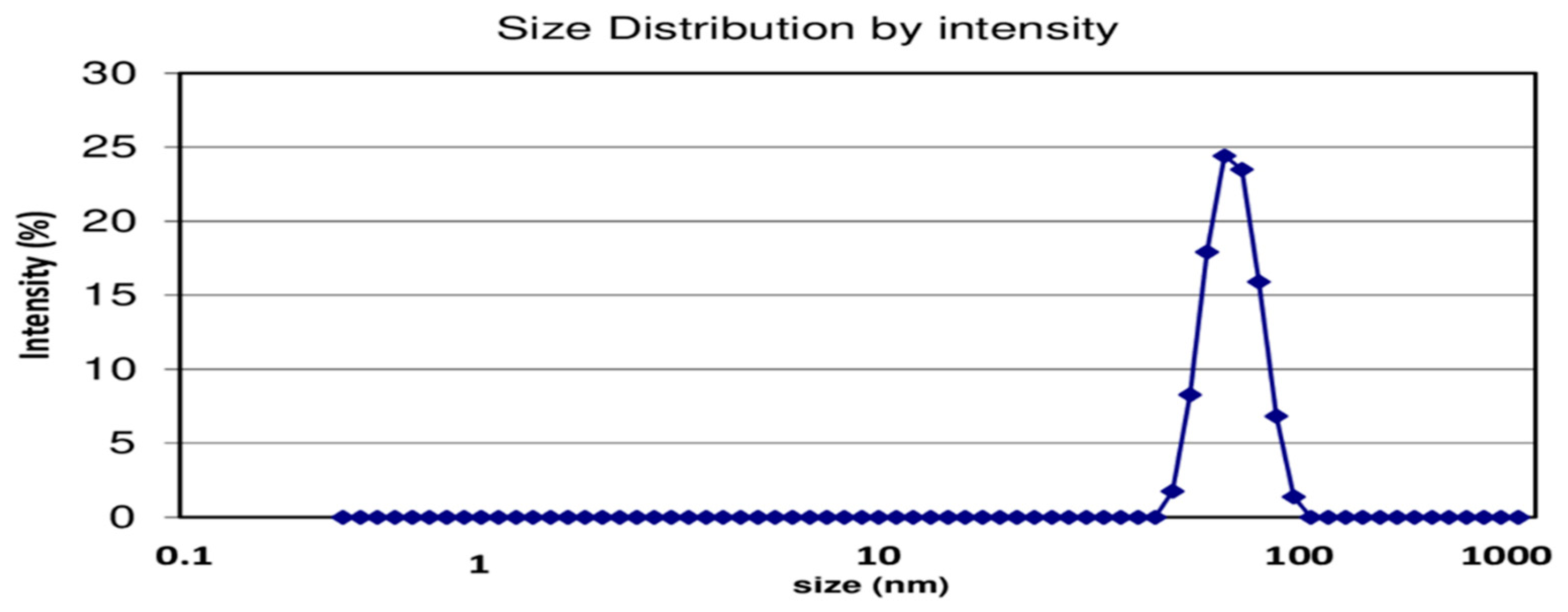
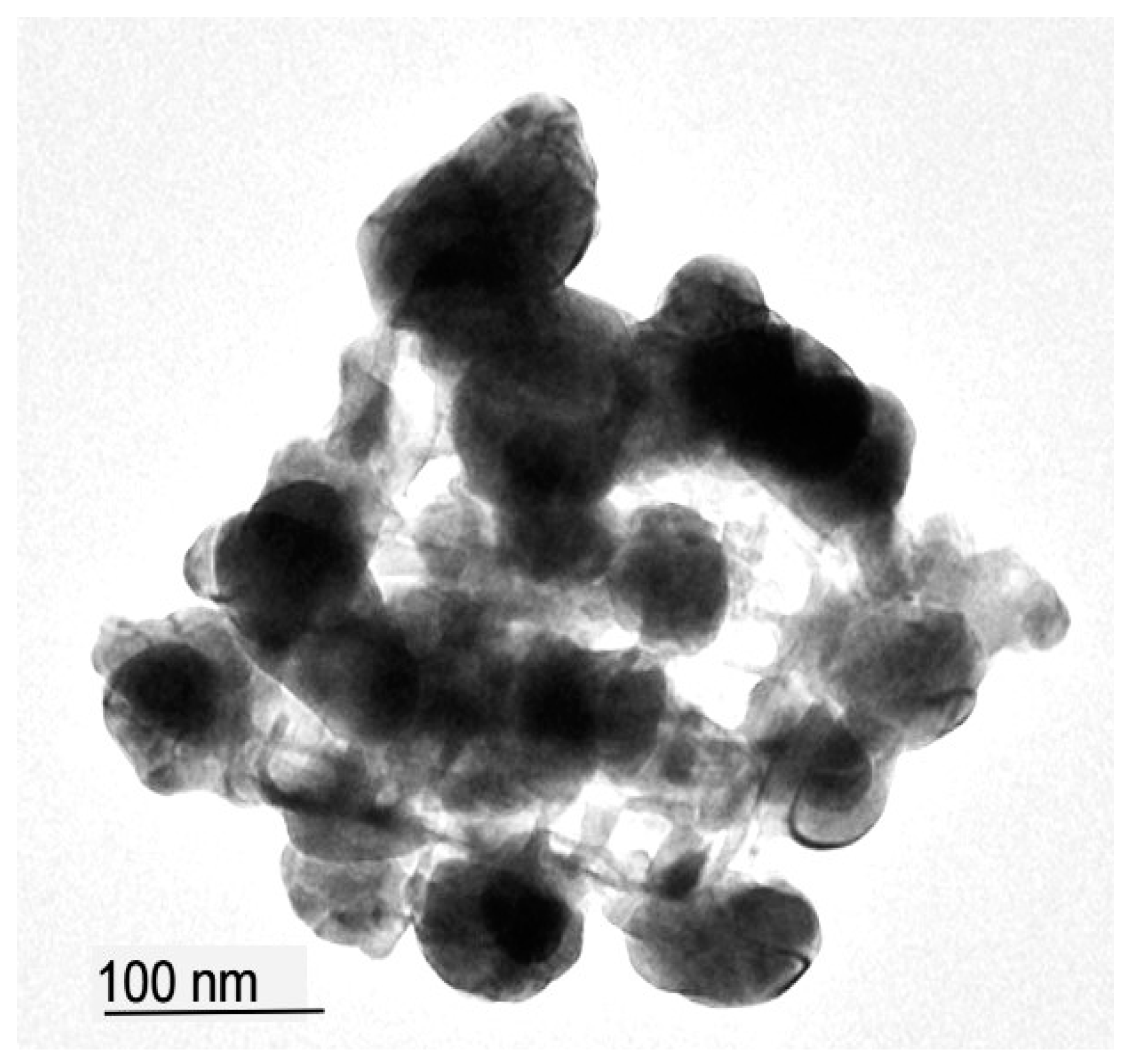
3.1. Synthesis and Characterization of NPs

The MLCNPs were formed as a milky color solution, and their powder form was attained by centrifugation. The synthesis of chitosan nanoparticles was confirmed through UV-visible spectroscopy, as shown in Figure 1. The reported UV-visible spectrum shows an absorption peak at 434 nm. The SEM analysis revealed that MLCNPs display a spherical shape and irregular surface morphology (Figure 2), while DLS show a mean particle size of 97.5 nm (Figure 3). The TEM micrograph of the nanoparticles is shown in Figure 4. It is clear from the TEM image that MLCNPs are spherical with a smooth surface. The synthesized particles possessed a substantial zeta potential of +1.04 mV (Figure 5). The percentage of drug-loaded chitosan was the DLC of 91.5 µg/mL, and the encapsulation efficiency was 97.56%.

3.2. In Vitro Drug Release Assay

The drug release concentration of the miltefosine drug from the nanoparticles was divided into two phases. In the initial stage, 30% of the drug was rapidly released from the NPs after 6 h at pH 7.4. In the second stage, the drug was constantly released from the nanoparticles, resulting in around 96% of the loaded drug up to 48 h (Figure 6).

3.3. Hemolysis Assay

The hemolysis activity of MLCNPs and conventional miltefosine was compared. The result showed that MLCNPs have 6% less hemolytic activity than conventional miltefosine. The OD values and hemolysis (%) are presented in Table 1.

3.4. MTT Assay

The cytotoxicity of conventional miltefosine and MLCNPs was assessed at six different concentrations (50, 40, 30, 20, 10 and 5 µg/mL) for 24, 48 and 72 h. The mean viability percentage of promastigotes exposed to different MLCNPs and conventional miltefosine concentrations is illustrated in Table 2. The IC50 value of MLCNPs (0.0218 µg/mL) was higher than the conventional miltefosine (0.3548 µg/mL).

3.5. In Vivo Study

At 4 weeks post-inoculation, when lesions were established, the mice were treated for 2 weeks, and a visible reduction in lesion size was noted, as shown in Figure 7. The mean and SD of each group’s pre- and post-treatment lesion sizes were determined, and p-values were determined as presented in Table 3. All the groups exhibited decreased lesion size after drug therapy except the placebo group. Among the groups, the orally administrated MLCNP group was considered significant (p = 0.01).
The Mann–Whitney test was performed for the lesion size comparison between the two groups. Post-treatment lesion comparisons of conventional miltefosine- and MLCNP (oral)-treated groups were statistically significant (p = 0.019). The p-value differences between pre- and post-treated groups are also statistically significant (p = 0.020), as shown in Table 4. Similarly, the amastigote count on the last day of treatment was significantly less under the light microscope than the pre-treated parasite burden. The comparison between conventional miltefosine-treated and intralesional-injected MLCNP-treated groups was also performed but detected as less effective. The p-value difference is 0.065 and considered insignificant (Table 5).
Furthermore, comparisons based on the route of administration of MLCNPs were performed. The difference in the p-value between the pre- and post-treated groups was 0.019, as calculated in Table 6. The results revealed that the oral route of administration for MLCNPs is much more significant than the intralesional route.

4. Discussion

The high toxicity and emergence of resistance against available drugs (antimonial) for leishmaniasis is a worldwide grave concern. Recently, various methods have been tried to develop alternative medicine against Leishmania species. Amphotericin B has been prepared among the novel approaches, but these drugs remain limited due to their high cost [18]. A nanoparticle-based drug delivery system is considered an essential tool for treating leishmaniasis due to its direct interaction with residing Leishmania parasites. Among the NP-based drug delivery systems, chitosan polymers are considered the best option due to their ideal characteristics such as an enhanced permeation, high uptake of the drug intracellularly, sustained release, increased biocompatibility and stability [19].
In the current study, chitosan NPs were synthesized by the ionotropic gelation method. Chitosan-based nanoparticles were broadly synthesized using the same technique as in [20]. The first reported chitosan nanoparticle preparation by the ionic gelation method was done in 1997 in Spain. Studies showed that chitosan nanoparticles synthesized by this method were relatively small, ranging from 100 nm to 150 nm in diameter [21]. The UV-visible spectrum was recorded for the determination of the optical properties of the chitosan polymer by using UV-visible spectroscopy. The peak observed for the chitosan was at 250 nm in the UV region. These results are in good agreement with the previous study. The DLS results of this study revealed the average size of MLCNPs to be 97.5 nm with a spherical shape and irregular surface morphology without aggregation. The SEM analysis of MLCNPs showed a spherical morphology with a smooth surface. Spherical nanoparticles were shown to incorporate maximum drug accumulation [22]. TEM images of the MLCNPs showed that they were spherical and about 90–98 nm in size. The concentration of chitosan, molecular weight and TPP significantly affected the size of the nanoparticles. By increasing the concentration of chitosan or TPP, the size of nanoparticles also increased [23]. Our TEM results align with the previously reported study [24].
The encapsulation efficiency of MLCNPs observed in our study was 97.56%. Our results align with other antileishmanial drugs encapsulated in chitosan nanoparticles prepared through the ionic gelation method [25].
Similarly, the drug loading content of MLCNPs was 91.5 µg/mL. This high amount of DLC may be due to the zwitterionic and amphiphilic nature of the drug used in this experiment [26]. The zeta potential of MLCNPs was +1.04 mV. In a previously published study, the zeta potentials of liposomal-based formulation were in the range of −8.2 ± 3.50 mV, which is less than the chitosan nanoparticles of the current study [27].
In vitro, the MTT colorimetric assay was performed to analyze the biological activity of MLCNPs and conventional miltefosine against L. tropica. In the current study, the IC50 value of conventional miltefosine was 0.3548 µg/mL, and in another study, it was reported to be 0.19 µg/mL [28]. In contrast, the MLCNPs reported in the present study were 0.0218 µg/mL, less than conventional miltefosine, indicating the high efficacy of MCLNPs. The hemolysis assay revealed that MLCNPs induced less hemolysis and were considered safer than conventional miltefosine. Our study’s hemolysis activity of conventional miltefosine was similar to an earlier reported study [29].
The effectiveness of miltefosine encapsulated in polymer nanoparticles was studied for treating CL in BALB/c mice by oral and intralesional routes of administration. The efficacy of conventional miltefosine (2.5 mg/kg/day) and our newly synthesized nanoparticles (MLCNPs) was compared. Although both the conventional miltefosine and MLCNPs were able to reduce the lesion size over time, the activity of MLCNPs was much better. It indicates that chitosan nanoparticles are targeted drug delivery systems, which transport miltefosine specifically to macrophages for anti-leishmanial activity. In the case of orally administrated conventional miltefosine, some quantity of the drug is left over in the body. A study reported that a high concentration of miltefosine efficiently reduced lesion size compared to a lower concentration of miltefosine [30]. Based on this, the higher efficacy of MLCNPs is because they provide a high concentration of miltefosine to infected macrophages compared to conventional miltefosine.

5. Conclusions

The current study describes a potential novel therapy for curing CL caused by L. tropica in the form of MLCNPs. The chitosan nanoparticles encapsulate the miltefosine efficiently. The overall results reveal that MLCNPs have higher efficacy than conventional miltefosine, which is proved both in vitro and in vivo. Regarding the route of administration, the efficacy of MLCNPs given orally was more significant than intralesional injections. Moreover, the hemolysis assay showed significantly less hemolysis activity of MLCNPs than conventional miltefosine. In summary, all these features make these newly synthesized nanoparticles an ideal drug delivery system and they might be used as an alternative therapeutic option for curing leishmaniasis.

Author Contributions

Conceptualization, R.U.K., H.B. and M.Z.K.; Methodology, Q.U. and A.S.; formal analysis, S.U., A.I. and M.K.; Investigation, R.I., R.U.K., S.U., Q.U., M.Z.K., A.I. and H.B.; Data curation, M.K., R.U.K., A.I., R.I., S.U. and A.S.; writing—original draft preparation, R.U.K., S.U., Q.U., M.K., R.I. and A.I.; Project administration, M.K., R.U.K. and H.B. All authors have read and agreed to the published version of the manuscript.

Funding

This research received no external funding.

Institutional Review Board Statement

The study was conducted in accordance with the Declaration of Helsinki. All experiments and procedures were approved by Ethics Committee of Institute of Basic Medical Sciences, Khyber Medical University Peshawar, Pakistan under reference No. Dir/KMU-EB/IV/000517.

Informed Consent Statement

Not applicable.

Data Availability Statement

Data will be available upon request.

Conflicts of Interest

The authors declare no conflict of interest.

References

  1. Ballart, C.; Torrico, M.C.; Vidal, G.; Torrico, F.; Lozano, D.; Gállego, M.; Pinto, L.; Rojas, E.; Aguilar, R.; Dobaño, C.; et al. Clinical and immunological characteristics of tegumentary leishmaniasis cases in Bolivia. PLoS Negl. Trop. Dis. 2021, 15, e0009223. [Google Scholar] [CrossRef]
  2. Garrido-Jareño, M.; Sahuquillo-Torralba, A.; Chouman-Arcas, R.; Castro-Hernández, I.; Molina-Moreno, J.M.; Llavador-Ros, M.; Gómez-Ruiz, M.D.; López-Hontangas, J.L.; Botella-Estrada, R.; Salavert-Lleti, M.; et al. Cutaneous and mucocutaneous leishmaniasis: Experience of a Mediterranean hospital. Parasites Vectors 2020, 13, 24. [Google Scholar] [CrossRef]
  3. Khan, R.U.; Khan, M.; Sohail, A.; Ullah, R.; Iqbal, A.; Ahmad, B.; Khan, I.U.; Tariq, A.; Ahmad, M.; Said, A.; et al. Efficacy of pentamidine-loaded chitosan nanoparticles as a novel drug delivery system for Leishmania tropica. Trop. Biomed. 2022, 39, 1–7. [Google Scholar]
  4. Alves, F.; Bilbe, G.; Blesson, S.; Goyal, V.; Monnerat, S.; Mowbray, C.; Muthoni Ouattara, G.; Pécoul, B.; Rijal, S.; Rode, J.; et al. Recent Development of Visceral Leishmaniasis Treatments: Successes, Pitfalls, and Perspectives. Clin. Microbiol. Rev. 2018, 31, e00048-18. [Google Scholar] [CrossRef]
  5. Khan, N.H.; Bari, A.U.; Hashim, R.; Khan, I.; Muneer, A.; Shah, A.; Wahid, S.; Yardley, V.; O’Neil, B.; Sutherland, C.J. Cutaneous Leishmaniasis in Khyber Pakhtunkhwa Province of Pakistan: Clinical Diversity and Species-Level Diagnosis. Am. J. Trop. Med. Hyg. 2016, 95, 1106–1114. [Google Scholar] [CrossRef] [PubMed]
  6. Mostafavi, M.; Farajzadeh, S.; Sharifi, I.; Khazaeli, P.; Sharifi, H. Leishmanicidal effects of amphotericin B in combination with selenium loaded on niosome against Leishmania tropica. J. Parasit. Dis. Off. Organ Indian Soc. Parasitol. 2019, 43, 176–185. [Google Scholar] [CrossRef] [PubMed]
  7. Neira, L.F.; Mantilla, J.C.; Escobar, P. Anti-leishmanial activity of a topical miltefosine gel in experimental models of New World cutaneous leishmaniasis. J. Antimicrob. Chemother. 2019, 74, 1634–1641. [Google Scholar] [CrossRef]
  8. Ware, J.M.; O’Connell, E.M.; Brown, T.; Wetzler, L.; Talaat, K.R.; Nutman, T.B.; Nash, T.E. Efficacy and Tolerability of Miltefosine in the Treatment of Cutaneous Leishmaniasis. Clin. Infect. Dis. Off. Publ. Infect. Dis. Soc. Am. 2021, 73, e2457–e2562. [Google Scholar] [CrossRef]
  9. Baranwal, A.; Chiranjivi, A.K.; Kumar, A.; Dubey, V.K.; Chandra, P. Design of commercially comparable nanotherapeutic agent against human disease-causing parasite, Leishmania. Sci. Rep. 2018, 8, 8814. [Google Scholar] [CrossRef] [PubMed]
  10. Oliveira, S.S.; Ferreira, C.S.; Branquinha, M.H.; Santos, A.L.; Chaud, M.V.; Jain, S.; Cardoso, J.C.; Kovačević, A.B.; Souto, E.B.; Severino, P. Overcoming multi-resistant leishmania treatment by nanoencapsulation of potent antimicrobials. J. Chem. Technol. Biotechnol. 2021, 96, 2123–2140. [Google Scholar] [CrossRef]
  11. Esboei, B.R.; Mohebali, M.; Mousavi, P.; Fakhar, M.; Akhoundi, B. Potent antileishmanial activity of chitosan against Iranian strain of Leishmania major (MRHO/IR/75/ER): In vitro and in vivo assay. J. Vector Borne Dis. 2018, 55, 111–115. [Google Scholar] [CrossRef]
  12. Lazaridou, M.; Christodoulou, E.; Nerantzaki, M.; Kostoglou, M.; Lambropoulou, D.A.; Katsarou, A.; Pantopoulos, K.; Bikiaris, D.N. Formulation and In-Vitro Characterization of Chitosan-Nanoparticles Loaded with the Iron Chelator Deferoxamine Mesylate (DFO). Pharmaceutics 2020, 12, 238. [Google Scholar] [CrossRef]
  13. Siripattanapipong, S.; Boontanom, P.; Leelayoova, S.; Mungthin, M.; Tan-Ariya, P. In vitro growth characteristics and morphological differentiation of Leishmania martiniquensis promastigotes in different culture media. Acta Trop. 2019, 197, 105039. [Google Scholar] [CrossRef]
  14. Sharma, M.; Sharma, R.; Jain, D.K.; Saraf, A. Enhancement of oral bioavailability of poorly water soluble carvedilol by chitosan nanoparticles: Optimization and pharmacokinetic study. Int. J. Biol. Macromol. 2019, 135, 246–260. [Google Scholar] [CrossRef] [PubMed]
  15. Sethi, A.; Ahmad, M.; Huma, T.; Khalid, I.; Ahmad, I. Evaluation of Low Molecular Weight Cross Linked Chitosan Nanoparticles, to Enhance the Bioavailability of 5-Flourouracil. Dose-Response 2021, 19, 15593258211025353. [Google Scholar] [CrossRef]
  16. Esfandiari, F.; Motazedian, M.H.; Asgari, Q.; Morowvat, M.H.; Molaei, M.; Heli, H. Paromomycin-loaded mannosylated chitosan nanoparticles: Synthesis, characterization and targeted drug delivery against leishmaniasis. Acta Trop. 2019, 197, 105045. [Google Scholar] [CrossRef]
  17. Riaz, A.; Hendricks, S.; Elbrink, K.; Guy, C.; Maes, L.; Ahmed, N.; Kiekens, F.; Khan, G.M. Preparation and Characterization of Nanostructured Lipid Carriers for Improved Topical Drug Delivery: Evaluation in Cutaneous Leishmaniasis and Vaginal Candidiasis Animal Models. AAPS PharmSciTech 2020, 21, 185. [Google Scholar] [CrossRef]
  18. Valle, I.V.; Machado, M.E.; Araújo, C.; da Cunha-Junior, E.F.; da Silva Pacheco, J.; Torres-Santos, E.C.; da Silva, L.; Cabral, L.M.; do Carmo, F.A.; Sathler, P.C. Oral pentamidine-loaded poly(d,l-lactic-co-glycolic) acid nanoparticles: An alternative approach for leishmaniasis treatment. Nanotechnology 2019, 30, 455102. [Google Scholar] [CrossRef] [PubMed]
  19. Hadidi, M.; Pouramin, S.; Adinepour, F.; Haghani, S.; Jafari, S.M. Chitosan nanoparticles loaded with clove essential oil: Characterization, antioxidant and antibacterial activities. Carbohydr. Polym. 2020, 236, 116075. [Google Scholar] [CrossRef] [PubMed]
  20. Ashvini, H.; Balla, A.; Mutta, S. Clarithromycin-loaded chitosan nanoparticles: Preparation, characterisation and antibacterial activity on Streptococcus pneumonia. Indian J. Pharm. Sci. 2019, 81, 302–308. [Google Scholar] [CrossRef]
  21. Grenha, A.; Seijo, B.; Serra, C.; Remuñan-López, C. Chitosan nanoparticle-loaded mannitol microspheres: Structure and surface characterization. Biomacromolecules 2007, 8, 2072–2079. [Google Scholar] [CrossRef]
  22. Varshosaz, J.; Arbabi, B.; Pestehchian, N.; Saberi, S.; Delavari, M. Chitosan-titanium dioxide-glucantime nanoassemblies effects on promastigote and amastigote of Leishmania major. Int. J. Biol. Macromol. 2018, 107, 212–221. [Google Scholar] [CrossRef]
  23. Shawer, R.; El-Leithy, E.S.; Abdel-Rashid, R.S.; Eltaweil, A.S.; Baeshen, R.S.; Mori, N. Preparation of Lambda-Cyhalothrin-Loaded Chitosan Nanoparticles and Their Bioactivity against Drosophila suzukii. Nanomaterials 2022, 12, 3110. [Google Scholar] [CrossRef]
  24. Chandra Hembram, K.; Prabha, S.; Chandra, R.; Ahmed, B.; Nimesh, S. Advances in preparation and characterization of chitosan nanoparticles for therapeutics. Artif. Cells Nanomed. Biotechnol. 2016, 44, 305–314. [Google Scholar] [CrossRef]
  25. Sohail, A.; Khan, R.U.; Khan, M.; Khokhar, M.; Ullah, S.; Ali, A.; Bilal, H.; Khattak, S.; Khan, M.; Ahmad, B. Comparative efficacy of amphotericin B-loaded chitosan nanoparticles and free amphotericin B drug against Leishmania tropica. Bull. Natl. Res. Cent. 2021, 45, 187. [Google Scholar] [CrossRef]
  26. Khokhar, M.; Shereen, M.A.; Khan, M.; Khan, R.U.; Sohail, A.; Khan, I.U.; Khan, I.U.; Khattak, S. In vitro efficacy of polymer coated miltefosine drug against leishmania tropica. J. Parasit. Dis. Off. Organ Indian Soc. Parasitol. 2022, 46, 366–376. [Google Scholar] [CrossRef]
  27. Gürbüz Çolak, N.; Çetin Uyanikgil, E.; Özbel, Y.; Töz, S. The Designing of a Gel Formulation with Chitosan Polymer Using Liposomes as Nanocarriers of Amphotericin B for a Non-invasive Treatment Model of Cutaneous Leishmaniasis. Acta Parasitol. 2022, 67, 1354–1363. [Google Scholar] [CrossRef]
  28. Ali, H.Z. Cytotoxicity of Miltefosine against Leishmania major Promastigotes. Adv. Biores. 2012, 3, 90–94. [Google Scholar]
  29. Mahmoudzadeh-Niknam, H.; Khalili, G.; Abrishami, F.; Najafy, A.; Khaze, V. The route of Leishmania tropica infection determines disease outcome and protection against Leishmania major in BALB/c mice. Korean J. Parasitol. 2013, 51, 69–74. [Google Scholar] [CrossRef] [PubMed]
  30. Rebello, K.M.; Andrade-Neto, V.V.; Gomes, C.R.B.; de Souza, M.V.N.; Branquinha, M.H.; Santos, A.L.S.; Torres-Santos, E.C.; d’Avila-Levy, C.M. Miltefosine-Lopinavir Combination Therapy Against Leishmania infantum Infection: In vitro and in vivo Approaches. Front. Cell. Infect. Microbiol. 2019, 9, 229. [Google Scholar] [CrossRef] [PubMed]
Figure 1. UV-visible spectrum of chitosan nanoparticles.
Msf 21 00019 g001
Figure 2. SEM images of the MLCNPs with spherical morphology magnified 20,000× (a) and 30,000× (b).
Msf 21 00019 g002
Figure 3. Average size distribution intensity of MLCNPs using dynamic light scattering technique.
Msf 21 00019 g003
Figure 4. TEM image of the MLCNPs. Scale bar—100 nm.
Msf 21 00019 g004
Figure 5. Zeta potential of MLCNPs.
Msf 21 00019 g005
Figure 6. In vitro miltefosine release pattern from chitosan nanoparticles at pH 7.4.
Msf 21 00019 g006
Figure 7. L. tropica-infected BALB/c mice; (a) lesion development after 4 weeks of inoculation and (b) reduction of lesion size after 2 weeks of MLCNP treatment.
Msf 21 00019 g007
Table 1. Hemolysis assay of MLCNPs and conventional miltefosine drug.
Scheme 570ConcentrationsOD at 570 nm (Mean ± SD)Hemolysis (%)
MLCNPs150 µg/mL0.032 ± 0.00251.60%
200 µg/mL0.043 ± 0.00152.45%
250 µg/mL0.051 ± 0.00103.20%
Con MFS150 µg/mL0.062 ± 0.00205.96%
200 µg/mL0.075 ± 0.00156.92%
250 µg/mL0.082 ± 0.00307.40%
PBS-0.003 ± 0.00050%
Triton 100×-1.223 ± 0.0251100%
Table 2. Mean viability percentages for promastigotes exposed to different concentrations of MLCNPs and conventional miltefosine.
Concentrations 24 h48 h72 h
MLCNPsCon MFSMLCNPsCon MFSMLCNPsCon MFS
50 µg/mL16 ± 4.228 ± 1.114 ± 0.721 ± 0.710 ± 0.318 ± 1.3
40 µg/mL17 ± 2.530 ± 2.0516 ± 0.325 ± 1.812 ± 0.820 ± 2.0
30 µg/mL20.5 ± 4.533 ± 3.0818 ± 1.130 ± 0.915 ± 1.825 ± 1.5
20 µg/mL24 ± 4.144 ± 1.821 ± 1.540 ± 1.523 ± 2.530 ± 0.5
10 µg/mL31.5 ± 3.452 ± 1.626 ± 1.645 ± 1.425 ± 2.141 ± 1.3
05 µg/mL35 ± 3.860 ± 1.430 ± 1.352 ± 0.929 ± 1.644 ± 1.5
Miltefosine. Data are expressed as the mean ± SD (n = 3).
Table 3. Lesion size (mean ± SD) comparison before and after treatment of each group.
GroupsNBefore TreatmentAfter Treatmentp-Value
Con MFS46.9 (±0.26) mm6.6 (±0.23) mm0.09
MLCNPs (oral)46.9 (±0.46) mm5.9 (±0.33) mm0.01
MLCNPs (IL)47.2 (±0.50) mm6.4 (±0.45) mm0.06
Placebo46.8 (±0.50) mm7.2 (±0.29) mm0.24
Table 4. Lesion size comparison of the conventional miltefosine group and MLCNP (oral) group.
Lesion SizeGroupsMean RankSum of Ranksp-Valuep-Value
Difference
Before (Trt)Con MFS4.6318.500.8850.020
MLCNPs (oral)4.3817.50
After (Trt)Con MFS6.5026.000.019
MLCNPs (oral)2.5010.00
Table 5. Lesion size comparison of conventional miltefosine group and MLCNP (intralesional) group.
Lesion SizeGroupsMean RankSum of Ranksp-Valuep-Value
Difference
Before (Trt)Con MFS4.6318.500.8850.065
MLCNPs (IL)4.3817.50
After (Trt)Con MFS4.8819.500.661
MLCNPs (IL)4.1316.50
Table 6. Lesion size comparison of MLCNP (oral) and MLCNP (intralesional) groups.
Lesion SizeGroupsMean RankSum of Ranksp-Valuep-Value
Difference
Before (Trt)MLCNPs (oral)4.2517.000.7700.019
MLCNPs (IL) 4.7519.00
After (Trt)MLCNPs (oral)3.1312.500.108
MLCNPs (IL) 5.8823.50
Disclaimer/Publisher’s Note: The statements, opinions and data contained in all publications are solely those of the individual author(s) and contributor(s) and not of MDPI and/or the editor(s). MDPI and/or the editor(s) disclaim responsibility for any injury to people or property resulting from any ideas, methods, instructions or products referred to in the content.

