Proceeding Paper

Novel Natural and Synthetic Anticandidal Therapeutic Peptides to Combat Drug-Resistant Infections †

Department of Biotechnology, CGC Mohali, Punjab 140307, India
Presented at the 3rd International Electronic Conference on Antibiotics (ECA 2023), 1–15 December 2023; Available online: https://eca2023.sciforum.net/.
Med. Sci. Forum 2024, 24(1), 12; https://doi.org/10.3390/ECA2023-16385
Published: 30 November 2023
(This article belongs to the Proceedings of The 3rd International Electronic Conference on Antibiotics)

Abstract

:
Candida species are considered as common flora of the healthy human mycobiome and occur on skin, mucosal surfaces of gastrointestinal and genitourinary tracks. Pathogenic Candida spp., reported to cause skin, vaginal and oral infections. Extensive use of antifungal agents has increased the drug resistance among pathogenic strains of Candida. To this effect, recently naturally occurring AMPs and synthetically modified peptides, are effectively being used as promising antifungal agents. Short peptides display better permeability to cross the yeast membrane, thus short antifungal peptide were designed using sequences from APD database. Natural peptides are potential source of antifungal agents. Considering above facts, we studied anticandidal potential of synthetic and natural peptides.

1. Introduction

The increasing drug resistance seen in fungal pathogenic strains in recent years, specifically in clinical strains, requires immediate attention so that alternative antifungal agents can be developed. Among the fungal genera, members of the genus Candida are the most common causal organisms of human infection. They usually reside as a commensal in the genitourinary and gastrointestinal tracts as well as in the oral and conjunctival flora [1], causing both superficial and invasive infections under immunocompromised conditions. Superficial infection are known as candidiasis, which affects the mucous membranes or skin and is usually treated with topical antifungal drugs, with a low success rate. However, invasive fungal infections have recently been reported to be life-threatening due to inefficient prognostic methods and unsuitable antifungal therapies. Only three classes of conventional antifungal drugs, viz. fluconazole, caspofungin and amphotericin B, are used extensively for candidiasis treatment [2]. However, there are Candida strains which have been reported to be increasingly resistant to these antibiotics [3,4]. This increased occurrence of drug-resistant candidiasis desperately requires alternative antifungal agents so the resistance problem can be overcome.
Nowadays, various natural and synthetic antimicrobial peptides (AMPs) have been reported to inhibit Candida spp. and are considered promising alternative candidates for the treatment of drug-resistant Candida spp. Infections [5,6,7,8]. Naturally occurring AMPs generally consist of 10–50 amino acids with different structural groups, including sheet, helix, extended and looped structures [9]. Though some peptides, such as melittin and protegrin, exhibit potent activity, they also possess toxic effects on mammalian cells. Hence, researchers have also focused on synthetic antimicrobial peptides with enhancement activity, reduced cytotoxicity and resistance to protease enzymes. The main objective of the present study was to use available AMP sequences to construct short peptides with anticandidal properties and resistance to environmental degradation. Effective peptide modifications were included to enhance antimicrobial potency against Candida spp. and low toxicity towards mammalian cells. The minimum inhibitory concentration of peptides towards pathogenic Candida species and their biofilms as well as their hemolysis ability was studied.

2. Methodology

2.1. Peptide Design and Synthesis

Peptides were designed using peptide sequences from the APD database [10]. The antifungal activity predictive tool Antifp [11] was used to predict their antifungal potential in terms of protein binding potential, amphipathicity and charge. Peptide helical wheel projections were analyzed using the bioinformatics program Netwheels [12] (Castro et al., 2018). Peptides were synthesized by CSIR-IMTECH, Chandigarh, in solid phase using the Fmoc methodology. All peptides were >90% pure (as determined by RP-HPLC) and had the expected molecular weight.

2.2. Test Organisms

The reference Candida strains used in this study were procured from the Microbial Type Culture Collection and Gene Bank (MTCC), CSIR-Institute of Microbial Technology (IMTECH), Chandigarh, India, and clinical isolates were provided by the National Culture Collection of Pathogenic Fungi (NCCPF), Postgraduate Institute of Medical Education and Research, Chandigarh, India. The test strains used were Candida albicans (MTCC 184), C. albicans (MTCC 227) and one clinical isolate of C. albicans (400054). All strains were maintained on yeast malt agar (YMA), subcultured regularly (every 30 days or so) and stored at 4 °C. Glycerol stocks were preserved at −80 °C. The anticandidal activity of peptides was analyzed using agar well diffusion assay.

2.3. Biofilm Formation

A test strain biofilm was formed on pre-sterilized 96-well flat-bottom polystyrene microtiter plates in triplicate. The overnight grown culture OD600 was adjusted to 0.4 and 10 µL of cell suspension was inoculated in 190 µL YM broth in each well. The microtiter plate was incubated for 24–48 h at 37 °C. After incubation, the medium was removed by inverting the plate and any other planktonic cells present were removed by gentle flush with sterile distilled water. Then, 200 μL of crystal violet solution (0.2%) was added to all wells. After 15 min, the excess crystal violet was removed and plates were washed twice and air-dried. Finally, the cell-bound crystal violet was dissolved in 70% ethanol. Biofilm growth was monitored in terms of OD595 nm using an ELISA microplate reader (Thermo, Waltham, MA, USA). Antifungal agents, including fluconazole and amphotericin B, were used as controls in this study.

2.4. Purification and Characterization

For the characterization of the antimicrobial peptide produced by the Bacillus sp. strain, the SVDS-15 culture was grown in 1000 mL of NB for 24 h on a rotary shaker at 30 °C. Subsequently, cells were separated by centrifuge and the peptide was subsequently eluted in methanol. The peptide was redissolved in Milli-Q water and subjected to gel filtration (G50 Sephadex). Further purification was achieved using HPLC (1260 Infinity, Agilent Technologies, Santa Clara, CA, USA) with a semi-preparative C18 column (Pursuit 10C18 250 × 21.2 mm) with acetonitrile and aqueous trifluoroacetic acid as a solvent system. The molecular mass of the HPLC-purified peptide was analyzed using matrix-assisted laser desorption ionization (MALDI) [13]. A de novo sequence was generated manually using a fragmentation pattern.

2.5. Determination of Antimicrobial Activity and MIC Values of Peptides

The MICs of purified peptides were evaluated by using a microtiter plate dilution assay. Test strains were grown to mid-log phase (5 × 10⁵ CFU/mL) in a 96-well plate with different concentrations of peptides for 24–48 h at 37 °C (final volume of 200 μL). OD600 was measured after 24–48 h using an ELISA microplate reader (Thermo Fisher Scientific, Waltham, MA, USA). The lowest concentration that inhibited the growth of the test strain and did not show any increase in absorption after 48 h was considered as the MIC. Antifungal agents such as fluconazole and amphotericin B were included in this study as controls.

2.6. Hemolysis and Time-Kill Assay

Hemolysis assay was performed using rabbit blood (New Zealand white). For the experiment, the blood was centrifuged and the cells were washed and resuspended in phosphate-buffered saline (PBS). Different concentrations of peptide (50, 100 and 200 μg/mL) were mixed with blood cells and adjusted to a concentration of 2 × 10⁸ cells/mL in PBS. Triton X-100 was used as a positive control. Centrifuge tubes were incubated in a CO2 incubator at 37 °C and readings were recorded at different time intervals (1 h, 12 h and 24 h).
A time-kill assay of the purified peptide was performed [14]. C. albicans (MTCC 183) culture of 0.2 OD600 was centrifuged, washed and resuspended in PBS. The culture was treated with 5X concentration of AMP. After treatment, pellet cells were dried, coated with gold and observed under a scanning electron microscope (ZEISS, Jena, Germany) [15].

2.7. Emulgel Formulation and Skin Irritation Studies

For emulgel formulation, a gel base was prepared using carbopol 934 (Hi-media, Thane, India). The oil-phase emulsion contained cetomacrogol 1000, white soft paraffin, cetostearyl alcohol, light liquid paraffin and propylene glycol. The oil phase and an aqueous phase containing 0.5% w/v of purified peptide were mixed together for emulgel preparation [14]. The viscosity of the gel, its extrudability, appearance and pH were determined. The antimicrobial activity of the peptide was performed in vitro before performing skin irritation studies on mice.
For skin irritation studies of the SVDS-15 formulation, BALB/c female mice (eight weeks old) were used [16]. A plain gel and a 20% SLS solution were used as negative and positive controls, respectively. The SVDS-15 formulation was topically applied to a hairless skin area of the mice (approximately 1 cm2). The experiment results were recorded at 24, 48 and 72 h.

3. Results and Discussion

3.1. Design and Synthesis of Peptides

In this study, the amino acid sequences of peptides were used as a framework to design short antimicrobial peptides. The effect of positively charged residue distribution on the biological viability of the antimicrobial peptide was studied by replacing several amino acid residues with Lys (K) to increase a net positive charge of the peptide. Short peptides containing 10–17 amino acids in their composition are known to display better permeability to cross the yeast membrane [17]. The designed peptides varied in total net positive charge, which ranged from +2 to +5. The peptide sequences and their characteristics, such as hydrophobicity, amphipathicity, hydrophilicity and charge were analyzed; details are listed in Table 1.
Further, the amphipathic orientation of the amino acids present in these synthetic peptides was determined using helical wheel projection, which allowed for the understanding of possible in silico interactions with the membrane.

3.2. Growth and Biofilm Optimization

All Candida strains were grown in YM broth containing varying concentrations of glucose (0.2–1%) and incubated for 24–48 h at 37 °C for biofilm studies. After incubation, the plates were stained with crystal violet. All strains grew well in YM broth containing glucose and formed excellent biofilms, with ODs ranging from 0.8 to 1.5. Glucose presence affected the biofilm formation of different MTCC strains as well as of clinical strains. Maximum biofilm formation was achieved by using 0.5% glucose in YM broth. C. albicans strains consistently formed more biofilm than other species.

3.3. Effect of Synthetic Peptides on Growth and Biofilm

In order to determine the effect of the synthetic peptides on and biofilm growth and inhibition, Candida strains with the ability to form biofilms were treated with increasing concentrations of peptides (50–500 μg/mL). SK01, SK03 and SK07 showed an inhibition of biofilms formed by MTCC and clinical strains. Increasing concentrations of the peptide resulted in an increased disruption of biofilms and complete biofilm inhibition was observed with concentrations ranging from 200 to 800 μg/mL. Synthetic peptide SK08 and pep2 showed good anticandidal activity against various test strains. The MIC values against Candida albicans (MTCC 184), C. albicans (MTCC 227) and C. albicans (400054) were 200, 400 and 1000 μg/mL, respectively. Recently, three synthetic peptides (PNR20, PNR20–1 and 35,409) have been reported to exhibit antifungal activity against various Candida spp. [18] (Torres et al., 2023). Synthetic peptides KU2 and KU3 showed MIC values ranging from 8 to 16 mg/L [6].

3.4. Characterization of SVDS-15 Peptide

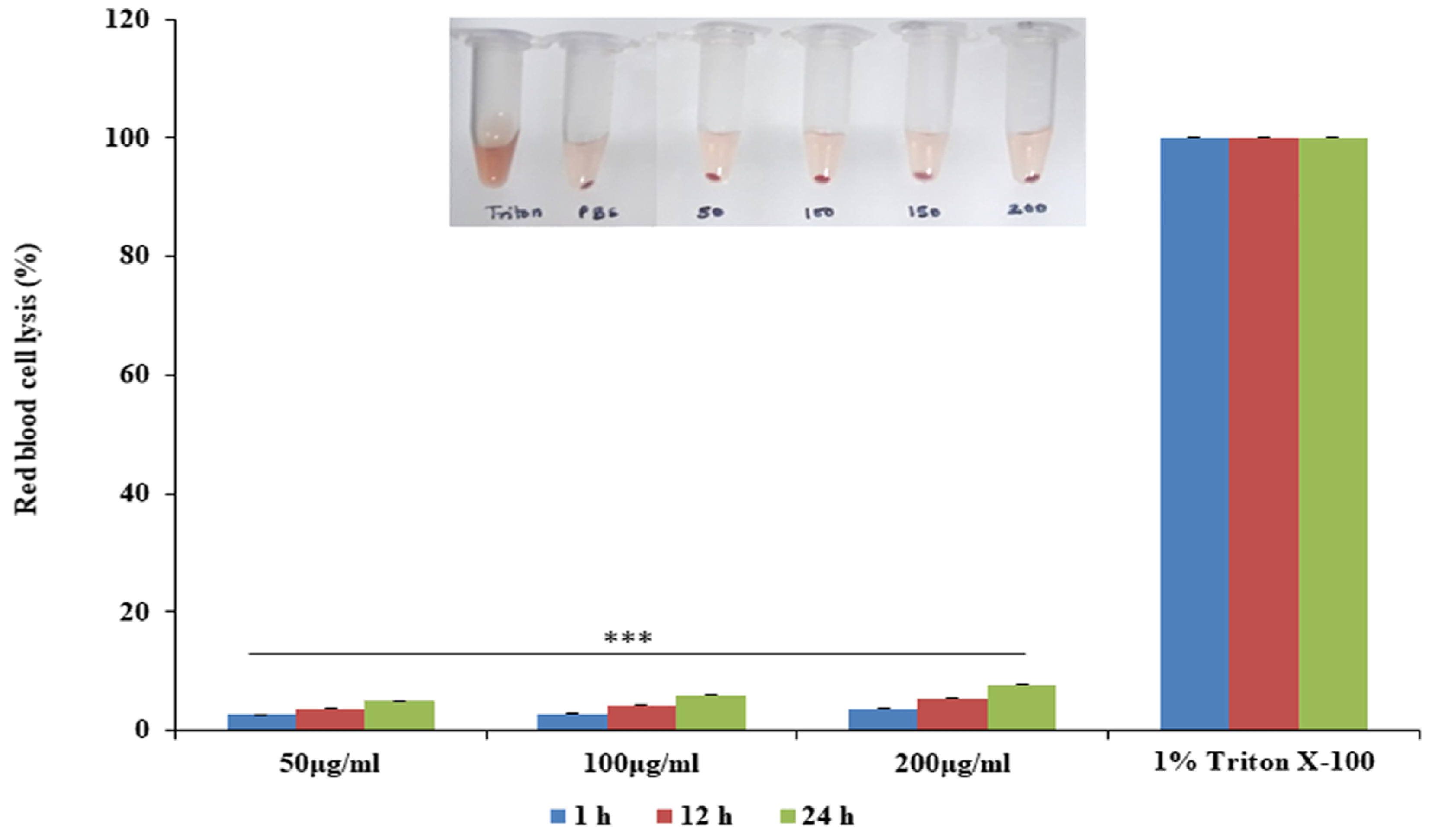
The antimicrobial peptide-producing Bacillus sp. strain SVDS-15 was selected for this study as it showed strong antimicrobial activity against all Candida strains. Considering the anticandidal potential of the peptide, further purification was carried out from CFB using a combination of chromatographic techniques. The SVDS-15 peptide showed very low MIC values against the test strains. Its MIC values against Candida albicans (MTCC 184), C. albicans (MTCC 227) and Candida (400054) were 12, 20 and 30 μg/mL, respectively. MALDI mass spectrometry of HPLC-purified SVDS-15 peptide revealed that it has a molecular mass as 1296 Da and belongs to the loloatin antimicrobial class (Figure 1). Loloatins have been reported to exhibit in vitro antimicrobial activity against methicillin-resistant Staphyloccoccus aureus, vancomycin-resistant enterococci and drug-resistant Streptococcus pneumoniae [19]. Moreover, the purified SVDS-15 peptide was found to be non-hemolytic in nature. Consequently, it could be used as a potent therapeutic agent. The peptide was found to be non-hemolytic in nature as it did not cause the lysis of blood cells (Figure 2).

3.5. Scanning Electron Microscopy of SVDS-15 Peptide

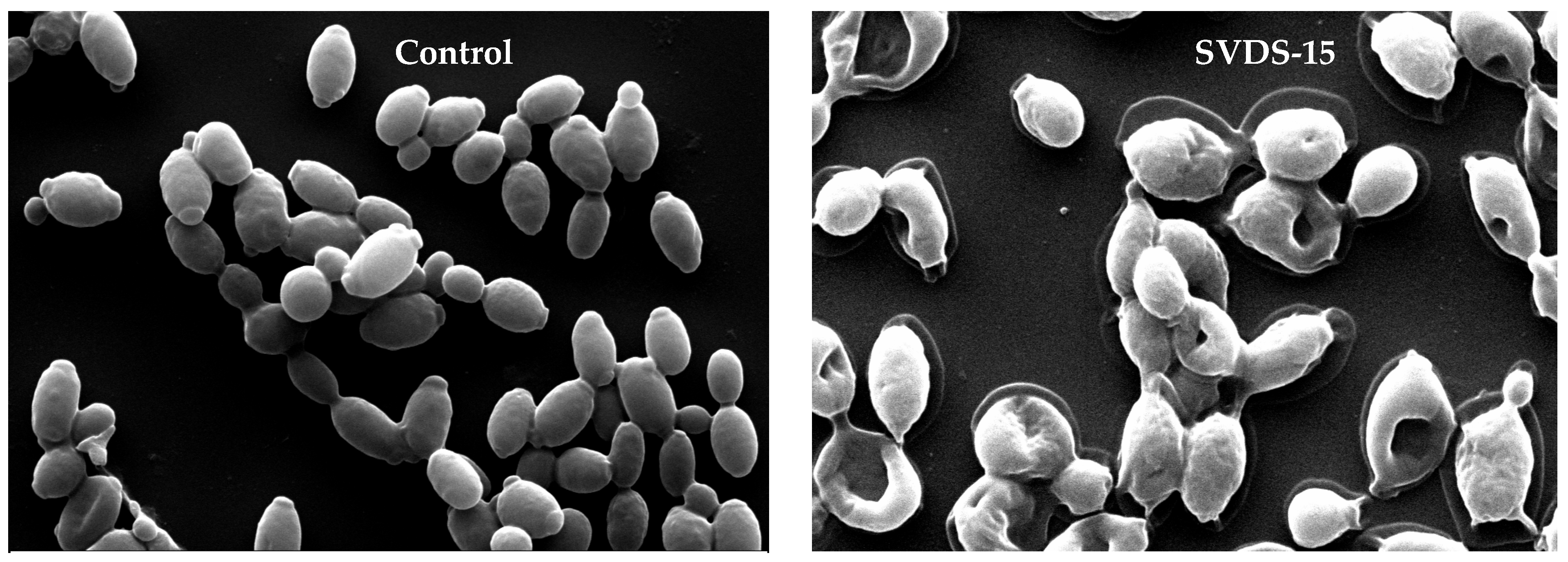
SEM images of C. albicans (MTCC 183) cells treated with SVDS-15 revealed that the peptide disintegrated the cell membranes of cells and caused the dispersion of intracellular contents. However, the control cell membrane was intact and the cytoplasm was homogenous after peptide treatment (Figure 3). Similar observations of cell membrane lysis and release of cell content by peptide treatment has been reported earlier [14,20].

3.6. Emulgel Formulation of Peptides

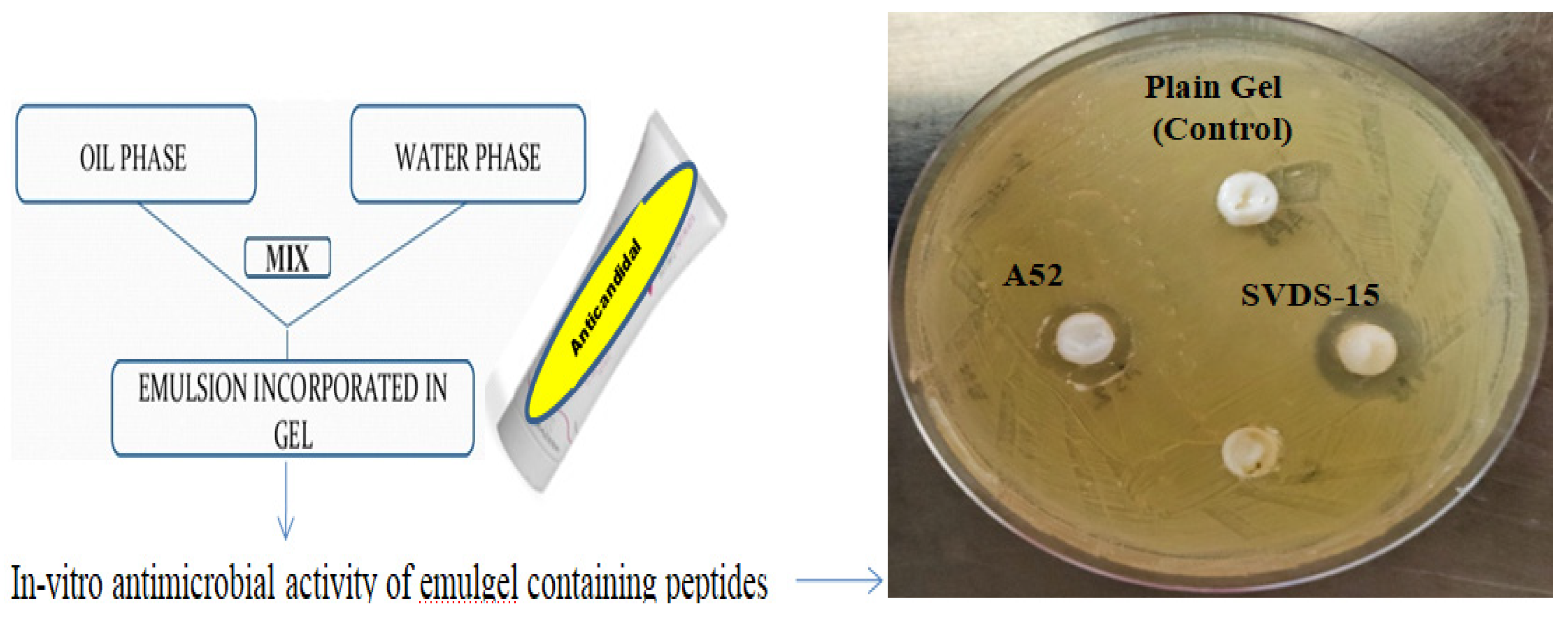
The optimized peptide emulgel contained cetomacrogol 1000 (2.5%), propylene glycol (15.0%), white soft paraffin (10.0%), cetostearyl alcohol (7.0%), liquid paraffin (2.5%), carbopol 934 (0.5%), isopropyl alcohol (1.5%) vitamin E TPGS (5.0%) and water (55.5%). The emulgel formulations showed in vitro antimicrobial activity against C. albicans MTCC 183 (Figure 4). The emulgel formulation was creamy and opaque in appearance, with a pH of 6.8 ± 0.2 and good extrudability. Individual component testing was also performed and they did not exhibit any activity against C. albicans (MTCC 183) cells. Emulgel formulations of antimicrobial peptides have been successfully prepared and reported [14,21,22]. The viscosity of the emulgels was found to be 1451 and 1522cP for A52 and SVDS-15, respectively.

3.7. Skin Irritation Study of Emulgels

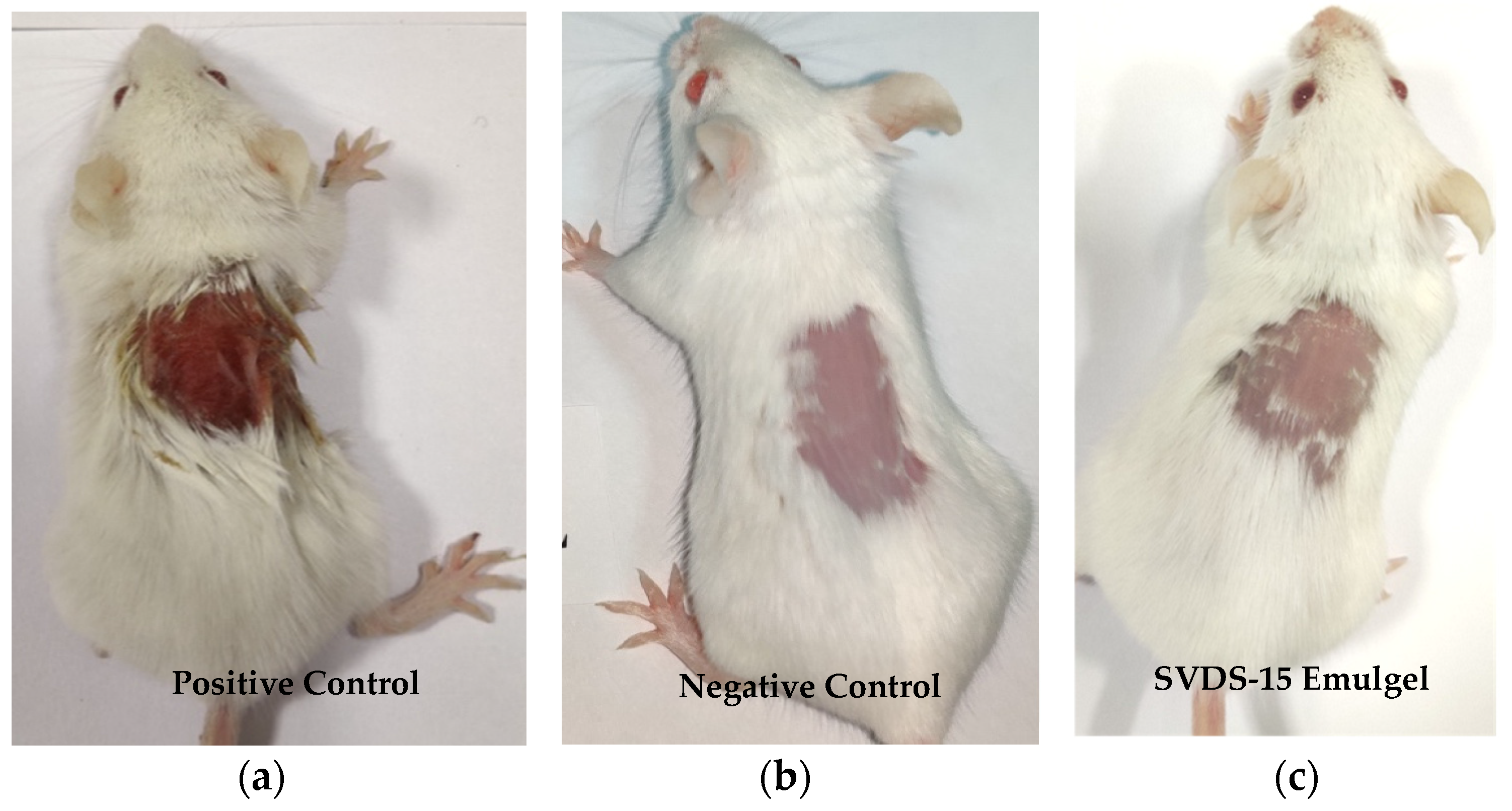
The skin irritancy results of the emulgel formulation were determined using the Draize test and the results showed that the emulgel formulation did not exhibit any skin irritation, even at 72 h after application. However, the positive (SLS treated) control caused hardening, redness and dryness in mouse skin (Figure 5). Many natural peptides which show no toxicity towards animals have been reported in previous studies [20,23]. The results of the skin irritation study showed that our emulgel formulations can be safely applied on the skin.

4. Conclusions

The increasing drug resistance of pathogenic fungal strains, specifically of Candida strains, requires immediate attention for the development of alternative antifungal agents. The main objective of this study was to develop potent antifungal agents using synthetic as well as natural peptides. The SVDS-15 peptide was successfully purified and its emulgel was developed for topical application. Synthetic (SK08 and pep2) and natural (SVDS-15) peptides could be used as potent therapeutic agents against various resistant Candida strains.

Funding

This research was supported by grant from the Department of Science and Technology (DST), New Delhi, India (SR/WOS-A/LS-220/2017).

Institutional Review Board Statement

Institutional Animal Ethics Committee for the Purpose of Control and Supervision of Experiments on Animals has reviewed and approved study. The approval number of the experiment is 18/08.

Informed Consent Statement

Not applicable.

Data Availability Statement

Data sharing is not applicable to this article.

Acknowledgments

Research support from the Department of Science and Technology (India) and the guidance of Suresh Korpole, CSIR-IMTECH Chandigarh (India), are duly acknowledged.

Conflicts of Interest

The author declare no conflict of interest.

References

  1. Naglik, J.R.; Moyes, D.L.; Wächtler, B.; Hube, B. Candida albicans interactions with epithelial cells and mucosal immunity. Microbes Infect. 2011, 13, 963–976. [Google Scholar] [CrossRef] [PubMed]
  2. Lum, K.; Tay, S.; Le, C.F.; Lee, V.S.; Sabri, N.H.; Velayuthan, R.D.; Hassan, H.; Sekaran, S.D. Activity of Novel Synthetic Peptides against Candida albicans. Sci. Rep. 2015, 5, 9657. [Google Scholar] [CrossRef] [PubMed]
  3. Eksi, F.; Gayyurhan, E.D.; Balci, I. In vitro susceptibility of Candida species to four antifungal agents assessed by the reference broth microdilution method. Sci. World J. 2013, 2013, 236903. [Google Scholar] [CrossRef] [PubMed]
  4. Ademe, M.; Girma, F. Candida auris: From multidrug resistance to pan-resistant strains. Infect Drug Resist. 2020, 13, 1287–1294. [Google Scholar]
  5. Singh, P.K.; Sharma, S.; Kumari, A.; Korpole, S. A non-pediocin low molecular weight antimicrobial peptide produced by Pediococcus pentosaceus strain IE-3 shows increased activity under reducing environment. BMC Microbial. 2014, 14, 226. [Google Scholar] [CrossRef]
  6. Freitas, C.G.; Felipe, M.S. Candida albicans and Antifungal Peptides. Infect. Dis. Ther. 2023, 12, 2631–2648. [Google Scholar] [CrossRef]
  7. Raman, N.; Lee, M.R.; Lynn, D.M.; Palecek, S.P. Antifungal activity of 14-helical β-peptides against planktonic cells and biofilms of Candida species. Pharmaceuticals 2015, 8, 483–503. [Google Scholar] [CrossRef]
  8. Lyu, Y.; Yang, Y.; Lyu, X.; Dong, N.; Shan, A. Antimicrobial activity, improved cell selectivity and mode of action of short PMAP-36-derived peptides against bacteria and Candida. Sci. Rep. 2016, 6, 27258. [Google Scholar] [CrossRef]
  9. Zasloff, M. Antimicrobial peptides of multicellular organisms. Nature 2002, 415, 389–395. [Google Scholar] [CrossRef] [PubMed]
  10. Wang, G.; Li, X.; Wang, Z. APD3: The antimicrobial peptide database as a tool for research and education. Nucleic Acids Res. 2016, 44, D1087–D1093. [Google Scholar] [CrossRef] [PubMed]
  11. Agrawal, P.; Bhalla, S.; Chaudhary, K.; Kumar, R.; Sharma, M.; Raghava, G.P.S. In Silico Approach for Prediction of Antifungal Peptides. Front Microbiol. 2018, 9, 323. [Google Scholar] [CrossRef]
  12. Mól, A.R.; Castro, M.S.; Fontes, W. NetWheels: A web application to create high quality peptide helical wheel and net projections. bioRxiv 2018, 416347. [Google Scholar] [CrossRef]
  13. Baindara, P.; Singh, N.; Ranjan, M.; Nallabelli, N.; Chaudhry, V.; Pathania, G.L.; Sharma, N.; Kumar, A.; Patil, P.B.; Korpole, S. Laterosporulin10: A novel defensin like Class IId bacteriocin from Brevibacillus sp. strain SKDU10 with inhibitory activity against microbial pathogens. Microbiology 2016, 162, 1286–1299. [Google Scholar] [CrossRef] [PubMed]
  14. Sharma, D.; Singh, S.S.; Baindara, P.; Sharma, S.; Khatri, N.; Grover, V.; Korpole, S. Surfactin like broad spectrum antimicrobial lipopeptide co-produced with sublancin from Bacillus subtilis strain A52: Dual reservoir of bioactives. Front. Microbiol. 2020, 11, 1167. [Google Scholar] [CrossRef]
  15. Raje, M.; Dhiman, R.; Majumdar, S.; Dass, T.; Dikshit, K.L.; Kaur, R. Charged nylon membrane substrate for convenient and versatile high resolution microscopic analysis of Escherichia coli & mammalian cells in suspension culture. Cytotechnology 2006, 51, 111–117. [Google Scholar]
  16. Draize, J.H.; Woodard, G.; Calvery, H.O. Methods for the study of irritation and toxicity of substances applied topically to the skin and mucous membranes. J. Pharmacol. Exp. Ther. 1944, 82, 377–390. [Google Scholar]
  17. Gong, Z.; Karlsson, A.J. Translocation of cell-penetrating peptides into Candida fungal pathogens. Protein Sci. 2017, 26, 1714–1725. [Google Scholar] [CrossRef]
  18. Torres, R.; Barreto-Santamaría, A.; Arévalo-Pinzón, G.; Firacative, C.; Gómez, B.L.; Escandón, P.; Patarroyo, M.A.; Muñoz., J.E. In Vitro Antifungal Activity of Three Synthetic Peptides against Candida auris and Other Candida Species of Medical Importance. Antibiotics 2023, 12, 1234. [Google Scholar] [CrossRef] [PubMed]
  19. Gerard, J.M.; Haden, P.; Kelly, M.T.; Andersen, R.J. Loloatins A-D, cyclic decapeptide antibiotics produced in culture by a tropical marine bacterium. J. Nat. Prod. 1999, 62, 80–85. [Google Scholar] [CrossRef]
  20. Singh, S.S.; Sharma, D.; Baindara, P.; Choksket, S.; Harshvardhan; Mandal, S.M.; Grover, V.; Korpole, S. Characterization and antimicrobial studies of iturin-like and bogorol-like lipopeptides from Brevibacillus spp. strains GI9 and SKDU10. Front. Microbiol. 2021, 12, 729026. [Google Scholar] [CrossRef]
  21. Miller, M.A.; Zachary, J.F. Mechanisms and Morphology of Cellular Injury, Adaptation, and Death. Pathol. Basis Vet. Dis. 2017, 2, 43.e19. [Google Scholar]
  22. Neff, J.A.; Bayramov, D.F.; Patel, E.A.; Miao, J. Novel antimicrobial peptides formulated in chitosan matrices are effective against biofilms of multidrug-resistant wound pathogens. Mil. Med. 2020, 185, 637–643. [Google Scholar] [CrossRef] [PubMed]
  23. Gordya, N.; Yakovlev, A.; Kruglikova, A.; Tulin, D.; Potolitsina, E.; Suborova, T.; Bordo, D.; Rosano, C.; Chernysh, S. Natural antimicrobial peptide complexes in the fighting of antibiotic resistant biofilms: Calliphora vicina medicinal maggots. PLoS ONE 2017, 12, e0173559. [Google Scholar] [CrossRef] [PubMed]
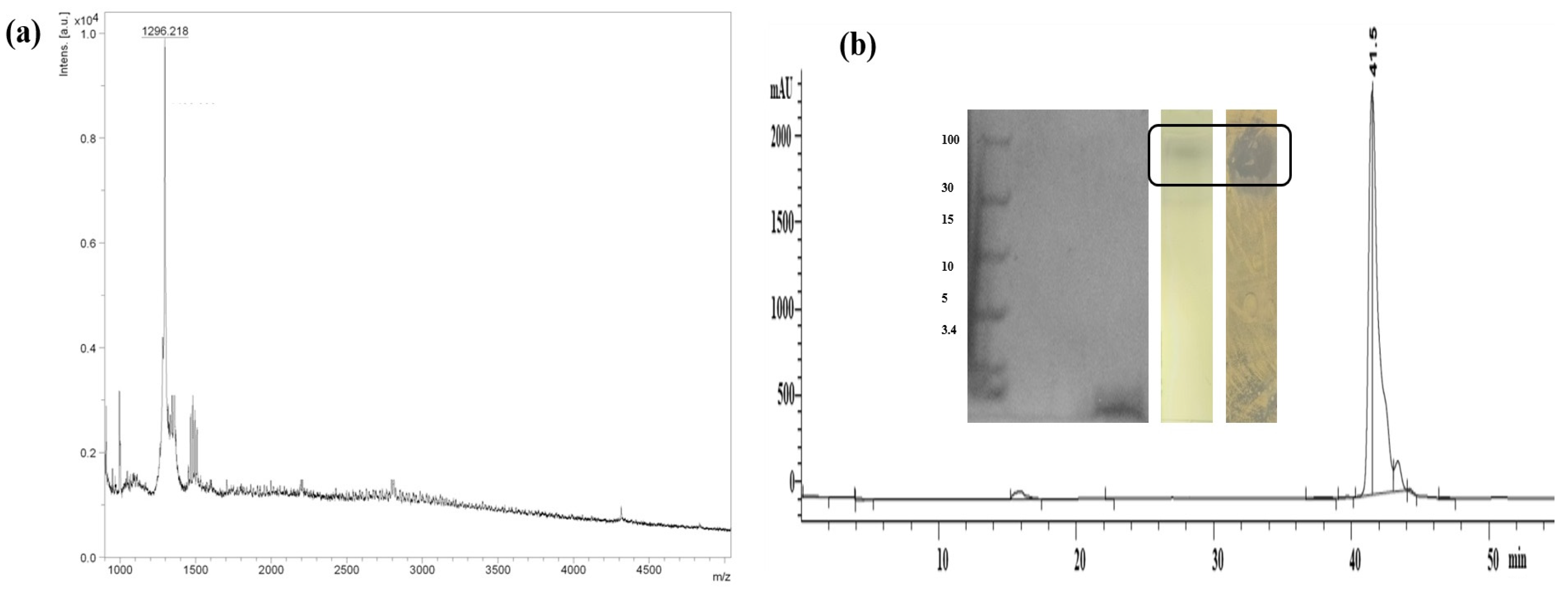
Figure 1. (a) MALDI mass spectrometry of SVDS-15; (b) RP-HPLC profile of SVDS-15 peptide inset showing tricine–SDS-PAGE of peptide, TLC stained with phosphomolybdic acid and bioautography demonstrating a clear inhibition zone.
Msf 24 00012 g001
Figure 2. Hemolysis assay of SVDS-15 (*** p < 0.0005).
Msf 24 00012 g002
Figure 3. Scanning electron microscopy images of C. albicans MTCC 183 (magnification 10,000×) after treatment with SVDS-15 peptide (5×).
Msf 24 00012 g003
Figure 4. Preparation of emulgel and in vitro assessment of its antimicrobial activity against C. albicans (MTCC 183).
Msf 24 00012 g004
Figure 5. Skin irritation study results at 72 h: (a) positive control, 20% SLS treated; (b) negative control, plain gel; and (c) SVDS emulgel.
Msf 24 00012 g005
Table 1. Physicochemical properties of designed peptides (predicted using Antifp software [11] (http://webs.iiitd.edu.in/raghava/antifp)).
PeptidePeptide SequenceHydrophobicityAmphipathicityHydrophilicityChargeMol. Wt.Structure
SK01MACVNQCPKAIDRFIVK−0.120.65−0.0621937Defensin-like beta
SK02KQVYKACMNGKHLYC−0.210.91−0.193.51786Defensin-like beta
SK03GIRWLVYRLRKV−0.250.92−0.1741559Helical
SK04HGLENKMYRHV−0.310.940.1221384Helical
SK05ATCHCSIHVSK−0.090.6−0.3321186Helical
SK06CMNGTQVYCR−0.220.37−0.4111175Helical
SK07KILKVARAWLAK−0.131.120.0241396.7Helical
SK08Fmoc-KILKVARAWLAK−0.131.120.0241655.4Helical
Pep1VKILAVALKWRAKR−0.21.140.3151652.3Helical
Pep2VIHKRHDGVKRI−0.381.260.6241457.9Helical
Disclaimer/Publisher’s Note: The statements, opinions and data contained in all publications are solely those of the individual author(s) and contributor(s) and not of MDPI and/or the editor(s). MDPI and/or the editor(s) disclaim responsibility for any injury to people or property resulting from any ideas, methods, instructions or products referred to in the content.