Article

Exploring the Interactions between Human microRNAs and the Ilheus Virus Genome

by
Joyhare Barbosa Souza
and
Samir Mansour Moraes Casseb
*
Oncology Research Center, Federal University of Pará, R. Augusto Corrêa, 01-Guamá, Belém 66075-110, PA, Brazil
*
Author to whom correspondence should be addressed.
SynBio 2023, 1(3), 194-203; https://doi.org/10.3390/synbio1030014
Submission received: 7 September 2023 / Revised: 16 October 2023 / Accepted: 18 October 2023 / Published: 26 October 2023
(This article belongs to the Special Issue Feature Paper Collection in Synthetic Biology)

Abstract

:
MicroRNAs (miRNAs) are small non-coding RNA molecules that play a fundamental role in the regulation of gene expression in humans. There has been a growing interest in investigating the interactions between human miRNAs and viruses to better understand the underlying mechanisms of the immune response and viral pathogenesis. The Ilheus virus, an arbovirus transmitted by mosquitoes, is known to cause disease in humans, with symptoms ranging from mild fever to severe neurological complications. This scientific article aims to explore the potential role of human miRNAs in their association with the genome of the Ilheus virus. Previous research has indicated that miRNAs can affect viral replication and the host’s immune response, playing a critical role in modulating the virus–host interaction. Here, we will investigate the possible interactions between specific human miRNAs and regions of the Ilheus virus genome, focusing on identifying miRNAs that may impact viral replication or the host’s immune response. A search for potential human miRNAs associated with the viral genome of ILHV was conducted through database searches such as miRBase. For the elucidation of targets regulated by these miRNAs, the TargetScan program was adopted. Functional enrichment analysis, inferring the function of genes regulated by miRNAs, was provided by the DAVID software. To elucidate the secondary structure, tools hosted in the RNAFold repositories were employed. In summary, our research has identified miRNAs linked to crucial sections of the Ilheus virus genome. These miRNAs can potentially regulate genes associated with neurological and immune functions. This highlights the intricate interplay between human miRNAs and the Ilheus virus genome, suggesting a pivotal role for these molecules in the host’s response to viral infections.

1. Introduction

Within the expansive domain of the Flaviviridae family, a distinguished assemblage of arthropod-borne viruses, the Ilheus virus (ILHV) emerges as a prominent member of the Flavivirus genus. ILHV’s transmission primarily hinges upon mosquitoes of the Aedes genus, which share vectors with other globally consequential flaviviruses such as Aedes aegypti, the vector responsible for propagating formidable viruses including dengue, Zika, and yellow fever. A comprehensive exploration into the intricate web of molecular interactions between the Ilheus virus and its host organism occupies a paramount position. It is driven by its potential to unveil the fundamental mechanisms governing viral pathogenesis, delineate potential transmission routes, and facilitate the formulation of efficacious therapeutic strategies [1].
The Ilheus virus, classified within the Flavivirus genus, is a positive-sense, single-stranded RNA virus. It was initially isolated in the 1940s in the Brazilian city of Ilhéus, after which it was named. The virus is primarily maintained in a sylvatic cycle involving non-human primates and forest-dwelling mosquitoes, while human infections are considered incidental. Despite this, sporadic human cases have been reported across tropical and subtropical regions, indicating the potential for zoonotic transmission [2].
Central to this intricate dynamic is a promising avenue for unraveling the nuanced dialogues between ILHV and the human host: the probing of microRNAs (miRNAs) and their hypothesized influence upon the Ilheus virus genome. MiRNAs, diminutive non-coding RNA molecules, orchestrate a pivotal role in post-transcriptional gene expression regulation. Their intrinsic versatility in modulating a spectrum of cellular processes positions them pivotally within the regulatory framework governing fundamental phenomena, encompassing cell proliferation, differentiation, and immune responses [3,4].
In the context of viral infections, the pivotal role of miRNAs assumes multifaceted dimensions. Extensive evidence substantiates miRNAs’ pivotal function in orchestrating cellular and immune responses during viral infections. These multifarious molecules wield the potential to impact viral replication, temper the host’s inflammatory response, fine-tune the expression of signaling molecules, and even shape the presentation of viral antigens. This discernment unveils an additional stratum of complexity within the molecular apparatus that underpins the intricate interplay between viruses and their hosts [5].
MiRNAs also play a fundamental role in plants, regulating various crucial biological processes for their development and adaptation to the environment. These small RNA molecules are involved in the regulation of gene expression, affecting protein translation [6]. MiRNAs in plants can influence growth, cell differentiation, response to environmental stress, flowering, and resistance to pathogens [7]. By modulating the expression of target genes, miRNAs allow plants to adjust their physiology and morphology according to environmental conditions, making them essential elements in plant adaptation and survival in different ecosystems [8].
Notwithstanding the substantial headway achieved in apprehending the intersections of miRNAs and flaviviruses, glaring lacunae endure, especially concerning the interplay between human miRNAs and the Ilheus virus. The need for more data on miRNAs’ influence on ILHV-mediated responses curtails our comprehension of the associated pathogenesis and the ensuing immune reactions. Against this backdrop, the foremost objective of this study crystallizes: to meticulously investigate and discern human miRNAs that conceivably interface with the Ilheus virus genome. Embracing this pioneering and trenchant approach, our ambition transcends mere scientific inquiry, aspiring instead to scaffold an enriched and robust framework for comprehending the intricate molecular pas de deux characterizing the interplay between the virus and its host [4,9].
A convergence of compelling factors underpins the rationale for this investigation. First, the novel approach of scrutinizing the potential interaction between human microRNAs and the Ilheus virus genome introduces an uncharted dimension to our understanding of viral infections. Unveiling such interactions could reveal novel therapeutic targets and develop innovative strategies against Ilheus virus infections.
Moreover, the convergence of vectors between the Ilheus virus and other flaviviruses of significant medical importance and their shared geographical distribution suggests that insights gained from this study could transcend our understanding of ILHV alone. The implications span broader implications for managing mosquito-borne viral diseases, contributing to a holistic strategy for public health preparedness.
In a broader context, the emerging precision medicine paradigm underscores the potential utility of specific microRNAs as diagnostic biomarkers or personalized therapeutic targets. By unraveling the intricate interactions between human microRNAs and the Ilheus virus, this research could offer a pivotal step toward personalized antiviral interventions, setting a precedent for tailored treatment strategies.

2. Results

2.1. Human miRNAs Interact with Key Regions of the ILHV Genome

The binding sites of miRNAs in viral genomes often correspond to non-coding regions such as the 5′ and 3′ untranslated regions (NTRs), although miRNA binding sites have been elucidated in the coding regions of viral proteins as well. Table 1 presents the specific regions found in the viral genome under study, the miRNAs associated with each of these regions, and their respective sequences (Table 1).
The 5′ cap region of the viral genome is responsible for encoding the polyprotein that will give rise to the viral capsid. In turn, the viral genome’s precursor membrane (prM) region is associated with viral pathogenesis [3]. Through genomic annotation of the genome regions, four human miRNAs with their respective sequences were obtained.

2.2. Human miRNAs Regulate Cellular Pathways Associated with Viral Replication, Cell Cycle Control, and Immune Signaling

After functional enrichment analysis using the DAVID tool, we identified the targets regulated by the miRNAs previously associated with the ILHV genome, as listed in Table A1. This analysis revealed significant associations between miRNAs and their respective targets, highlighting the biological functions and cellular pathways in which these miRNAs exhibit regulatory activity. In addition to the predicted functions in the functional enrichment analysis, the literature reports that the genes ATL2, CKB, SOCKS7, SCP2, and SH2D1A are associated with viral infection and replication, with the miRNAs regulating these pathways having binding sites associated with the 5′ cap region in the ILHV genome [10].
Furthermore, five other genes have been linked to functions related to cell cycle control, such as CDH20, FZD3, SMG9, KCNIP1, and MARKS. The miRNAs associated with these targets have binding sites present in both the 5′ cap and prM regions of the ILHV viral genome [11].
There are also targets related to important neuronal functions through pathways involved in neuronal dendrite maintenance and elongation, such as the target MAP2. Differential expression of the SNAP25 gene has been associated with excitotoxicity events in critical regions of the central nervous system. Both of these genes are regulated by miRNAs whose binding sites are inferred in the 5′ cap and prM regions of the ILHV genome [12].
Moreover, the 5′ cap and prM regions also contain binding sites for miRNAs that regulate pathways associated with immune signaling and viral immune evasion mechanisms. The genes related to these pathways are TCF3, ARRB2, and EBI3 [13].

2.3. Secondary Structure of miRNAs

The results of the secondary structures of the miRNAs identified in the genome were generated using the RNAfold software, elucidating the conformation of these RNA molecules. The secondary structures infer the base-pairing regions, loops, and stem structures of the miRNAs (Figure 1).
The minimum free energy (MFE) results of the secondary structures of the studied miRNAs, obtained through the RNAfold software, provided crucial information about the stability of these RNA molecule conformations. MFE is a measure that reflects the energy required to maintain the most stable RNA secondary structure, and the lower the MFE value, the greater the stability of the structure (Figure 2).
The analysis of minimum free energy (MFE) in the secondary structures of miRNAs allows us to assess the probability of these structures forming stably within cells, which is essential for the proper functioning of these molecules in gene expression regulation.
For hsa-mir-139, the ensemble’s thermodynamic free energy is −6.63 kcal/mol, indicating considerable stability of the structure. Additionally, the frequency of the MFE structure in the ensemble is 26.19%, suggesting that this conformation is highly prevalent. The ensemble diversity, which is 6.88, highlights the variability of secondary structures present. The centroid, representing a typical secondary structure, has a minimum free energy of −4.90 kcal/mol, a significant value indicating the importance of the stability of this conformation.
The hsa-mir-6076 exhibits an impressive thermodynamic ensemble’s free energy of 18.37 kcal/mol, indicating considerable stability in the observed conformations. However, it is important to note that the frequency of the minimum free energy (MFE) structure in the ensemble is relatively low, representing only 2.13% of conformations, suggesting considerable diversity in secondary structures. The high ensemble diversity, with a value of 28.33, highlights the variety of conformations the miRNA can adopt.
For the miRNA associated with the prM region in the viral genome, hsa-mir-1285-5p, the thermodynamic ensemble’s free energy is extremely low, with a value of −82.31 kcal/mol, indicating exceptional stability in the observed conformations. The ensemble’s diversity is very high, with a value of 69.62, emphasizing the variety and complexity of different secondary conformations of the miRNA that have been observed.
This information is crucial for understanding how miRNAs interact with their mRNA targets and how they exert their regulatory function in gene expression. Additionally, the analysis of secondary structures can help identify functional features, such as protein binding sites or conserved regions.

3. Discussion

The regulation of targets by miRNAs is an important feature in various viruses, including ILHV [14]. Through comparative analyses, it has been observed that some miRNAs previously associated with the ILHV viral genome may also have homologs or similar targets in other viruses. This suggests a potential conservation of gene regulation mechanisms mediated by miRNAs across different viruses [15]. This interaction between viral miRNAs and their targets can play a significant role in viral pathogenesis and the host’s immune response.
It has been observed that the miRNAs hsa-mir-139, hsa-mir-6076, and hsa-mir-4693 have potential binding sites in the 5′ cap region of the ILHV genome. This region is responsible for protecting the viral genome against degradation and for the translation of viral proteins, such as the protein (C), which forms the viral capsid. The viral capsid protein (C) is a small helical protein with surfaces that bind to viral nucleic acids or host lipids, directing the incorporation of the viral genome into the cell. It is an essential protein in viral replication [16].
The target gene ATL2, regulated by hsa-mir-139, one of the miRNAs associated with the 5′ cap region, has been elucidated as a facilitator of Zika virus (ZIKAV) replication through the fusion of the virus with the cellular endoplasmic reticulum (ER) membrane [17]. Additionally, miRNA hsa-mir-139 also regulates the targets CKB, SOCS7, and MAP2. These targets have been associated with the replication of the hepatitis C virus (HCV) through interaction with the viral non-structural 4A protein (NS4A), induction of immune signaling suppression, enabling ZIKV replication, and important neurological functions such as dendrite maintenance and elongation, respectively [10,18,19].
It has been observed that miRNA hsa-mir-6076 also has a binding site in the 5′ cap region of the ILHV genome. TargetScan identified the gene SMG9 as a target regulated by this miRNA. The SMG9 gene is part of a group of genes that function in the Nonsense-Mediated mRNA Decay (NMD) pathway, which is a mRNA quality control mechanism aimed at maintaining the quality of gene expression [20]. It has been reported that the human T-cell lymphotropic virus 1 (HTLV-1) has the capacity for negative modulation of the SMG1 gene associated with this pathway [21].
Also associated with the 5′ cap region in the ILHV genome, miRNA hsa-mir-4693, according to functional enrichment analysis, has been linked as a regulator of the targets KCNIP1 and ARRB2. Both of these targets are associated with the regulation of regulatory subunits of type A cellular potassium channels, which modulate the excitability of neuronal membranes, agonist-mediated desensitization of G protein-coupled receptors, and mediate the processes of desensitization and resensitization of cellular receptors to hormones, neurotransmitters, and immune signaling, respectively [22,23].
In a study conducted by [24], it was observed that the human immunodeficiency virus (HIV) modulated cellular membrane channels regulated by the KCNIP1 gene through the integral viral protein, thereby permeabilizing the membrane. This contributed to the virus’s entry into the cellular model and disrupted cellular homeostasis. Additionally, [25] identified in an in vitro assay that RNA viruses, through the degradation of ARRB2, hindered cellular immune signaling and promoted viral immune evasion.
The hsa-mir-1285-5p has been inferred to have the potential to interact with the prM region in the ILHV genome. According to analyses through TargetScan, hsa-mir-1285-5p regulates genes associated with neurological functions and immune system signaling [26]. It is interesting to observe interactions between miRNAs that regulate neuronal functions and the genomes of neuropathogenic viruses. When a neuropathogenic virus infects the nervous system, it can interfere with these delicate regulation mechanisms by modulating host miRNAs or expressing its own viral miRNAs [27].
This complex interaction can indeed result in significant neurological dysfunctions, contributing to the development of neurological disorders associated with viral infection. Studying this interaction between miRNAs and neuropathogenic viruses is crucial for understanding the molecular basis of viral neuropathologies and can guide the development of targeted therapeutic strategies.

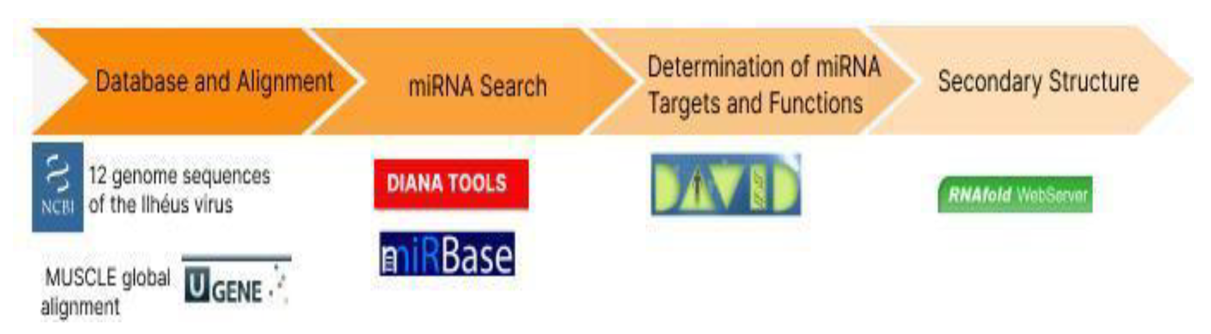
4. Materials and Methods

4.1. Database and Alignment

First, the 12 genome sequences of the Ilhéus virus from NCBI [28] were collected and prepared in FASTA format. Then, the Ugene software [29] was installed and configured, taking into account the preferences and parameters necessary for the analysis, as illustrated in Figure 3. The sequences were imported into a new project within Ugene, and the decision was made to use the MUSCLE global alignment algorithm [30]. Alignment parameters were properly configured, considering the substitution matrix and appropriate spacing options.
We considered the Virxicon [31] database to search for viral miRNAs with potential interaction with the human genome; however, according to the pre-established criteria in the study, we did not obtain data.
In this section, the miRbase database is not mentioned because it was not the database used to obtain sequences of the Ihéus virus; it was explored later (Section 4.2) in the search for human miRNAs with potential association with the viral genome. The selected databases yielded the largest amount of data for analysis among the adopted tools.

4.2. miRNA Search

To identify human miRNAs potentially associated with regions of the ILHV genome, bioinformatics tools such as the DAVID tool and miRBase were used [32,33]. Based on genomic annotations of each region of the ILHV genome, the tools obtained 147 and 123 pre-miRNAs, respectively, of which 35 were human, and 9 of them had elucidated targets. The coding regions of the capsid (5′ cap) and viral membrane precursor proteins (prM) were the regions that showed potential binding sites for human miRNAs.

4.3. Determination of miRNA Targets and Functions

To understand the functions of miRNAs associated with the ILHV genome, a search for biological targets was conducted using the TargetScan tool [34], followed by subsequent functional enrichment analysis using the DAVID tool [32]. This analysis revealed important cellular pathways associated with viral replication, cell cycle control, signaling, immune evasion, and neurological functions.
Initially, a total of 270 miRNAs with potential association with the ILHV viral genome were obtained through bioinformatics tools hosted in the DIANAtools repository [35]. Genomic annotation of viral genome regions was used to search for corresponding miRNAs on the platform. By searching for sequence homology in the miRBase tool, among the 270 miRNA sequences, 35 were human sequences, and 9 of them were associated with the regulation of important cellular pathways.

4.4. Secondary Structure

In the methodology for elucidating the secondary structure of the miRNAs under study using the web service software RNAfold [36], the miRNA sequences were first input into the program. RNAfold uses thermodynamics-based algorithms to predict the most stable secondary structure of RNA molecules. The software evaluated the different possible conformations, considering interactions between complementary bases, loops, and stem structures, as well as the minimum free energy (MFE). The results obtained provided a graphical representation of the predicted secondary structures, allowing visualization of base-pairing regions and conformation stability.
To validate the results, in addition to the aforementioned prediction, we utilized the miRDB, TargetScan, and miRTarBase databases, ensuring a higher reliability of the obtained data. Furthermore, we employed the RNA2Vec program [37] to investigate the miRNA–genome interactions of the Ilheus virus. This method enabled us to assess the similarity between different miRNAs and their functions, enhancing the comprehensiveness and accuracy of our analysis.

5. Conclusions

In summary, our investigations have revealed the presence of miRNAs associated with critical regions of the Ilhéus viral genome. These identified miRNAs demonstrate significant potential for regulating targets related to neurological functions and immune system signaling. These findings underscore the complex interaction between human miRNAs and the Ilheus virus genome, suggesting a fundamental role for these molecules in the host’s response to viral infection. These discoveries not only deepen our understanding of the molecular biology of arboviruses but also may open new perspectives for the development of therapeutic approaches and control strategies for arbovirus-caused infections, with a special focus on neurological complications and immune responses triggered by these viruses.

Author Contributions

Conceptualization, methodology, formal analysis, writing—original draft preparation, J.B.S.; writing—review and editing, visualization, supervision, project administration, S.M.M.C. All authors have read and agreed to the published version of the manuscript.

Funding

This research received no external funding.

Institutional Review Board Statement

Not applicable.

Informed Consent Statement

Not applicable.

Data Availability Statement

Not applicable.

Acknowledgments

We thank the Federal University of Pará through the Núcleo de Pesquisa em Oncologia for supporting the computer servers for the analysis.

Conflicts of Interest

The authors declare no conflict of interest.

Appendix A

Table A1. Target genes of miRNAs associated with the ILHV viral genome and their respective functions.
miRNATarget GeneFunction
hsa-mir-139ATL2Allows identical protein-binding activity. Involved in Golgi organization and organization of the tubular network membrane of the endoplasmic reticulum. Located in the tubular network membrane of the endoplasmic reticulum. It is an integral component of the membrane.
CKBCytoplasmic enzyme involved in energy homeostasis
SOCKS7Predicted to act upstream or within various processes, including brain development, adipocyte differentiation, and the insulin receptor signaling pathway.
MAP2Involved in microtubule assembly, which is an essential step in neurogenesis.
CDH20Calcium-dependent cell adhesion proteins; cadherins can thus contribute to the classification of heterogeneous cell types.
FZD3Promotes neurogenesis by maintaining sympathetic neuroblasts in the cell cycle in a beta-catenin-dependent manner (by similarity).
KLC2Microtubule-associated force-producing protein that plays a role in organelle transport.
MGAFunctions as a dual-specificity transcription factor, regulating the expression of target genes of the MAX network and T-box family. Functions as a repressor or activator.
TCF3Transcriptional regulator involved in early neuronal differentiation and mesenchymal-to-epithelial transition (by similarity).
hsa-mir-4693KCNIP1Regulatory subunit of type A potassium channels.
ARRB2Associated with agonist-mediated desensitization of G protein-coupled receptors.
hsa-mir-6076SMG9Encodes a regulatory subunit of the SMG1 complex, which plays a critical role in nonsense-mediated mRNA decay (NMD).
hsa-mir-1285-5pSCP2Plays a crucial role in peroxisomal oxidation of branched-chain fatty acids.
SNAP25Involved in the molecular regulation of neurotransmitter release.
MARCKSInvolved in cell motility, phagocytosis, membrane trafficking, and mitogenesis.
EBI3This gene was identified for its induced expression in B lymphocytes in response to Epstein–Barr virus infection. Associates with IL27 to form interleukin IL-27, which acts in innate immunity.
SH2D1AEncodes a protein that plays an important role in bidirectional stimulation of T and B cells.

Appendix B

Figure A1. Scheme illustrating regions of the ILHV genome and the human miRNAs potentially associated with them.
Synbio 01 00014 g0a1

References

  1. da Costa, V.G.; Saivish, M.V.; Lino, N.A.; Bittar, C.; de Freitas Calmon, M.; Nogueira, M.L.; Rahal, P. Clinical Landscape and Rate of Exposure to Ilheus Virus: Insights from Systematic Review and Meta-Analysis. Viruses 2022, 15, 92. [Google Scholar] [CrossRef] [PubMed]
  2. Vieira, C.J.; Andrade, C.D.; Kubiszeski, J.R.; Silva, D.J.; Barreto, E.S.; Massey, A.L.; Canale, G.R.; Bernardo, C.S.; Levi, T.; Peres, C.A.; et al. Detection of Ilheus virus in mosquitoes from southeast Amazon, Brazil. Trans. R. Soc. Trop. Med. Hyg. 2019, 113, 424–427. [Google Scholar] [CrossRef] [PubMed]
  3. Plante, J.A.; Plante, K.S.; Popov, V.L.; Shinde, D.P.; Widen, S.G.; Buenemann, M.; Nogueira, M.L.; Vasilakis, N. Morphologic and genetic characterization of ilheus virus, a potential emergent flavivirus in the americas. Viruses 2023, 15, 195. [Google Scholar] [CrossRef] [PubMed]
  4. Smith, J.L.; Jeng, S.; McWeeney, S.K.; Hirsch, A.J. A MicroRNA Screen Identifies the Wnt Signaling Pathway as a Regulator of the Interferon Response during Flavivirus Infection. J. Virol. 2017, 91, 10–1128. [Google Scholar] [CrossRef] [PubMed]
  5. Majumdar, A.; Basu, A. Involvement of host microRNAs in flavivirus-induced neuropathology: An update. J. Biosci. 2022, 47, 1–18. [Google Scholar] [CrossRef]
  6. Miskiewicz, J.; Szachniuk, M. Discovering Structural Motifs in miRNA Precursors from the Viridiplantae Kingdom. Molecules 2018, 23, 1367. [Google Scholar] [CrossRef]
  7. Li, Z.; Xu, R.; Li, N. MicroRNAs from plants to animals, do they define a new messenger for communication? Nutr. Metab. 2018, 15, 68. [Google Scholar] [CrossRef]
  8. Reinhart, B.J.; Weinstein, E.G.; Rhoades, M.W.; Bartel, B.; Bartel, D.P. MicroRNAs in plants. Genes. Dev. 2002, 16, 1616–1626. [Google Scholar] [CrossRef]
  9. Casseb, S.M.; Simith, D.B.; Melo, K.F.; Mendonca, M.H.; Santos, A.C.; Carvalho, V.L.; Cruz, A.C.; Vasconcelos, P.F. Drosha, DGCR8, and Dicer mRNAs are down-regulated in human cells infected with dengue virus 4, and play a role in viral pathogenesis. Genet. Mol. Res. 2016, 15, gmr.15027891. [Google Scholar] [CrossRef]
  10. Seong, R.-K.; Lee, J.K.; Shin, O.S. Zika Virus-Induction of the Suppressor of Cytokine Signaling 1/3 Contributes to the Modulation of Viral Replication. Pathogens 2020, 9, 163. [Google Scholar] [CrossRef]
  11. Li, C.; Hu, J.; Hao, J.; Zhao, B.; Wu, B.; Sun, L.; Peng, S.; Gao, G.F.; Meng, S. Competitive virus and host RNAs: The interplay of a hidden virus and host interaction. Protein Cell. 2014, 5, 348–356. [Google Scholar] [CrossRef]
  12. Johnson, B.W.; Cruz, C.; Felices, V.; Espinoza, W.R.; Manock, S.R.; Guevara, C.; Olson, J.G.; Kochel, T.J. Ilheus virus isolate from a human, Ecuador. Emerg. Infect. Dis. 2007, 13, 956–958. [Google Scholar] [CrossRef]
  13. Uozaki, H.; Chong, J.M.; Fujimoto, E.; Itoh, M.; Saito, M.; Sakuma, K.; Sudo, M.; Ushiku, T.; Niki, T.; Nagai, H.; et al. Soft and hard keratin expression in Epstein-Barr-virus-associated gastric carcinoma. Anticancer Res. 2005, 25, 3183–3190. [Google Scholar]
  14. Saivish, M.V.; Pacca, C.C.; da Costa, V.G.; Menezes, G.d.L.; da Silva, R.A.; Nebo, L.; da Silva, G.C.D.; Milhim, B.H.G.d.A.; Teixeira, I.d.S.; Henrique, T.; et al. Caffeic Acid Has Antiviral Activity against Ilhéus Virus In Vitro. Viruses 2023, 15, 494. [Google Scholar] [CrossRef]
  15. Kincaid, R.P.; Sullivan, C.S. Virus-Encoded microRNAs: An Overview and a Look to the Future. PLoS Pathog. 2012, 8, e1003018. [Google Scholar] [CrossRef]
  16. Byk, L.A.; Gamarnik, A.V. Properties and Functions of the Dengue Virus Capsid Protein. Annu. Rev. Virol. 2016, 3, 263–281. [Google Scholar] [CrossRef]
  17. Monel, B.; Rajah, M.M.; Hafirassou, M.L.; Sid Ahmed, S.; Burlaud-Gaillard, J.; Zhu, P.P.; Nevers, Q.; Buchrieser, J.; Porrot, F.; Meunier, C.; et al. Atlastin Endoplasmic Reticulum-Shaping Proteins Facilitate Zika Virus Replication. J. Virol. 2019, 93, 10–1128. [Google Scholar] [CrossRef] [PubMed]
  18. Hara, H.; Aizaki, H.; Matsuda, M.; Shinkai-Ouchi, F.; Inoue, Y.; Murakami, K.; Shoji, I.; Kawakami, H.; Matsuura, Y.; Lai, M.M.C.; et al. Involvement of Creatine Kinase B in Hepatitis C Virus Genome Replication through Interaction with the Viral NS4A Protein. J. Virol. 2009, 83, 5137–5147. [Google Scholar] [CrossRef] [PubMed]
  19. Avdoshina, V.; Mahoney, M.; Gilmore, S.F.; Wenzel, E.D.; Anderson, A.; Letendre, S.L.; Imamichi, T.; Fischer, N.O.; Mocchetti, I. HIV influences microtubule associated protein-2: Potential marker of HIV-associated neurocognitive disorders. AIDS 2020, 34, 979–988. [Google Scholar] [CrossRef] [PubMed]
  20. Karousis, E.D.; Mühlemann, O. Nonsense-Mediated mRNA Decay Begins Where Translation Ends. Cold Spring Harb. Perspect. Biol. 2018, 11, a032862. [Google Scholar] [CrossRef]
  21. Mocquet, V.; Neusiedler, J.; Rende, F.; Cluet, D.; Robin, J.P.; Terme, J.M.; Duc Dodon, M.; Wittmann, J.; Morris, C.; Le Hir, H.; et al. The human T-lymphotropic virus type 1 tax protein inhibits nonsense-mediated mRNA decay by interacting with INT6/EIF3E and UPF1. J. Virol. 2012, 86, 7530–7543. [Google Scholar] [CrossRef] [PubMed]
  22. Xu, Y.; Shi, W.; Song, R.; Long, W.; Guo, H.; Yuan, S.; Zhang, T. Divergent patterns of genic copy number variation in KCNIP1 gene reveal risk locus of type 2 diabetes in Chinese population. Endocr. J. 2018, 65, 537–545. [Google Scholar] [CrossRef]
  23. Wen, Q.; Li, Y.; Han, Z.; Liu, H.; Zhang, S.; Chen, Y.; He, J.; Du, X.; Fu, Y.; Zhang, L.; et al. β-Arrestin 2 Regulates Inflammatory Responses against Mycobacterium tuberculosis Infection through ERK1/2 Signaling. J. Immunol. 2021, 206, 2623–2637. [Google Scholar] [CrossRef]
  24. Herrero, L.; Monroy, N.; González, M.E. HIV-1 Vpu Protein Mediates the Transport of Potassium in Saccharomyces cerevisiae. Biochemistry 2012, 52, 171–177. [Google Scholar] [CrossRef] [PubMed]
  25. Zhang, Y.; Li, M.; Li, L.; Qian, G.; Wang, Y.; Chen, Z.; Liu, J.; Fang, C.; Huang, F.; Guo, D.; et al. β-arrestin 2 as an activator of cGAS-STING signaling and target of viral immune evasion. Nat. Commun. 2020, 11, 6000. [Google Scholar] [CrossRef] [PubMed]
  26. Aryal, M.; Lin, D.; Regan, K.; Du, S.; Shi, H.; Alvarado, J.J.; Ilina, T.V.; Andreotti, A.H.; Smithgall, T.E. The HIV-1 protein Nef activates the Tec family kinase Btk by stabilizing an intermolecular SH3-SH2 domain interaction. Sci. Signal. 2022, 15, eabn8359. [Google Scholar] [CrossRef]
  27. Fernández-Pato, A.; Virseda-Berdices, A.; Resino, S.; Ryan, P.; Martínez-González, O.; Pérez-García, F.; Martin-Vicente, M.; Valle-Millares, D.; Brochado-Kith, O.; Blancas, R.; et al. Plasma miRNA profile at COVID-19 onset predicts severity status and mortality. Emerg. Microbes Infect. 2022, 11, 676–688. [Google Scholar] [CrossRef] [PubMed]
  28. National Center for Biotechnology Information (NCBI). National Library of Medicine (US), National Center for Biotechnology Information, Bethesda, MD, USA. 1988. Available online: https://www.ncbi.nlm.nih.gov/ (accessed on 6 April 2017).
  29. Rose, R.; Golosova, O.; Sukhomlinov, D.; Tiunov, A.; Prosperi, M. Flexible design of multiple metagenomics classification pipelines with UGENE. Bioinformatics 2018, 35, 1963–1965. [Google Scholar] [CrossRef]
  30. Edgar, R.C. MUSCLE: Multiple sequence alignment with high accuracy and high throughput. Nucleic Acids Res. 2004, 32, 1792–1797. [Google Scholar] [CrossRef]
  31. Kudla, M.; Gutowska, K.; Synak, J.; Weber, M.; Bohnsack, K.S.; Lukasiak, P.; Villmann, T.; Blazewicz, J.; Szachniuk, M. Virxicon: A lexicon of viral sequences. Bioinformatics 2020, 36, 5507–5513. [Google Scholar] [CrossRef]
  32. Sherman, B.T.; Hao, M.; Qiu, J.; Jiao, X.; Baseler, M.W.; Lane, H.C.; Imamichi, T.; Chang, W. DAVID: A web server for functional enrichment analysis and functional annotation of gene lists (2021 update). Nucleic Acids Res. 2022, 50, W216–W221. [Google Scholar] [CrossRef] [PubMed]
  33. Griffiths-Jones, S.; Grocock, R.J.; Van Dongen, S.; Bateman, A.; Enright, A.J. miRBase: microRNA sequences, targets and gene nomenclature. Nucleic Acids Res. 2006, 34, D140–D144. [Google Scholar] [CrossRef] [PubMed]
  34. McGeary, S.E.; Lin, K.S.; Shi, C.Y.; Pham, T.; Bisaria, N.; Kelley, G.M.; Bartel, D.P. The biochemical basis of microRNA targeting efficacy. Science 2019, 366, eaav1741. [Google Scholar] [CrossRef] [PubMed]
  35. Vlachos, I.S.; Kostoulas, N.; Vergoulis, T.; Georgakilas, G.; Reczko, M.; Maragkakis, M.; Paraskevopoulou, M.D.; Prionidis, K.; Dalamagas, T.; Hatzigeorgiou, A.G. Hatzigeorgiou DIANA miRPath v.2.0: Investigating the combinatorial effect of microRNAs in pathways. Nucleic Acids Res. 2012, 40, W498–W504. Available online: https://dianalab.e-ce.uth.gr/html/universe/index.php?r=mirpath (accessed on 8 September 2023).
  36. Gruber, A.R.; Lorenz, R.; Bernhart, S.H.; Neuböck, R.; Hofacker, I.L. The Vienna RNA Websuite. Nucleic Acids Res. 2008, 36, W70–W74. [Google Scholar] [CrossRef]
  37. Yi, H.C.; You, Z.H.; Cheng, L.; Zhou, X.; Jiang, T.H.; Li, X.; Wang, Y.B. Learning distributed representations of RNA and protein sequences and its application for predicting lncRNA-protein interactions. Comput. Struct. Biotechnol. J. 2019, 18, 20–26. [Google Scholar] [CrossRef]
Figure 1. Secondary structures of miRNAs highlighting the probable base-pairing positions.
Synbio 01 00014 g001
Figure 2. Minimum free energy (MFE) graphs revealing the stability of miRNA secondary structures.
Synbio 01 00014 g002
Figure 3. Scheme of materials and methods used to generate results from ILHV sequences.
Synbio 01 00014 g003
Table 1. Human miRNAs associated with viral genome regions and their respective sequences.
RegionmiRNASequence
5′ caphsa-mir-139UCUGACUAAUUGUUCUAGUGGAGAUGUCUCCUGUUUGACUU
hsa-mir-6076
hsa-mir-4693
prMhsa-mir-1285-5pACCUUUUCCCAGUGCCUUCUUCUGCUUAUGUC
Disclaimer/Publisher’s Note: The statements, opinions and data contained in all publications are solely those of the individual author(s) and contributor(s) and not of MDPI and/or the editor(s). MDPI and/or the editor(s) disclaim responsibility for any injury to people or property resulting from any ideas, methods, instructions or products referred to in the content.

