Review

Organoid Models of Lymphoid Tissues

by
Ania Bogoslowski
1,*,†,
Joice Ren
1,†,
Clément Quintard
1 and
Josef M. Penninger
1,2,3,*
1
Department of Medical Genetics, Life Sciences Institute, University of British Columbia, Vancouver, BC V6T 1Z3, Canada
2
Helmholtz Institute for Infection Research, 38124 Braunschweig, Germany
3
Department of Laboratory Medicine, Eric Kandel Institute, Medical University of Vienna, 1030 Vienna, Austria
*
Authors to whom correspondence should be addressed.
These authors contributed equally to this work.
Organoids 2025, 4(2), 7; https://doi.org/10.3390/organoids4020007
Submission received: 11 February 2025 / Revised: 13 March 2025 / Accepted: 31 March 2025 / Published: 7 April 2025

Abstract

:
Lymphoid organs are critical for organizing the development of the immune system, generating immune tolerance, and orchestrating the adaptive immune response to foreign antigens. Defects in their structure and function can lead to immunodeficiency, hypersensitivity, cancer, or autoimmune diseases. To better understand these diseases and assess potential therapies, complex models that recapitulate the anatomy and physiology of these tissues are required. Organoid models possess a number of advantages, including complex 3D microarchitecture, scalability, and personalization, which make them ideal for modelling lymphoid organs and related pathologies. Organoids have been developed for both primary and secondary lymphoid tissues; however, these models possess several limitations, including immature phenotypes and incomplete stromal cell populations. Furthermore, these organoids are often heterogeneous in both structure and function. Several lymphoid organs, such as the spleen, do not yet have robust organoid models, offering opportunities for breakthroughs in the field. Overall, development of lymphoid organoids will pave the way for the rapid development and testing of novel therapies, organ modelling, and personalized medicine. This review summarizes current advances in models for the primary lymphoid organ—bone marrow and thymus—as well as the secondary lymphoid organs of the lymph node and spleen.

Graphical Abstract

1. Introduction

Effective translation of biomedical research to clinical medicine relies on preclinical models that accurately recapitulate human physiology [1,2,3]. Gaps between laboratory models and patients are in part responsible for the ubiquity of failed clinical trials, slowing improvements in patient care and scientific understanding [4]. These gaps are especially evident in diseases of the primary lymphoid organs, which are responsible for the development of naïve lymphocytes, and the secondary lymphoid organs, which facilitate interactions between these lymphocytes and antigens in adaptive immune responses. Both primary and secondary lymphoid organs possess intricate three-dimensional (3D) structures with distinct cellular niches and spatially separated developmental processes (Figure 1). This complexity is incompletely understood and challenging to replicate using in vitro models.
Pathologies in these organs are poorly characterized and often difficult to treat. Both primary lymphoid organs (bone marrow and thymus) and secondary lymphoid organs (spleen, lymph nodes, and mucosa-associated lymphoid tissue) can be the sites of malignancies including lymphomas, acute and chronic leukemias, myelomas, and thymomas. A number of promising treatments for these malignancies—such as histone deacetylase (HDAC) inhibitors for multiple myeloma [5], mammalian target of rapamycin (mTOR) inhibitors for diffuse large B cell lymphoma [6,7], or volasertib for acute myeloid leukemia [8,9]—have shown promising results in preclinical models but failed to materialize clinical benefits for patients. Given the tremendous burden of hematological malignancies, with a predicted 4.5 million cases by 2030 [10] and ten-year survival rates as low as 19% for acute leukemias [11,12], it is critical to develop preclinical models which better recapitulate human lymphoid physiology and disease. Conventional in vivo models of lymphoid organs are limited by fundamental between-species differences [13,14,15], whereas in vitro models often lack the 3D complexity and interactions of in situ tissues.
One exciting avenue for more accurate and tractable preclinical models is the creation of organoids—self-organized, three-dimensional structures that recapitulate the cellular interactions and spatial architecture of specific in vivo tissues [16]. Organoids possess several advantages for modelling lymphoid tissues. They can be made using human cells, whether sampled from patients or differentiated from human pluripotent stem cells, avoiding many of the limitations of animal models, such as species-specific differences [17]. It also allows for the creation of patient-specific organoids that can be applied in personalized medicine [18]. Organoids are also scalable, allowing for mid-throughput screening of potential drugs or toxins. They can recapitulate changes in tissue organization and cell–cell interactions more accurately than conventional in vitro models such as two-dimensional cell culture. Several lymphoid organoids have been created (Table 1), including for the bone marrow [19,20], the thymus [21,22], and secondary lymphoid tissues such as the spleen [23] and tonsils [24]. These have been helpful in understanding common pathologies and drug responses in these tissues, but they still possess a number of limitations, including immature cell types and limited immunocompetence [19,25]. Ultimately, improvements in organoid models will lay the groundwork for a better understanding of lymphoid organ physiology, immunity-related pathologies, and potential treatments.

2. Bone Marrow

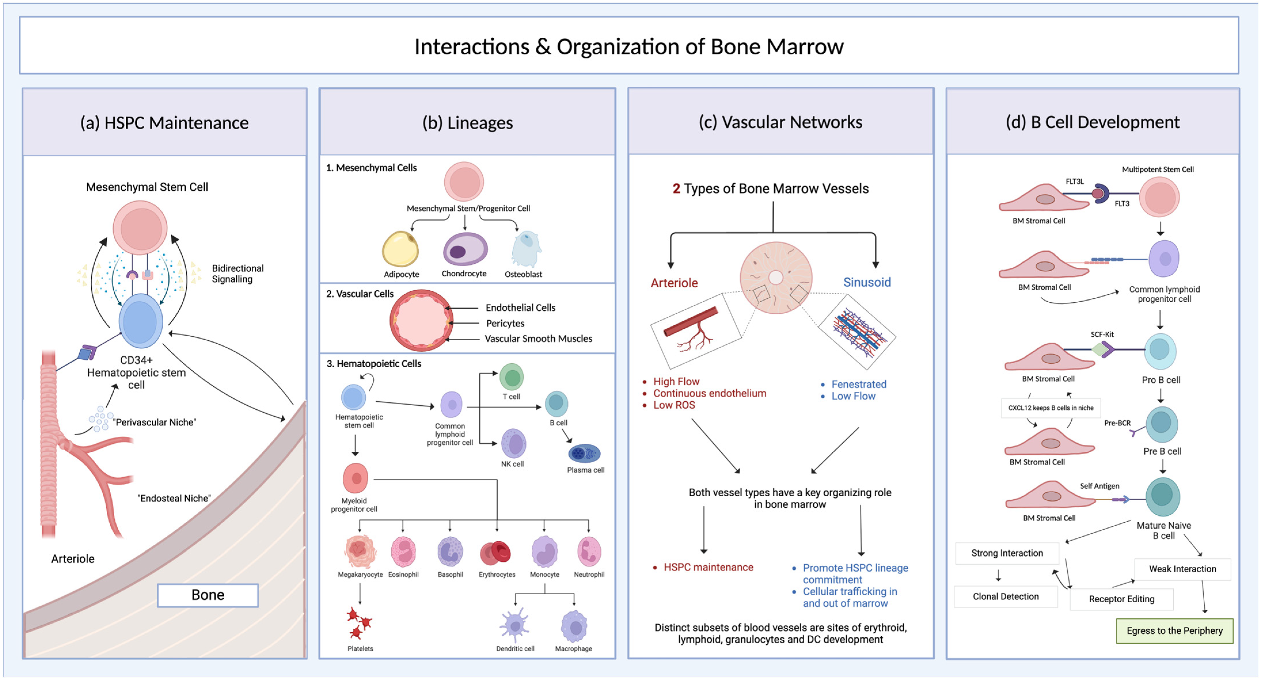
2.1. Bone Marrow Structure and Development

The bone marrow is the primary site of hematopoiesis, beginning in late fetal development and continuing across adult life. The bone marrow is composed of a variety of distinct, cellular-level microenvironments—or ‘niches’—in which cell–cell interactions and local soluble mediators support a particular cell type or developmental lineage [15]. Structurally, bone marrow is composed of three general groups of cells: (1) endothelial cells, (2) mesenchymal cells, and (3) hematopoietic cells. Each of these cell lineages is critical for bone marrow organization and function, and bone marrow pathologies have been linked to defects in all three cell types.
Endothelial cells are a critical component of the bone marrow, as blood vessels are critical in organizing bone marrow tissue in both fetal and adult life [50]. The vasculature of mature bone marrow can be divided into two general groups: (1) linear, arteriolar vessels that are organized in a columnar fashion and (2) branching, fenestrated sinusoids which are analogous to veins and found more centrally in bone [50,51]. Distinct bone marrow vessels differ in their gene expression, secreted angiocrine factors, and cell–cell interaction molecules—allowing them to define distinct niches. Arteriolar vessels help define the perivascular hematopoietic stem and progenitor cell (HSPC) niche in the bone marrow, in part due to their ability to maintain a low-reactive-oxygen-species environment [51,52]. In contrast, sinusoidal vessels are more permeable, promoting lineage commitment of HSPCs and facilitating cellular trafficking in and out of bone marrow [50]. Perivascular cells, such as pericytes and arteriolar smooth muscle cells, are present in varying degrees on bone marrow vessels; these cells also play a role in defining niches adjacent to vessels [52,53]. Blood vessels are also involved in specifying downstream hematopoietic development, as lineage-committed progenitors for erythroid, lymphoid, granulocytic, and dendritic cell (DC) development localize to specific, non-overlapping sinusoids and arterioles [52,53]. Altogether, the heterogeneous vasculature is an important player in localizing specific cell types and developmental processes in the bone marrow, through the formation of distinct and specialized perivascular niches.
Mesenchymal cells are also critical in defining bone marrow structure and supporting hematopoietic development. These cells are descended from mesenchymal stem and progenitor cells (MSPCs), which developmentally arise from pericytes or circulating precursors [54]. Canonically, they have a trilineage differentiation potential—as they can form adipogenic, chondrogenic, and osteogenic lines [55]—but more recent evidence has indicated their ability to differentiate into other cell types, including vascular smooth muscle [56]. New research has better characterized the niche in which bone marrow MSPCs reside—revealing that they are predominantly perivascular and prefer hypoxic conditions [57]. More detailed overviews of MSPCs and their descendants are available elsewhere, e.g., ref. [58].
The best-characterized niche within the bone marrow is that of HSPCs. These multipotent precursors require a delicate microenvironment to maintain their identity, and a variety of cell types have been implicated in this process. MSPCs and endothelial cells are especially critical in defining the HSPC niche. Mesenchymal cells are vital in expressing genes that support HSPC maintenance [59]. Furthermore, they have been shown to maintain HSPCs in vitro [60]. Endothelial cells and pericytes are also critical in establishing an HSPC niche, expressing soluble factors such as stem cell factor (SCF) and cell surface proteins such as Jag1 that signal to maintain HSPCs [61,62]. More recently, other non-hematopoietic cells such as osteoblasts [63] and adipocytes [64,65] have been shown to help define the HSPC niche [66].
Altogether, the bone marrow is a highly complex organ, with an anatomical organization at the microscopic rather than macroscopic level (Figure 2). Recent research has characterized the interplay between the three main lineages of cells in the bone marrow: endothelial, mesenchymal, and hematopoietic cell types. Given the immense importance of bone marrow biology to human health—and the risks posed by diseases of the bone marrow—it is critical to have models which accurately recapitulate bone marrow physiology and bone marrow-associated diseases.

2.2. Mouse Models of Bone Marrow

Many foundational studies of bone marrow anatomy and physiology came from in vivo studies of animal models. Mouse models have been used to demonstrate how the structure and function of bone marrow are impacted by immunodeficiencies [67,68,69], malignancy [70,71,72], drugs [73], or radiation [74]. These studies were also critical in devising the concept of a bone marrow niche [75]. Mouse models have also been used to model a variety of hematologic disease states, including acute myeloid leukemia (AML) [76,77], chronic myelogenous leukemia (CML) [78], acute lymphoblastic leukemia (ALL) [79], chronic lymphocytic leukemia (CLL) [80], multiple myeloma (MM) [81], myelodysplastic syndrome (MDS) [82], primary myelofibrosis (PMF) [83], aplastic anemia [84], severe combined immunodeficiency [67,85], or agammaglobulinemia [68].
Through improved molecular imaging methods, mouse models have also been used to uncover new aspects of bone marrow niches [52]. Wu and colleagues used an unbiased analytical pipeline on flow cytometry data from mouse bone marrow cells to identify novel surface markers that distinguish subsets of hematopoietic cells, including hematopoietic stem cells, multipotent and oligopotent progenitors, and cells of the erythroid, myeloid, and lymphoid lineages [52]. Using a combination of these novel markers and established lineage markers, they used confocal microscopy and an artificial intelligence algorithm to quantify the relationship of these populations to each other and the surrounding vasculature [52]. Confocal microscopy demonstrated that stem cells, oligopotent progenitors, and multipotent progenitors were distributed across the bone marrow as single cells [52]. In contrast, lineage-committed progenitors for erythroid, lymphoid, granulocytic, and DC development form clusters and localize to specific, non-overlapping sinusoids and arterioles [52,53].
However, despite the importance of in vivo studies, these models have a number of limitations for understanding human bone marrow physiology. Most significantly, there are a variety of differences between human and rodent bone marrow physiology. Adult human hematopoiesis occurs primarily in the ilium and the bones of the axial skeleton, whereas adult mouse hematopoiesis is spread across all bones alongside a contribution from the spleen [13,86]. There are also differences in the frequencies, gene expression, and phenotypes of the hematopoietic and non-hematopoietic cells between human and mouse bone marrow [15]. These differences make it difficult to generalize from studies of mouse models to humans and limit the applicability of these animal models in preclinical testing and drug screening [2]. Furthermore, researchers have a limited ability to capture and manipulate the precise 3D microstructure of bone marrow in animal models [87].

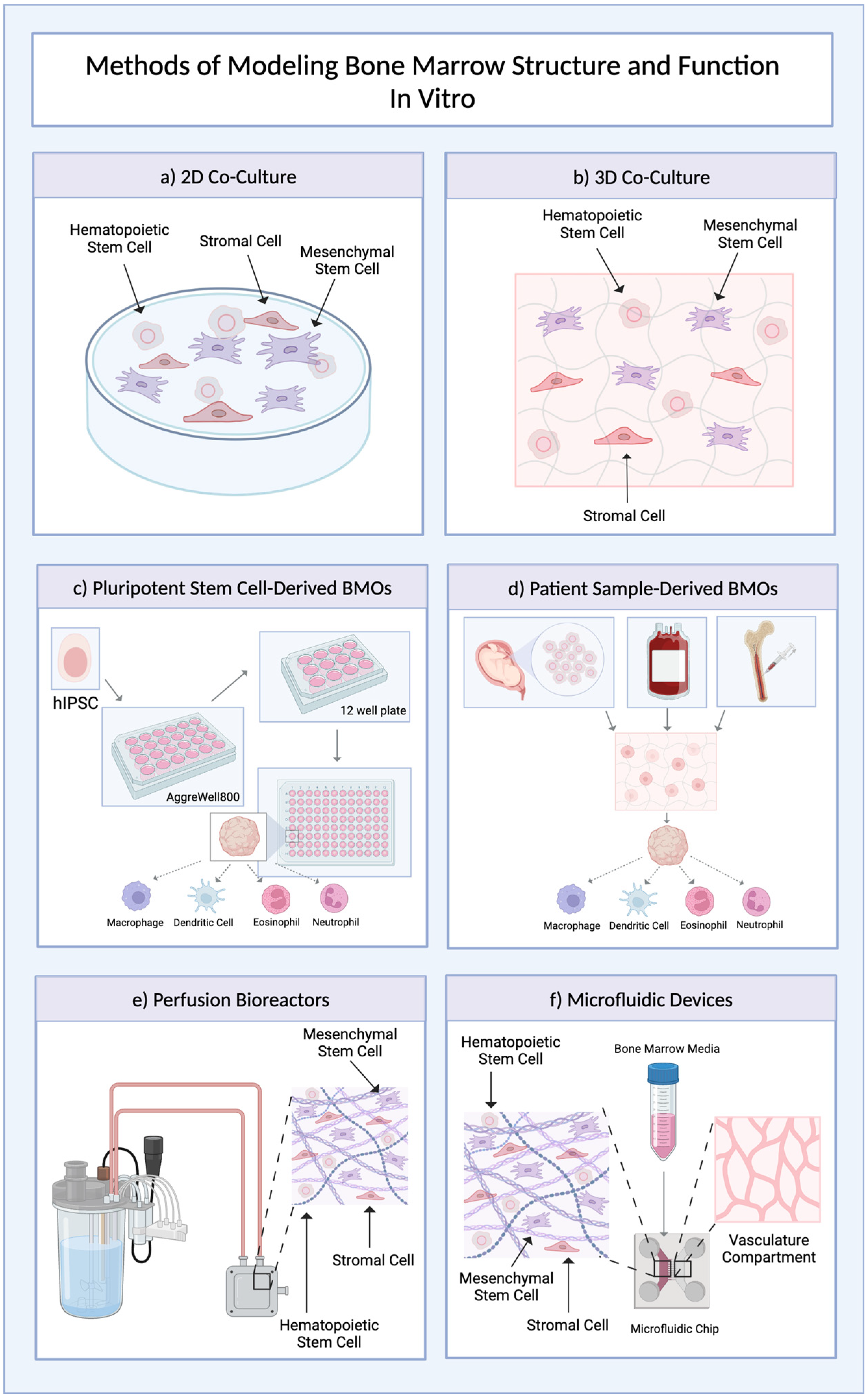
2.3. Bone Marrow Organoids

In vitro human bone marrow models allow for experimental exploration of bone marrow function (Figure 3), addressing species differences and more reliably translating to human health. Various cell types, particularly HSPCs and MSPCs, have been maintained in cell culture, allowing for insight into their cellular functions but providing only limited evidence about the overall structure and function of bone marrow [88,89,90,91]. Efficient protocols have been developed to differentiate defined bone marrow cells from human pluripotent stem cells, allowing for more reproducible phenotypes [92,93,94,95,96]. Nonetheless, these 2D models lack consideration for the heterogeneous cell composition and 3D microstructure of bone marrow [97].
Co-culture models, which combine different cell types to better recapitulate cell–cell interactions, can address some of these concerns. Long-term murine bone marrow cultures have been used to study murine hematopoiesis ex vivo [13,98,99]. More recent co-culture models often incorporate the interactions of human MSPCs and HSPCs, which are critical for bone marrow physiology and maintaining distinct niches [60,100,101]. Furthermore, specific co-culture systems have been developed to focus on particular bone marrow processes, including megakaryopoiesis/thrombopoiesis [102], angiogenesis [103], or monocyte/macrophage differentiation [104]. Nonetheless, these two-dimensional co-culture models still lack the microarchitecture and spatial organization of bone marrow. Three-dimensional bone marrow co-culture systems have emerged, allowing for the formation of multiple niches [105,106,107], including more accurate leukemia niches [108,109]. Complex 3D biomaterials, such as collagen [110,111], Matrigel [97,112], and poly-lactic-co-glycolic acid [113] have been engineered to support various bone marrow niches, allowing these cells to self-renew and maintain their properties. A variety of bone marrow pathologies, such as myeloma [114,115] and AML [116], have been modelled to mimic their endogenous microenvironment using these 3D biomaterials. However, the maintenance of both 2D and 3D co-cultures strays considerably from physiological conditions, often requiring supraphysiologic concentrations of cytokines or a need for a ‘feeder’ cell layer. Moreover, these cultures typically require the addition of serum to support their growth, resulting in considerable variability dependent on the serum batch [102,115]. Finally, even 3D co-culture systems are unable to fully incorporate the variety and complexity of known cell–cell and cell–extracellular matrix interactions of bone marrow [117]. Thus, while these systems provide insights about the function of various bone marrow cells, they do not present a holistic model of bone marrow.
Bone marrow organoids are self-organized, three-dimensional models that mimic the tissue microenvironment and spatial organization of bone marrow in vitro [111,118,119], making them well suited for preclinical applications such as drug screens, toxicity screens, and genetic modelling [19,20]. Bone marrow organoids have been used for studying diseases such as PMF, radiation-induced bone marrow suppression [120], AML, CML, ALL, and MM, as well as screening for drugs targeting bone marrow fibrosis [19,20]. Initial attempts to model bone marrow microarchitecture used biomaterial engineering to craft complex 3D structures to recreate specific aspects of bone marrow structure and function, such as HSPCs niches, MSPC niches, and megakaryopoiesis [19,20]. The definition of bone marrow niches in these models is impacted by a variety of factors, including surrounding cells, soluble factors, extracellular matrix components, scaffold structures, or perfusion [121]. Other researchers used patient-derived mesenchymal and/or endothelial cells, rather than engineered biomaterials, to create a structural scaffold that is populated by HSPCs [122,123]. Further, some groups have used bone marrow differentiation protocols to introduce immune cells into existing organoid models for lung [124] and liver tissues [125], more accurately recapitulating human physiology [125,126].
More recently, novel protocols have been developed for creating bone marrow organoids from human pluripotent stem cells [19,20]. These are organized within hydrogel structures, allowing for the formation of a 3D microarchitecture that mimics human bone marrow. These models contain the three cell lineages that form the bulk of bone marrow—hematopoietic, mesenchymal, and endothelial cells [19]—and have a spatial organization that recapitulates perivascular HSPC niches [19] and MSPC niches [19]. Flow cytometry and histology have demonstrated many cell types from these organoids, including myeloid progenitors, megakaryocyte progenitors, lymphoid progenitors, chondrogenic precursors, osteogenic precursors, endothelial cells, pericytes, vascular smooth muscle cells, granulocytes, and monocytes/macrophages [19,20]. Many of these cells recapitulated the morphology, gene expression, or differentiation potential of their in vivo bone marrow counterparts [19,20]. They also formed spatial relationships that are crucial for bone marrow function, such as platelet-derived growth factor receptor β (PDGFRβ)-positive pericytes enwrapping endothelial cells, interconnected vascular networks with lumens, and perivascular networks of CXCL12-abundant reticular cells [19,20,127]. When implanted within the renal capsule of immunodeficient mice, stem-cell-derived bone marrow organoids were engrafted, developing a functional vascular network that allows for the flow of blood as well as the migration of leukocytes from the organoid into the host circulation and host bone marrow. Recent advances in the production of pluripotent stem cells, including the advent of protocols for creating clinical-grade, stroma- and vector-free HSPCs from human iPSCs, offer the promise of additional uses [128]. However, the use of iPSCs resulted in an immature phenotype of bone marrow organoids with an organoid structure and cell composition resembling fetal bone marrow rather than adult bone marrow [19]. Frenz-Wiessner and colleagues found that while HSPCs from these iPSC-derived bone marrow organoids were able to transiently engraft into immunodeficient mice, these cells were unable to sustain long-term hematopoiesis [19]. This is a prominent limitation of all PSC-derived organoids; while it allows them to be uniquely accurate in modelling developmental processes within the bone marrow, it is a critical barrier for translating these models into clinical settings such as bone marrow replacement. Several strategies have been proposed to circumvent the immaturity of iPSC-derived models, including metabolic maturation [129], forced expression of aging-related genes [130], and in vivo transplantation [19,131]. In other organ systems, the addition of dynamic flow via microfluidic systems has helped promote maturity of PSC-derived organoid models, and this may also be another method to address this limitation [26,132]. Lastly, the hematopoietic potential has been shown to differ between iPSCs derived from cord blood, peripheral blood, and bone marrow aspirates, suggesting that the initial stem cell population used may help determine the overall maturity and functionality of these models [27].
Furthermore, many bone marrow organoid models are limited by their static nature―despite their 3D complexity, they lack the defined flow of blood and nutrients that helps shape bone marrow physiology [97]. To address this limitation, several 3D perfused bone marrow organoids have been developed, including perfusion chambers that surround organoids in flowing media and bioreactors that allow media to flow through the organoid [28]. These models combined umbilical cord blood mononuclear cells with patient-derived serum to support their growth [28,29,133,134]. Perfusion-based bone marrow organoids have been shown to recapitulate critical bone marrow niches, maintain cell populations including HSPCs, MSCs, lymphoid progenitors, myeloid progenitors, osteoblasts, and endothelial cells [28], and support erythropoiesis [28,29,133,134]. Novel approaches to recapitulate elements of the bone marrow niches, such as using the stromal vascular fraction of adipose tissue to create perivascular HSPC niches, have further allowed for more precise modelling of niches in these bioreactor systems [135].
Microfluidic devices, also known as bone-marrow-on-a-chip devices, aim to provide a more physiologically relevant environment by culturing cells within a micrometer-scale chamber that is constantly perfused [97,136]. They allow for the precise control of nutrient perfusion, concentration gradients, and shear forces. This is in contrast to perfusion-chamber-based models, such as an early bone-marrow-on-a-chip system developed by Torisawa et al., which combined in vivo and in vitro tissue engineering protocol. They subcutaneously implanted a type I collagen matrix with bone-differentiation-inducing factors into a mouse model [30]. Over time, this scaffold developed into a cylinder of bone-like tissue surrounding a bone marrow core with stromal cells, a vascular network, and hematopoietic cells. These structures were then extracted and placed into an in vitro microfluidic chamber designed to provide constant flow [30]. This bone marrow-on-a-chip device was able to maintain HSPCs without the addition of exogenous cytokines, and it effectively modelled the impacts of gamma radiation on HSPCs, hematopoietic progenitors, lymphoid cells, and myeloid cells [30]. Later models were developed by combining patient-derived MSCs and HSPCs in vitro, allowing for an entirely human bone-marrow-on-a-chip system that could sustain HSPC populations in a long-term culture.
Glaser et al. developed a bone-marrow-on-a-chip model including endosteal and perivascular HSPC niches on a microfluidic device with a fibrin hydrogel scaffold [31]. To model the perivascular niche, cord-blood-derived endothelial cells and adult-donor-derived bone marrow stromal cells were added to the scaffold of one chamber. Similarly, these endothelial cells were added along with human fetal osteoblasts to mimic the endosteal HSPC niche. A mixture of CD34+ HSPCs, endothelial cells, and the respective stromal cells were then injected into both chambers. Using this model, HSPC maintenance, chemotherapy responses, and hematopoiesis in response to stimulation with cytokines like G-CSF were recapitulated. When only one of these two niches was present, there was a reduced capacity to maintain HSPCs, concordant with the role of both niches in stem cell maintenance in vivo [31].
Microfluidic devices have also been developed to model bone marrow pathologies, including multiple myeloma [137] and acute lymphoblastic leukemia [138]. Chou et al. developed a bone-marrow-on-a-chip model that can accurately recapitulate 5-fluorouracil and radiation toxicity at patient-relevant doses, physiologic processes such as intravasation of mature granulocytes, and the pathophysiology of patient-specific rare genetic disorders [139]. Recently, the Huh group has developed a microfluidic model of the bone marrow that precisely emulates the perivascular HSPC niche [140]. This model used a microfluidic chip that combined human umbilical vein endothelial cell (HUVEC)-lined side channels with a central chamber containing HUVECs and human bone marrow donor-derived CD34+ HSPCs and MSPCs, which allowed for the self-organization of bone marrow sinusoid-like vessels with a perivascular niche for HSPCs [140]. The results from these microfluidic models highlight the utility of the bone-marrow-on-a-chip approach for understanding the pathophysiology and treatment of individual patients [139]. However, organ-on-a-chip models often have a lower yield of cells and can only be maintained for shorter periods than perfusion-chamber-based models [31,97].
However, both static and perfused bone marrow organoids are often difficult to generate and optimize, requiring a delicate balance of conditions. Furthermore, no bone marrow organoids to date have been able to recapitulate mature B cells, osteoid lineage cells, and adipocytes, which also play roles in bone marrow physiology [19,20,123]. Immaturity is the main concern with pluripotent stem-cell-derived organoids; this is also observed in heart, lung, kidney, and other organoids [141,142,143,144,145]. This limitation is particularly relevant for hematopoietic models, as the microstructure, gene expression, cell composition, and function of bone marrow differs considerably between fetuses, children, and adults [86,146,147,148]. Furthermore, many bone marrow pathologies, including AML, CML, CLL, MDS, and MM, predominantly affect older adults [149,150,151]. Lastly, bone marrow organoids cannot effectively model the impacts of systemic inputs such as endocrine and neural stimulation. These systemic inputs may be more effectively modelled by implanting organoids subcutaneously into humanized and/or immunodeficient mice [97].
A number of unknowns also remain with respect to the phenotype of bone marrow organoid-derived hematopoietic cells. While gene expression profiles, cell surface markers, and microscopy have shown that these cells recapitulate genetic properties of in vivo hematopoietic cells, there is limited understanding of whether this extends to function [19,20]. Some cell types can respond to bacterial lipopolysaccharide (LPS) with the production of the pro-inflammatory cytokines IL-6, IL-8, and G-CSF [19]. Nonetheless, most functions of immune cells (migration, phagocytosis, degranulation, antigen presentation, etc.), as well as their overall ability to respond to challenges to various pathogens, remain understudied. Future research may be able to evaluate the functions of bone marrow organoid-derived immune cells. For instance, neutrophils could be tested for reactive oxygen species production, phagocytic capacity, and formation of neutrophil extracellular traps (NETs) in response to immunologically relevant stimuli [152]. Similarly, phenotypic assays could be used to test the function of iPSC-derived macrophages [153], DCs [154], and NK cells [155] in isolation to probe whether these bone marrow organoid-derived cells recapitulate the functions of their endogenous counterparts.
Altogether, bone marrow organoids offer a scalable and tractable model system for understanding bone marrow physiology, pathology, pharmacology, and cell biology. This lays the groundwork for more accurate in vitro modelling of bone marrow pathologies and may allow for more precise drug screening. Perfusion-based bone marrow organoids, including those using bioreactors and microfluidic devices, allow for the integration of vascular networks and diffusion gradients. These also have a number of limitations, including a lack of protocols exclusively using off-the-shelf reagents, low yields, and challenges in maintaining these organoids at a large scale. Further improvements are therefore required to generate mature, perfused bone marrow organoids, even representing an alternative and novel tool for adoptive cell therapies and bone marrow transplantation.

3. The Thymus

3.1. Thymic Development and Anatomy

Located just above the heart, the thymus is the primary site of T cell development and education—and the origin of the eponymous “T” in T cells. The thymus can generate lymphocytes with an incredibly diverse T cell receptor (TCR) repertoire, capable of recognizing a wide variety of foreign antigens. While maintaining this diverse receptor specificity, the thymus also blocks the development of autoreactive T cells through the process of central tolerance [156]. The thymus has a complex 3D organization which ensures that developing thymocytes are sequentially exposed to distinct microenvironments and are tested through positive and negative selection (Figure 4), facilitating this balance of diversity without autoreactivity [157].
Anatomically, the thymus is divided into lobules, each consisting of an outer cortex and an inner medulla. Early thymic progenitors, originating from the bone marrow, exit the circulation to seed the thymus along the junction between the cortex and medullary regions [158]. Signalling through the Notch receptor commits these lymphoid cells to the T cell lineage, and they migrate to the outer cortex and begin to proliferate [159,160,161,162]. The subsequent stages of thymocyte development proceed in a highly spatially regulated manner—beginning at the outer cortex and proceeding inward through the inner cortex and medulla to form mature T cells that can emigrate to the periphery [163,164].
The thymus is composed of immune cells and non-immune stromal cells. Immune cells are CD45+ and include thymocytes, dendritic cells, macrophages, and B cells. These cells originate from hematopoietic stem cells and continuously colonize the thymus throughout development [165]. Non-immune structural cells include the medullary and cortical thymic epithelial cells (TECs), as well as the capsule, endothelium, fibroblasts, and mesenchymal cells. TECs develop from the endoderm in a Foxn1-dependant process [164,166], whereas other stromal cells develop independent of Foxn1 from neural crest mesenchyme [165]. The origin of thymus stromal cells versus haemopoietic cells needs to be considered when designing thymus organoid models.

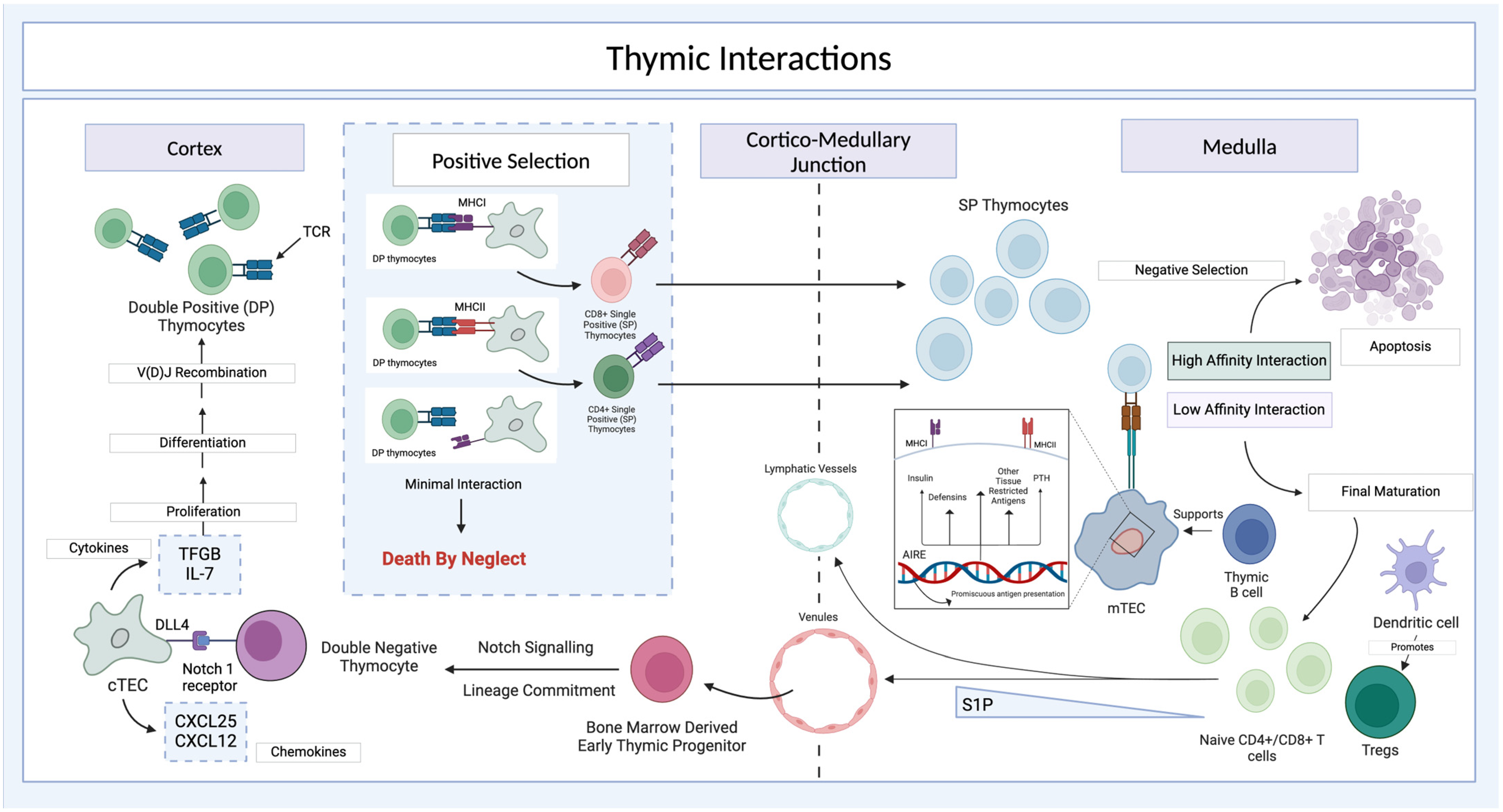
3.2. Thymic Cortex and Medulla

The structure of the thymic cortex and medulla are highly distinct, with differences in both immune and stromal cells. These structural and cell population differences allow them to accomplish separate functions at distinct stages in T cell development.
While both the cortex and medulla contain TECs, the gene expression of these cells differs significantly between the two regions, allowing them to serve distinct functions. Cortical TECs (cTECs) are specialized for creating a microenvironment for the early stages of T cell development [167]. Through a combination of cell surface proteins (e.g., DLL4 for Notch signalling), cytokines (e.g., TGF-β, IL-7), and chemokines (e.g., CCL25, CXCL12), cTECs promote the rapid proliferation of thymocytes as well as the process of V(D)J recombination to generate novel TCRs [163,168,169,170,171,172]. cTECs also express both MHC I and MHC II, allowing them to interact with double-positive (CD4+CD8+) thymocytes [169]. cTECs and double-positive (DP) thymocytes continue interacting until thymocytes develop into single-positive (SP) ones; T cells that react to peptides presented on MHC I become CD8+ cytotoxic T cells, and those that react to peptides presented on MHC II become CD4+ helper T cells [173]. Developing DP thymocytes which cannot respond to either MHC I or II—which constitute the majority of cells at this stage—undergo cell death by neglect [174]. Thus, cTECs help orchestrate the process of positive selection, ensuring that developing thymocytes can respond to peptides presented on the host’s MHC molecules.
Once a T cell passes positive selection in the cortex, it migrates to the medulla where it encounters medullary TECs (mTECs). These cells express the transcription factor Autoimmune Regulator (AIRE), which allows them to present antigens that are otherwise tissue-specific to developing thymocytes. This is critical for negative selection of autoreactive lymphocytes, which occurs in the medulla and triggers apoptosis to prevent these self-reactive T cells from reaching the periphery [175].
In addition to differences in epithelial stromal cells, bone-marrow-derived cells differ considerably between the two compartments. While the cortex is predominantly composed of thymocytes and TECs, along with sparse macrophages to facilitate turnover of apoptotic cells, the medulla contains a rich variety of hematopoietic lineage cells, including B cells, dendritic cells (DCs), and macrophages. Thymic B cells assist mTECs in presenting self-antigens to thymocytes as part of negative selection [176,177]. Thymic DCs are composed of several different subsets, but play roles in negative selection, induction of regulatory T cells (Tregs), and responding to peripheral signals such as infection [178,179,180]. Similarly, macrophages in the medulla—in addition to efferocytosing the apoptotic thymocytes—can present antigens to help with negative selection and are critical in repair following thymic injury [180,181,182]. There are also distinct subsets of bone-marrow-derived cells that define a unique environment at the corticomedullary junction, including plasmacytoid dendritic cells (pDCs), SIRP1α+ DCs, and CX3CR1+ macrophages [180].
Any studies aiming to create a functional thymus organoid will need to maintain this distinct organization and cellular diversity of the thymic cortex and medulla.

3.3. Thymus Organoids

The thymus is the site of T cell development and maturation; hence, robust thymic organoid models allow for the interrogation of thymic deficiencies or diseases. While animal models of normal and disrupted thymic development, such as the immunocompromised nude mice, have been critical in unraveling the function of this organ, they are in part limited by differences between species [17]. While in vitro models of the thymus have been challenging to create, recent advances have allowed for more accurate and scalable models of thymic tissue (Figure 5). While individual cultures of various thymic cell types have been successful, the recapitulation of the complex microarchitecture and regions of the thymus has remained elusive for many years. TECs are challenging to maintain ex vivo, as cultures lose their characteristic ability to support T cell development in the absence of a complex 3D structure [34]. The OP9-DL1 in vitro system was developed to support T cell development, but it relied on genetically modified bone marrow stroma rather than mimicking the in vivo thymic structure [183]. Furthermore, the reliability of these early models was affected by their dependence on serum, due to the potential for variation between batches [97].
To circumvent these limitations, several groups have worked to generate 3D models of the thymus in vitro. When murine thymic components were separated and reaggregated ex vivo, this generated a 3D model that supported the selection of CD4+ and CD8+ T cells [32]. Later studies expanded upon this, independently culturing stromal cells such TECs and non-TEC stromal cells prior to reaggregation [33]. This allows for the genetic or pharmacological manipulation of specific cell types in these reaggregate cultures. Similarly, another group developed a 3D model of the thymus structure by using decellularized murine thymic tissue as a scaffold for robust TEC culture; this model supported the reconstitution of a functional and diverse T cell repertoire when implanted into athymic nude mice [184]. However, all of these models are limited in that they are derived from rodents, not human tissues, and they still do not fully recapitulate the in vivo thymic cell composition. Pinto et al. worked on models that better mimic aspects of stromal cell support, such as promiscuous antigen presentation by mTECs, but these typically lacked the ability to support T cell development [185].
Another 3D thymic organoid system used murine stromal cells co-cultured with HSPCs. The bone marrow stromal cells were genetically modified to express members of the Notch ligand family (DLL1 or DLL4), allowing them to mimic the role of the thymic stroma in promoting T cell development. These artificial thymic organoid (ATO) assays were able to mimic the maturation of naïve CD4+ and CD8+ T cells from CD34+ cells [21] and induced pluripotent stem cells (iPSCs) [22]. They were also used to generate T cells with engineered TCRs targeting tumor-associated antigens, suggesting that this method may be useful for adoptive cell therapy. Further research applied these ATO systems to produce chimeric antigen receptor (CAR) T cells with potent anti-tumor activity [186,187].
These ATOs have also been used to evaluate T cell developmental deficiencies. Pille et al. knocked out the WAS gene in human HSPCs, which is responsible for the immunodeficiency Wiskott–Aldrich syndrome, and they used these cells to build ATO models [188]. These models showed a distinct defect in T cell development—with normal progression to DP thymocytes and appropriate TCR specificities but a reduction in the transition of DP thymocytes to CD8+ SP T cells [188]. Furthermore, the ATO assay has been used to explore the effects of mutations in the Pre T-cell Receptor Alpha (PTCRA) gene, which are associated with T cell repertoire biases towards γδ T cells; these patient-derived mutant cells exhibit a specific defect in the production of TCRαβ+ cells. Furthermore, the severity of this defect was directly related to the severity of the underlying PTCRA mutation [189]. In preclinical settings, the ATO assay has also been utilized to test CRISPR/Cas9-mediated gene editing of patient-derived iPSCs or CD34+ cells [190,191,192,193,194].
However, one limitation of the ATO assay is that it can only model thymic defects that are intrinsic to T cells, not stromal cells. The MS5 stromal cells used in this organoid model are bone-marrow-derived and from a single cell line; as such, they are not suitable for modelling defects in TECs or other stromal cells of the thymus [21,195]. An alternative system was necessary to study defects in TECs directly where TECs are derived from pluripotent stem cells. Many differentiation protocols exist that efficiently make immature TEC progenitors [34,196,197,198]; however, these frequently fail to generate mature TECs in vitro. For TEC maturation, the protocols rely on transplantation into mice (often under the kidney capsules) to complete the differentiation process.
To more fully model human T cell development—from HSPCs to mature T cells—in the context of a living organism, a protocol was developed to implant human thymus organoids into hematopoietic-humanized mice [199]. The authors used iPSCs embedded in alginate to generate thymic epithelial progenitor cells using a four-step approach and confirmed this identity through expression of EpCAM and KRT8. In 3D culture, these cells expressed AIRE, the main regulator of self-antigen expression in mTECs. When the iPSC-TECs were injected into a decellularized thymic scaffold along with CD45+ cells, they were able to interact with and activate T cells. This translated to the successful transplantation of iPSC-TEC thymic organoids into the kidney capsule of mice, which were then able to mount an immune response against an allogeneic tumor graft. Models such as this have allowed for a more detailed understanding and manipulation of the role of TECs, but ultimately, they still rely upon a mouse model rather than establishing a fully human in vitro system.
In contrast to differentiation protocols, which require in vivo transplantation to complete TEC differentiation, a recent protocol has successfully generated mature TECs from iPSCs entirely in vitro [12]. This protocol used these TECs, along with hematopoietic progenitor cells and mesenchymal cells, to generate a thymic organoid [35]. These thymic organoids can support T cell development, express AIRE, and even generate T regulatory cells [35]. Another study from the Lutolf group followed on the heels of this work but used embryonic thymus tissue instead of iPSCs to generate organoids [36]. RNA sequencing demonstrated that TECs from these organoids were far more similar to adult TECs than those grown in TEC monolayers [36]. These organoids were also able to promote T cell development in vitro and after engraftment in vivo [36]. The Clevers group used a different approach, reprogramming adult murine thymus cells to a state of indefinite expansion driven by Wnt and epidermal growth factor [200]. These reprogrammed TECs were able to form an organoid structure that supported T cell development in vitro and in vivo, and it was readily modifiable by CRISPR [200]. These novel organoid approaches represent major steps forward in modelling the thymus in vitro and further emphasize the importance of stromal cell populations that recapitulate the complex in vivo organ structure.
Most thymic organoid models have focused on TECs when building a stromal compartment; however, endothelial cells and mesenchymal cells such as fibroblasts and pericytes are also important components of a mature thymus. In organogenesis, mesenchymal cells regulate TEC function through production of Wnt3, Wnt11, and BMP4 [201]. Rather than adding these molecules exogenously, a more advanced thymic organoid model would include functional mesenchymal cells that would produce necessary levels of these factors. Furthermore, fibroblasts have been shown to play a role in tolerance induction and in the generation of chemokine gradients which direct thymocytes to specific areas of the thymus throughout development [202]. Similarly, a major role for thymic endothelial cells is the regulation of entry into and egress out of the thymus. For researchers designing a more advanced thymus model that incorporates thymocytes in flow, an endothelium expressing the necessary adhesion molecules, P-selectin, ICAM-1, and VCAM-1, would be a valuable step towards understanding immune cell trafficking within the thymus [203].
Thymic organoid models have opened a new avenue of probing human thymic function that is genetically tractable, scalable, and, in part, able to recapitulate aspects of various diseases and therapies. Artificial thymic organoid systems can be adapted to various cell sources (patient-derived cells, HSPCs, or iPSCs) and have been used to study T cell development, contributing to a better understanding of T cell deficiencies, failures of central tolerance, and malignancies of the thymus. Clinically, scalable thymic organoids could be exploited for T cell generation for adoptive cell therapies such as CAR T cells [204]. Since most ATOs rely on genetically modified mouse stromal cell lines to support thymopoiesis, iPSC-generated mature TECs are essential to accurately mimic the thymus environment. For many years, maturation of TECs has only been accomplished with transplantation in vivo, but several groups have recently succeeded in generating fully in vitro thymic organoids with mature TECs. However, TECs are only one piece of the thymic organoid puzzle, and finding ways to recapitulate other aspects of thymic structure, such as its unique vasculature or other hematopoietic cells, will help produce even more accurate models.

4. Lymph Nodes

4.1. Lymph Node Structure and Function

Lymph nodes are small, highly organized secondary lymphoid organs located along lymphatic draining paths. The human body has about 600 lymph nodes, each of which is specialized to receive and respond to antigens from a specific regional localization [205]. These antigens arrive via the lymphatic system, a series of vessels that collect interstitial fluid, deliver it to lymph nodes, and ultimately return it to the venous blood supply [206].
Broadly, lymph nodes consist of a B-cell-rich outer cortex, a more central T-cell-rich paracortex, and an inner medulla that allows for cell exit through efferent lymphatics [207]. These highly regulated, spatially distinct compartments are maintained through the specialized vasculature and stromal cells of lymph nodes. In addition to the influx of immune cells from tissues via afferent lymphatics, lymph nodes also recruit immune cells from the circulation via high endothelial venules (HEVs). These HEVs express integrins and L-selectin ligands, which allow for leukocyte rolling, binding, and extravasation [208]. Once they exit the circulation, B cells and T cells migrate to their respective compartments through select chemotactic signals from stromal cells. Fibroblastic reticular cells (FRCs) within the paracortex secrete high levels of CCL19, which acts as a chemoattractant for T cells via its receptor CCR7 [209]. Likewise, follicular dendritic cells (FDCs) within the outer cortex secrete CXCL13, which draws B cells to this region via their CXCR5 receptor [210]. (Of note is the fact that FDCs are not a subpopulation of DCs; while they share many morphological features with dendritic cells (DCs), they are embryologically derived from the same progenitors as other lymph node stromal cells.) The organization of the B cell zone gains additional complexity during responses to cognate antigens, with the formation of secondary structures known as germinal centers (GCs) to support clonal expansion and somatic antibody hypermutation [211].
Antigens that are presented to lymphocytes within lymph nodes can enter via afferent lymphatics in one of two forms: (1) displayed on the surface of antigen-presenting cells, mainly DCs, or (2) as free, soluble antigens. DCs are specialized for antigen presentation to T cells and can therefore provide both the specific antigen signal as well as co-stimulation through CD40 and B7 to induce a T cell response. DCs that enter the lymph node via lymphatics are directed by the CCL19/21 gradient towards the paracortex [209]. This trafficking is critical for the function of lymph nodes; given the vast diversity of T cell receptor specificities in the human body, this organization maximizes the chance that a DC presenting a particular antigen will encounter a cognate T cell to initiate an appropriate immune response.
In addition to this normal flow of resident antigen-presenting cells from tissues to lymph nodes, lymph nodes can also actively recruit immune cells. For instance, neutrophils can be recruited to prevent lymphatic spread of infection [37]. Lymph nodes can also mount responses against soluble antigens, which are caught by macrophages within the subcapsular sinus of the lymph node—a narrow region of lymphatic flow adjacent to the outer connective tissue capsule and just overlying the B cell zone [38]. Rather than being phagocytosed and presented on the surface of these macrophages, these antigens are instead carried without phagocytosis to the B cells within the lymph node [39]. They are therefore able to encounter their cognate B cells and, along with corresponding signals from CD4+ helper T cells, initiate a B cell response.
The lymph node is a highly organized organ which relies on dedicated stromal cells such as FRCs and FDCs, defined chemotactic signalling molecules (CXCL13, CCL19, and CCL21), specialized blood and lymphatic vessels, and a variety of immune cell types to function. Therefore, lymph nodes present an incredibly complex—yet vital—system to replicate in vitro.

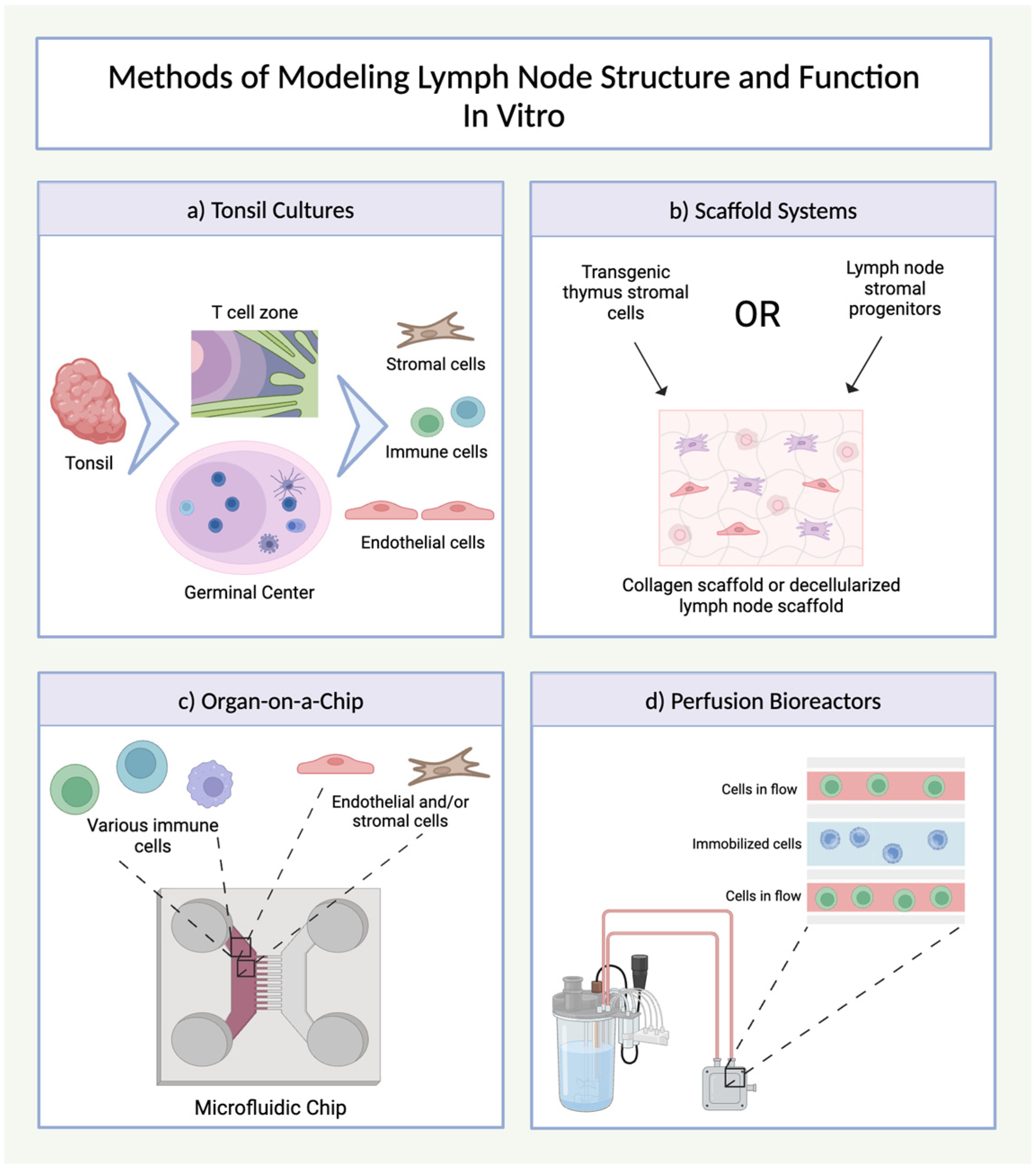
4.2. Lymph Node Organoids

The development of accurate and functional in vitro models of lymph nodes is crucial for understanding immune responses and effectively translating foundational research into clinical medicine (Figure 6). However, recreating the intricate microenvironments of these organs in vitro poses numerous challenges. The diverse cellular composition and complex structures of lymph nodes, described above, require tightly regulated cell–cell interactions, soluble factors, extracellular matrix, and dynamic fluid flow. To date, no human pluripotent stem-cell-derived protocol exists to generate a complete human lymph node organoid. However, a range of in vitro models of lymph nodes and other secondary lymphoid tissue have been developed—from ex vivo cultures of human and mouse lymphoid tissues to complex synthetic scaffolds that mimic in vivo conditions and to organ-on-a-chip models. These models typically rely on ex vivo cell lines and often require in vivo transplantation for long-term maintenance and population by mature lymphoid cells. Therefore, a truly in vitro lymph node organoid that recapitulates the in vivo functions of lymph nodes remains elusive.
The first attempts at modelling human secondary lymphoid tissues used ex vivo cell cultures. One important benefit of this approach is that the model reflects the diversity of cell types and interactions within the in vivo lymphoid organ. These models are often constructed with cultures of the palatine tonsils—a tertiary lymphoid organ found within the throat—as its cellular and morphological features mimic those of lymph nodes [212]. Thus, as they are readily accessible, tonsil explants can be considered as a representative source of cells for human ex vivo lymph node organoids [213]. Traditionally, these organoids were created by combining direct culturing of small pieces of tonsil tissue—which allowed for the growth of tonsillar epithelial cells—with a suspension of tonsil-derived B and T cells [214]. More complex systems have been developed to envelop samples of tonsillar tissue within a collagen scaffold and provide perfusion through a bioreactor that mimics in vivo flow conditions [215].
These early ex vivo models have played critical roles in improving our understanding of lymphoid biology. For instance, a variety of foundational studies on the modes and risk of transmission of HIV was performed in ex vivo secondary lymphoid organ cultures. Maher et al. used ex vivo tonsillar organoid models with time-lapse confocal microscopy to assess how lymphoid tissue morphology impacts cellular transmission of HIV-1 [214]. Soto-Rivera and colleagues built on this system to create a model of HIV-1 transmission within the mucosa-associated lymphoid tissue of the female reproductive tract by developing a combined model that co-cultured dissociated tonsillar lymphoid cells and cervical punch biopsies [216]. These ex vivo secondary lymphoid models have also been used to better understand the structure and function of secondary lymphoid tissues, including antigenic stimulation [217], FDCs [40], and immunoglobulin secretion [41].
Researchers have also developed more advanced ex vivo systems to accurately recapitulate certain substructures that are found within lymph nodes. Wagar and colleagues recently developed a new method for culturing secondary lymphoid organ cells that recapitulate features of GCs, the specialized structures within lymph nodes where B cells undergo affinity maturation and differentiate into antibody-secreting plasma cells [24]. The protocol involves digesting tonsil biopsies (or lung-draining lymph nodes from human organ donors) to obtain a single-cell suspension, which is then cultured in specialized media that promotes organoid formation [24]. These organoids self-organize into structures containing T and B cell zones reminiscent of those found in lymph nodes. These organoids were also able to support the differentiation of B cells into GC-like B cells, characterized by the expression of canonical markers like CD38, upon stimulation with influenza antigens [24]. Furthermore, the organoids facilitated the generation of antigen-specific antibodies, highlighting their potential for modelling humoral immune responses. The ability of this organoid system to generate GC-like structures and support antibody production makes it a valuable in vitro model for studying GC reactions and, by extension, the overall function of lymph nodes in adaptive immunity. These B cell follicle organoids have also been useful for understanding lymph node reactions to glycoconjugate vaccines [42].
Other groups have used scaffolds to guide the 3D organization of lymphoid cells to more accurately recapitulate lymph node structures. In 2004, Suematsu and Watanabe demonstrated that a collagen-based scaffold seeded with thymic stromal cells could trigger the formation of lymph-node-like lymphoid tissue when implanted into the renal subcapsular space of mice [43]. Specifically, this scaffold was composed of sponge-like bovine collagen and thymus stromal cells to mimic lymph node stromal cells engineered to express the lymphoid chemokine LTα. In addition to their structural resemblance to lymph nodes—with distinct T cell zones, B cell zones (with follicles), and HEV-like vessels—these models also recapitulated lymph node function, as they were able to mount an adaptive immune response and secrete antigen-specific IgG1 antibodies [43]. Other scaffolds have been developed by using chemical and/or enzymatic means to remove all cells from lymph nodes, leaving only the extracellular matrix. The first successful decellularization of lymph nodes into a functional scaffold was published in 2005 using the detergent sodium dodecyl sulfate [44]. In 2019, the Brendolan group established functional lymph node organoids by combining mouse-derived lymph node stromal progenitors with scaffolds built from decellularized splenic stromal cell culture [45]. These organoids were successfully transplanted into the renal subcapsular space of mice, where they integrated into the lymphatic vasculature and showed significant lymphatic perfusion. They observed clusters of CD3+ T cells and B220+ B cells, as well as CD31+ (blood vessel) and Prox1+ (lymphatic vessel) endothelium [45]. These organoids also promoted lymphatic drainage and reduced microscopic lymphedema when used to replace surgically resected lymph nodes in mice. Furthermore, these organoids supported an adaptive immune response by inducing T cell proliferation and generating antigen-specific antibody-secreting cells upon immunization [45]. While these results rely on mouse cells, the general approach of developing a complex ECM scaffold from decellularized stromal cell culture appears to be a feasible and reliable method.
Lymph node function is also critically dependent on fluid dynamics. Flow of lymph from afferent lymphatics to the medullary cords allows for the delivery of antigens and factors from draining tissues, as well as the exposure to fluid shear stress, which can influence cell behavior and differentiation. To better mimic this dynamic environment, flow-based systems such as bioreactors and organs-on-a-chip have been incorporated into in vitro lymph node models. Shanti et al. designed an organ-on-a-chip system to mimic lymphatic flow in defined regions, i.e., the subcapsular sinus, reticular conduit network, and the outer cortex and paracortex [218]. Further, these defined regions on the chip were also co-cultured with cell-line-derived DCs and T cells to demonstrate proliferation and viability [218]. Birmingham et al. modelled shear flow in the subcapsular sinus with a microfluidic chip and were able to identify changes in lymphatic E-selectin expression, which impacted cell adhesion of monocytes and cancer cells [219]. Microfluidic chips can also be critical tools for elucidating the numerous and complex signalling pathways that govern lymph node function. To study CXCL12 gradients influencing cell mobility, a flow-free microfluidic chip was designed that controlled for fibronectin and CXCL12 gradients [220]. Using this system, they identified that Jurkat cells respond differently to CXCL12 gradients depending on the fibronectin concentration [220]. Using similar systems, other groups have also focused on chemokine gradients to explore DC responses to CCR7 and CXCR4 [221,222,223].
In addition to organ-on-a-chip models, lymph node organoids were used to model vascular and lymphatic flow through the use of bioreactors. Giese et al. described a miniaturized bioreactor with constant media perfusion, in which a mix of human B cells, peripheral blood mononuclear cells, and myeloid DCs were maintained in an agarose matrix for up to 30 days [224]. Analysis of the supernatants showed interleukin-2 and TNF-alpha responses following antigen stimulation, and immunohistochemistry showed clustering of cells in GC-like formation [224]. This system was the first lymph node organoid that incorporated human peripheral blood cells. Additionally, using human tonsil explants in a 3D perfusion system, Bonaiti et al. were able to recapitulate immune activation in response to influenza [215].
Since lymph nodes have a complex and highly organized structure—consisting of numerous innate and adaptive immune cells, specialized vascular cells, and stromal cells—it is incredibly challenging to replicate their structures and functions with in vitro protocols. No fully stem-cell-derived protocols for lymph node organoids have yet been developed, but researchers have approached this problem from several angles. First, ex vivo cultures of either lymph nodes themselves or tonsillar tissue can be an effective method to model these organs. Second, complex bioscaffolding can facilitate the organization of lymphoid cells into the complex structures of lymph nodes, especially when coupled with additional in vivo transplantations. Furthermore, dynamic organoid models like organ-on-a-chip models and perfusion bioreactors can help capture the critical role of blood and lymphatic flow within these lymph node models. Many of the existing models focus on a single aspect of lymph node structure, such as GCs, the subcapsular sinus, vaccine responses, or chemokine signalling. Future work in this area must build on these strengths to develop fully PSC-derived lymph node organoids, ensuring models that better mimic human physiology. These models will also be critical in understanding lymph node physiology and applying these discoveries to treat disease or develop and test new therapies.

5. Spleen

5.1. Spleen Structure and Function

The spleen is an intraperitoneal secondary lymphoid organ that is specialized for filtering and surveilling the blood for damage, senescent cells, and foreign antigens. Both histologically and functionally, the spleen is divided into two compartments: red pulp and white pulp. By volume, the majority of the spleen consists of red pulp, which is the primary site of erythrocyte filtration [225]. Macrophages that are situated within the splenic cords of the red pulp detect erythrocytes that are senescent or damaged and eliminate them via phagocytosis. The red pulp also plays an important storage function, acting as a reserve of erythrocytes, platelets, and iron that can be mobilized in response to specific stresses like hemorrhage [226,227].
The lymphoid compartment within the white pulp of the spleen also plays a unique role in the function of this organ. This lymphocyte-rich region is organized around blood vessels within the spleen, is structured in specific subregions, and utilizes similar signalling pathways to those present in lymph nodes [228,229]. While the anatomical relationships between these lymphoid compartments differ between lymph nodes and the spleen, their functions, signalling molecules, and microenvironments are very similar [229]. The main difference arises from the source of the antigens; while lymph nodes survey antigens carried via lymphatics from peripheral tissues or visceral organs, splenic cells sample antigens from the blood directly and are therefore able to respond to sepsis and pathogens attempting to spread via the blood flow [46].
The heterogeneous and specialized structures of the spleen are challenging to recapitulate in vitro. An effective model would need to capture the spleen’s structural complexity, such as the distinct vasculature, as well as its diverse functions in erythrocyte filtration, adaptive immune responses to blood-borne antigens, and myeloid-cell-mediated responses [47,48].

5.2. Spleen Organoid Models

Similar to lymph nodes, existing spleen organoid models are limited in number and in scope. This is likely compounded by the lack of focus on splenic function. Accordingly, some groups have aimed to create generic secondary lymphoid organoids—which capture the general concepts and organization shared by the spleen, lymph nodes, and other secondary lymphoid organs such as mucosa-associated lymphoid tissue—without capturing the unique structure or function of the spleen. A description of these secondary lymphoid organoids is included in Section 4.2 above. There are many avenues for future development in spleen-specific organoids, as robust stem-cell-derived splenic organoids have not yet been developed. Nonetheless, a few splenic organoid models have been created using tissue from human donors or mice.
Using spleen tissue as a cell source allows researchers to include the full breadth of physiologically relevant cell types and maintaining some or all of the original tissue structure. Finetti et al. described a methodology to keep mouse spleen slices alive for up to 48 h with no significant loss of cell viability [49]. Furthermore, the protocol used by Wagar et al. to generate tonsil and lymph node organoids (reaggregating single-cell suspensions created from donor tissues) was also effective for spleen samples, resulting in organoids that could mount an adaptive immune response to both live-attenuated and wild-type H1N1 influenza virus [24].
Gee and colleagues recently created spleen organoids using human tissue that then acted as a functional spleen when transplanted into a mouse [23]. The authors used enzymatic digestion and mechanical disruption of human donor spleens to generate multicellular clusters, which were then seeded onto a polyglycolic acid–poly-l lactic acid scaffold to generate “spleen organoid units” [23]. Cells from these engineered spleens exhibited proliferation upon implantation into mice, and after one month, they were able to scavenge damaged erythrocytes [23]. The immune cells in these engineered spleens expressed prototypic markers (such as CD11c, CD4) expected within splenic tissue, but the authors did not test the ability of these cells to respond to antigens [23].
Other groups attempted to model the function of splenic red pulp in clearing senescent erythrocytes. Using a microfabricated filtering unit, one group mimicked spleen sinusoidal flow to assess erythrocyte deformability [230]. Another model focused on mimicking the blood flow through the spleen by using a two-layer microfluidic device that had open–slow and closed–fast channels [231]. Although no endothelial cells were included, the authors were able to show differences in cell movement dynamics between uninfected and Plasmodium yoelii-infected human erythrocytes [231].
In summary, there are very few existing organoid models of splenic function. Some models focused on the flow and uptake of erythrocytes within the spleen, but models of the splenic white pulp are few. For example, innate functions in the spleen have yet to be modeled, such as the populations of neutrophils that protect against Streptococcus infection [232]. In order to properly study the pathogen-clearing ability of the spleen, models need to recapitulate functional innate immune cells along with flow resembling in vivo blood flow that brings blood-borne bacteria into the spleen. For designing an entirely in vitro system, bone marrow organoid-derived immune cells would be an ideal source. These cells could be pumped through a decellularized spleen scaffold and allowed to populate this as resident cells. This is just one of the attainable directions in which spleen organoid research could progress. In terms of assessing immune functions, the current models are limited and need to be further developed. The limited progress in the spleen organoid field may be attributed to the perceived similarity between the adaptive functions of the spleen and the lymph nodes, and this may lead researchers to assume there is less potential for scientific advances in this particular area. However, the unique microarchitecture, targeted pathogens, and pathologies found in the spleen necessitate unique organoid models.

6. Conclusions

In recent years, there has been an explosion in the development of organoid models for lymphoid tissues, fueled by advances in biomaterial engineering, microfluidics, and stem cell biology. These have resulted in robust organoid models for the thymus and bone marrow, recently followed by advances in the in vitro modelling of secondary lymphoid tissues including lymph nodes, spleen, and tonsils. These models allow for scalable and highly tractable systems for investigating fundamental biology and assessing disease mechanisms and new therapies. These immune organoids also hold promise for applications in personalized medicine, such as identifying drug side effects. For example, as lymph node organoids improve and become easier to generate, they could be used as a high-throughput way to test vaccine responses before moving to human trials. Further, several groups have successfully engrafted lymph-node-like organoids to replace excised lymph nodes and maintain lymphatic flow in mouse models. Once this technique has been optimized, it could be implemented in patients to treat lymphedema secondary to lymph node dissections. Bone marrow organoids could be developed for use as transplantation sources to cure bone-marrow-derived cancers without the need for a matching donor. A robust bone marrow organoid model could also be used to investigate how certain bone-marrow-derived cancers arise and screen for potential therapies in a high-throughput manner.
While current thymus models are impressive, the existing thymus models have not yet delved into modelling the aging thymus. Following adolescence, the thymus begins to involute, and the production of thymocytes begins to drop. Further, this is associated with reduced central tolerance and decreased negative selection. This phenomenon is a problem for older cancer patients in remission if they were treated with chemotherapy, which depletes existing immune cell populations [233]. Older patients have much lower T cell production and will have a reduced ability to replace their circulating T cells, and even after recovery from cancer, they will have a decreased immunity to infection. Modelling thymic aging in mice, while not difficult, is time-consuming and expensive due to mouse housing costs. The development of a thymic organoid that could model aging-associated thymic involution would help elucidate more about the field.
Existing models for these organs are associated with a variety of limitations. First, there are still a number of barriers for generating mature cells of lymphoid and other immune lineages when using only pluripotent stem cells and off-the-shelf reagents. Thus, these models rely on using differentiated cells from donors, equivalent cells from animal models, and existing cell lines, otherwise the cells present in the current organoid models retain an overall immature phenotype. It is therefore critical to develop differentiation protocols for mature hematopoietic cells, especially lymphoid cells, which are particularly challenging to differentiate without the aid of transplantation into an in vivo model.
Furthermore, there has often been a divergence between static organoid models, which are increasingly incorporating complex structures and cellular interactions, and perfusion-based organoids such as microfluidic and bioreactor models, which can model dynamic processes but often lack the cellular complexity of static models. To generate models that truly recapitulate human physiology, it will be critical to merge dynamic systems with the complexity of organoid models using methods such as organoids-on-a-chip.
Moreover, in many cases, our understanding of the specific signalling pathways and precise processes involved in establishing these organs is still limited. Existing models also do not faithfully capture the unique structural elements that distinguish different secondary lymphoid organs—such as the periarteriolar lymphoid sheath of the splenic white pulp or the medullary cords of lymph nodes.
Lymphoid organoid models hold immense promise for revolutionizing our understanding of the human immune system, enabling the development of novel therapeutics and personalized medicine approaches.

Author Contributions

Conceptualization, A.B. and J.R.; resources, A.B., J.R. and C.Q.; writing—original draft preparation, A.B. and J.R.; writing—review and editing, A.B., J.R. and C.Q.; visualization, A.B. and J.R.; supervision, J.M.P.; project administration, A.B.; funding acquisition, A.B., C.Q. and J.M.P. All authors have read and agreed to the published version of the manuscript.

Funding

A.B. and C.Q. were funded by a Michael Smith Health Research BC Trainee Award. C.Q. and J.M.P. were funded by the 21CVD03 Leducq Foundation with the Transatlantic Network of Excellence grant “ReVAMP—Recalibrating Mechanotransduction in Vascular Malformations” (2022–2027). J.M.P was further funded by the Canada 150 Research Chairs Program F18-01336.

Data Availability Statement

No new data were created or analyzed in this study. Data sharing is not applicable to this article.

Conflicts of Interest

The authors declare no conflicts of interest.

References

  1. Carneiro, C.F.D.; Drude, N.; Hülsemann, M.; Collazo, A.; Toelch, U. Mapping Strategies towards Improved External Validity in Preclinical Translational Research. Expert Opin. Drug Discov. 2023, 18, 1273–1285. [Google Scholar] [CrossRef] [PubMed]
  2. Pound, P.; Ritskes-Hoitinga, M. Is It Possible to Overcome Issues of External Validity in Preclinical Animal Research? Why Most Animal Models Are Bound to Fail. J. Transl. Med. 2018, 16, 304. [Google Scholar] [CrossRef] [PubMed]
  3. Contopoulos-Ioannidis, D.G.; Ntzani, E.E.; Ioannidis, J.P.A. Translation of Highly Promising Basic Science Research into Clinical Applications. Am. J. Med. 2003, 114, 477–484. [Google Scholar] [CrossRef]
  4. Fogel, D.B. Factors Associated with Clinical Trials That Fail and Opportunities for Improving the Likelihood of Success: A Review. Contemp. Clin. Trials Commun. 2018, 11, 156–164. [Google Scholar] [CrossRef] [PubMed]
  5. Stahl, M.; Gore, S.D.; Vey, N.; Prebet, T. Lost in Translation? Ten Years of Development of Histone Deacetylase Inhibitors in Acute Myeloid Leukemia and Myelodysplastic Syndromes. Expert Opin. Investig. Drugs 2016, 25, 307–317. [Google Scholar] [CrossRef]
  6. Witzig, T.E.; Tobinai, K.; Rigacci, L.; Ikeda, T.; Vanazzi, A.; Hino, M.; Shi, Y.; Mayer, J.; Costa, L.J.; Bermudez Silva, C.D.; et al. Adjuvant Everolimus in High-Risk Diffuse Large B-Cell Lymphoma: Final Results from the PILLAR-2 Randomized Phase III Trial. Ann. Oncol. 2018, 29, 707–714. [Google Scholar] [CrossRef]
  7. Xu, Z.-Z.; Wang, W.-F.; Fu, W.-B.; Wang, A.-H.; Liu, Z.-Y.; Chen, L.-Y.; Guo, P.; Li, J.-M. Combination of Rituximab and Mammalian Target of Rapamycin Inhibitor Everolimus (RAD001) in Diffuse Large B-Cell Lymphoma. Leuk. Lymphoma 2014, 55, 1151–1157. [Google Scholar] [CrossRef]
  8. Janning, M.; Fiedler, W. Volasertib for the Treatment of Acute Myeloid Leukemia: A Review of Preclinical and Clinical Development. Future Oncol. 2014, 10, 1157–1165. [Google Scholar] [CrossRef]
  9. Dohner, H.; Symeonidis, A.; Deeren, D.; Demeter, J.; Sanz, M.A.; Anagnostopoulos, A.; Esteve, J.; Fiedler, W.; Porkka, K.; Kim, H.J.; et al. Adjunctive Volasertib in Patients with Acute Myeloid Leukemia Not Eligible for Standard Induction Therapy: A Randomized, Phase 3 Trial. Hemasphere 2021, 5, e617. [Google Scholar] [CrossRef]
  10. An, Z.-Y.; Fu, H.; He, Y.; Zhu, X.; Huang, Q.-S.; Wu, J.; Liu, K.; Zhang, X. Projected Global Trends in Hematological Malignancies: Incidence, Mortality, and Disability-Adjusted Life Years from 2020 to 2030. Blood 2023, 142 (Suppl. 1), 3810. [Google Scholar] [CrossRef]
  11. Zhang, N.; Wu, J.; Wang, Q.; Liang, Y.; Li, X.; Chen, G.; Ma, L.; Liu, X.; Zhou, F. Global Burden of Hematologic Malignancies and Evolution Patterns over the Past 30 Years. Blood Cancer J. 2023, 13, 82. [Google Scholar] [CrossRef]
  12. Pulte, D.; Jansen, L.; Brenner, H. Changes in Long Term Survival after Diagnosis with Common Hematologic Malignancies in the Early 21st Century. Blood Cancer J. 2020, 10, 1–8. [Google Scholar] [CrossRef]
  13. Purton, L.E.; Scadden, D.T. The Hematopoietic Stem Cell Niche in Stembook; Harvard Stem Cell Institute: Cambridge, MA, USA, 2008; Available online: https://www.stembook.org/articles/the-hematopoietic-stem-cell-niche/ (accessed on 10 February 2025).
  14. Parekh, C.; Crooks, G.M. Critical Differences in Hematopoiesis and Lymphoid Development between Humans and Mice. J. Clin. Immunol. 2013, 33, 711–715. [Google Scholar] [CrossRef] [PubMed]
  15. Nombela-Arrieta, C.; Manz, M.G. Quantification and Three-Dimensional Microanatomical Organization of the Bone Marrow. Blood Adv. 2017, 1, 407–416. [Google Scholar] [CrossRef]
  16. Kim, J.; Koo, B.K.; Knoblich, J.A. Human Organoids: Model Systems for Human Biology and Medicine. Nat. Rev. Mol. Cell Biol. 2020, 21, 571–584. [Google Scholar] [CrossRef] [PubMed]
  17. Mestas, J.; Hughes, C.C.W. Of Mice and Not Men: Differences between Mouse and Human Immunology. J. Immunol. 2004, 172, 2731–2738. [Google Scholar] [CrossRef]
  18. Takahashi, T. Organoids for Drug Discovery and Personalized Medicine. Annu. Rev. Pharmacol. Toxicol. 2019, 59, 447–462. [Google Scholar] [CrossRef]
  19. Frenz-Wiessner, S.; Fairley, S.D.; Buser, M.; Goek, I.; Salewskij, K.; Jonsson, G.; Illig, D.; zu Putlitz, B.; Petersheim, D.; Li, Y.; et al. Generation of Complex Bone Marrow Organoids from Human Induced Pluripotent Stem Cells. Nat. Methods 2024, 21, 868–881. [Google Scholar] [CrossRef]
  20. Khan, A.O.; Rodriguez-Romera, A.; Reyat, J.S.; Olijnik, A.A.; Colombo, M.; Wang, G.; Wen, W.X.; Sousos, N.; Murphy, L.C.; Grygielska, B.; et al. Human Bone Marrow Organoids for Disease Modeling, Discovery, and Validation of Therapeutic Targets in Hematologic Malignancies. Cancer Discov. 2023, 13, 364–385. [Google Scholar] [CrossRef]
  21. Seet, C.S.; He, C.; Bethune, M.T.; Li, S.; Chick, B.; Gschweng, E.H.; Zhu, Y.; Kim, K.; Kohn, D.B.; Baltimore, D.; et al. Generation of Mature T Cells from Human Hematopoietic Stem and Progenitor Cells in Artificial Thymic Organoids. Nat. Methods 2017, 14, 521–530. [Google Scholar] [CrossRef]
  22. Montel-Hagen, A.; Seet, C.S.; Li, S.; Chick, B.; Zhu, Y.; Chang, P.; Tsai, S.; Sun, V.; Lopez, S.; Chen, H.C.; et al. Organoid-Induced Differentiation of Conventional T Cells from Human Pluripotent Stem Cells. Cell Stem Cell 2019, 24, 376–389.e8. [Google Scholar] [CrossRef] [PubMed]
  23. Gee, K.; Isani, M.A.; Fode, A.; Maselli, K.M.; Zuber, S.M.; Fowler, K.L.; Squillaro, A.I.; Nucho, L.M.A.; Grikscheit, T.C. Spleen Organoid Units Generate Functional Human and Mouse Tissue-Engineered Spleen in a Murine Model. Tissue Eng. Part. A 2020, 26, 411–418. [Google Scholar] [CrossRef] [PubMed]
  24. Wagar, L.E.; Salahudeen, A.; Constantz, C.M.; Wendel, B.S.; Lyons, M.M.; Mallajosyula, V.; Jatt, L.P.; Adamska, J.Z.; Blum, L.K.; Gupta, N.; et al. Modeling Human Adaptive Immune Responses with Tonsil Organoids. Nat. Med. 2021, 27, 125–135. [Google Scholar] [CrossRef] [PubMed]
  25. Karagiannis, P.; Takahashi, K.; Saito, M.; Yoshida, Y.; Okita, K.; Watanabe, A.; Inoue, H.; Yamashita, J.K.; Todani, M.; Nakagawa, M.; et al. Induced Pluripotent Stem Cells and Their Use in Human Models of Disease and Development. Physiol. Rev. 2019, 99, 79–114. [Google Scholar] [CrossRef]
  26. Saglam-Metiner, P.; Devamoglu, U.; Filiz, Y.; Akbari, S.; Beceren, G.; Goker, B.; Yaldiz, B.; Yanasik, S.; Biray Avci, C.; Erdal, E.; et al. Spatio-Temporal Dynamics Enhance Cellular Diversity, Neuronal Function and Further Maturation of Human Cerebral Organoids. Commun. Biol. 2023, 6, 1–18. [Google Scholar] [CrossRef]
  27. Cho, Y.K.; Kim, H.K.; Kwon, S.S.; Jeon, S.H.; Cheong, J.W.; Nam, K.T.; Kim, H.S.; Kim, S.; Kim, H.O. In Vitro Erythrocyte Production Using Human-Induced Pluripotent Stem Cells: Determining the Best Hematopoietic Stem Cell Sources. Stem Cell Res. Ther. 2023, 14, 1–15. [Google Scholar] [CrossRef]
  28. Allenby, M.C.; Tahlawi, A.; Morais, J.C.F.; Li, K.; Panoskaltsis, N.; Mantalaris, A. Ceramic Hollow Fibre Constructs for Continuous Perfusion and Cell Harvest from 3D Hematopoietic Organoids. Stem Cells Int. 2018, 2018, 6230214. [Google Scholar] [CrossRef] [PubMed]
  29. Allenby, M.C.; Okutsu, N.; Brailey, K.; Guasch, J.; Zhang, Q.; Panoskaltsis, N.; Mantalaris, A. A Spatiotemporal Microenvironment Model to Improve Design of a Three-Dimensional Bioreactor for Red Cell Production. Tissue Eng. Part A 2022, 28, 38–53. [Google Scholar] [CrossRef]
  30. Torisawa, Y.S.; Spina, C.S.; Mammoto, T.; Mammoto, A.; Weaver, J.C.; Tat, T.; Collins, J.J.; Ingber, D.E. Bone Marrow–on–a–Chip Replicates Hematopoietic Niche Physiology in Vitro. Nat. Methods 2014, 11, 663–669. [Google Scholar] [CrossRef]
  31. Glaser, D.E.; Curtis, M.B.; Sariano, P.A.; Rollins, Z.A.; Shergill, B.S.; Anand, A.; Deely, A.M.; Shirure, V.S.; Anderson, L.; Lowen, J.M.; et al. Organ-on-a-Chip Model of Vascularized Human Bone Marrow Niches. Biomaterials 2022, 280, 121245. [Google Scholar] [CrossRef]
  32. Anderson, G.; Jenkinson, E.J. Investigating Central Tolerance with Reaggregate Thymus Organ Cultures. Methods Mol. Biol. 2007, 380, 185–196. [Google Scholar] [CrossRef]
  33. Chung, B.; Montel-Hagen, A.; Ge, S.; Blumberg, G.; Kim, K.; Klein, S.; Zhu, Y.; Parekh, C.; Balamurugan, A.; Yang, O.O.; et al. Engineering the Human Thymic Microenvironment to Support Thymopoiesis in Vivo. Stem Cells 2014, 32, 2386. [Google Scholar] [CrossRef] [PubMed]
  34. Bosticardo, M.; Notarangelo, L.D. Human Thymus in Health and Disease: Recent Advances in Diagnosis and Biology. Semin. Immunol. 2023, 66, 101732. [Google Scholar] [CrossRef] [PubMed]
  35. Ramos, S.A.; Armitage, L.H.; Morton, J.J.; Alzofon, N.; Handler, D.; Kelly, G.; Homann, D.; Jimeno, A.; Russ, H.A. Generation of Functional Thymic Organoids from Human Pluripotent Stem Cells. Stem Cell Rep. 2023, 18, 829–840. [Google Scholar] [CrossRef]
  36. Hübscher, T.; Lorenzo-Martín, L.F.; Barthlott, T.; Tillard, L.; Langer, J.J.; Rouse, P.; Blackburn, C.C.; Holländer, G.; Lutolf, M.P. Thymic Epithelial Organoids Mediate T-Cell Development. Development 2024, 151, dev202853. [Google Scholar] [CrossRef] [PubMed]
  37. Bogoslowski, A.; Butcher, E.C.; Kubes, P. Neutrophils Recruited through High Endothelial Venules of the Lymph Nodes via PNAd Intercept Disseminating Staphylococcus Aureus. Proc. Natl. Acad. Sci. USA 2018, 115, 2449–2454. [Google Scholar]
  38. Louie, D.A.P.; Liao, S. Lymp Node Subcapsular Sinus Macrophages as the Frontline of Lymphatic Immune Defense. Front. Immunol. 2019, 10, 438090. [Google Scholar] [CrossRef]
  39. Phan, T.G.; Green, J.A.; Xu, Y.; Cyster, J.G. Immune Complex Relay by Subcapsular Sinus Macrophages and Noncognate B Cells Drives Antibody Affinity Maturation. Nat. Immunol. 2009, 10, 786–793. [Google Scholar] [CrossRef]
  40. Skibinski, G.; Skibinska, A.; Deckers, M.; James, K. Tonsil Stromal-Cell Lines Expressing FDC-like Properties: Isolation, Characterization, and Interaction with B Lymphocytes. J. Immunol. Res. 1998, 6, 273–284. [Google Scholar] [CrossRef]
  41. Van Laar, J.M.; Melchers, M.; Teng, Y.K.O.; Van Der Zouwen, B.; Mohammadi, R.; Fischer, R.; Margolis, L.; Fitzgerald, W.; Grivel, J.C.; Breedveld, F.C.; et al. Sustained Secretion of Immunoglobulin by Long-Lived Human Tonsil Plasma Cells. Am. J. Pathol. 2007, 171, 917–927. [Google Scholar] [CrossRef]
  42. Moeller, T.D.; Shah, S.B.; Lai, K.; Lopez-Barbosa, N.; Desai, P.; Wang, W.; Zhong, Z.; Redmond, D.; Singh, A.; DeLisa, M.P. Profiling Germinal Center-like B Cell Responses to Conjugate Vaccines Using Synthetic Immune Organoids. ACS Cent. Sci. 2023, 9, 787–804. [Google Scholar] [CrossRef] [PubMed]
  43. Suematsu, S.; Watanabe, T. Generation of a Synthetic Lymphoid Tissue–like Organoid in Mice. Nat. Biotechnol. 2004, 22, 1539–1545. [Google Scholar] [CrossRef] [PubMed]
  44. Cuzzone, D.A.; Albano, N.J.; Aschen, S.Z.; Ghanta, S.; Mehrara, B.J. Decellularized Lymph Nodes as Scaffolds for Tissue Engineered Lymph Nodes. Lymphat. Res. Biol. 2015, 13, 186–194. [Google Scholar] [CrossRef] [PubMed]
  45. Lenti, E.; Bianchessi, S.; Proulx, S.T.; Palano, M.T.; Genovese, L.; Raccosta, L.; Spinelli, A.; Drago, D.; Andolfo, A.; Alfano, M.; et al. Therapeutic Regeneration of Lymphatic and Immune Cell Functions upon Lympho-Organoid Transplantation. Stem Cell Rep. 2019, 12, 1260–1268. [Google Scholar] [CrossRef]
  46. Altamura, M.; Caradonna, L.; Amati, L.; Pellegrino, N.M.; Urgesi, G.; Miniello, S. SPLENECTOMY AND SEPSIS: THE ROLE OF THE SPLEEN IN THE IMMUNE-MEDIATED BACTERIAL CLEARANCE. Immunopharmacol. Immunotoxicol. 2001, 23, 153–161. [Google Scholar] [CrossRef]
  47. Golub, R.; Tan, J.; Watanabe, T.; Brendolan, A. Origin and Immunological Functions of Spleen Stromal Cells. Trends Immunol. 2018, 39, 503–514. [Google Scholar] [CrossRef]
  48. Schmidt, E.E.; MacDonald, I.C.; Groom, A.C. Comparative Aspects of Splenic Microcirculatory Pathways in Mammals: The Region Bordering the White Pulp. Scanning Microsc. 1993, 7, 17. [Google Scholar]
  49. Finetti, F.; Capitani, N.; Manganaro, N.; Tatangelo, V.; Libonati, F.; Panattoni, G.; Calaresu, I.; Ballerini, L.; Baldari, C.T.; Patrussi, L. Optimization of Organotypic Cultures of Mouse Spleen for Staining and Functional Assays. Front. Immunol. 2020. [Google Scholar] [CrossRef]
  50. Itkin, T.; Gur-Cohen, S.; Spencer, J.A.; Schajnovitz, A.; Ramasamy, S.K.; Kusumbe, A.P.; Ledergor, G.; Jung, Y.; Milo, I.; Poulos, M.G.; et al. Distinct Bone Marrow Blood Vessels Differentially Regulate Haematopoiesis. Nature 2016, 532, 323–328. [Google Scholar] [CrossRef]
  51. Bessy, T.; Itkin, T.; Passaro, D. Bioengineering the Bone Marrow Vascular Niche. Front. Cell Dev. Biol. 2021, 9, 645496. [Google Scholar] [CrossRef]
  52. Wu, Q.; Zhang, J.; Kumar, S.; Shen, S.; Kincaid, M.; Johnson, C.B.; Zhang, Y.S.; Turcotte, R.; Alt, C.; Ito, K.; et al. Resilient Anatomy and Local Plasticity of Naive and Stress Haematopoiesis. Nature 2024, 627, 839–846. [Google Scholar] [CrossRef] [PubMed]
  53. Zhang, J.; Wu, Q.; Johnson, C.B.; Pham, G.; Kinder, J.M.; Olsson, A.; Slaughter, A.; May, M.; Weinhaus, B.; D’Alessandro, A.; et al. In Situ Mapping Identifies Distinct Vascular Niches for Myelopoiesis. Nature 2021, 590, 457–462. [Google Scholar] [CrossRef]
  54. Dennis, J.E.; Caplan, A.I. Bone Marrow Mesenchymal Stem Cells; Humana Press: Totowa, NJ, USA, 2004; pp. 107–117. [Google Scholar] [CrossRef]
  55. Lee, C.C.I.; Christensen, J.E.; Yoder, M.C.; Tarantal, A.F. Clonal Analysis and Hierarchy of Human Bone Marrow Mesenchymal Stem and Progenitor Cells. Exp. Hematol. 2010, 38, 46–54. [Google Scholar] [CrossRef]
  56. Hegner, B.; Lange, M.; Kusch, A.; Essin, K.; Sezer, O.; Schulze-Lohoff, E.; Luft, F.C.; Gollasch, M.; Dragun, D. MTOR Regulates Vascular Smooth Muscle Cell Differentiation from Human Bone Marrow-Derived Mesenchymal Progenitors. Arterioscler. Thromb. Vasc. Biol. 2009, 29, 232–238. [Google Scholar] [CrossRef] [PubMed]
  57. Kolf, C.M.; Cho, E.; Tuan, R.S. Mesenchymal Stromal Cells. Biology of Adult Mesenchymal Stem Cells: Regulation of Niche, Self-Renewal and Differentiation. Arthritis Res. Ther. 2007, 9, 204. [Google Scholar] [CrossRef]
  58. Gao, Q.; Wang, L.; Wang, S.; Huang, B.; Jing, Y.; Su, J. Bone Marrow Mesenchymal Stromal Cells: Identification, Classification, and Differentiation. Front. Cell Dev. Biol. 2022, 9, 787118. [Google Scholar] [CrossRef]
  59. Frenette, P.S.; Pinho, S.; Lucas, D.; Scheiermann, C. Mesenchymal Stem Cell: Keystone of the Hematopoietic Stem Cell Niche and a Stepping-Stone for Regenerative Medicine. Annu. Rev. Immunol. 2013, 31, 285–316. [Google Scholar] [CrossRef] [PubMed]
  60. Jing, D.; Fonseca, A.V.; Alakel, N.; Fierro, F.A.; Muller, K.; Bornhauser, M.; Ehninger, G.; Corbeil, D.; Ordemann, R. Hematopoietic Stem Cells in Co-Culture with Mesenchymal Stromal Cells-Modeling the Niche Compartments In Vitro. Haematologica 2010, 95, 542–550. [Google Scholar] [CrossRef]
  61. Ding, L.; Saunders, T.L.; Enikolopov, G.; Morrison, S.J. Endothelial and Perivascular Cells Maintain Haematopoietic Stem Cells. Nature 2012, 481, 457–462. [Google Scholar] [CrossRef]
  62. Poulos, M.G.; Guo, P.; Kofler, N.M.; Pinho, S.; Gutkin, M.C.; Tikhonova, A.; Aifantis, I.; Frenette, P.S.; Kitajewski, J.; Rafii, S.; et al. Endothelial Jagged-1 Is Necessary for Homeostatic and Regenerative Hematopoiesis. Cell Rep. 2013, 4, 1022–1034. [Google Scholar] [CrossRef]
  63. Wu, J.Y.; Scadden, D.T.; Kronenberg, H.M. Role of the Osteoblast Lineage in the Bone Marrow Hematopoietic Niches. J. Bone Miner. Res. 2009, 24, 759–764. [Google Scholar] [CrossRef] [PubMed]
  64. Robles, H.; Park, S.J.; Joens, M.S.; Fitzpatrick, J.A.J.; Craft, C.S.; Scheller, E.L. Characterization of the Bone Marrow Adipocyte Niche with Three-Dimensional Electron Microscopy. Bone 2019, 118, 89–98. [Google Scholar] [CrossRef] [PubMed]
  65. Cuminetti, V.; Arranz, L. Bone Marrow Adipocytes: The Enigmatic Components of the Hematopoietic Stem Cell Niche. J. Clin. Med. 2019, 8, 707. [Google Scholar] [CrossRef]
  66. Boulais, P.E.; Frenette, P.S. Making Sense of Hematopoietic Stem Cell Niches. Blood 2015, 125, 2621–2629. [Google Scholar] [CrossRef] [PubMed]
  67. Belizario, J.E. Immunodeficient Mouse Models: An Overview. AACE Clin. Case Rep. 2021, 7, 79–85. [Google Scholar] [CrossRef]
  68. Fiedler, K.; Sindrilaru, A.; Terszowski, G.; Kokai, E.; Feyerabend, T.B.; Bullinger, L.; Rodewald, H.R.; Brunner, C. Neutrophil Development and Function Critically Depend on Bruton Tyrosine Kinase in a Mouse Model of X-Linked Agammaglobulinemia. Blood 2011, 117, 1329–1339. [Google Scholar] [CrossRef]
  69. Blundell, M.P.; Bouma, G.; Calle, Y.; Jones, G.E.; Kinnon, C.; Thrasher, A.J. Improvement of Migratory Defects in a Murine Model of Wiskott–Aldrich Syndrome Gene Therapy. Mol. Ther. 2008, 16, 836–844. [Google Scholar] [CrossRef]
  70. Peng, C.; Li, S. Chronic Myeloid Leukemia (CML) Mouse Model in Translational Research. Methods Mol. Biol. 2016, 1438, 225–243. [Google Scholar] [CrossRef]
  71. O’Neil, J.; Calvo, J.; McKenna, K.; Krishnamoorthy, V.; Aster, J.C.; Bassing, C.H.; Alt, F.W.; Kelliher, M.; Look, A.T. Activating Notch1 Mutations in Mouse Models of T-ALL. Blood 2006, 107, 781–785. [Google Scholar] [CrossRef]
  72. Deshpande, A.J.; Cusan, M.; Rawat, V.P.S.; Reuter, H.; Krause, A.; Pott, C.; Quintanilla-Martinez, L.; Kakadia, P.; Kuchenbauer, F.; Ahmed, F.; et al. Acute Myeloid Leukemia Is Propagated by a Leukemic Stem Cell with Lymphoid Characteristics in a Mouse Model of CALM/AF10-Positive Leukemia. Cancer Cell 2006, 10, 363–374. [Google Scholar] [CrossRef]
  73. Aydemir, N.; Bilaloǧlu, R. Genotoxicity of Two Anticancer Drugs, Gemcitabine and Topotecan, in Mouse Bone Marrow In Vivo. Mutat. Res. Genet. Toxicol. Environ. Mutagen. 2003, 537, 43–51. [Google Scholar] [CrossRef]
  74. Till, J.E.; Mcculloch, E.A. A Direct Measurement of the Radiation Sensitivity of Normal Mouse Bone Marrow Cells. Radiat. Res. 1961, 14, 213–222. [Google Scholar]
  75. Wolf, N.S.; Trentin, J.J. Hemopoietic colony studies: V. Effect of hemopoietic organ stroma on differentiation of pluripotent stem cells. J. Exp. Med. 1968, 127, 205–214. [Google Scholar] [CrossRef] [PubMed]
  76. Reddy, P.; Negrin, R.; Hill, G.R. Mouse Models of Bone Marrow Transplantation. Biol. Blood Marrow Transplant. 2008, 14 (Suppl. 1), 129. [Google Scholar] [CrossRef]
  77. Almosailleakh, M.; Schwaller, J. Murine Models of Acute Myeloid Leukaemia. Int. J. Mol. Sci. 2019, 20, 453. [Google Scholar] [CrossRef]
  78. Ilaria, R.L. Animal Models of Chronic Myelogenous Leukemia. Hematol. Oncol. Clin. N. Am. 2004, 18, 525–543. [Google Scholar] [CrossRef]
  79. Jacoby, E.; Chien, C.D.; Fry, T.J. Murine Models of Acute Leukemia: Important Tools in Current Pediatric Leukemia Research. Front. Oncol. 2014, 4, 95. [Google Scholar] [CrossRef]
  80. Simonetti, G.; Bertilaccio, M.T.S.; Ghia, P.; Klein, U. Mouse Models in the Study of Chronic Lymphocytic Leukemia Pathogenesis and Therapy. Blood 2014, 124, 1010–1019. [Google Scholar] [CrossRef]
  81. Rossi, M.; Botta, C.; Arbitrio, M.; Grembiale, R.D.; Tagliaferri, P.; Tassone, P. Mouse Models of Multiple Myeloma: Technologic Platforms and Perspectives. Oncotarget 2018, 9, 20119. [Google Scholar] [CrossRef]
  82. Beachy, S.H.; Aplan, P.D. Mouse Models of Myelodysplastic Syndromes. Hematol. Oncol. Clin. N. Am. 2010, 24, 361–375. [Google Scholar] [CrossRef]
  83. Jacquelin, S.; Kramer, F.; Mullally, A.; Lane, S.W. Murine Models of Myelofibrosis. Cancers 2020, 12, 2381. [Google Scholar] [CrossRef]
  84. Scheinberg, P.; Chen, J. Aplastic Anemia: What Have We Learned From Animal Models and From the Clinic. Semin. Hematol. 2013, 50, 156–164. [Google Scholar] [CrossRef] [PubMed]
  85. Leblond, V.; Autran, B.; Cesbron, J.Y. The SCID Mouse Mutant: Definition and Potential Use as a Model for Immune and Hematological Disorders. Hematol. Cell Ther. 1997, 39, 213–221. [Google Scholar] [CrossRef]
  86. Blebea, J.S.; Houseni, M.; Torigian, D.A.; Fan, C.; Mavi, A.; Zhuge, Y.; Iwanaga, T.; Mishra, S.; Udupa, J.; Zhuang, J.; et al. Structural and Functional Imaging of Normal Bone Marrow and Evaluation of Its Age-Related Changes. Semin. Nucl. Med. 2007, 37, 185–194. [Google Scholar] [CrossRef] [PubMed]
  87. Abarrategi, A.; Mian, S.A.; Passaro, D.; Rouault-Pierre, K.; Grey, W.; Bonnet, D. Modeling the Human Bone Marrow Niche in Mice: From Host Bone Marrow Engraftment to Bioengineering Approaches. J. Exp. Med. 2018, 215, 729–743. [Google Scholar] [CrossRef] [PubMed]
  88. Delorme, B.; Charbord, P. Culture and Characterization of Human Bone Marrow Mesenchymal Stem Cells. Methods Mol. Med. 2007, 140, 67–81. [Google Scholar] [CrossRef]
  89. Bonab, M.M.; Alimoghaddam, K.; Talebian, F.; Ghaffari, S.H.; Ghavamzadeh, A.; Nikbin, B. Aging of Mesenchymal Stem Cell in Vitro. BMC Cell Biol. 2006, 7, 14. [Google Scholar] [CrossRef]
  90. Frisch, B.J.; Calvi, L.M. Hematopoietic Stem Cell Cultures and Assays. Methods Mol. Biol. 2014, 1130, 315–324. [Google Scholar] [CrossRef]
  91. Audet, J.; Zandstra, P.W.; Eaves, C.J.; Piret, J.M. Advances in Hematopoietic Stem Cell Culture. Curr. Opin. Biotechnol. 1998, 9, 146–151. [Google Scholar] [CrossRef]
  92. Zhang, H.; Reilly, M.P. Human Induced Pluripotent Stem Cell? Derived Macrophages for Unraveling Human Macrophage Biology. Arterioscler. Thromb. Vasc. Biol. 2017, 37, 2000–2006. [Google Scholar] [CrossRef]
  93. Morishima, T.; Watanabe, K.; Niwa, A.; Fujino, H.; Matsubara, H.; Adachi, S.; Suemori, H.; Nakahata, T.; Heike, T. Neutrophil Differentiation from Human-Induced Pluripotent Stem Cells. J. Cell Physiol. 2011, 226, 1283–1291. [Google Scholar] [CrossRef]
  94. Nianias, A.; Themeli, M. Induced Pluripotent Stem Cell (IPSC)–Derived Lymphocytes for Adoptive Cell Immunotherapy: Recent Advances and Challenges. Curr. Hematol. Malig. Rep. 2019, 14, 261–268. [Google Scholar] [CrossRef] [PubMed]
  95. Diederichs, S.; Tuan, R.S. Functional Comparison of Human-Induced Pluripotent Stem Cell-Derived Mesenchymal Cells and Bone Marrow-Derived Mesenchymal Stromal Cells from the Same Donor. Stem Cells Dev. 2014, 23, 1594–1610. [Google Scholar] [CrossRef] [PubMed]
  96. Buchrieser, J.; James, W.; Moore, M.D. Human Induced Pluripotent Stem Cell-Derived Macrophages Share Ontogeny with MYB-Independent Tissue-Resident Macrophages. Stem Cell Rep. 2017, 8, 334–345. [Google Scholar] [CrossRef] [PubMed]
  97. de Janon, A.; Mantalaris, A.; Panoskaltsis, N. Three-Dimensional Human Bone Marrow Organoids for the Study and Application of Normal and Abnormal Hematoimmunopoiesis. J. Immunol. 2023, 210, 895–904. [Google Scholar] [CrossRef]
  98. Dexter, T.M.; Allen, T.D.; Lajtha, L.G. Conditions Controlling the Proliferation of Haemopoietic Stem Cells in Vitro. J. Cell Physiol. 1977, 91, 335–344. [Google Scholar] [CrossRef]
  99. Ogawa, M. Differentiation and Proliferation of Hematopoietic Stem Cells. Blood 1993, 81, 2844–2853. [Google Scholar] [CrossRef]
  100. Perdomo-Arciniegas, A.M.; Vernot, J.P. Co-Culture of Hematopoietic Stem Cells with Mesenchymal Stem Cells Increases VCAM-1-Dependent Migration of Primitive Hematopoietic Stem Cells. Int. J. Hematol. 2011, 94, 525–532. [Google Scholar] [CrossRef]
  101. Walenda, T.; Bork, S.; Horn, P.; Wein, F.; Saffrich, R.; Diehlmann, A.; Eckstein, V.; Ho, A.D.; Wagner, W. Co-Culture with Mesenchymal Stromal Cells Increases Proliferation and Maintenance of Haematopoietic Progenitor Cells. J. Cell Mol. Med. 2009, 14, 337–350. [Google Scholar] [CrossRef]
  102. Hatami, J.; Andrade, P.Z.; Alves de Matos, A.P.; Djokovic, D.; Lilaia, C.; Ferreira, F.C.; Cabral, J.M.S.; da Silva, C.L. Developing a Co-Culture System for Effective Megakaryo/Thrombopoiesis from Umbilical Cord Blood Hematopoietic Stem/Progenitor Cells. Cytotherapy 2015, 17, 428–442. [Google Scholar] [CrossRef]
  103. Gurel Pekozer, G.; Torun Kose, G.; Hasirci, V. Influence of Co-Culture on Osteogenesis and Angiogenesis of Bone Marrow Mesenchymal Stem Cells and Aortic Endothelial Cells. Microvasc. Res. 2016, 108, 1–9. [Google Scholar] [CrossRef] [PubMed]
  104. Salati, S.; Lisignoli, G.; Manferdini, C.; Pennucci, V.; Zini, R.; Bianchi, E.; Norfo, R.; Facchini, A.; Ferrari, S.; Manfredini, R. Co-Culture of Hematopoietic Stem/Progenitor Cells with Human Osteblasts Favours Mono/Macrophage Differentiation at the Expense of the Erythroid Lineage. PLoS ONE 2013, 8, e53496. [Google Scholar] [CrossRef]
  105. Leisten, I.; Kramann, R.; Ventura Ferreira, M.S.; Bovi, M.; Neuss, S.; Ziegler, P.; Wagner, W.; Knüchel, R.; Schneider, R.K. 3D Co-Culture of Hematopoietic Stem and Progenitor Cells and Mesenchymal Stem Cells in Collagen Scaffolds as a Model of the Hematopoietic Niche. Biomaterials 2012, 33, 1736–1747. [Google Scholar] [CrossRef]
  106. Dhami, S.P.S.; Kappala, S.S.; Thompson, A.; Szegezdi, E. Three-Dimensional Ex Vivo Co-Culture Models of the Leukaemic Bone Marrow Niche for Functional Drug Testing. Drug Discov. Today 2016, 21, 1464–1471. [Google Scholar] [CrossRef]
  107. Huang, X.; Zhu, B.; Wang, X.; Xiao, R.; Wang, C. Three-Dimensional Co-Culture of Mesenchymal Stromal Cells and Differentiated Osteoblasts on Human Bio-Derived Bone Scaffolds Supports Active Multi-Lineage Hematopoiesis in Vitro: Functional Implication of the Biomimetic HSC Niche. Int. J. Mol. Med. 2016, 38, 1141–1151. [Google Scholar] [CrossRef] [PubMed]
  108. Salman, Z.; Balandrán-Juárez, J.C.; Pelayo, R.; Guzman, M.L. A Novel Three-Dimensional Co-Culture System to Study Leukemia in the Bone Marrow Microenvironment. Blood 2015, 126, 1864. [Google Scholar] [CrossRef]
  109. Barbaglio, F.; Belloni, D.; Scarfò, L.; Sbrana, F.V.; Ponzoni, M.; Bongiovanni, L.; Pavesi, L.; Zambroni, D.; Stamatopoulos, K.; Caiolfa, V.R.; et al. Three-Dimensional Co-Culture Model of Chronic Lymphocytic Leukemia Bone Marrow Microenvironment Predicts Patient-Specific Response to Mobilizing Agents. Haematologica 2021, 106, 2334–2344. [Google Scholar] [CrossRef]
  110. Elizabeth, E.; Lewis, L. Modelling the Mesenchymal Stem Cell Niche In Vitro Using Magnetic Nanoparticles. Ph.D. Thesis, University of Glasgow, Glasgow, UK, 2016. [Google Scholar]
  111. Olijnik, A.A.; Rodriguez-Romera, A.; Wong, Z.C.; Shen, Y.; Reyat, J.S.; Jooss, N.J.; Rayes, J.; Psaila, B.; Khan, A.O. Generating Human Bone Marrow Organoids for Disease Modeling and Drug Discovery. Nat. Protoc. 2024, 19, 2117–2146. [Google Scholar] [CrossRef]
  112. Cuddihy, M.J.; Wang, Y.; MacHi, C.; Bahng, J.H.; Kotov, N.A. Replication of Bone Marrow Differentiation Niche: Comparative Evaluation of Different Three-Dimensional Matrices. Small 2013, 9, 1008–1015. [Google Scholar] [CrossRef]
  113. Ventura Ferreira, M.S.; Jahnen-Dechent, W.; Labude, N.; Bovi, M.; Hieronymus, T.; Zenke, M.; Schneider, R.K.; Neurs, S. Cord Blood-Hematopoietic Stem Cell Expansion in 3D Fibrin Scaffolds with Stromal Support. Biomaterials 2012, 33, 6987–6997. [Google Scholar] [CrossRef]
  114. de la Puente, P.; Muz, B.; Gilson, R.C.; Azab, F.; Luderer, M.; King, J.; Achilefu, S.; Vij, R.; Azab, A.K. 3D Tissue-Engineered Bone Marrow as a Novel Model to Study Pathophysiology and Drug Resistance in Multiple Myeloma. Biomaterials 2015, 73, 70–84. [Google Scholar] [CrossRef] [PubMed]
  115. Belloni, D.; Heltai, S.; Ponzoni, M.; Villa, A.; Vergani, B.; Pecciarini, L.; Marcatti, M.; Girlanda, S.; Tonon, G.; Ciceri, F.; et al. Modeling Multiple Myeloma-Bone Marrow Interactions and Response to Drugs in a 3D Surrogate Microenvironment. Haematologica 2018, 103, 707–716. [Google Scholar] [CrossRef]
  116. Houshmand, M.; Soleimani, M.; Atashi, A.; Saglio, G.; Abdollahi, M.; Zarif, M.N. Mimicking the Acute Myeloid Leukemia Niche for Molecular Study and Drug Screening. Tissue Eng. Part. C Methods 2017, 23, 72–85. [Google Scholar] [CrossRef] [PubMed]
  117. Isern, J.; Méndez-Ferrer, S. Stem Cell Interactions in a Bone Marrow Niche. Curr. Osteoporos. Rep. 2011, 9, 210–218. [Google Scholar] [CrossRef]
  118. Zhao, Z.; Chen, X.; Dowbaj, A.M.; Sljukic, A.; Bratlie, K.; Lin, L.; Fong, E.L.S.; Balachander, G.M.; Chen, Z.; Soragni, A.; et al. Organoids. Nat. Rev. Methods Primers 2022, 2, 1–21. [Google Scholar] [CrossRef]
  119. Forsyth, R.; Ergün, S.; Rausch, M.; Iqbal, N.; Pathak, S.; Owston, H.E.; Ganguly, P. Organoid Models and Next-Generation Sequencing for Bone Marrow and Related Disorders. Organoids 2023, 2, 123–139. [Google Scholar] [CrossRef]
  120. Chen, K.; Li, Y.; Wu, X.; Tang, X.; Zhang, B.; Fan, T.; He, L.; Pei, X.; Li, Y. Establishment of Human Hematopoietic Organoids for Evaluation of Hematopoietic Injury and Regeneration Effect. Stem Cell Res. Ther. 2024, 15, 133. [Google Scholar] [CrossRef]
  121. Doherty-Boyd, W.S.; Donnelly, H.; Tsimbouri, M.P.; Dalby, M.J. Building Bones for Blood and beyond: The Growing Field of Bone Marrow Niche Model Development. Exp. Hematol. 2024, 135, 104232. [Google Scholar] [CrossRef]
  122. Isern, J.; Martín-Antonio, B.; Ghazanfari, R.; Martín, A.M.; López, J.A.; delToro, R.; Sánchez-Aguilera, A.; Arranz, L.; Martín-Pérez, D.; Suárez-Lledó, M.; et al. Self-Renewing Human Bone Marrow Mesenspheres Promote Hematopoietic Stem Cell Expansion. Cell Rep. 2013, 3, 1714–1724. [Google Scholar] [CrossRef]
  123. Giger, S.; Hofer, M.; Miljkovic-Licina, M.; Hoehnel, S.; Brandenberg, N.; Guiet, R.; Ehrbar, M.; Kleiner, E.; Gegenschatz-Schmid, K.; Matthes, T.; et al. Microarrayed Human Bone Marrow Organoids for Modeling Blood Stem Cell Dynamics. APL Bioeng. 2022, 6, 50. [Google Scholar] [CrossRef]
  124. Popova, G.; Soliman, S.S.; Kim, C.N.; Keefe, M.G.; Hennick, K.M.; Jain, S.; Li, T.; Tejera, D.; Shin, D.; Chhun, B.B.; et al. Human Microglia States Are Conserved across Experimental Models and Regulate Neural Stem Cell Responses in Chimeric Organoids. Cell Stem Cell 2021, 28, 2153–2166.e6. [Google Scholar] [CrossRef] [PubMed]
  125. Nie, Y.; Yang, X.; Liu, Y.; Deng, X.; Hayashi, Y.; Plummer, R.; Li, Q.; Luo, N.; Kasai, T.; Okumura, T.; et al. Integration of Kupffer Cells into Human IPSC-Derived Liver Organoids for Modeling Liver Dysfunction in Sepsis Ll Integration of Kupffer Cells into Human IPSC-Derived Liver Organoids for Modeling Liver Dysfunction in Sepsis. Cell Rep. 2024, 43, 113918. [Google Scholar] [CrossRef]
  126. Bogoslowski, A.; An, M.; Penninger, J.M. Incorporating Immune Cells into Organoid Models: Essential for Studying Human Disease. Organoids 2023, 2, 140–155. [Google Scholar] [CrossRef]
  127. Kunisaki, Y. Pericytes in Bone Marrow. Adv. Exp. Med. Biol. 2019, 1122, 101–114. [Google Scholar] [CrossRef]
  128. Piau, O.; Brunet-Manquat, M.; L’Homme, B.; Petit, L.; Birebent, B.; Linard, C.; Moeckes, L.; Zuliani, T.; Lapillonne, H.; Benderitter, M.; et al. Generation of Transgene-Free Hematopoietic Stem Cells from Human Induced Pluripotent Stem Cells. Cell Stem Cell 2023, 30, 1610–1623.e7. [Google Scholar] [CrossRef]
  129. Feyen, D.A.M.; McKeithan, W.L.; Bruyneel, A.A.N.; Spiering, S.; Hörmann, L.; Ulmer, B.; Zhang, H.; Briganti, F.; Schweizer, M.; Hegyi, B.; et al. Metabolic Maturation Media Improve Physiological Function of Human IPSC-Derived Cardiomyocytes. Cell Rep. 2020, 32, 107925. [Google Scholar] [CrossRef]
  130. Ho, R.; Sances, S.; Gowing, G.; Amoroso, M.W.; O’Rourke, J.G.; Sahabian, A.; Wichterle, H.; Baloh, R.H.; Sareen, D.; Svendsen, C.N. ALS Disrupts Spinal Motor Neuron Maturation and Aging Pathways within Gene Co-Expression Networks. Nat. Neurosci. 2016, 19, 1256–1267. [Google Scholar] [CrossRef] [PubMed]
  131. Wang, M.; Gage, F.H.; Schafer, S.T. Transplantation Strategies to Enhance Maturity and Cellular Complexity in Brain Organoids. Biol. Psychiatry 2023, 93, 616–621. [Google Scholar] [CrossRef]
  132. Kolanowski, T.J.; Busek, M.; Schubert, M.; Dmitrieva, A.; Binnewerg, B.; Pöche, J.; Fisher, K.; Schmieder, F.; Grünzner, S.; Hansen, S.; et al. Enhanced Structural Maturation of Human Induced Pluripotent Stem Cell-Derived Cardiomyocytes under a Controlled Microenvironment in a Microfluidic System. Acta Biomater. 2020, 102, 273–286. [Google Scholar] [CrossRef]
  133. Allenby, M.C.; Panoskaltsis, N.; Tahlawi, A.; Dos Santos, S.B.; Mantalaris, A. Dynamic Human Erythropoiesis in a Three-Dimensional Perfusion Bone Marrow Biomimicry. Biomaterials 2019, 188, 24–37. [Google Scholar] [CrossRef]
  134. Bourgine, P.E.; Klein, T.; Paczulla, A.M.; Shimizu, T.; Kunz, L.; Kokkaliaris, K.D.; Coutu, D.L.; Lengerke, C.; Skoda, R.; Schroeder, T.; et al. In Vitro Biomimetic Engineering of a Human Hematopoietic Niche with Functional Properties. Proc. Natl. Acad. Sci. USA 2018, 115, E5688–E5695. [Google Scholar] [CrossRef] [PubMed]
  135. Born, G.; Nikolova, M.; Scherberich, A.; Treutlein, B.; García-García, A.; Martin, I. Engineering of Fully Humanized and Vascularized 3D Bone Marrow Niches Sustaining Undifferentiated Human Cord Blood Hematopoietic Stem and Progenitor Cells. J. Tissue Eng. 2021, 12, 20417314211044855. [Google Scholar] [CrossRef] [PubMed]
  136. Sieber, S.; Wirth, L.; Cavak, N.; Koenigsmark, M.; Marx, U.; Lauster, R.; Rosowski, M. Bone Marrow-on-a-Chip: Long-Term Culture of Human Haematopoietic Stem Cells in a Three-Dimensional Microfluidic Environment. J. Tissue Eng. Regen. Med. 2018, 12, 479–489. [Google Scholar] [CrossRef]
  137. Zhang, W.; Lee, W.Y.; Siegel, D.S.; Tolias, P.; Zilberberg, J. Patient-Specific 3D Microfluidic Tissue Model for Multiple Myeloma. Tissue Eng. Part. C Methods 2014, 20, 663–670. [Google Scholar] [CrossRef] [PubMed]
  138. Ma, C.; Witkowski, M.T.; Harris, J.; Dolgalev, I.; Sreeram, S.; Qian, W.; Tong, J.; Chen, X.; Aifantis, I.; Chen, W. Leukemia-on-a-Chip: Dissecting the Chemoresistance Mechanisms in B Cell Acute Lymphoblastic Leukemia Bone Marrow Niche. Sci. Adv. 2020, 6, eaba5536. [Google Scholar] [CrossRef]
  139. Chou, D.B.; Frismantas, V.; Milton, Y.; David, R.; Pop-Damkov, P.; Ferguson, D.; MacDonald, A.; Vargel Bölükbaşı, Ö.; Joyce, C.E.; Moreira Teixeira, L.S.; et al. On-Chip Recapitulation of Clinical Bone Marrow Toxicities and Patient-Specific Pathophysiology. Nat. Biomed. Eng. 2020, 4, 394–406. [Google Scholar] [CrossRef]
  140. Georgescu, A.; Oved, J.H.; Galarraga, J.H.; Cantrell, T.; Mehta, S.; Dulmovits, B.M.; Olson, T.S.; Fattahi, P.; Wang, A.; Candarlioglu, P.L.; et al. Self-Organization of the Hematopoietic Vascular Niche and Emergent Innate Immunity on a Chip. Cell Stem Cell 2024, 31, 1847–1864.e6. [Google Scholar] [CrossRef]
  141. Machiraju, P.; Greenway, S.C. Current Methods for the Maturation of Induced Pluripotent Stem Cell-Derived Cardiomyocytes. World J. Stem Cells 2019, 11, 33–43. [Google Scholar] [CrossRef]
  142. Jung, J.H.; Yang, S.R.; Kim, W.J.; Rhee, C.K.; Hong, S.H. Human Pluripotent Stem Cell-Derived Alveolar Organoids: Cellular Heterogeneity and Maturity. Tuberc. Respir. Dis. 2023, 87, 52–64. [Google Scholar] [CrossRef]
  143. Ergir, E.; Oliver-De La Cruz, J.; Fernandes, S.; Cassani, M.; Niro, F.; Pereira-Sousa, D.; Vrbský, J.; Vinarský, V.; Perestrelo, A.R.; Debellis, D.; et al. Generation and Maturation of Human IPSC-Derived 3D Organotypic Cardiac Microtissues in Long-Term Culture. Sci. Rep. 2022, 12, 17409. [Google Scholar] [CrossRef]
  144. Nishinakamura, R. Human Kidney Organoids: Progress and Remaining Challenges. Nat. Rev. Nephrol. 2019, 15, 613–624. [Google Scholar] [CrossRef] [PubMed]
  145. Chang, M.; Bogacheva, M.S.; Lou, Y.R. Challenges for the Applications of Human Pluripotent Stem Cell-Derived Liver Organoids. Front. Cell Dev. Biol. 2021, 9, 748576. [Google Scholar] [CrossRef]
  146. Liu, Y.; Chen, Q.; Jeong, H.W.; Koh, B.I.; Watson, E.C.; Xu, C.; Stehling, M.; Zhou, B.; Adams, R.H. A Specialized Bone Marrow Microenvironment for Fetal Haematopoiesis. Nat. Commun. 2022, 13, 1327. [Google Scholar] [CrossRef]
  147. Coşxkun, S.; Chao, H.; Vasavada, H.; Heydari, K.; Gonzales, N.; Zhou, X.; de Crombrugghe, B.; Hirschi, K.K. Development of the Fetal Bone Marrow Niche and Regulation of HSC Quiescence and Homing Ability by Emerging Osteolineage Cells. Cell Rep. 2014, 9, 581–590. [Google Scholar] [CrossRef] [PubMed]
  148. Brady, K.; Dickinson, S.C.; Guillot, P.V.; Polak, J.; Blom, A.W.; Kafienah, W.; Hollander, A.P. Human Fetal and Adult Bone Marrow-Derived Mesenchymal Stem Cells Use Different Signaling Pathways for the Initiation of Chondrogenesis. Stem Cells Dev. 2014, 23, 541–554. [Google Scholar] [CrossRef]
  149. Newell, L.F.; Cook, R.J. Advances in Acute Myeloid Leukemia. BMJ 2021, 375, n2026. [Google Scholar] [CrossRef] [PubMed]
  150. Castagnetti, F.; Gugliotta, G.; Baccarani, M.; Breccia, M.; Specchia, G.; Levato, L.; Abruzzese, E.; Rossi, G.; Iurlo, A.; Martino, B.; et al. Differences among Young Adults, Adults and Elderly Chronic Myeloid Leukemia Patients. Ann. Oncol. 2015, 26, 185–192. [Google Scholar] [CrossRef]
  151. Kipps, T.J.; Stevenson, F.K.; Wu, C.J.; Croce, C.M.; Packham, G.; Wierda, W.G.; O’Brien, S.; Gribben, J.; Rai, K. Chronic Lymphocytic Leukaemia. Nat. Rev. Dis. Primers 2017, 3, 16096. [Google Scholar] [CrossRef]
  152. Trump, L.R.; Nayak, R.C.; Singh, A.K.; Emberesh, S.; Wellendorf, A.M.; Lutzko, C.M.; Cancelas, J.A. Neutrophils Derived from Genetically Modified Human Induced Pluripotent Stem Cells Circulate and Phagocytose Bacteria In Vivo. Stem Cells Transl. Med. 2019, 8, 557–567. [Google Scholar] [CrossRef]
  153. Gutbier, S.; Wanke, F.; Dahm, N.; Rümmelin, A.; Zimmermann, S.; Christensen, K.; Köchl, F.; Rautanen, A.; Hatje, K.; Geering, B.; et al. Large-Scale Production of Human IPSC-Derived Macrophages for Drug Screening. Int. J. Mol. Sci. 2020, 21, 4808. [Google Scholar] [CrossRef]
  154. Monkley, S.; Krishnaswamy, J.K.; Göransson, M.; Clausen, M.; Meuller, J.; Thörn, K.; Hicks, R.; Delaney, S.; Stjernborg, L. Optimised Generation of IPSC-Derived Macrophages and Dendritic Cells That Are Functionally and Transcriptionally Similar to Their Primary Counterparts. PLoS ONE 2020, 15, e0243807. [Google Scholar] [CrossRef] [PubMed]
  155. Cichocki, F.; Bjordahl, R.; Gaidarova, S.; Mahmood, S.; Abujarour, R.; Wang, H.; Tuininga, K.; Felices, M.; Davis, Z.B.; Bendzick, L.; et al. IPSC-Derived NK Cells Maintain High Cytotoxicity and Enhance in Vivo Tumor Control in Concert with T Cells and Anti–PD-1 Therapy. Sci. Transl. Med. 2020, 12, eaaz5618. [Google Scholar] [CrossRef] [PubMed]
  156. Xing, Y.; Hogquist, K.A. T-Cell Tolerance: Central and Peripheral. Cold Spring Harb. Perspect. Biol. 2012, 4, a006957. [Google Scholar] [CrossRef]
  157. Petrie, H.T.; Zúñiga-Pflücker, J.C. Zoned out: Functional Mapping of Stromal Signaling Microenvironments in the Thymus. Annu. Rev. Immunol. 2007, 25, 649–679. [Google Scholar] [CrossRef]
  158. Zlotoff, D.A.; Bhandoola, A. Hematopoietic Progenitor Migration to the Adult Thymus. Ann. N. Y. Acad. Sci. 2011, 1217, 122–138. [Google Scholar] [CrossRef] [PubMed]
  159. Pui, J.C.; Allman, D.; Xu, L.; DeRocco, S.; Karnell, F.G.; Bakkour, S.; Lee, J.Y.; Kadesch, T.; Hardy, R.R.; Aster, J.C.; et al. Notch1 Expression in Early Lymphopoiesis Influences B versus T Lineage Determination. Immunity 1999, 11, 299–308. [Google Scholar] [CrossRef]
  160. Radtke, F.; Fasnacht, N.; MacDonald, H.R. Notch Signaling in the Immune System. Immunity 2010, 32, 14–27. [Google Scholar] [CrossRef]
  161. Shortman, K.; Egerton, M.; Spangrude, G.J.; Scollay, R. The Generation and Fate of Thymocytes. Semin. Immunol. 1990, 2, 3–12. [Google Scholar]
  162. Gameiro, J.; Nagib, P.; Verinaud, L. The Thymus Microenvironment in Regulating Thymocyte Differentiation. Cell Adhes. Migr. 2010, 4, 382–390. [Google Scholar] [CrossRef]
  163. Han, J.; Zúñiga-Pflücker, J.C. A 2020 View of Thymus Stromal Cells in T Cell Development. J. Immunol. 2021, 206, 249–256. [Google Scholar] [CrossRef]
  164. Bleul, C.C.; Boehm, T. Chemokines Define Distinct Microenvironments in the Developing Thymus. Eur. J. Immunol. 2000, 30, 3371–3379. [Google Scholar] [CrossRef] [PubMed]
  165. Rodewald, H.R. Thymus Organogenesis. Annu. Rev. Immunol. 2008, 26, 355–388. [Google Scholar] [CrossRef] [PubMed]
  166. Su, D.M.; Navarre, S.; Oh, W.J.; Condie, B.G.; Manley, N.R. A Domain of Foxn1 Required for Crosstalk-Dependent Thymic Epithelial Cell Differentiation. Nat. Immunol. 2003, 4, 1128–1135. [Google Scholar] [CrossRef]
  167. Abramson, J.; Anderson, G. Thymic Epithelial Cells. Annu. Rev. Immunol. 2017, 35, 85–118. [Google Scholar] [CrossRef]
  168. Hozumi, K.; Mailhos, C.; Negishi, N.; Hirano, K.I.; Yahata, T.; Ando, K.; Zuklys, S.; Holländer, G.A.; Shima, D.T.; Habu, S. Delta-like 4 Is Indispensable in Thymic Environment Specific for T Cell Development. J. Exp. Med. 2008, 205, 2507. [Google Scholar] [CrossRef] [PubMed]
  169. Ohigashi, I.; Kozai, M.; Takahama, Y. Development and Developmental Potential of Cortical Thymic Epithelial Cells. Immunol. Rev. 2016, 271, 10–22. [Google Scholar] [CrossRef]
  170. Di Santo, J.P.; Aifantis, I.; Rosmaraki, E.; Garcia, C.; Feinberg, J.; Fehling, H.J.; Fischer, A.; Von Boehmer, H.; Rocha, B. The Common Cytokine Receptor γ Chain and the Pre-T Cell Receptor Provide Independent but Critically Overlapping Signals in Early α/β T Cell Development. J. Exp. Med. 1999, 189, 563. [Google Scholar] [CrossRef]
  171. Alves, N.L.; Goff, O.R.-L.; Huntington, N.D.; Sousa, A.P.; Ribeiro, V.S.G.; Bordack, A.; Vives, F.L.; Peduto, L.; Chidgey, A.; Cumano, A.; et al. Characterization of the Thymic IL-7 Niche in Vivo. Proc. Natl. Acad. Sci. USA 2009, 106, 1512–1517. [Google Scholar] [CrossRef]
  172. Hewitt, S.L.; Chaumeil, J.; Skok, J.A. Chromosome Dynamics and the Regulation of V(D)J Recombination. Immunol. Rev. 2010, 237, 43–54. [Google Scholar] [CrossRef]
  173. Anderson, G.; Takahama, Y. Thymic Epithelial Cells: Working Class Heroes for T Cell Development and Repertoire Selection. Trends Immunol. 2012, 33, 256–263. [Google Scholar] [CrossRef]
  174. Starr, T.K.; Jameson, S.C.; Hogquist, K.A. Positive and Negative Selection of T Cells. Annu. Rev. Immunol. 2003, 21, 139–176. [Google Scholar] [CrossRef] [PubMed]
  175. Perniola, R. Twenty Years of AIRE. Front. Immunol. 2018, 9, 296687. [Google Scholar] [CrossRef]
  176. Perera, J.; Huang, H. The Development and Function of Thymic B Cells. Cell Mol. Life Sci. 2015, 72, 2657. [Google Scholar] [CrossRef]
  177. Yamano, T.; Steinert, M.; Klein, L. Thymic B Cells and Central T Cell Tolerance. Front. Immunol. 2015, 6, 155427. [Google Scholar] [CrossRef]
  178. Martín-Gayo, E.; Sierra-Filardi, E.; Corbí, A.L.; Toribio, M.L. Plasmacytoid Dendritic Cells Resident in Human Thymus Drive Natural Treg Cell Development. Blood 2010, 115, 5366–5375. [Google Scholar] [CrossRef] [PubMed]
  179. Proietto, A.I.; Van Dommelen, S.; Zhou, P.; Rizzitelli, A.; D’Amico, A.; Steptoe, R.J.; Naik, S.H.; Lahoud, M.H.; Liu, Y.; Zheng, P.; et al. Dendritic Cells in the Thymus Contribute to T-Regulatory Cell Induction. Proc. Natl. Acad. Sci. USA 2008, 105, 19869–19874. [Google Scholar] [CrossRef]
  180. Wang, H.; Zúñiga-Pflücker, J.C. Thymic Microenvironment: Interactions Between Innate Immune Cells and Developing Thymocytes. Front. Immunol. 2022, 13, 885280. [Google Scholar] [CrossRef]
  181. Kurd, N.S.; Lutes, L.K.; Yoon, J.; Chan, S.W.; Dzhagalov, I.L.; Hoover, A.R.; Robey, E.A. A Role for Phagocytosis in Inducing Cell Death during Thymocyte Negative Selection. Elife 2019, 8, e48097. [Google Scholar] [CrossRef]
  182. Hashimoto, D.; Gabriel, J.; Colet, R.; Murashima, A.; Fujimoto, K.; Ueda, Y.; Suzuki, K.; Hyuga, T.; Hemmi, H.; Kaisho, T.; et al. Radiation Inducible MafB Gene Is Required for Thymic Regeneration. Sci. Rep. 2021, 11, 1–17. [Google Scholar] [CrossRef]
  183. Holmes, R.; Zuñiga-Pflücker, J.C. The OP9-DL1 System: Generation of T-Lymphocytes from Embryonic or Hematopoietic Stem Cells In Vitro. Cold Spring Harb. Protoc. 2009, 2009, pdb.prot5156. [Google Scholar] [CrossRef]
  184. Fan, Y.; Tajima, A.; Goh, S.K.; Geng, X.; Gualtierotti, G.; Grupillo, M.; Coppola, A.; Bertera, S.; Rudert, W.A.; Banerjee, I.; et al. Bioengineering Thymus Organoids to Restore Thymic Function and Induce Donor-Specific Immune Tolerance to Allografts. Mol. Ther. 2015, 23, 1262–1277. [Google Scholar] [CrossRef] [PubMed]
  185. Pinto, S.; Schmidt, K.; Egle, S.; Stark, H.-J.; Boukamp, P.; Kyewski, B. An Organotypic Coculture Model Supporting Proliferation and Differentiation of Medullary Thymic Epithelial Cells and Promiscuous Gene Expression. J. Immunol. 2013, 190, 1085–1093. [Google Scholar] [CrossRef] [PubMed]
  186. Wang, Z.; McWilliams-Koeppen, H.P.; Reza, H.; Ostberg, J.R.; Chen, W.; Wang, X.; Huynh, C.; Vyas, V.; Chang, W.C.; Starr, R.; et al. 3D-Organoid Culture Supports Differentiation of Human CAR+ IPSCs into Highly Functional CAR T Cells. Cell Stem Cell 2022, 29, 515–527.e8. [Google Scholar] [CrossRef]
  187. Li, S.; Wang, C.S.; Montel-Hagen, A.; Chen, H.C.; Lopez, S.; Zhou, O.; Dai, K.; Tsai, S.; Satyadi, W.; Botero, C.; et al. Strength of CAR Signaling Determines T Cell versus ILC Differentiation from Pluripotent Stem Cells. Cell Rep. 2023, 42, 112241. [Google Scholar] [CrossRef] [PubMed]
  188. Pille, M.; Avila, J.; Sanchez, G.S.; Goetgeluk, G.; De Munter, S.; Jansen, H.; Billiet, L.; Weening, K.; Xue, H.; Bonte, S.; et al. The Wiskott–Aldrich Syndrome Protein Is Required for Positive Selection during T-Cell Lineage Differentiation. Front. Immunol. 2023, 14, 1188099. [Google Scholar] [CrossRef]
  189. Materna, M.; Delmonte, O.M.; Bosticardo, M.; Momenilandi, M.; Conrey, P.E.; Muylder, B.C.D.; Bravetti, C.; Bellworthy, R.; Cederholm, A.; Staels, F.; et al. The Immunopathological Landscape of Human Pre-TCRa Deficiency: From Rare to Common Variants. Science (1979) 2024, 383, eadh4059. [Google Scholar] [CrossRef]
  190. Brault, J.; Liu, T.; Bello, E.; Liu, S.; Sweeney, C.L.; Meis, R.J.; Koontz, S.; Corsino, C.; Choi, U.; Vayssiere, G.; et al. CRISPR-Targeted MAGT1 Insertion Restores XMEN Patient Hematopoietic Stem Cells and Lymphocytes. Blood 2021, 138, 2768–2780. [Google Scholar] [CrossRef]
  191. Brault, J.; Liu, T.; Liu, S.; Lawson, A.; Choi, U.; Kozhushko, N.; Bzhilyanskaya, V.; Pavel-Dinu, M.; Meis, R.J.; Eckhaus, M.A.; et al. CRISPR-Cas9-AAV versus Lentivector Transduction for Genome Modification of X-Linked Severe Combined Immunodeficiency Hematopoietic Stem Cells. Front. Immunol. 2023, 13, 1067417. [Google Scholar] [CrossRef]
  192. Castiello, M.C.; Brandas, C.; Ferrari, S.; Porcellini, S.; Sacchetti, N.; Canarutto, D.; Draghici, E.; Merelli, I.; Barcella, M.; Pelosi, G.; et al. Exonic Knockout and Knockin Gene Editing in Hematopoietic Stem and Progenitor Cells Rescues RAG1 Immunodeficiency. Sci. Transl. Med. 2024, 16, eadh8162. [Google Scholar] [CrossRef]
  193. Gardner, C.L.; Pavel-Dinu, M.; Dobbs, K.; Bosticardo, M.; Reardon, P.K.; Lack, J.; DeRavin, S.S.; Le, K.; Bello, E.; Pala, F.; et al. Gene Editing Rescues In Vitro T Cell Development of RAG2-Deficient Induced Pluripotent Stem Cells in an Artificial Thymic Organoid System. J. Clin. Immunol. 2021, 41, 852–862. [Google Scholar] [CrossRef]
  194. McAuley, G.E.; Yiu, G.; Chang, P.C.; Newby, G.A.; Campo-Fernandez, B.; Fitz-Gibbon, S.T.; Wu, X.; Kang, S.H.L.; Garibay, A.; Butler, J.; et al. Human T Cell Generation Is Restored in CD3δ Severe Combined Immunodeficiency through Adenine Base Editing. Cell 2023, 186, 1398–1416.e23. [Google Scholar] [CrossRef] [PubMed]
  195. Hubin, F.; Humblet, C.; Belaid, Z.; Lambert, C.; Boniver, J.; Thiry, A.; Defresne, M.-P. Murine Bone Marrow Stromal Cells Sustain In Vivo the Survival of Hematopoietic Stem Cells and the Granulopoietic Differentiation of More Mature Progenitors. Stem Cells 2005, 23, 1626–1633. [Google Scholar] [CrossRef] [PubMed]
  196. Okabe, M.; Ito, S.; Nishio, N.; Tanaka, Y.; Isobe, K.I. Thymic Epithelial Cells Induced from Pluripotent Stem Cells by a Three-Dimensional Spheroid Culture System Regenerates Functional T Cells in Nude Mice. Cell. Reprogramming 2015, 17, 368–375. [Google Scholar] [CrossRef]
  197. Parent, A.V.; Russ, H.A.; Khan, I.S.; Laflam, T.N.; Metzger, T.C.; Anderson, M.S.; Hebrok, M. Generation of Functional Thymic Epithelium from Human Embryonic Stem Cells That Supports Host T Cell Development. Cell Stem Cell 2013, 13, 219–229. [Google Scholar] [CrossRef]
  198. Sun, X.; Xu, J.; Lu, H.; Liu, W.; Miao, Z.; Sui, X.; Liu, H.; Su, L.; Du, W.; He, Q.; et al. Directed Differentiation of Human Embryonic Stem Cells into Thymic Epithelial Progenitor-like Cells Reconstitutes the Thymic Microenvironment in Vivo. Cell Stem Cell 2013, 13, 230–236. [Google Scholar] [CrossRef] [PubMed]
  199. Zeleniak, A.; Wiegand, C.; Liu, W.; McCormick, C.; Ravikumar, K.; Alavi, A.; Guan, H.; Bertera, S.; Lakomy, R.; Tajima, A.; et al. De Novo Construction of T Cell Compartment in Humanized Mice Engrafted with IPSC-Derived Thymus Organoids. Nat. Methods 2022, 19, 1306–1319. [Google Scholar] [CrossRef]
  200. Lim, S.; Van Son, G.J.F.; Luh Wisma, N.; Yanti, E.; Lee, H.-G.; Begthel, H. Derivation of Functional Thymic Epithelial Organoid Lines from Adult Murine Thymus. Cell Rep. 2024, 43, 114019. [Google Scholar] [CrossRef]
  201. Sultana, D.A.; Tomita, S.; Hamada, M.; Iwanaga, Y.; Kitahama, Y.; Van Khang, N.; Hirai, S.; Ohigashi, I.; Nitta, S.; Amagai, T.; et al. Gene Expression Profile of the Third Pharyngeal Pouch Reveals Role of Mesenchymal MafB in Embryonic Thymus Development. Blood 2009, 113, 2976. [Google Scholar] [CrossRef]
  202. James, K.D.; Jenkinson, W.E.; Anderson, G. Non-Epithelial Stromal Cells in Thymus Development and Function. Front. Immunol. 2021, 12, 634367. [Google Scholar] [CrossRef]
  203. Scimone, M.L.; Aifantis, I.; Apostolou, I.; Von Boehmer, H.; Von Andrian, U.H. A Multistep Adhesion Cascade for Lymphoid Progenitor Cell Homing to the Thymus. Proc. Natl. Acad. Sci. USA 2006, 103, 7006–7011. [Google Scholar] [CrossRef]
  204. Montel-Hagen, A.; Crooks, G.M. From Pluripotent Stem Cells to T Cells. Exp. Hematol. 2019, 71, 24–31. [Google Scholar] [CrossRef]
  205. Moore, J.E.; Bertram, C.D. Lymphatic System Flows. Annu. Rev. Fluid. Mech. 2018, 50, 459–482. [Google Scholar] [CrossRef] [PubMed]
  206. Margaris, K.N.; Black, R.A. Modelling the Lymphatic System: Challenges and Opportunities. J. R. Soc. Interface 2012, 9, 601–612. [Google Scholar] [CrossRef]
  207. Ansel, K.M.; Cyster, J.G. Chemokines in Lymphopoiesis and Lymphoid Organ Development. Curr. Opin. Immunol. 2001, 13, 172–179. [Google Scholar] [CrossRef]
  208. Von Andrian, U.H.; Mempel, T.R. Homing and Cellular Traffic in Lymph Nodes. Nat. Rev. Immunol. 2003, 3, 867–878. [Google Scholar] [CrossRef] [PubMed]
  209. Fletcher, A.L.; Acton, S.E.; Knoblich, K. Lymph Node Fibroblastic Reticular Cells in Health and Disease. Nat. Rev. Immunol. 2015, 15, 350–361. [Google Scholar] [CrossRef] [PubMed]
  210. León, B.; Lund, F.E. Compartmentalization of Dendritic Cell and T-Cell Interactions in the Lymph Node: Anatomy of T-Cell Fate Decisions. Immunol. Rev. 2019, 289, 84–100. [Google Scholar] [CrossRef]
  211. Victora, G.D.; Nussenzweig, M.C. Germinal Centers. Annu. Rev. Immunol. 2012, 30, 429–457. [Google Scholar] [CrossRef]
  212. Nave, H.; Gebert, A.; Pabst, R. Morphology and Immunology of the Human Palatine Tonsil. Anat. Embryol. 2001, 204, 367–373. [Google Scholar] [CrossRef]
  213. Pipi, E.; Nayar, S.; Gardner, D.H.; Colafrancesco, S.; Smith, C.; Barone, F. Tertiary Lymphoid Structures: Autoimmunity Goes Local. Front. Immunol. 2018, 9, 363085. [Google Scholar] [CrossRef]
  214. Maher, D.M.; Zhang, Z.Q.; Schacker, T.W.; Southern, P.J. Ex Vivo Modeling of Oral HIV Transmission in Human Palatine Tonsil. J. Histochem. Cytochem. 2005, 53, 631–642. [Google Scholar] [CrossRef]
  215. Bonaiti, E.; Muraro, M.G.; Robert, P.A.; Jakscha, J.; Dirnhofer, S.; Martin, I.; Berger, C.T. Tonsil Explants as a Human in Vitro Model to Study Vaccine Responses. Front. Immunol. 2024, 15, 1425455. [Google Scholar] [CrossRef]
  216. Soto-Rivera, J.; Patterson, B.K.; Chen, Y.; Shen, C.; Ratner, D.; Ding, M.; Tumne, A.; Gupta, P. Study of HIV-1 Transmission across Cervical Mucosa to Tonsil Tissue Cells Using an Organ Culture. Am. J. Reprod. Immunol. 2013, 69, 52–63. [Google Scholar] [CrossRef] [PubMed]
  217. Klinman, N.R. The Mechanism of Antigenic Stimulation of Primary and Secondary Clonal Precursor Cells. J. Exp. Med. 1972, 136, 241–260. [Google Scholar] [CrossRef] [PubMed]
  218. Shanti, A.; Samara, B.; Abdullah, A.; Hallfors, N.; Accoto, D.; Sapudom, J.; Alatoom, A.; Teo, J.; Danti, S.; Stefanini, C. Multi-Compartment 3D-Cultured Organ-on-a-Chip: Towards a Biomimetic Lymph Node for Drug Development. Pharmaceutics 2020, 12, 464. [Google Scholar] [CrossRef]
  219. Birmingham, K.G.; O’Melia, M.J.; Bordy, S.; Reyes Aguilar, D.; El-Reyas, B.; Lesinski, G.; Thomas, S.N. Lymph Node Subcapsular Sinus Microenvironment-On-A-Chip Modeling Shear Flow Relevant to Lymphatic Metastasis and Immune Cell Homing. iScience 2020, 23, 101751. [Google Scholar] [CrossRef]
  220. Sonmez, U.M.; Wood, A.; Justus, K.; Jiang, W.; Syed-Picard, F.; LeDuc, P.R.; Kalinski, P.; Davidson, L.A. Chemotactic Responses of Jurkat Cells in Microfluidic Flow-Free Gradient Chambers. Micromachines 2020, 11, 384. [Google Scholar] [CrossRef] [PubMed]
  221. Ricart, B.G.; John, B.; Lee, D.; Hunter, C.A.; Hammer, D.A. Dendritic Cells Distinguish Individual Chemokine Signals through CCR7 and CXCR4. J. Immunol. 2011, 186, 53–61. [Google Scholar] [CrossRef]
  222. Haessler, U.; Kalinin, Y.; Swartz, M.A.; Wu, M. An Agarose-Based Microfluidic Platform with a Gradient Buffer for 3D Chemotaxis Studies. Biomed. Microdevices 2009. [Google Scholar] [CrossRef]
  223. Haessler, U.; Pisano, M.; Wu, M.; Swartz, M.A. Dendritic Cell Chemotaxis in 3D under Defined Chemokine Gradients Reveals Differential Response to Ligands CCL21 and CCL19. Proc. Natl. Acad. Sci. USA 2011, 108, 5614–5619. [Google Scholar] [CrossRef]
  224. Giese, C.; Lubitz, A.; Demmler, C.D.; Reuschel, J.; Bergner, K.; Marx, U. Immunological Substance Testing on Human Lymphatic Micro-Organoids In Vitro. J. Biotechnol. 2010, 148, 38–45. [Google Scholar] [CrossRef]
  225. Chadburn, A. The Spleen: Anatomy and Anatomical Function. Semin. Hematol. 2000, 37 (Suppl. 1), 13–21. [Google Scholar] [CrossRef] [PubMed]
  226. Luu, S.; Woolley, I.J.; Andrews, R.K. Platelet Phenotype and Function in the Absence of Splenic Sequestration (Review). Platelets 2021, 32, 47–52. [Google Scholar] [CrossRef]
  227. Udroiu, I. Storage of Blood in the Mammalian Spleen: An Evolutionary Perspective. J. Mamm. Evol. 2017, 24, 243–260. [Google Scholar] [CrossRef]
  228. Cesta, M.F. Normal Structure, Function, and Histology of the Spleen. Toxicol. Pathol. 2006, 34, 455–465. [Google Scholar] [CrossRef] [PubMed]
  229. Mebius, R.E.; Kraal, G. Structure and Function of the Spleen. Nat. Rev. Immunol. 2005, 5, 606–616. [Google Scholar] [CrossRef]
  230. Picot, J.; Ndour, P.A.; Lefevre, S.D.; El Nemer, W.; Tawfik, H.; Galimand, J.; Da Costa, L.; Ribeil, J.A.; de Montalembert, M.; Brousse, V.; et al. A Biomimetic Microfluidic Chip to Study the Circulation and Mechanical Retention of Red Blood Cells in the Spleen. Am. J. Hematol. 2015. [Google Scholar] [CrossRef]
  231. Rigat-Brugarolas, L.G.; Elizalde-Torrent, A.; Bernabeu, M.; De Niz, M.; Martin-Jaular, L.; Fernandez-Becerra, C.; Homs-Corbera, A.; Samitier, J.; Del Portillo, H.A. A Functional Microengineered Model of the Human Splenon-on-a-Chip. Lab. Chip 2014, 14, 1715–1724. [Google Scholar] [CrossRef]
  232. Deniset, J.F.; Surewaard, B.G.; Lee, W.-Y.; Kubes, P. Splenic Ly6Ghigh Mature and Ly6Gint Immature Neutrophils Contribute to Eradication of S. Pneumoniae. J. Exp. Med. 2017, 214, 1333–1350. [Google Scholar]
  233. Liang, Z.; Dong, X.; Zhang, Z.; Zhang, Q.; Zhao, Y. Age-related Thymic Involution: Mechanisms and Functional Impact. Aging Cell 2022, 21, e13671. [Google Scholar] [CrossRef]
Figure 1. Structural features and cellular interactions in lymphoid tissues. The major primary and secondary lymphoid organs rely on interactions between specialized blood vessels, stromal cells, and a variety of immune cells (center). Each lymphoid organ is defined by specific cell–cell interactions, as well as distinct microanatomical regions, as shown in the four quadrants. Created using BioRender.com, accessed on 30 March 2025.
Organoids 04 00007 g001
Figure 2. Important features of bone marrow structure and function. (a) The critical function of bone marrow is to maintain hematopoietic stem cells. This is primarily achieved through three interactions: 1. those with mesenchymal stem cells, 2. signalling from bone marrow arterioles (the “perivascular niche”), and 3. signalling from bone and other stromal cells (the “endosteal niche”). (b) Bone marrow consists of three main lineages which are vital to its function: mesenchymal cells, vascular cells, and hematopoietic cells. (c) The vasculature of the bone marrow is predominantly composed of arterioles and sinusoids. These have distinct functions in vascular flow within the marrow, as well as in defining niches for hematopoiesis. (d) For B cell development, bone marrow stromal cells are critical, providing signals for lineage commitment and B cell selection. Created using BioRender.com, accessed on 30 March 2025.
Organoids 04 00007 g002
Figure 3. Approaches to modelling bone marrow structure and function in vitro. (a) Two-dimensional co-culture of HSPCs, MSPCs, and endothelial cells in a dish. These models are scalable, relatively cheap, and can maintain hematopoietic stem cells but lack many of the interactions and complexity of in vivo bone marrow. (b) Three-dimensional co-culture of HSPCs, MSPCs, and endothelial cells in an extracellular matrix-like gel. These models are better at recapitulating cellular interaction but lack the heterogeneity of bone marrow. (c) Induced pluripotent stem-cell-derived bone marrow organoids can independently produce a number of hematopoietic lineages but resemble fetal rather than adult bone marrow. (d) Human bone marrow donor-derived bone marrow organoids are critical tools to study hematologic malignancies. (e) Perfusion-bioreactor-based bone marrow organoid with constant flow through a three-dimensional model of bone marrow with extracellular matrix, HSPCs, MSPCs, and endothelial cells. (f) Bone-marrow-on-a-chip model with vascular and hematopoietic compartments. Created using BioRender.com, accessed on 30 March 2025.
Organoids 04 00007 g003
Figure 4. The structure and cellular composition of the thymus is critical for supporting T cell development. Throughout the diverse processes within the thymus—including lineage commitment, proliferation, T cell receptor re-arrangements and generation, positive selection, negative selection, and thymic egress—distinct cells, vessels, structures, and signalling molecules are required. Briefly, early thymic progenitors enter the thymus at the cortico-medullary junction, where they are committed to T cell development via notch signalling. These thymocytes then process through T cell development, including T cell receptor rearrangement and positive selection in the thymic cortex and negative selection and maturation in the thymic medulla. Mature T cells are drawn to enter the systemic circulation through venules and lymphatic vessels at the cortico-medullary junction by a gradient of sphingosine-1-phosphate. Created using BioRender.com, accessed on 30 March 2025.
Organoids 04 00007 g004
Figure 5. Current methods in thymus organoid generation. (a) Reaggregation in 3D. Thymus tissue from mice or human donors is digested and thymocytes, and thymic epithelial cells are isolated. Cells are reaggregated in wells to generate organoids. (b) Scaffold-based 3D model. Decellularized mouse thymus is seeded with thymic epithelial cells. To gain maturity and thymocyte influx, this type of organoid is often transplanted in vivo in mice. (c) Artificial thymic organoids (ATOs). Mouse-derived bone marrow stromal cells are transformed into thymic-like epithelial cells by the expression of TEC-specific genes. These are combined with iPSC-derived thymocytes into a thymic organoid model. (d) iPSC-derived TECs. iPSCs are used to generate thymic epithelial cells, which then generate a thymic organoid. In this model, maturation can be achieved in vitro or through in vivo transplantation. Created using BioRender.com, accessed on 30 March 2025.
Organoids 04 00007 g005
Figure 6. Current methods in lymph node organ generation. (a) Tonsil cultures. Tonsils are digested and used to generate organoids which mimic T cell zone organization and germinal centers. These organoids contain stromal cells, immune cells, endothelial cells, and other existing components of the organ. (b) A collagen scaffold or decellularized lymph node scaffold is populated with transgenic thymus stromal cells modified to express LTα or lymph node stromal progenitors. After transplantation, this organoid is populated by lymphocytes that organize into zones. (c) Organ-on-a-chip. Depending on the researcher’s goals, specific target cells and stromal/endothelial cells are seeded on a chip where the role of fluidics can be assessed. (d) Perfusion bioreactors. Typically, antigen-presenting cells are immobilized in a matrix to mimic lymph node tissues, while lymphocytes are perfused in flow to mimic lymph node vasculature. Created using BioRender.com, accessed on 30 March 2025.
Organoids 04 00007 g006
Table 1. Summary of organoid and related models for primary and secondary lymphoid organs.
Lymphoid OrganModel
Description
Cell SourceAdvantagesLimitationsRefs
Bone
Marrow
iPSC-derived organoidsHuman iPSCs
-
Can generate multiple hematopoietic lineages, including lymphoid and myeloid progenitors
-
Recapitulate perivascular HSPC niche and MSPC niche
-
Entirely off-the-shelf
-
Genetically tractable
-
Resembles fetal rather than adult bone marrow
-
Engraftment into a xenotransplantation model was only transient
-
Limited lymphoid function
-
Lacks perfusion
[19,20]
Perfusion-bioreactor-based organoidsHuman umbilical cord blood
-
Maintains diverse cell populations
-
Supports erythropoiesis
-
Mimics in vivo perfusion
-
Difficult to optimize
-
Requires serum and cord blood
-
Low yield
[26,27,28,29]
Bone-marrow-on-a-chipHuman umbilical-cord-blood-derived endothelial cells and HSPCs, as well as human fetal osteoblast (hFOB) cells
-
Controls nutrient perfusion, gradients, and shear forces
-
Supports long-term culture of HSPCs without cytokines or serum
-
Can recapitulate endosteal and perivascular HSPC niches
-
Able to model responses to gamma radiation, chemotherapy, and G-CSF stimulation
-
Lower cell yield
-
Variable inclusion of stromal components such as mineralized bone
-
Lacks genetic tractability
-
Shorter maintenance than perfusion models
[30,31]
ThymusReaggregate thymus organ culturesMurine thymic cells
-
Supports CD4+ and CD8+ T cell selection
-
Allows for genetic/pharmacological manipulation
-
Rodent-derived
-
Lacks full in vivo thymic composition
[32,33]
Decellularized thymic scaffoldsMurine thymic tissue, TECs
-
Isolates the role of thymic stromal tissue
-
Supports functional T cell reconstitution of nude mice in vivo
-
Rodent-derived
-
Requires in vivo transplantation
[34]
Artificial thymic organoids (ATOs)Murine bone marrow stromal cells and human HSPC/iPSCs
-
Supports human naïve CD4+/CD8+ T cell maturation
-
Used for CAR T cell development
-
Able to model T cell developmental deficiencies, genetic diseases, and gene therapy
-
Uses a genetically modified bone marrow stromal cell line rather than true TECs
-
Lacks other stromal components of the thymus, such as thymic B cells or DCs
-
Models only T-cell-intrinsic defects
[21,22]
ATOs with iPSC-derived TECsiPSC- or embryonic-tissue-derived TECs, HSPCs
-
Expresses human AIRE, supports T cell activation
-
Able to model human thymic diseases of both T cell and TEC origin
-
Immune response observed post-transplantation
-
Complex differentiation, low yield
-
Lacks complete thymic structure
[35,36]
Lymph NodesEx vivo tonsillar culturesHuman/mouse tonsil tissue
-
Mimics in vivo cell diversity
-
Useful for immunology studies
-
Requires fresh ex vivo tissue rather than off-the-shelf components and reagents
-
Limited longevity
-
Variability in structure and composition
[37,38,39]
Tonsil-derived B cell follicle organoidsHuman tonsil biopsy or lung lymph node samples
-
Forms T/B cell zones
-
Supports antigen-driven B cell differentiation
-
Supports antibody production
-
Relies on ex vivo tissues
-
Limited longevity
[24]
Synthetic or decellularized scaffolds with stromal cellsCollagen scaffolds, thymic stromal cells
-
Mimics lymphoid architecture and function
-
Supports adaptive immune responses
-
Requires murine sourcing and/or implantation
-
Cell clustering does not entirely mimic lymph node microarchitecture
[40,41,42]
Lymph-node-on-a-chip modelsDCs, T cells, or Jurkat cells
-
Mimics lymphatic flow and cytokine/chemokine gradients
-
Recapitulates differential flow in regions of LN, such as the subcapsular sinus and paracortex
-
Relies on individual cell lines, limiting genetic congruence and tractability
-
Lacks 3D complexity
-
Different models focus on specific lymph node functions (chemokine responses, shear stress)
[43,44,45]
SpleenEx vivo spleen slice maintenanceMouse spleen tissue
-
Preserves tissue structure and cell diversity
-
Able to model both red and white pulp
-
Short-lived model
-
Animal-derived
-
No perfusion
[46]
Donor-tissue-derived organoidsHuman/mouse spleen
-
Includes all physiologically relevant cell types
-
Responds to viral infection
-
Dependent on donor tissue
-
Inconsistent structure
-
Lacks perfusion
[24]
Scaffold-based spleen organoidsDonor-spleen-derived cell clusters
-
Functional post-implantation in mice
-
Expresses splenic markers
-
Requires in vivo implantation into animal models
-
No modelling of white pulp function
[47]
Microfluidic spleen modelsHuman erythrocytes
-
Mimics splenic blood flow, able to model red pulp function
-
Lacks endothelial cells
-
Limited to erythrocyte dynamics
-
No modelling of white pulp function
[48,49]
Disclaimer/Publisher’s Note: The statements, opinions and data contained in all publications are solely those of the individual author(s) and contributor(s) and not of MDPI and/or the editor(s). MDPI and/or the editor(s) disclaim responsibility for any injury to people or property resulting from any ideas, methods, instructions or products referred to in the content.

