Perspective

Purple-Colored Urine Induced by Cefiderocol: A Case Report and Comprehensive Literature Review

1
Department of Internal Medicine, Staten Island University Hospital, Northwell Health, Staten Island, NY 10305, USA
2
Department of Infectious Diseases, Staten Island University Hospital, Northwell Health, Staten Island, NY 10305, USA
*
Author to whom correspondence should be addressed.
These authors contributed equally to this work.
Bacteria 2025, 4(1), 9; https://doi.org/10.3390/bacteria4010009
Submission received: 29 November 2024 / Revised: 21 January 2025 / Accepted: 31 January 2025 / Published: 6 February 2025

Abstract

:
Background: Purple urine discoloration, known as purple urine bag syndrome (PUBS), has various etiologies ranging from benign to serious conditions. We present a case of cefiderocol-induced PUBS and review the literature. Methods: A 56-year-old bedridden patient developed purplish urine discoloration three days after initiating cefiderocol treatment for severe sepsis caused by carbapenem-resistant Acinetobacter baumannii/nosocomialis isolated from an infected sacral pressure ulcer. A comprehensive literature review of PubMed and Google Scholar was performed, with articles screened by two independent reviewers. Results: Our patient’s urine color cleared one day after cefiderocol discontinuation. Eight additional cases of cefiderocol-induced PUBS were identified in the literature. In all reported cases, urine discoloration resolved spontaneously within 1 to 3 days of cefiderocol cessation. Conclusions: Cefiderocol-induced PUBS is being increasingly recognized. While generally benign and self-limiting, it is crucial to exclude other potentially life-threatening diagnoses before attributing PUBS to cefiderocol.

1. Introduction

Urine discoloration encompasses a wide differential diagnosis, ranging from benign causes such as certain foods (e.g., beets) and medications (e.g., ibuprofen, rifampin, sennosides, phenazopyridine) to more pathological conditions such as infections, trauma, renal stones, or malignancies [1]. Purple urine discoloration, known as purple urine bag syndrome (PUBS), can arise from various underlying causes, including malignancy, acute intermittent porphyria, and specific foods or medications [2,3,4]. Recently, a particular drug, cefiderocol, was associated with purplish urine discoloration [5]. Cefiderocol is a novel siderophore cephalosporin with broad-spectrum activity against multidrug-resistant Gram-negative bacteria [4,5]. It is stable against all four classes of β-lactamases and demonstrates excellent in vitro activity against various clinically significant Gram-negative bacteria [4]. Increasing cases of cefiderocol-induced urine discoloration have been reported, with animal studies showing chromaturia and rare occurrences in human subjects [6]. Since the drug’s introduction, case reports have described purplish urine discoloration following cefiderocol administration [4,7], attributed to cefiderocol–ferric ion complexes excreted renally in an alkaline pH environment [8]. This discoloration is benign, with urine color typically clearing two days after stopping the medication [4]. Therefore, recognizing this phenomenon is crucial, as it does not necessarily indicate an underlying condition and could otherwise lead to unnecessary clinical investigations.
In this paper, we document a rare case of cefiderocol-induced purple urine in an adult patient. We also conducted a comprehensive literature review of published cases documenting this association.

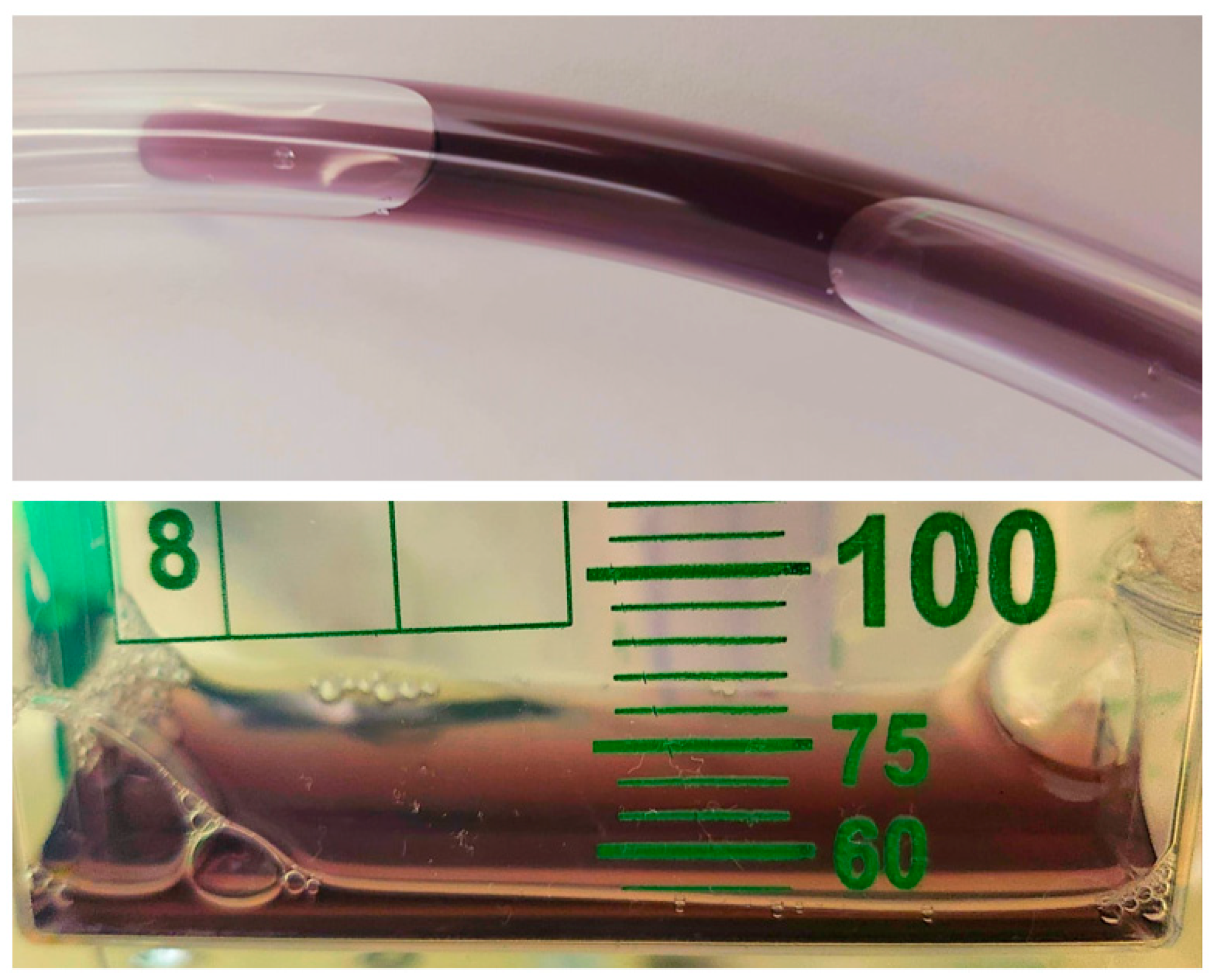
2. Case Presentation

The patient was a 56-year-old female with a complex medical history presented with worsening pain and enlargement of a stage IV sacral pressure ulcer. Her past medical history includes ischemic cardiomyopathy with reduced ejection fraction requiring frequent intubations, a cerebrovascular accident leading to paraplegia and resultant sacral pressure ulcer, type II diabetes mellitus, and Rutherford category 6 peripheral artery disease of the lower extremities, post multiple bypass surgeries and endovascular interventions. The patient has a history of recurrent hospital admissions for sepsis from various infections, including osteomyelitis, complicated urinary tract infections, and pneumonia, leading to colonization with resistant bacteria.
On admission, the patient met the criteria for sepsis, presenting with a heart rate of 101 bpm, a white blood cell count of 23.6 × 10³/µL, and a confirmed source of infection. Physical examination revealed a full-thickness sacral wound measuring approximately 10 × 40 × 6 cm, characterized by dark necrotic tissue, serosanguinous and purulent discharge with a foul odor, and areas of granulation tissue. A chest X-ray showed no acute cardiopulmonary changes, and urinalysis and culture were negative.
The patient was admitted to the general medical floor for the management of severe sepsis secondary to a suspected infected sacral wound ulcer. Empiric antibiotic therapy with intravenous vancomycin, cefepime, and metronidazole was initiated. On day 2 of admission, the patient underwent surgical debridement of the sacral wound down to the muscles and ligaments. The wound appeared mostly pink with scattered nonviable tissue. Bacterial identification in the wound was performed using wound cultures obtained on day 4. The organisms were directly identified via polymerase chain reaction. Then, the clinical specimen was inoculated onto selective and differential media, including blood agar for the growth of Gram-positive organisms, MacConkey agar for the growth of Gram-negative organisms, and specialized plates (bile esculin azide agar supplemented with Vancomycin) for resistant organisms such as vancomycin-resistant Enterococcus. The following organisms were identified: carbapenem-resistant Acinetobacter baumannii/nosocomialis, Proteus mirabilis, and vancomycin-resistant Enterococcus faecium. The organisms were identified using the minimum inhibitory concentration (MIC) method or the Kirby–Bauer method. The carbapenem-resistant Acinetobacter baumannii/nosocomialis was also resistant to the aminoglycosides tobramycin, amikacin, and gentamicin. Additionally, Proteus mirabilis was noted to be resistant to ampicillin/sulbactam. Based on these results, antibiotic therapy was adjusted to intravenous cefiderocol, ampicillin/sulbactam, and daptomycin. The patient underwent two additional surgical debridements.
Three days into therapy, purple discoloration of the urine was noted in the Foley catheter drainage bag (Figure 1). Despite a positive fluid balance, urine output was low. Renal function remained stable with creatinine levels between 0.4 and 0.5 mg/dL. Laboratory investigations revealed no liver injury, hemolysis, or rhabdomyolysis. Urinalysis showed a pH of 6.5, with 2–3 white blood cells/hpf and 3–4 red blood cells/hpf. There was no presence of bilirubin, urobilinogen, ketones, nitrites, bacteria, or leukocyte esterase in the urine. Urine cultures were negative, and renal ultrasound showed no hydronephrosis or bladder pathology.
Despite appropriate management, the patient’s hemodynamic status deteriorated on day 8. Given the poor prognosis, the healthcare proxy decided to transition to comfort measures only, discontinuing all life-prolonging therapies, including cefiderocol. On day 9, the urine began to clear but had not returned to baseline color. Unfortunately, the patient passed away later that day.

3. Methods

We performed a comprehensive literature review utilizing the PubMed and Google Scholar databases to identify studies exploring the association between purple urine and cefiderocol use, from database inception through November 2024. The search was limited to English-language publications. Two independent reviewers systematically screened the identified articles using Microsoft EndNote X9, assessing both titles and abstracts for potential relevance. In cases where abstracts lacked sufficient detail, the full text was retrieved and reviewed. Full text articles meeting the inclusion criteria were thoroughly evaluated. A standardized, piloted data extraction form was used to collect data on patients’ characteristics (e.g., demographics, past medical history), laboratory results, treatment plan, and outcome data. The exclusion criteria included review articles and abstracts without available full texts. Articles deemed eligible by both reviewers were included in the analysis, while those selected by only one reviewer were assessed by a third reviewer to determine eligibility. A risk of bias assessment was conducted independently by the same two reviewers using appropriate tools based on the study design.

4. Results

After comprehensive screening, we included six articles encompassing data on eight patients, as shown in the PRISMA flow diagram in Figure 2. The data from these cases are compared in Table 1. The patients were equally distributed between males and females, with five adults (mean age 67 ± 9.75 years) from five different case reports, and three pediatric cases (mean age 9 ± 3.61 years) from a single case series [5].
Cefiderocol was administered for various pathogens: Pseudomonas in three cases [4,6,9], Acinetobacter (similar to our case) in one case [8], both Pseudomonas and Acinetobacter in one case [10], Escherichia coli in two pediatric cases [5], and Stenotrophomonas maltophilia in one pediatric case [5].
In two cases, urine discoloration was preceded by IV iron administration [6,9], in one case by upper gastrointestinal bleed [8], and in the three pediatric cases, it was preceded by packed red blood cell transfusions [5].
Urine discoloration resolved spontaneously within 1 to 3 days after cefiderocol discontinuation or, in some instances, even with continued administration following iron therapy completion. Notably, in our case, the patient experienced an unfavorable outcome and ultimately passed away, precluding full documentation of urine clearing.
Table 1. Published cases of purple urine associated with cefiderocol use.
First Author, Year of PublicationPatient Age
(In Years)
GenderPast Medical HistoryPresenting SymptomsSource of the InfectionIsolated Organism/sIsolation Site of Organism/s Administered AntibioticsIron Supplementation/Blood TransfusionReported Urine Color/Time to Urine DiscolorationResolution Time
Lewis et al., 2022 [6]63FemaleNon-cystic fibrosis bronchiectasis status post bilateral lung transplantationComplication of the transplant surgeryHospital-acquired pneumoniaMDR Pseudomonas aeruginosaDeep tracheal aspirateCefiderocolIV Ferric gluconate 250 mgDark red/on day 14 of cefiderocol initiation; after first dose of iron infusionUrine color normalized after completion of iron repletion
Shaik et al., 2023 [4]64MaleDown syndrome, hypothyroidism, adrenal insufficiency, seizure disorder, and chronic respiratory failureIncreased lung secretions and desaturationVentilator-associated pneumoniaDTR- PseudomonasEndotracheal aspirateIV vancomycin, cefiderocol, IV amikacin, micafunginNoPurple/on day 5 of cefiderocol initiationUrine color normalized after cefiderocol discontinuation
Lupia et al., 2023 [8]82MaleChronic kidney failureNot documentedNosocomial pneumoniaCarbapenem resistant Acinetobacter baumanniiRectal colonizationCefiderocolNo, but had an upper gastrointestinal bleedingDark brown chromaturia/on day 7 of cefiderocol initiation and day 1 of the bleedingUrine color normalized 48 h after cefiderocol discontinuation
Smith et al., 2023 [10]Early 70sFemaleDM complicated by peripheral neuropathy, peripheral arterial disease and bilateral diabetic foot ulcers1-week history of altered mental status and progressive purulent discharge from a non-healing diabetic foot ulcer on the right heelChronic osteomyelitis of the calcaneum of the right footXDR Pseudomonas aeruginosa, XDR Acinetobacter baumannii, and Enterococcus faecalisTissue cultureIV vancomycin and meropenem. Then, switched to ampicillin-sulbactam and cefiderocolNoBrown/on day 7 of cefiderocol initiationUrine color normalized 3 days after cefiderocol discontinuation for another suspected side effect
Lescroart et al., 2023 [9]56MaleMyocardial infarction complicated by cardiogenic shock status post left ventricular assist device followed by heart transplantComplication of the procedureLeft ventricular assist device driveline and pocket infectionCAZ-AVI resistant Pseudomonas aeruginonaDrive line site culturesCaspofungin, and cefiderocol One dose of IV ferric carboxymaltose 500 mgBlack urine/on day 22 of cefiderocol initiation; after first dose of iron infusionUrine color normalized after 3 days despite the continuation of cefiderocol
Shapiro et al., 2024 [5]10FemaleRefractory AMLFebrile neutropeniaPerianal cellulitisNDM Escherichia coliBlood cultureCefepime, metronidazole and vancomycin, which was changed to cefiderocol, polymyxin-B and tigecyclineReceived 1 unit of packed red blood cells on day 10Pinkish hue on day 2 of cefiderocol initiation then red on day 14 (4 days after receiving 1 unit of packed red cells)Urine color normalized on the day of cefiderocol discontinuation
Shapiro et al., 2024 [5]12FemaleRefractory B-cell ALL received CAR-T cell therapy with a course complicated by severe immunosuppression and graft failureRespiratory failurePulmonary mucormycosis and bacterial
pneumonia
Stenotrophomonas maltophilia resistant to trimethoprim-sulfamethoxazoleSputum cultureCefiderocol (2 courses)Received 3 units of packed red blood cells 6 days prior to starting cefiderocolReddish orange/on day 2 of cefiderocol initiation during both coursesUrine color normalized on the day of cefiderocol discontinuation
Shapiro et al., 2024 [5]5MaleMetastatic medulloblastomaHigh feverNot documentedCarbapenem-resistant, NDM-producing Escherichia coliBlood cultureIV linezolid, cefepime and gentamicin, then meropenem, then cefiderocolReceived 3 units of packed red blood cells 6 days prior to starting cefiderocolPurple/on day 2 of cefiderocol initiationUrine color normalized on day 11 despite the continuation of cefiderocol
Present Report, 2024 56FemaleIschemic cardiomyopathy, HFrEF, advanced PAD, CVA, paraplegia, DM, recurrent UTIsFoul smelling discharges from the sacral woundInfected stage IV sacral pressure ulcerCarbapenem-resistant Acinetobacter baumannii/nosocomialis, Proteus mirabilisWound cultureIV vancomycin, cefepime, and metronidazole then ampicillin/sulbactam, cefiderocol, and daptomycinNoPurple/on day 3 of cefiderocol initiationGiven the poor prognosis patient was transitioned to comfort measures only. Urine began to clear one day after cefiderocol discontinuation
CAZ-AVI: Ceftazidime-Avibactam; CVA: cerebrovascular accident; DM: diabetes mellitus; DTR: difficult-to-treat resistance; HFrEF: heart failure with reduced ejection fraction; IV: intravenous; MDR: multidrug-resistant; UTIs: urinary tract infections; XDR: Extensively Drug Resistant.

5. Discussion

Cefiderocol is a novel injectable cephalosporin with siderophore properties. Similarly to other β-lactam antibiotics, its primary mechanism of action involves targeting Gram-negative bacterial cell wall synthesis through binding to penicillin-binding proteins. However, cefiderocol stands out due to its unique ability to penetrate the bacterial periplasmic space, facilitated by its siderophore-like characteristics, and its exceptional stability against β-lactamase enzymes [11,12]. The drug is predominantly excreted unchanged by renal pathways [8]. Following a single 1 g dose of radiolabeled cefiderocol infused over an hour, approximately 98.6% of the radioactivity appeared in the urine (90.6% as unchanged cefiderocol), while only 2.8% was excreted in feces [13].
Cefiderocol’s siderophore property enables it to form a complex with ferric iron, which is then excreted in urine, leading to a purple discoloration. This could explain why, in five of the eight reported cases, urine discoloration was preceded by intravenous iron administration or red blood cell transfusions, as each unit of red blood cells contains approximately 200–250 mg of iron [5]. In these cases, the iron product was administered three to four days before the discoloration appeared, suggesting that the iron may have combined with cefiderocol and subsequently been excreted in the urine. However, it remains unclear why, in the remaining cases, including ours, the urine turned purple despite the absence of any iron-containing products. Therefore, further studies are needed to clarify this association.
In our case, cefiderocol was selected for its efficacy against carbapenem-resistant Acinetobacter baumannii. Three days after starting cefiderocol, our patient developed purple urine discoloration. Although PUBS is often linked with urinary tract infections in long-term bedridden patients with indwelling urinary catheters [4], urine analysis and culture in our patient were negative. Other known risk factors for PUBS include prolonged catheterization, female gender, immobility, chronic constipation, renal insufficiency, dementia, alkaline urine, and the use of specific polyvinyl chloride-based urine bags [7,14,15]. Although our patient had risk factors like immobility and an indwelling catheter, laboratory findings—including blood counts and bilirubin levels—were within normal limits, and urinalysis showed no pigments, effectively ruling out blood loss, hemolysis, or rhabdomyolysis as possible causes. The urine discoloration was attributed to cefiderocol, as it developed three days after initiation and began to clear within a day of discontinuation, strongly indicating cefiderocol as the causative factor.
A key point to clarify is that the discoloration does not originate from the urine in its natural state. The characteristic purple discoloration is observed exclusively in the urine contained within the catheter and its collecting bag. The biochemical pathway involves the metabolism of the amino acid tryptophan by intestinal bacteria, which convert it to indole. Indole is then metabolized in the liver via the cytochrome P450 isoenzyme CYP2E1 into 3-hydroxyindole and subsequently sulfonated to form indoxyl sulfate. This metabolite is excreted by the kidneys. Bacteria colonizing the catheter, such as Escherichia coli or Klebsiella pneumoniae, produce the enzyme indoxyl sulfatase, which converts indoxyl sulfate into two pigments, Indirubin and Indigo. These pigments adhere to the catheter and the collecting bag. Oxidation of these pigments results in the purple discoloration. Notably, variations in pigmentation may occur between the urine within the catheter and that in the collection bag [7,16,17,18]. Table 2 summarizes the possible causes and contributing factors for the development of PUBS.
As of 29 October 2024, VigiAccess, the World Health Organization’s global database of reported adverse drug reactions, documented a total of 22 cases of cefiderocol-associated chromaturia [19]; however, our literature review identified only 8 cases reported in published studies.

6. Clinical Implications

Drug-induced urine discoloration is typically harmless, but it may lead to unnecessary clinical inquiries and cause concern for patients, their families, and healthcare providers. Careful evaluation of clinical data, paying special attention to accompanying symptoms like pain, fever, or dysuria, is essential for narrowing down potential causes. Although there is no established guideline for managing asymptomatic drug-induced urine discoloration, a reasonable approach includes performing basic urine tests, such as urinalysis and urine culture, to rule out urinary tract infections [1,4]. If the tests are negative, and the clinical context allows, discontinuing the medication and monitoring for resolution of the discoloration is recommended [7]. More cases are necessary to establish whether this discoloration is a rare adverse event or merely an incidental occurrence, and to evaluate if it is dose-dependent.

7. Conclusions

We presented a rare side effect of cefiderocol—purple urine discoloration—with a total of nine cases reported in the literature, including our own. This underscores the importance of considering medication-related adverse effects in the differential diagnosis of urine discoloration to avoid unnecessary diagnostic testing. Further studies are needed to better elucidate this phenomenon.

Author Contributions

E.B.S.: conceptualization, methodology, data collection, literature review, abstract screening, and manuscript writing. H.I.: data collection, literature review, abstract screening, and manuscript writing. E.M.: literature review and manuscript writing. A.G.: Supervision, and critical/medical review of the manuscript. All authors read and approved the final manuscript.

Funding

The authors received no financial support for the research, authorship, and/or publication of this article.

Institutional Review Board Statement

This study was conducted in accordance with the fundamental principles of the Declaration of Helsinki.

Informed Consent Statement

Informed consent was obtained from the patient’s healthcare proxy, as the patient had unfortunately passed away.

Data Availability Statement

Data sharing is not applicable to this article as no datasets were generated or analyzed during the current study.

Conflicts of Interest

The authors declare that they have no competing interests to report.

List of Abbreviations

CAZ-AVI: Ceftazidime-Avibactam; CVA: cerebrovascular accident; DM: diabetes mellitus; DTR: difficult-to-treat resistance; HFrEF: heart failure with reduced ejection fraction; IV: intravenous; MDR: multidrug-resistant; PUBS: purple urine bag syndrome; UTIs: urinary tract infections; XDR: extensively drug resistant.

References

  1. Singh, A.K.; Agrawal, P.; Singh, A.K.; Singh, O. Differentials of abnormal urine color: A review. Ann. Appl. Biosci. 2014, 1, R21–R25. [Google Scholar]
  2. Peterson, L.M.; Reed, H.S. Hematuria. Prim. Care Clin. Off. Pract. 2019, 46, 265–273. [Google Scholar] [CrossRef] [PubMed]
  3. Saraireh, M.; Gharaibeh, S.; Araydah, M.; Al Sharie, S.; Haddad, F.; Alrababah, A. Violet discoloration of urine: A case report and a literature review. Ann. Med. Surg. 2021, 68, 102570. [Google Scholar] [CrossRef] [PubMed]
  4. Shaik, M.R.; Shaik, N.A.; Hossain, S.; Yunasan, E.; Khachatryan, A.; Chow, R. Purplish Discoloration of Urine in a Patient Receiving Cefiderocol: A Rare Adverse Effect. J. Community Hosp. Intern. Med. Perspect. 2023, 13, 43. [Google Scholar] [CrossRef] [PubMed]
  5. Shapiro, K.; Ungar, S.P.; Krugman, J.; McGarrity, O.; Cross, S.J.; Indrakumar, B.; Hatcher, J.; Ratner, A.J.; Wolf, J. Cefiderocol Red Wine Urine Syndrome in Pediatric Patients: A Multicenter Case Series. Pediatr. Infect. Dis. J. 2024, 43, 142–144. [Google Scholar] [CrossRef] [PubMed]
  6. Lewis, T.C.; Arnouk, S. Dark Red Urine in a Patient on Cefiderocol and Ferric Gluconate. Ann. Pharmacother. 2022, 56, 1082–1083. [Google Scholar] [CrossRef] [PubMed]
  7. Karray, O.; Batti, R.; Talbi, E.; Ayed, H.; Chakroun, M.; Ouarda, M.A.; Bouzouita, A.; Cherif, M.; Slama, M.R.B.; Amel, M. Purple urine bag syndrome, a disturbing urine discoloration. Urol. Case Rep. 2018, 20, 57–59. [Google Scholar] [CrossRef] [PubMed]
  8. Lupia, T.; Salvador, E.; Corcione, S.; De Rosa, F.G. Dark Brown urine in a patient treated with Cefiderocol. Infez. Med. 2023, 31, 265. [Google Scholar]
  9. Lescroart, M.; Noe, G.; Coutance, G. The black urine challenge. Transpl. Infect. Dis. 2023, 25, e14103. [Google Scholar] [CrossRef] [PubMed]
  10. Smith, M.; Foong, K.S. Cefiderocol-associated brown chromaturia. BMJ Case Rep. CP 2023, 16, e258207. [Google Scholar] [CrossRef] [PubMed]
  11. Zhanel, G.G.; Golden, A.R.; Zelenitsky, S.; Wiebe, K.; Lawrence, C.K.; Adam, H.J.; Idowu, T.; Domalaon, R.; Schweizer, F.; Zhanel, M.A. Cefiderocol: A siderophore cephalosporin with activity against carbapenem-resistant and multidrug-resistant gram-negative bacilli. Drugs 2019, 79, 271–289. [Google Scholar] [CrossRef]
  12. Doi, Y. Treatment options for carbapenem-resistant gram-negative bacterial infections. Clin. Infect. Dis. 2019, 69 (Supp. S7), S565–S575. [Google Scholar] [CrossRef]
  13. Miyazaki, S.; Katsube, T.; Shen, H.; Tomek, C.; Narukawa, Y. Metabolism, excretion, and pharmacokinetics of [14C]-Cefiderocol (S-649266), a siderophore cephalosporin, in healthy subjects following intravenous administration. J. Clin. Pharmacol. 2019, 59, 958–967. [Google Scholar] [CrossRef] [PubMed]
  14. Khan, F.; Chaudhry, M.A.; Qureshi, N.; Cowley, B. Purple urine bag syndrome: An alarming hue? A brief review of the literature. Int. J. Nephrol. 2011, 2011, 419213. [Google Scholar] [CrossRef] [PubMed]
  15. Peters, P.; Merlo, J.; Beech, N.; Giles, C.; Boon, B.; Parker, B.; Dancer, C.; Munckhof, W.; Teng, H. The purple urine bag syndrome: A visually striking side effect of a highly alkaline urinary tract infection. Can. Urol. Assoc. J. 2011, 5, 233. [Google Scholar] [CrossRef] [PubMed]
  16. El Chamieh, C.; Liabeuf, S.; Massy, Z. Uremic toxins and cardiovascular risk in chronic kidney disease: What have we learned recently beyond the past findings? Toxins 2022, 14, 280. [Google Scholar] [CrossRef] [PubMed]
  17. El Chamieh, C. Influence Des Toxines Urémiques Sur La Morbi-Mortalité Cardiovasculaire Des Patients en Maladie Rénale Chronique Dans La Cohorte CKD-REIN. Université Paris-Saclay. 2024. Available online: https://theses.hal.science/tel-04625539 (accessed on 11 January 2025).
  18. Gautam, G.; Kothari, A.; Kumar, R.; Dogra, P. Purple urine bag syndrome: A rare clinical entity in patients with long term indwelling catheters. Int. Urol. Nephrol. 2007, 39, 155–156. [Google Scholar] [CrossRef] [PubMed]
  19. World Health Organisation. VigiAccess. Available online: https://www.vigiaccess.org/ (accessed on 29 October 2024).
Figure 1. Photographs of urine samples demonstrating purplish discoloration of urine.
Bacteria 04 00009 g001
Figure 2. Flow diagram of article selection for cefiderocol-induced purple urine.
Bacteria 04 00009 g002
Table 2. Summary of possible causes and contributing factors for purple urine bag syndrome.
CategoryDetailsExamples/Mechanisms
Predisposing FactorsPatient-related conditions that increase risk- Female sex
- Dementia
- Chronic constipation
- Chronic renal disease
- Alkaline urine
- Increased urine bacterial load
Environmental FactorsFeatures of the medical setup that enhance PUBS risk- Use of specific polyvinyl chloride-based urine bags
CatheterizationType and duration of urinary catheterization- Permanent/prolonged urinary catheterization (urethral or suprapubic)
Bacterial CausesSpecific bacteria capable of metabolizing indoxyl sulfate- Proteus mirabilis
- Klebsiella pneumoniae
- Pseudomonas aeruginosa
- Morganella morganii
- Escherichia coli
Biochemical PathwayMetabolism of tryptophan leading to pigment formation- Tryptophan → Indole (intestinal bacteria)
- Indole → 3-hydroxyindole (liver, via CYP2E1)
- 3-hydroxyindole → Indoxyl sulfate (via sulfonation)
- Indoxyl sulfate → Indirubin (red) and Indigo (blue) by bacterial enzymes in the catheter
Oxidative ProcessChemical oxidation leading to purple urine coloration- Indirubin (red) + Indigo (blue) combine upon oxidation to create the characteristic purple hue
Medication-RelatedPotential drug side effects- Cefiderocol (antibiotic)
Urinary EnvironmentLocal conditions in the urinary tract that facilitate pigment production and bacterial activity- Catheter-associated biofilm formation
- Stasis of urine
- Alkaline pH enhancing enzymatic activity of bacteria
Disclaimer/Publisher’s Note: The statements, opinions and data contained in all publications are solely those of the individual author(s) and contributor(s) and not of MDPI and/or the editor(s). MDPI and/or the editor(s) disclaim responsibility for any injury to people or property resulting from any ideas, methods, instructions or products referred to in the content.

