Review

Bacterial Sialidases: Biological Significance and Application

The Stephan Angeloff Institute of Microbiology, Bulgarian Academy of Sciences, Acad. Georgi Bonchev Str., Bl. 26, 1113 Sofia, Bulgaria
*
Author to whom correspondence should be addressed.
Appl. Biosci. 2025, 4(2), 17; https://doi.org/10.3390/applbiosci4020017
Submission received: 20 January 2025 / Revised: 17 February 2025 / Accepted: 24 February 2025 / Published: 1 April 2025

Abstract

:
This review summarizes recent findings on the diverse roles of bacterial sialidases in microbial biology. Bacterial sialidases, also known as neuraminidases, are exog α-lycosidases that cleave terminal sialic acid residues from a number of complex compounds designated as sialoglycoconjugates (glycoproteins, glycolipids and oligosaccharides). Metabolically, they are involved in sialic acid catabolism, providing energy, carbon and nitrogen sources. Catabolic degradation of sialic acids is a physiological feature that can be considered an important virulence factor in pathogenic microorganisms. Sialidases play a pivotal role in host–pathogen interactions and promotion of bacterial colonization. The activity of these enzymes enables bacterial adhesion, biofilm formation, tissue invasion, and also provides immune evasion by exposing cryptic receptors and modifying immune components. Many different perspectives are being developed for the potential application of sialidases. In the field of medicine, they are being explored as appropriate targets for antimicrobials, vaccines, diagnostic preparations and in tumor immunotherapy. In the field of enzymatic synthesis, they are used for the regioselective production of oligosaccharide analogs, enzymatic separation of isoenzymes and as a tool for structural analysis of sialylated glycans, among other applications.

1. Introduction

Sialidases, or neuraminidases, are exo-α-glycosidases (EC 3.2.1.18) that catalyze the removal of terminal sialic acid residues from compounds such as glycoproteins, glycolipids and oligosaccharides by the hydrolysis of α-glycosidic bonds between the 2C atom of the sialic acid and the 3C, 6C of 8C atom of the carbohydrate component (α2-3, α2-6 and α2-8 linkages) [1,2]. Sialic acids are found in some viruses, bacteria, protozoa and fungi, and are present in almost all tissues of animals from echinoderms to humans [3,4]. They are α-keto-acid sugars with a nine-carbon backbone, participating in the regulation of many physiologically important cellular and molecular processes, including signaling and adhesion [5] (Figure 1).
Removal of sialic acids, which represent the first protective barrier, reveals the receptors to which pathogens or toxins bind, thus contributing to the degradation of cellular components and disruption of tissue integrity [6]. Sialidases have traditionally been studied in pathogenic microorganisms, where these enzymes are considered virulence factors, “paving the way” for other hydrolytic enzymes [5,6].

2. Structural Properties, Catalytic Mechanism and Substrate Specificity

Most bacterial sialidases are secretory proteins that contain signal peptides cleaved during the secretory process. They consist of a sialidase domain for catalytic activity and additional domains that aid in substrate binding or enzyme localization. Although the similarity in the amino acid sequence among the bacterial sialidases is low (less than 30%), the catalytic part contains some conserved motifs common to most sialidases [7] (Figure 2a). The first conserved region, located near the N-terminus of the protein molecule is the FRIP motif (Phe-Arg-Ile-Pro, with some variations). The second motif, the so-called ‘Asp-box’ (due to the highest conservatism of asparagine) is a sequence Ser/Thr-X-Asp-X-Gly-X-Thr-Trp/Phe (X represents any amino acid), repeated three to five times in the catalytic domain [7,8]. The Asp-box motifs are remote from the active site, in topologically equivalent locations [8]. Their function is not yet fully understood, but it is assumed that they have a structural role and other functions, such as participation in the secretory process [8] or in catalysis [9]. In large sialidases, the amino acid sequences between the N-terminus and the second Asp-box, as well as between the fifth Asp-box and the C-terminus, are longer, which is thought to contribute to their broader substrate specificity [10]. The arginine residue in FRIP motif is part of the arginine triad in the active site, directly involved in the catalytic process [11].
In general, enzymatic hydrolysis of the glycosidic bond proceeds either with retention or inversion of the anomeric configuration—the first occurs via a two-step mechanism in which the catalytic residues act as an acid/base and a nucleophile, respectively, and the second—via a one-step mechanism in which the substrate and a water molecule bind simultaneously. The arginine triad, the Tyr/Glu nucleophilic pair and the Asp acid/base are key residues involved in the catalytic cleavage mechanism. Initially, the positively charged arginines in the catalytic pocket interact with the negatively charged carboxylate group of the sialic acid. The Tyr residue, hydrogen-bonded to Glu, attaches to the anomeric center (C-2), leading to a semiplanar oxocarbenium transition state with the adjacent carbohydrate and the formation of covalent aryl-glycoside intermediate. The Asp residue activates the water molecule attaching the anomeric C-2 center in a trans-addition to form another semiplanar oxocarbenium transition state. Finally, free sialic acid is released from the active site of the sialidase, completing the hydrolysis with retention of the anomeric configuration [12]. All sialidases have a hydrophobic “pocket” in their active sites, which accommodates the N-acetyl group of the substrate, but the residues forming it are different and likely determine the substrate specificity, catalytic efficiency and kinetics of the enzyme [7,11].
The catalytic domain of bacterial sialidases have a beta-propeller structure, with the Asp-boxes forming the turn between the third and fourth strands of each sheet on the periphery of the propeller [8]. Besides the catalytic part, many bacterial sialidases possess additional carbohydrate-binding (lectin-like) domains that most likely facilitate enzyme-substrate binding [1]. Sialidase from C. chauvoei contains two additional non-catalytic domains. One of them regulates the binding of the enzyme to the non-sialic carbohydrate region, and the other is responsible for recognizing and binding to sialic acid itself [13]. Similarly to the above example, the catalytic domain of sialidase from V. cholerae is flanked by two lectin-like regions showing structural similarity to plant and animal lectins [2] (Figure 2b).
Figure 2. Example of primary structure of bacterial sialidases: (a) Partial alignment of the sialidase amino acid sequences of Salmonella enterica, C. perfringens, Manheimia haemolytica, P. multocida, M. viridifaciens, Corynebacterium pseudotuberculosis and C. glutamicum. The FRIP and the ASP-box motifs are outlined in black, the conservative residues of the arginine triad are in red font. The other conserved residues or their conserved variations are highlighted in gray. (b) The deduced amino acid sequence of V. cholerae non-O1 strain 13 sialidase compared to sialidase enzymes of pathogenic O1 strains. Designations: non-O1_13—V. cholerae non-O1 strain 13 (KJ875802), O1_Amaz—V. cholerae O1 strain Amazonia 3509 (EU272902), O395—V. cholerae O1 strain O395 (ACP09894), IEC224—V. cholerae O1 strain IEC 224 (NC 016944), and ET_N16961—V. cholerae O1 biovar El Tor strain N16961 (NP 231419). The full identity is gray-highlighted. The FRIP motif is shown in blue square and Asp-boxes in red squares; the lectin domains are dashed-underlined. The arginine triad (224, 635, 712) of the catalytic site is shown with red bold arrows; other conserved residues—the glutamine (619), the tyrosine (740), and glutamine (756) participating in the active site are shown with black arrows (From: Eneva et al., 2015 [14]).
Applbiosci 04 00017 g002
The substrate specificity of a given sialidase is usually defined as the ability of the enzyme to discriminate between the type of bond between the sialic acid and the galactose residue. Typically, substrates used to determine this specificity are N-acetylneuraminosyl α2-3 lactose, N-acetylneuraminosyl α2-6 lactose and colominic acid (in which the bonds between the sialic residues are α2-8) [15]. Most bacterial sialidases can catalyze the hydrolysis of a wide range of sialoconjugates containing α2-3, α2-6 or α2-8 linked sialic acids but each has a preference for a particular linkage and type of glycoconjugate. In most cases, the α,2-3 linkage is hydrolyzed to the greatest extent [1]. This is true for species of the genus Clostridium, in which sialidases have a stronger affinity to α2-3 than α2-6 and α2-8 linkages [7]. However, sialidases from M. viridifaciens and C. diphtheriae prefer α2-6 [16,17]. In the sialidase from A. ureafaciens, the ratio of the degree of hydrolysis of bonds is α2-6 > α2-3 > α2-8 [7]. The only sialidase preferentially hydrolyzing the α2-8 bonds in colominic acid is from B. fragilis [18].

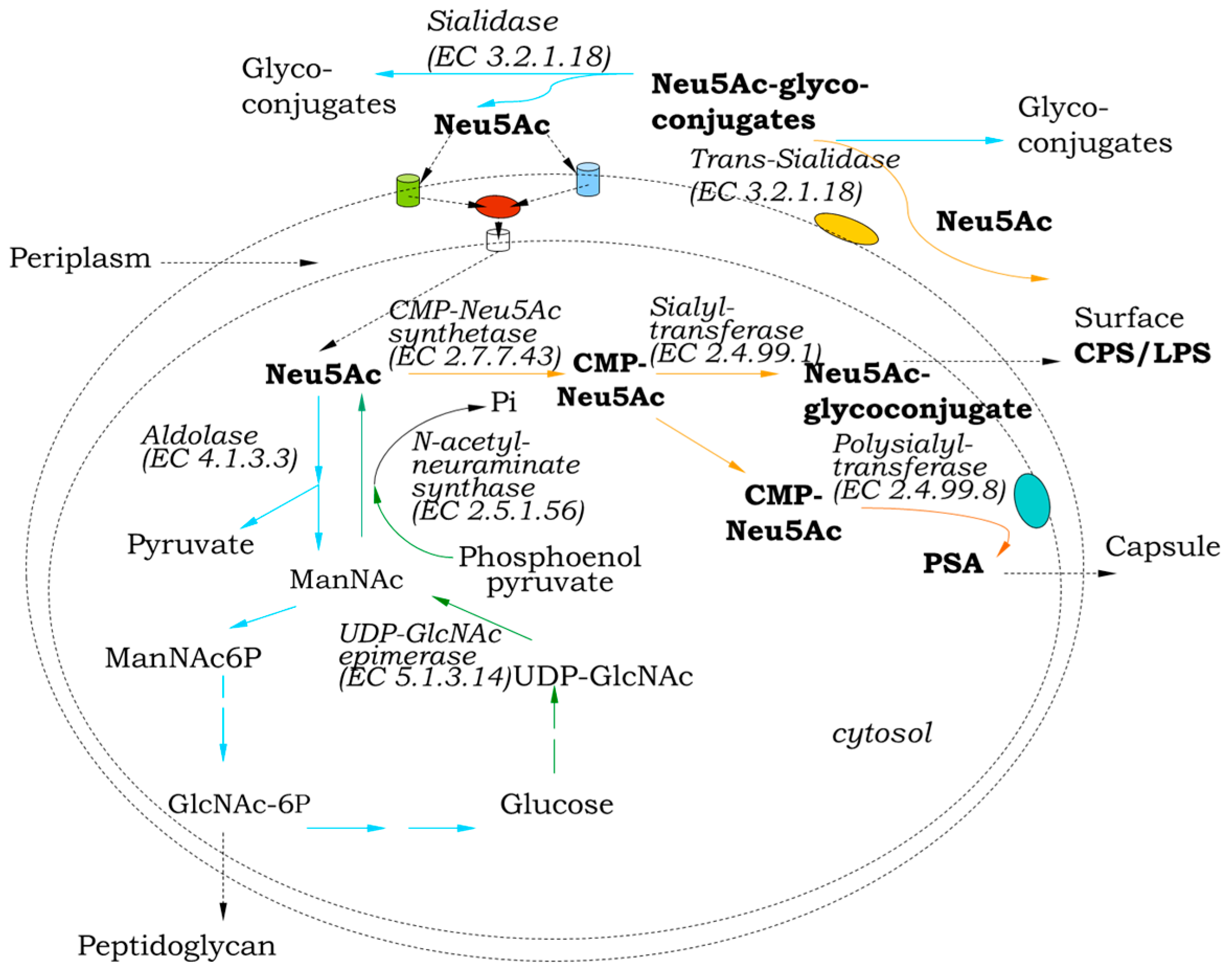
3. Role in Sialo Metabolism

Many bacterial species use sialic acid (N-acetylneuraminic acid (Neu5A) as a carbon, nitrogen and energy source [19,20]. A number of key enzymes are involved in this process (Figure 3). Sialidases, which cleave these terminal residues from sialoconjugate substrates are the first step of their catabolic degradation. In bacteria that assimilate sialic acid as a carbon source, these residues are transported across the cell membrane to the interior of the cell by means of specific transmembrane proteins. In the cytosol they are broken down by NanA (sialate aldolase, Neu5Ac lyase) to form ManNAc (N-acetylmannosamine) and pyruvate, which are, respectively, directed to glycolysis and the tricarboxylic acid cycle [20,21]. ManNAc is phosphorylated by NanK (kinase) to ManNAc6P (N-acetylmannosamine-6-phosphate), which in turn is converted by NanE (epimerase) to GlcNAc6P (N-acetylglucosamine-6-phosphate). Two additional enzymes (NagA and NagB) complete the catabolic cycle of sialic acids, with the final product fructose-6-phosphate, which enters central metabolism. Another key enzyme in the biosynthesis of sialoglycoconjugates is the sialyltransferase (E.C.2.4.99.1), which catalyzes the transfer of sialic residues from their activated form, citidine 5′-monophosphate sialic acid (CMP-Neu5Ac) to appropriate acceptors usually containing terminal galactose, N-acetylgalactosamine (GalNAc) or another sialic acid [22]. Trans-sialidase (E.C. 3.2.1.18) is a multifunctional enzyme combining two activities and two distinct active sites on one molecule. This enzyme would deliver sialic acid residues directly from the host or environment, binding them to appropriate glycans on the cell surface [17]. In the absence of suitable acceptor, it acts as sialidase transferring the sialic acid residues to water molecules, therefore classified in the same enzyme group [23].
In some prokaryotes, a complete set of enzymes can be observed, but it is most often presented in various abbreviated variants, depending on its particular biological sense [4,6]. Bacterial genes encoding NanA, NanK and NanE are clustered together, while those encoding NagA and NagB are mostly scattered throughout the genome [20]. The cluster is present only in members of Gammaproteobacteria and Fusobacteria among Gram-negative bacteria and Bacillales, Clostridia and Lactobacillales among Gram-positive bacteria, as well as Mycoplasma. Interestingly, the gene order and configuration of the cluster show great variability even within members of the same genus [20]. NanA, the key enzyme in sialic acid catabolism, can be present independently of NanE and NanK in four other bacterial groups, namely α-Proteobacteria, Planctomycetes, Verrucomicrobia and Bacteroidetes [20]. The majority of the bacteria carrying a Nan cluster use sialidases in releasing sialic acids of the host, including multiple species of Clostridium, Bacteroides, some strains of Bifidobacterium longum, V. cholerae, Ruminococcus gnavus and Akkermansia muciniphila [6]. A complete pathway of sialic acid catabolism, including a predicted sialidase gene, has been found in B. fragilis strains. However, some bacteria appear to have incomplete packages of enzymes for utilizing host sialic acids. For example, B. thetaiotaomicron VPI-5482 lacks the Nan operon and cannot consume the released sialic acid, despite encoding sialidase; conversely, Clostridium difficile strain 630 possesses the Nan operon but does not produce sialidase [6]. The presence or absence of sialidase-encoding genes in bacterial genomes of particular pathogenic or commensal species is often strain-specific [6,20]. Some bacterial strains produce more than one sialidase as isoenzymes with different substrate specificities and biochemical properties [5].
Concerning the way of acquisition of sialic acids, the following groups of bacteria can be distinguished: scavengers, which cleave sialic acid and subsequently metabolize it in the cytoplasm; spitters, which cannot utilize the cleaved sialic acid but metabolize the monosaccharides located below it (N-acetylglucosamine, galactose, N-acetylgalactosamine); and swallowers, which resemble spitters but are able to catabolize sialic acid itself [21]. In some cases, bacteria that do not produce sialidase but use sialic acids for their metabolic needs benefit from the sialidase of other microorganisms inhabiting the same ecological niche, as well as from sialidases produced by the host itself [24]. For example, cross-feeding is known between commensals in the gut Bifidobacterium breve and B. bifidum [6,25], as well as between G. vaginalis and Fusobacterium nucleatum, which promotes colonization and contributes to vaginal dysbiosis [26].

4. Influence of External Factors on Sialidase Synthesis

The majority of bacterial sialidases are inducible enzymes and their biosynthesis depends on the presence in the medium of fetuin, transferrin, and other high-molecular complex compounds containing α-glycosidic linked sialic residues [19]. Sialic acid itself is also an effective inducer of sialidase production, especially in S. pneumonia, P. aeruginosa, C. perfringens and V. cholerae non-O1/13 [27,28]. The glucose content in the medium also affects sialidase production, especially since the latter is subject to catabolic repression. It is known that high glucose concentrations suppress the expression of sialidase-encoding loci in S. pneumoniae and C. perfringens [27,29]. On the other hand, the addition of cAMP to the medium has a positive effect on sialidase biosynthesis in Salmonella typhimurium LT2 [30].
Interesting observations have been made concerning sialidase synthesis under stress conditions. There is evidence of increased sialidase activity in P. aeruginosa, C. perfringens and V. cholerae in sepsis [31]. In this regard, sialic acids released under the action of sialidase neutralize oxyradicals that arise during stress [32].
Enhancement in sialidase production in P. aeruginosa was observed under hyperosmolar conditions. The elevated transcription is regulated by genes responsible for alginate ex-pression, which is responsible for the mucoid phenotype. This suggests that sialidase, like alginate, may be specifically expressed under the conditions expected to be present in CF (cystic fibrosis) lung [33].

5. Role in Pathogenicity

Catabolic degradation of sialic acids is a physiological feature that can be considered as a factor of pathogenicity, increasing the virulence potential of the producers, as well as their ability to thrive and survive in tissues [34]. Thus, some enzymes like sialidases, common to sugar metabolism with a purely nutritional function, appear “dangerous” when their activity enhances the parasitic and pathogenic potential of the respective producers. The role of sialidase in providing nutrients to H. parasuis—a swine pathogen, may be an important factor in its obligate parasitism [35]. The main characteristics of sialidase, as a factor of virulence and pathogenicity, are considered to be the following: a large amount of extracellular enzymes with high specific activity; inducible activity at the site of infection; and specificity of substrate displayed at the site of colonization [19,36]. In some microorganisms, a correlation between sialidase activity and their pathogenicity is observed. For example, the increase or decrease in enzyme production in representatives of Streptococcus, Pasteurella, Clostridium, Erysipelothrix and Mycoplasma is followed by a corresponding increase and decrease in their virulence [37,38]. The role of sialidase in the pathogenic process can be expressed in different ways as a result of desialylation.

5.1. Pathogen Penetration and Adhesion

Removal of sialic acids from salivary glycoproteins by sialidase leads to a reduction in their protective capacity against various pathogens. Once inside the host, most pathogenic bacteria are directed to the respiratory or intestinal tract, which is coated with a mucin substance sensitive to sialidase action. Sialidase helps reduce the viscosity of mucins, breaking intercellular contacts and thus facilitating the penetration and spread of pathogens in other tissues. The penetration of pathogens into neighboring cells and tissues can occur at a high speed (10 cm per hour), and it has been suggested that neuraminidase, among other hydrolases, plays a key role in the processes of degradation of the mucous layer and detachment of epithelial cells [1,26]. For example, the sialidase produced by C. chauvoei contributes to the desialylation of liver and intestinal mucosal cells and this, along with the production of a toxin, leads to the destruction of the cells [39]. The poultry pathogens M. synoviae and Ornithobacterium rhinotracheale have been shown to cleave sialic residues from tracheal and serum glycoproteins (including immunoglobulin G and transferrin). This is thought to contribute to the pathogenicity of these bacteria to turkeys and poultry [38,40]. Many pathogens attach to galactose residues on the surface of host endothelial cells. Such microorganisms secrete sialidase to remove the sialic residues covering the galactose receptors [41,42]. A correlation was observed between the process of desialylation and the ability of E. rhusiopathiae to attach to cells of a rat aortic cell line. The removal of sialic residues was inhibited using N-acetylneuramin-lactose (a substrate of neuraminidase) [43]. Adhesion to various cell types of bacteria, such as Actinomyces naeslundii, S. pneumoniae and Arcanobacterium pyogenes, is increased by sialidase pretreatment [44].

5.2. Disclosure of Receptors

Since sialic acids cover many of the cellular structures, their removal by the pathogens’ sialidases leads to the disclosure of cryptic receptors that can be targets, both for the own immune cells and for some products synthesized by the pathogens. The removal of sialic acids from the surface of erythrocytes leads to the disclosure of the underlying galactose residues, acting as receptors for liver hepatocytes and macrophages which participate in the destruction of red blood cells [45]. The desialylation of erythrocytes results also in a decrease in their negative surface charge and an increase in aggregation between them, which in turn causes the appearance of thrombosis [1]. The disclosure of cryptic antigens under the action of bacterial sialidase is also related with phenomena such as shortening the half-life of glucoconjugates circulating in the blood; inactivation of certain hormones (erythropoietin); loss of cell surface receptor specificity for hormones, enzymes, and cells [19]. Streptococcus pneumoniae sialidase NanA is known to be involved in desialylation of blood glycoproteins and their remodeling, which in turn triggers events such as coagulation, thrombosis, sepsis and subsequent severe organ damage [46].
The role of sialidase from V. cholerae in the pathogenic process has been well studied. During infection, the enzyme removes two sialic residues from trisialogangliosides present in the intestinal mucus, thereby “unmasking” the cholera toxin receptor. The binding of the toxin to the receptor leads to the activation of adenylate cyclase and an increase in the amount of cAMP. This alters the levels of sodium and chloride transport in the epithelial cells, resulting in severe diarrhea [47,48]. In addition to exposing the receptor for cholera toxin, V. cholerae sialidase cleaves sialic acids from the mucus-rich environment in the gut, using them as a carbon and energy source, thus enabling the bacteria to penetrate deeper into the tissues [34].

5.3. Biofilm Formation

Bacterial sialidases contribute to increasing the virulence of some pathogens by promoting biofilm production [40,41,42,43,44,45,46,47,48,49,50,51,52]. One of the mains roles of pneumococcal NanA is to facilitate exposure of galactose residues, and it is thought that the prevalence of this sugar in the nasopharynx contributes to the growth of S. pneumoniae in a biofilm [53]. Streptococcus pneumoniae nanA expression is upregulated in lung tissue and in biofilm-grown cells [51]. Despite biochemical, structural, and phylogenetic differences, the sialidases of S. pneumoniae and P. aeruginosa demonstrate a common role in biofilm formation and in the pathogenesis of respiratory tract infection [51]. Although the involvement of sialidase in biofilm formation has been demonstrated, the precise role of the enzyme in this process has not yet been elucidated. Biofilm formation in sialidase-deficient mutant strains of P. aeruginosa, S. pneumoniae and P. gingivalis is severely impaired [40,53,54]. The supply of exogenous sialic acid enhances the ability of bacterium to form biofilm, suggesting that the added sialic residues alter the hydrophobicity of the cell surface and influence fimbriae formation [53,54]. Similarly, the addition of sialic acid, but no other monosaccharide, stimulated pneumococcal biofilm formation [51]. Gardnerella spp. is a dominant component of the bacterial vaginosis (BV) biofilm and putative initial anaerobic colonizer of the vaginal epithelium, creating conditions for the attachment of other bacteria in a process known as coaggregation. Sialidases increase the potential of G. vaginalis to enter into close contact to vaginal epithelial cells by altering the characteristics of mucus discharges, catalyzing their conversion into food for bacteria and expose adhesion receptors on polysaccharides [26].

5.4. Modulation of Immune Mechanisms

Sialidases in some pathogenic microorganisms can play a protective role against the immune defense of the host. Bacterial sialidases increase the susceptibility of some immunoglobulins to proteolytic degradation. For example, sialidase of M. synoviae and C. perfringens cleave SAα(2–6)gal moiety from heavy chain of chicken IgG [38]. Treatment of IgA1 molecules with streptococcal sialidase increases the possibility of their proteolytic degradation by streptococcal IgA1-protease. Probably, during the infectious process, the two enzymes act together [55]. Similarly, sialidases, galactosidases and hexosaminidases from species associated with BV are able to cleave sialic and mannose residues from immunoglobulins, making them susceptible to further proteolytic degradation [56].
In another case, sialidase NanA from S. pneumoniae, with the joint action of two other surface-located exoglycosidases—BgA (β-gactosidase) and StrH (N-acetylglucosaminidase) prevents the opsonophagocytic destruction of bacterial cells by neutrophils [57]. This immune reaction is a multistage process, the first stage of which is the so-called opsonization. It is carried out by the complement system, and the end result is the covalent deposition of C3b (an element of the complement system) on the surface of the bacterial cell. The cooperative action of the three mentioned enzymes inhibits specifically the covalent binding of C3b (a component of the complement system) on the microbial cell.
It has been shown that bacteria such as Capnocytophaga canimorsus resist the immune response precisely through the presence of cell-associated sialidase. The latter allows these bacteria to carry out intensive deglycosylation of glycoproteins located on the surface of immune cells and even “feed” on them [58].
Some microorganisms use their sialidase to alter the structure of components of the immune system. Sialidase from C. perfringens has been shown to catalyze the conversion of the cytokine TGF-β (a multifunctional protein regulating a number of immune processes) from its inactive to active form [59,60]. This occurs through the removal of sialic residues from latency-associated glycopeptide (LAP), which leads to conformational changes with subsequent breakdown of the bond between LAP and TGF-β1 and release of active TGF-β1. Thus, some pathogens effectively regulate the amount of this factor, thereby providing themselves with an “immunologically favorable” environment for their development.
Sialidases are able to affect the binding of immune cells to the corresponding target. It is known that sialidase treatment of immunocompetent cells results in enhanced phagocytic response and antigen binding [61]. This effect is largely due to the cleavage of sialic acids from the cell surface of lymphoid cells, followed by a lowering of their surface negative charge. This leads to some deformation of the cell shape, which, in turn, facilitates the contact of immune cells and target cells. On the other hand, there are data that suggest that treatment with bacterial sialidase reduces the surface negativities of macrophages, which, in turn, significantly impairs phagocytosis [62]. It has been suggested that some pathogens may use their sialidase to modify the surface of immune cells, thereby evading host defenses.
An interesting fact is that the treatment of HIV and its target immune cells with sialidase from Arthrobacter ureafaciens has been found to have a beneficial effect on the viral life cycle, in particular the processes of attachment and entry into the cell [63]. Once again, this is likely due to the desialylation process, which lowers the negative surface charge and facilitates the contact between the virus particle and the cell.
It should be mentioned that bacterial sialidases may also play an indirect role in evading host immune defenses. The removed sialic acids can be attached to the cell surface of the bacteria (sialylation). Capsule components and membrane lipopolysaccharides of some pathogens are commonly subjected to sialylation. The first step in this process is the synthesis of CMP-NeuAc (activated form of sialic acid), the reaction catalyzed by CMP-sialic acid synthetases. This compound is then added to appropriate acceptor (oligosialic chains or sialoglucoconjugate) by specific sialyltransferases and is subsequently transported to the cell surface (see Figure 3). In result, antigenic determinants are masked or structures similar to those of the host are obtained (molecular mimicry). All this helps to evade immune defense mechanisms [24]. The role of sialidase in this process has been elucidated mostly with respect to the trans-sialidase of eukaryote parasite Trypanosoma cruzi [64]. This enzyme is able to transfer sialic acid cleaved from host tissues to suitable lactose or lactosamine acceptors on the cell surface of trypanosomes. A similar activity has been suggested for the sialidase from C. diphtheriae [17].

5.5. Synergism Between Bacterial and Viral Sialidases

Although bacterial and viral neuraminidases differ in some structural features (viral sialidases do not possess Asp-boxes and FRIP motifs), they share a common catalytic activity. This fact explains the observation of some biological phenomena. When influenza virus growth is strongly suppressed upon treatment with virus-specific sialidase inhibitors (e.g., zanamivir), the addition of the corresponding bacterial sialidase fully restores the viral life cycle [65]. In a peculiar way, microbial enzymes “replace” their inactivated viral analogs, cleaving surface sialic residues to which the newly formed viral particles are attached and thus facilitating their release and spread in neighboring cells. Producers of these enzymes can be the most common commensals that inhabitants of the oral cavity and upper respiratory tract [65].
Sometimes, during the pathogenic process, a cooperative action between viral and bacterial sialidase has been observed. Pneumococcal adherence to the epithelial cells, nasal colonization and middle ear mucosa could be mediated by co-action of NanA sialidase activity of S. pneumoniae and influenza virus A sialidase during coinfection [52]. The desialylation of mucosal cells by the influenza virus sialidase increases susceptibility to secondary infections often caused by S. pneumoniae [51]. In this case, it can be commented that there is a lethal synergism between viral and bacterial neuraminidase. The viral enzymes that desialylate the cell surface expose cryptic receptors to which the bacteria attach, and their own sialidase helps them adhere to the trachea and descend into the lungs [66]. It has been shown that sequential influenza with A virus and S. pneumoniae infection caused more pronounced changes in carbohydrate structures than either pathogen alone. It has been proven that the cooperative action of the two enzymes either enhance or substitute for this activity [67].

5.6. Sialidases as Targets for Inhibition

In recent years, there has been a great demand for new and effective therapeutics to deal with the increasing number of multidrug-resistant pathogens. Due to functional and structural diversity, sialidases are attractive targets for the development of novel therapies against viruses, bacteria, fungi and parasites. The use of sialidase inhibitors to reduce bacterial adhesion, invasion and mucosal damage may be an interesting new therapeutic approach in the treatment of BV [68]. Oral administration of a sialidase inhibitor opens the prospect of treating intestinal inflammation resulting from E. coli dysbiosis by regulating sialic acid catabolism [69]. To date, little attention has been paid to the inhibition of sialidases in commensal bacteria, but this has potential interest to be applied in the regulation of obesity [70]. The elucidation of the precise structure of sialidase from Porphyromonas gingivalis determines it as an excellent target for the design of effective inhibitors with high potential to combat periodontitis [71]. The importance of P. aeruginosa sialidase in biofilm production suggests that the enzyme may be a potential therapeutic target in patients with cystic fibrosis [50].
The most promising sialidase inhibitors are based on 2-deoxy-2,3-didehydro-N-acetylneuraminic (DANA), a transition-state sialic acid analog [72]. Various derivatives have been created that inhibit viral and bacterial sialidases with varying efficacy [12]. Among them are the anti-influenza preparations zanamivir (Relenza) and oseltamivir (Tamiflu), blocking viral sialidases and preventing virion release and spread from the infected cells [72,73]. They differ, however, in their action against bacterial sialidase. Zanamivir displayed weak activity against pneumococcal sialidase isoforms NanA and NanB due to single amino acid substitutions in the active site. In contrast, oseltamivir completely inhibited the NanA isoform [74]. It is crucial to keep in mind that sialidase inhibitors with poor effect on viruses might have a significant effect on bacteria or protozoa (as is the case for DANA and vice versa) [3]. On the other hand, zanamivir has been described as effective against sialidase from G. vaginalis [75] and P. timonensis [68,76]. Some of the DANA derivatives show excellent inhibitory activity against bacterial sialidase. Triazole-linked derivatives were described as effective against sialidase from V. cholerae and A. ureafaciens, while sulfo-sialic acid analogs demonstrated strong suppression towards C. perfringens and Streptococcus 6646 K sialidase [12,73]. Poly-DANA inhibits the catalytic activity of sialidase from S. pneumoniae (NanA) and the symbiotic microorganism B. thetaiotaomicron (BtSA) at the picomolar and low nanomolar levels [72].
Also, a variety of natural compounds have been reported as inhibitors against bacterial sialidases, including curcumin and flavonoid derivatives, prenylated isoflavone and chromenone derivatives as well as artocarpin and katsumadain A [12]. These compounds have been reported to inhibit the activity of sialidase from V. cholerae, C. perfringens and S. pneumoniae. Recent studies have investigated the inhibition of sialidases from V. cholerae non-O1, A. nicotianae and O. paurometabola by rutin, fisetin and quinic acid [77], and by extracts from Rosa damascena, essential oil from Origanum vulgare ssp. hirtum and acetone exudate from Helichrysum arenarium [78]. D-mannose regulates nan1 gene expression; therefore, this sugar can be used in the development of potential new antibacterial agents acting as competitive inhibitors of sialidase [79].

6. Sialidase Production in Saprophytes

There is evidence of sialidase production in some saprophytic bacteria and fungi inhabiting sediments, mud, water environment, soils and decaying wood [4,80,81,82]. Although the exact role of the enzyme in these representatives is not clear, it is most likely that mucin-containing substances rich in sialic acids and present in natural biotopes favor the production of the enzyme. This, in turn, draws attention to the ecological importance of these enzymes. Among non-pathogenic prokaryotes with known sialidase expression are the environmental bacteria M. viridifaciens, Arthrobacter sialophilus, A. ureafaciens, A. nicotianae and O. paurometabola [3,8,28,83,84,85,86,87].

7. Application of Bacterial Sialidases

7.1. In Medicine

The structural and functional features of sialidases, their diverse substrate specificity as well as their role in cellular metabolism determine their potential for use as therapeutic compounds.

7.1.1. In Tumor Immunotherapy

Over the years, bacterial sialidases have been used with variable success as an adjuvant in the immunotherapy of oncological diseases. Tumor cells are known to be hypersialylated. The sialic residues on the surface of the malignant cells “hide” the tumor antigens located below them. In addition, oversialylation leads to the interaction of sialic residues with immunoglobulin-like lectin (Siglec) receptors on the surface of NK cells, which leads to the inhibition of the latter’s function [88]. Sialidase treatment of malignant cells increases their immunogenicity and makes them accessible to immune cells [89]. As “vaccines”, in this case, tumor cells or their cell membranes pre-treated with V. cholerae sialidase were used [90,91]. Data from other studies, however, did not confirm the effectiveness of such an approach [92]. Nowadays, interest in sialidase in the context of antitumor therapy has been renewed. Bacterial sialidases have been reported to be effective against mammalian adenocarcinoma AN3 and involved in tumor mass reduction [93]. It is known that the antitumor activity of some T cells is hampered by the presence of molecules with known immune checkpoint functions on the surface of target cells, with sialic acids playing a significant role. Pre-removal of sialic residues by sialidase from C. perfringens stimulates proliferation and improves the functional activity of T cells. Construction of genetically engineered T cells expressing C. perfringens sialidase has superior effector function and cytotoxicity in vitro [94]. A bioengineered E. coli that recognizes tumor sialoglycans and is equipped with sialidase and hemolysin has the potential to destroy malignant cells from solid tumors [95]. It also can be mentioned that a similar approach is used in the application of genetically manipulated oncolytic adenovirus expressing a hemagglutinin-sialidase from the Newcastle disease virus, thus proving to be suitable for eradication of tumor cells [96]. Attempts to develop more precise therapy against tumors include the attachment of sialidase to an antibody-like polymer targeting PD-L1 and is effective against the MDA-MB-231 mammary adenocarcinoma [97]. In general, the difficulties accompanying the immunotherapy of tumors by means of sialidase are related to the fact that malignant cells rapidly restore the removed sialic residues [98].

7.1.2. The Potential of Bacterial Sialidases as Antiviral Agents and Vaccines

The ability of bacterial sialidases to cleave sialic acids suggests their use as antiviral agents, although not immediately implemented in practice. Many viruses attach to sialic receptors on the surface of cells. Among them are representatives of the families Orthomyxoviridae, Paramyxoviridae, Coronaviridae, Reoviridae, Picornaviridae, Parvoviridae and Adenoviridae [99]. Sialidase is an initial step in preventing viral infections through the respiratory tract by degrading the sialic acid receptors of the host mucosal epithelial cells [100]. Pretreatment of cells with sialidase leads to a strong reduction in the infectivity of the transmissible gastroenteritis virus, infectious bronchitis virus and rotaviruses [99]. The antiviral preparation DAS 181 represents an inhaled bacterial sialidase derived from A. viscosus and is used to destroy surface-located sialic residues, that is, receptors for influenza viruses, breaking down both α2-3 and α2-6 bonds. Research with DAS 181 is ongoing (phase III clinical trials) and there are promising data regarding its effectiveness in immunocompromised patients with parainfluenza virus infection [101,102,103,104]. Similarly, has been reported that pretreatment of cells from human mucociliary airway epithelium with sialidase from V. cholerae greatly impedes attachment and entry of parainfluenza virus 3 into cells [105]. Purified sialidase from C. perfringens demonstrated a potency to inhibit Newcastle disease virus (NDV) replication in chicken embryos in ovo model by hydrolyzing sialic acid from the surface of host cells [100].
It should be mentioned that bacterial sialidases possess well-expressed antigenic properties, which allows them to obtain high-titer antisera after immunization of experimental animals. Over the years, such antisialidase sera have been obtained against V. cholerae, S. pneumoniae (former Diplococcus pneumoniae) and E. rhusiopathiae [80,106]. Vaccination with purified NanA from S. pneumoniae affords some protection against nasopharyngeal colonization and otitis media [51]. Clostridium perfringens type A 107 sialidase is used as a component of an avian flu vaccine containing three highly pathogenic (HP) H5N1 strains. It contributes to the formation of secretory IgA, which can effectively prevent the entry of the virus. The vaccine is administered intranasally to poultry [107]. Sialidase from the fish pathogen E. tarda has shown good potential as an immunotherapeutic agent. Its introduction into flounder fish as a subunit vaccine resulted in the development of 69% immunity against infection, which characterizes it as a protective immunogen [108].

7.1.3. As Diagnostic Preparations

An efficient method has been developed for the separation of liver and bone alkaline phosphatases after treatment with bacterial sialidase [109,110]. The two isoenzymes differ in their carbohydrate content as well as in their manner of sialic acid linkages. Pretreatment with V. cholerae sialidase leads to a change in their electrophoretic mobility and a more efficient separation in gel electrophoresis.
Sialidase from C. perfringens was used to pretreat tissues for immunohistochemical demonstration of certain carbohydrate structures (sialyl Lewis antigen) on Langerhans cells of human oral mucosa [111].
Monoclonal antibodies against C. perfringens sialidase are used for rapid diagnosis of gas gangrene [112].
A sensitive, quantitative and rapid assay is needed for the diagnosis and therapy monitoring for BV. A biochemiluminescent sialidase assay using a firefly luciferin-derived substrate was developed [113]. Another fluorescence spectrometry-based method has been recently developed to assess neuraminidase activity in bacterial vaginosis microflora. Imaging of sialic acid on the cell membrane is by using novel boron and nitrogen co-doped fluorescent carbon dots (BN-CDs) [114].

7.1.4. Others

There are data on the positive effect of recombinant sialidase from V. cholerae on the recovery of damaged nerve fibers in spinal cord injuries. The effect consists in the removal of surface sialic residues, which are receptors for some endogenous inhibitors of the repair process (e.g., the so-called myelin-associated glycoprotein) [115].

7.2. In Enzymatic Synthesis of Sialylated Glycans

Bacterial sialidase are also exploited in the enzymatic synthesis of a number of sialylated oligosaccharides. The chemical synthesis of these glycans is a complex and multi-step process, at the end of which the final products are in limited quantity and of unsatisfactory chemical purity [22]. These difficulties can be avoided by using enzymatic synthesis of sialylated glycans. It is known that many bacterial sialidases, in addition to their glycosidase activity, also possess the ability to transfer sialic residues to given acceptors in a highly specific manner, generating certain regioisomers with α2-3 or α2-6 linkages [116]. Sialidase SialH from H. parasuis preferentially catalyzes the formation of 3′ sialyllactose when casein glycomacropeptide is used as sialyl-linkage donor and lactose as acceptor substrate [5]. The formation of a particular regioisomer depends on both the origin of the respective enzyme and the donor of the sialic residue. Sialidase from C. perfringes, A. ureafaciens and V. cholerae exhibit transglycosylation activity with lactose and N-acetyl-lactosamine as acceptor substrates. They mostly form 6′- sialyllactose when α-2,8-sialic acid dimer was used as the sialyl donor and lactose as the acceptor substrate. When p-nitrophenyl-α-Neu5Ac was the donor, V. cholerae and C. perfringens neuraminidases synthesize 6′- sialyllactose as well as 3′- sialyllactose, while A. ureafaciens neuraminidase only produced 6′- sialyllactose [117]. Sialidase from C. perfringens and V. cholerae display good potential for synthesizing sialyl T and sialyl Tn antigens when the transglycosylation activity of S. typhimurium sialidase was used for the production of sialylated Lewis antigens [116].
Bacterial sialidases are also used for the synthesis of regioselective analogs of human milk oligosaccharides (HMO) [5]. HMOs represent a distinct group of bioactive glycans of human milk, the structure of which includes glucose, galactose, N-acetylglucosamine, fucose and sialic acid. HMOs inhibit the attachment of pathogens to the host cell. The more sialylated and fucosylated they are, the more protective potential they demonstrate. Sialidases from A. ureafaciens and B. infantis have been used to prepare sialylated oligosaccharides, which are analogs of HMOs. Casein glycomacropeptide (a cheese whey byproduct) was used as a donor of sialyl residues [5].
The advantage of neuraminidases over sialyl transferases (commonly used to sialylate glycans) is that they are usually secreted into the environment and are more readily available than sialyltransferases, which are tightly bounded to the cell. In addition, sialyltransferases require the expensive substrate CMP-Neu5Ac as donor of sialic residue, while neuraminidases are able to synthesize regioselective sialoglycoconjugates using cheaper sialic acid donors, such as various natural carbohydrates, glycoproteins, glycolipids and synthetic sialosides [7].

7.3. As a Tool for Structural Analysis

Sialidases cleave terminal sialic residues in a specific manner from a number of N-glycans, O-glycans and oligosaccharides included in the composition of natural complex macromolecules. The specificity of sialidases is a remarkable phenomenon that allows, when used in combination with other exo- and endoglycosidases, the determination of the structure of unknown glycoproteins, glycolipids and oligosaccharides. The regioselective hydrolysis activity of bacterial sialidases, in combination with other glycosidases, can be used as a tool for structural analysis of sialylated glycans. For example, the combined action of sialidase from A. ureafaciens degrading α2-6/3/8 linkages and sialidase NAN1 from S. pneumoniae hydrolyzing α2-3 bonds together with enzymes like β-galactosidases, α-fucosidases and N-acetylglucosaminidases, among others, cleaving certain monosaccharides in a strictly specific manner, helps in the structural analysis of complex molecules such as human serum IgG [118]. The broad spectrum of activity in sialidases from V. cholerae and A. ureafaciens makes them best suited for studies where the complete removal of all sialic groups is required before analyzing the underlying residues. The structural characterization of oligosaccharides from glycoproteins received significant development after the introduction of mass spectrometry in research. Sample preparation for this sensitive method requires enzymatic treatment with sialidases to separate the sugar and protein components. Comparing the profiles of the sugar component and the products after its desialylation provides information about the sialic acid content, the types of linkages and the level of branching [7,118,119].

7.4. As a Tool for Bioconversion of Polysialogangliosides to GM1

Sialidases from Brevibacterium casei and Oerskovia xanthineolytica YZ-2 are used for large-scale production of GM1 from polysialogangliosides [120,121]. The advantage is that GM1 is produced specifically, since sialidase does not act on sialic linkage in GM1. GM1 is a compound implicated in many human diseases, such as Alzheimer, Parkinson’s disease and stroke, among others. Its extraction is difficult, as it represents less than 20 percent of all gangliosides, the rest being polysialogangliosides with two or more sialic residues. To prepare GM1 from polysialogangliosides, either acid hydrolysis or sialidase treatment is required. When B. casei was cultured in synthetic medium containing crude gangliosides (10% w/v) in a fermenter, most of the polysialogangliosides were converted to GM1. The GM1 content increased from 9% in the crude gangliosides to 45% with a yield of 70% (w/w). On the other hand, the relative content of GM1 increased from 16.3% in crude ganglioside to 83.7% when O. xanthineolytica YZ-2 was cultivated in a bioreactor. A technique for the conversion of polysialogangliosides to monosialogangliosides using cell immobilization of B. casei has also been developed [122]. The crude ganglioside is pumped into the reactor and the conversion ratio can reach 313.5% using B. casei sialidase.

8. Future Perspectives

There are some less-studied aspects concerning bacterial sialidases. For example, the question of the presence of a carbohydrate moiety in the molecules of bacterial sialidases is understudied. In this regard, single articles have appeared in recent years, determining the glycoprotein nature of sialidases from V. cholerae non-O1 and O. paurometabola [22,123]. Also, it is important to deepen research on the optimization of sialidase production in bacteria (suitable nutrient medium, aeration, pH, cultivation temperature, inoculum concentration, etc.) given the practical interest in this enzyme. More complete data are still lacking regarding the precise role, function and biological significance of sialidase in saprophytes. Most of the sialidase producers are microorganisms that live in close contact with animal host tissues (pathogens or commensals). The enzyme has been identified also in a small number of saprophytes (Table 1).
In contrast to viral sialidases, studies on the inhibition of bacterial sialidases as pathogenicity factors are still less extensive. Research in this direction is likely to intensify with the discovery of new inhibitors, both synthetic and natural.
Some more in-depth studies regarding the application of bacterial sialidases in medical practice would be of essential importance. As mentioned above, sialidase preparations are applied to eliminate receptors for viruses on the cell surface. A similar approach is also used in host-directed therapy, where various anti-receptor antibodies, entry inhibitors, etc. are employed as therapeutic compounds to block the entry of the pathogen into the cell [124]. Regarding anti-tumor therapy, a number of preparations defined as Host-Directed Therapeutics are applied as licensed products; usually, they inhibit various immune checkpoint structures, thus augmenting the anti-tumor activity of immune cells [125]. A similar approach is promising for the application of bacterial sialidases in anti-tumor therapy. The removal of sialic residues, which play the role of checkpoint structures, increases the cytotoxic activity of immune cells. However, it is difficult to comment on the widespread use of sialidases as therapeutic agents, as such research is still in its early stages, and it will take some time for their potential to be realized in practice.
Table 1. List of bacterial sialidase producers, mentioned in the text.
ProducerReferences
A.
Pathogenic/Opportunistic
Clostridium perfringens[3,6,11,94,111,112]
Clostridium chauvoei[13,39]
Streptococcus pneumoniae[3,26,51,53,57]
Vibrio cholerae[2,3,6,14,34,47,48,61,77,78,89,115,123]
Pseudomonas aeruginosa[3,49,50,79]
Corynebacterium diphtheriae[17]
Pasteurella multocida[14,103]
Erysipelothrix rhusiopathiae[3,43]
Mycoplasma gallisepticum, M. synoviae[38]
Haemophilus parasuis[35]
Propionibacterium acnes[126]
Prevotella timonensis[68,76]
Gadnerella vaginalis[26,75]
Porphyromonas gingivalis[26,54,71]
Edwardsiella tarda[107]
B.
Commensal
Bacteroides fragilis[3,6]
Bacteroidetes thetaiotaomicron[6,70,72]
Bifidobacterium bifidum[6,25]
Bifidobacterium infantis[5]
Actinomyces viscosus[3]
C.
Saprophytic
Micromonospora viridifaciens[8,16,83]
Arthrobacter ureafaciens[3,5,63,85]
Arthrobacter sialophilus[84]
Arthrobacter nicotianae[77,78,86]
Oerskovia paurometabola[22,75,77,78]

9. Conclusions

Bacterial sialidases are enzymes that participate in a number of vital processes in microorganisms and in their interaction with the host or the environment. They are a key part of sialo metabolism. In some bacteria, in addition to trophic function, they have a role in the realization of pathogenesis, in the modulation of the immune response and the physiology of the host. Research on bacterial sialidases contributes to their diverse applications for a wide range of purposes in biochemistry, biology and medicine. Understanding their structure, function and mechanisms opens avenues for developing novel treatments targeting microbial infections, cancer and viral diseases.

Author Contributions

Conceptualization, S.E. and Y.G.; writing—original draft preparation, S.E.; writing—review and editing, I.L., R.E. and Y.G.; visualization, R.E. All authors have read and agreed to the published version of the manuscript.

Funding

This research received no external funding.

Conflicts of Interest

The authors declare no conflicts of interest.

References

  1. Traving, C.; Schauer, R. Structure, function and metabolism of sialic acids. Cell. Mol. Life Sci. 1998, 54, 1330–1349. [Google Scholar] [CrossRef]
  2. Vimr, E.; Kalivoda, K.; Deszo, E.; Steenbergen, S. Diversity of microbial sialic acid metabolism. Microbiol. Mol. Biol. Rev. 2004, 68, 132–153. [Google Scholar] [CrossRef]
  3. Schwerdtfeger, S.; Melzig, M. Sialidases in biological systems. Pharmazie 2010, 65, 551–561. [Google Scholar] [CrossRef]
  4. Eneva, R.; Engibarov, S.; Abrashev, R.; Krumova, E.; Angelova, M. Sialic acids, sialoconjugates and enzymes of their metabolism in fungi. Biotechnol. Biotechnol. Equip. 2021, 35, 346–357. [Google Scholar] [CrossRef]
  5. Muñoz-Provencio, D.; Yebra, M. Gut microbial sialidases and their role in the metabolism of human milk sialylated glycans. Int. J. Mol. Sci. 2023, 24, 9994. [Google Scholar] [CrossRef]
  6. Juge, N.; Tailford, L.; Owen, C. Sialidases from gut bacteria: A mini-review. Biochem. Soc. Trans. 2016, 44, 166–175. [Google Scholar] [CrossRef]
  7. Kim, S.; Oh, D.; Kang, H.; Kwon, O. Features and applications of bacterial sialidases. Appl. Microbiol. Biotechnol. 2011, 91, 1–15. [Google Scholar] [CrossRef]
  8. Gaskell, A.; Crennell, S.; Taylor, G. The three domains of a bacterial sialidase: A beta propeller, an immunoglobulin module and galactose-binding jelly-roll. Structure 1995, 3, 1197–1205. [Google Scholar] [CrossRef]
  9. Quistgaard, E.; Thirup, S. Sequence and structural analysis of the Asp-box motif and Asp-box beta-propellers; a widespread propeller-type characteristic of the Vps10 domain family and several glycoside hydrolase families. BMC Struct. Biol. 2009, 9, 46. [Google Scholar] [CrossRef]
  10. Van Dijk, A.; Cyplenkova, N.; Dekker, P.; Efimova, Y. Novel Sialidases. Patent Application Number 20100167344, 1 July 2010.
  11. Newstead, S.; Potter, J.; Wilson, J.; Xu, G.; Chien, C.; Watts, A.; Withers, S.; Taylor, G. The structure of Clostridium perfringens NanI sialidase and its catalytic intermediates. J. Biol. Chem. 2008, 283, 9080–9088. [Google Scholar] [CrossRef]
  12. Keil, J.; Rafn, G.; Turan, I.; Aljohani, M.; Sahebjam-Atabaki, R.; Sun, X. Sialidase Inhibitors with Different Mechanisms. J. Med. Chem. 2022, 65, 13574–13593. [Google Scholar] [CrossRef]
  13. Vilei, E.; Johansson, A.; Schlatter, Y.; Redhead, K.; Frey, J. Genetic and functional characterization of the NanA sialidase from Clostridium chauvoei. Vet. Res. 2011, 42, 1–9. [Google Scholar] [CrossRef]
  14. Eneva, R.; Engibarov, S.; Petrova, P.; Abrashev, R.; Strateva, T.; Kolyovska, V.; Abrashev, I. High production of neuraminidase by a Vibrio cholerae non-O1 strain—The first possible alternative to toxigenic producers. Appl. Biochem. Biotechnol. 2015, 176, 412–427. [Google Scholar] [CrossRef]
  15. Mochalova, L.; Korchagina, E.; Kurova, V.; Shtyria, I.; Gambaryan, A.; Bovin, N. Fluorescent assay for studying the substrate specificity of neuraminidase. Anal. Biochem. 2005, 341, 190–193. [Google Scholar] [CrossRef]
  16. Sakurada, K.; Ohta, T.; Hasegawa, M. Cloning, expression, and characterization of the Micromonospora viridifaciens neuraminidase gene in Streptomyces lividans. J. Bacteriol. 1992, 174, 6896–6903. [Google Scholar] [CrossRef]
  17. Kim, S.; Oh, D.; Kwon, O.; Kang, H. Identification and functional characterization of the NanH extracellular sialidase from Corynebacterium diphtheriae. J. Biochem. 2010, 147, 523–533. [Google Scholar] [CrossRef]
  18. Yamamoto, T.; Ugai, H.; Nakayama-Imaohji, H.; Tada, A.; Elahi, M.; Houchi, H.; Kuwahara, T. Characterization of a recombinant Bacteroides fragilis sialidase expressed in Escherichia coli. Anaerobe 2018, 50, 69–75. [Google Scholar] [CrossRef]
  19. Corfield, T. Bacterial sialidases: Roles in pathogenecty and nutrition. Glycobiology 1992, 2, 509–521. [Google Scholar] [CrossRef]
  20. Almagro-Moreno, S.; Boyd, E. Insights into the evolution of sialic acid catabolism among bacteria. BMC Evol. Biol. 2009, 26, 118. [Google Scholar] [CrossRef]
  21. Vimr, E. Unified Theory of Bacterial Sialometabolism: How and Why Bacteria Metabolize Host Sialic Acids. Int. Sch. Res. Not. 2013, 2013, 816713. [Google Scholar] [CrossRef]
  22. Chen, X.; Varki, A. Advances in the Biology and Chemistry of Sialic Acids. ACS Chem. Biol. 2010, 5, 163–176. [Google Scholar] [CrossRef]
  23. Todeschini, A.; Mendonça-Previato, L.; Previato, J.; Varki, A.; Halbeek, H. Trans-sialidase from Trypanosoma cruzi catalyzes sialoside hydrolysis with retention of configuration. Glycobiology 2000, 10, 213–221. [Google Scholar] [CrossRef]
  24. Severi, E.; Hood, D.; Thomas, G. Sialic acid utilization by bacterial pathogens. Microbiology 2007, 153, 2817–2822. [Google Scholar] [CrossRef]
  25. Yokoi, T.; Nishiyama, K.; Kushida, Y.; Uribayashi, K.; Kunihara, T.; Fujimoto, R.; Yamamoto, Y.; Ito, M.; Miki, T.; Haneda, T.; et al. O-acetylesterase activity of Bifidobacterium bifidum sialidase facilities the liberation of sialic acid and encourages the proliferation of sialic acid scavenging Bifidobacterium breve. Environ. Microbiol. Rep. 2022, 14, 637–645. [Google Scholar] [CrossRef]
  26. Chen, L.; Li, J.; Xiao, B. The role of sialidases in the pathogenesis of bacterial vaginosis and their use as a promising pharmacological target in bacterial vaginosis. Front. Cell Infec. Microbiol. 2024, 14, 1367233. [Google Scholar] [CrossRef]
  27. Gualdi, L.; Hayre, J.; Gerlini, A.; Bidossi, A.; Colomba, L.; Trappetti, C.; Pozzi, G.; Docquier, J.; Andrew, P.; Ricci, S.; et al. Regulation of neuraminidase expression in Streptococcus pneumoniae. BMC Microbiol. 2012, 12, 200. [Google Scholar] [CrossRef]
  28. Eneva, R.; Engibarov, S.; Gocheva, Y.; Mitova, S.; Arsov, A.; Petrov, K.; Abrashev, R.; Lazarkevich, I.; Petrova, P. Safe sialidase production by the saprophyte Oerskovia paurometabola: Gene sequence and enzyme purification. Molecules 2022, 27, 8922. [Google Scholar] [CrossRef]
  29. Li, J.; Evans, D.; Freedman, J.; McClane, B.A. NanR regulates nanI sialidase expression by Clostridium perfringens F4969, a human enteropathogenic strain. Infect. Immunn. 2017, 85, e00241-17. [Google Scholar] [CrossRef]
  30. Hoyer, L.; Roggentin, P.; Schauer, R.; Vimr, E. Purification and properties cloned Salmonela typhimurium LT-2 sialidase with virus-typical kinetic preference for sialyl alpha 2–3 linkages. J. Biochem. 1991, 110, 462–467. [Google Scholar] [CrossRef]
  31. Bateman, R.; Sharpe, M.; Singer, M.; Ellis, C. The effect of sepsis on the erythrocyte. Int. J. Mol. Sci. 2017, 18, 1932. [Google Scholar] [CrossRef]
  32. Iijima, R.; Takahashi, H.; Namme, R.; Ikegami, S.; Yamazaki, M. Novel biological function of sialic acid (N-acetylneuraminic acid) as a hydrogen peroxide scavenger. FEBS Lett. 2004, 561, 163–166. [Google Scholar] [CrossRef]
  33. Cacalano, G.; Kays, M.; Saiman, L.; Prince, A. Production of the Pseudomonas aeruginosa neuraminidase is increased under hyperosmolar conditions and is regulated by genes involved in alginate expression. J. Clin. Investig. 1992, 89, 1866–1874. [Google Scholar] [CrossRef]
  34. Almagro-Moreno, S.; Boyd, E. Sialic acid catabolism confers a competitive advantage to pathogenic Vibrio cholerae in the mouse intestine. Infect. Immun. 2009, 77, 3807–3816. [Google Scholar] [CrossRef]
  35. Lichtensteiger, C.; Vimr, E. Neuraminidase (sialidase) activity of Haemophilus parasuis. FEMS Microbiol. Lett. 1997, 152, 269–274. [Google Scholar] [CrossRef]
  36. Sudhakara, P.; Sellamuthu, I.; Aruni, A. Bacterial sialoglycosidases in virulence and pathogenesis. Pathogens 2019, 8, 39. [Google Scholar] [CrossRef]
  37. Abrashev, I.; Dulgerova, G. Neuraminidases (sialidases) from bacterial origin. Exp. Pathol. Parasitol. 2000, 4, 35–40. [Google Scholar]
  38. Berčič, R.; Cizelj, I.; Dušanić, D.; Narat, M.; Zorman-Rojs, O.; Dovč, P.; Benčina, D. Neuraminidase of Mycoplasma synoviae desialylates heavy chain of the chicken immunoglobulin G and glycoproteins of chicken tracheal mucus. Avian Pathol. 2011, 40, 299–308. [Google Scholar] [CrossRef]
  39. Useh, N.; Arimie, D.; Balogun, E.; Ibrahim, N.; Nok, A.; Esievo, K. Desialylation modulates alkaline phosphatase activity in zebu cattle experimentally infected with Clostridium chauvoei: A novel report. Jord. J. Bio Sci. 2012, 5, 265–268. [Google Scholar]
  40. Kastelic, S.; Bercic, R.L.; Cizelj, I.; Bencina, M.; Makrai, L.; Zorman-Rojs, O.; Narat, M.; Bisgaard, M.; Christiansen, H.; Bencina, D. Ornithobacterium rhinotracheale has neuraminidase activity causing desialylation of chicken and turkey serum and tracheal mucus glycoproteins. Vet. Microbiol. 2013, 162, 707–712. [Google Scholar] [CrossRef]
  41. Wong, A.; Grau, M.; Singh, A.; Woodiga, S.; King, S. Role of neuraminidase producing bacteria in exposing cryptic carbohydrate receptors for Streptococcus gordonii adherence. Infect. Immun. 2018, 86, e00068-18. [Google Scholar] [CrossRef]
  42. Hussain, M.; Hassan, M.; Shaik, N.; Iqbal, Z. The role of galactose in human health and disease. Cent. Eur. J. Med. 2012, 7, 409–419. [Google Scholar] [CrossRef]
  43. Wang, Q.; Chang, B.; Riley, T. Erysipelothrix rhusiopathiae. Vet. Microbiol. 2010, 140, 405–417. [Google Scholar] [CrossRef]
  44. Jost, B.; Songer, J.; Billington, S. Identification of a second Arcanobacterium pyogenes neuraminidase and involvement of neuraminidase activity in host cell adhesion. Infect. Immun. 2002, 70, 1106–1112. [Google Scholar] [CrossRef]
  45. Schauer, R. Sialic acids as regulators of molecular and cellular interactions. Curr. Opin. Struct. Biol. 2009, 19, 507–514. [Google Scholar] [CrossRef]
  46. Grewal, P.; Aziz, P.; Uchiyama, S.; Rubio, G.; Lardone, R.; Le, D.; Varki, N.; Nizet, V.; Marth, J. Inducing host protection in pneumococcal sepsis by preactivation of the Ashwell-Morell receptor. Proc. Natl. Acad. Sci. USA 2013, 110, 20218–20223. [Google Scholar] [CrossRef]
  47. Galen, J.; Ketley, J.; Fasano, A.; Richardson, S.; Wasserman, S.; Kaper, J. Role of Vibrio cholerae neuraminidase in the function of cholera toxin. Infect. Immun. 1992, 60, 406–415. [Google Scholar] [CrossRef]
  48. Moustafa, I.; Connaris, H.; Taylor, M.; Zaitsev, V.; Wilson, J.; Kiefel, M.; von Itzstein, M.; Tailor, G. Sialic acid recognition by Vibrio cholerae neuraminidase. J. Biol. Chem. 2004, 279, 40819–40826. [Google Scholar] [CrossRef]
  49. Soong, G.; Muir, A.; Gomez, M.I.; Waks, J.; Reddy, B.; Planet, P.; Singh, P.K.; Kanetko, Y.; Wolfgang, M.C.; Hsiao, Y.S.; et al. Bacterial neuraminidase facilitates mucosal infection by participating in biofilm production. J. Clin. Investig. 2006, 116, 2297–2305. [Google Scholar] [CrossRef]
  50. Xu, G.; Ryan, C.; Kiefel, M.; Wilson, J.; Taylor, G. Structural studies on the Pseudomonas aeruginosa sialidase-like enzyme PA2794 suggest substrate and mechanistic variations. J. Mol. Biol. 2009, 386, 828–840. [Google Scholar] [CrossRef]
  51. Parker, D.; Soong, G.; Planet, P.; Brower, J.; Ratner, A.J.; Prince, A. The NanA Neuraminidase of Streptococcus pneumoniae is involved in biofilm formation. Infect. Immun. 2009, 77, 3722. [Google Scholar] [CrossRef]
  52. Wren, J.; Blevins, L.; Pang, B.; Roy, A.; Oliver, M.; Reimche, J.; Wozniak, J.; Alexander-Miller, M.; Swords, W. Pneumococcal neuraminidase A (NanA) promotes biofilm formation and synergizes with influenza A virus in nasal colonization and middle ear infection (e01044-16). Infect. Immun. 2017, 85, 10–1128. [Google Scholar] [CrossRef]
  53. Blanchette, K.; Shenoy, A.; Milner, J., 2nd; Gilley, R.; McClure, E.; Hinojosa, C.; Kumar, N.; Daugherty, S.; Tallon, L.; Ott, S.; et al. Neuraminidase A-exposed galactose promotes Streptococcus pneumoniae biofilm formation during colonization. Infect. Immun. 2016, 84, 2922–2932. [Google Scholar] [CrossRef]
  54. Li, C.; Kurniyati, K.; Hu, B.; Bian, J.; Sun, J.; Zhang, W.; Liu, J.; Pan, Y.; Li, C. Abrogation of neuraminidase reduces biofilm formation, capsule biosynthesis, and virulence of Porphyromonas gingivalis. Infect. Immun. 2012, 80, 3–13. [Google Scholar] [CrossRef]
  55. Reinholdt, J.; Tomana, M.; Mortensen, S.; Kilian, M. Molecular aspects of immunoglobulin-A1 degradation by oral streptococci. Infect. Immun. 1990, 58, 1186–1194. [Google Scholar] [CrossRef]
  56. Lewis, W.; Robinson, L.; Perry, J.; Bick, J.; Peipert, J.; Allsworth, J.; Lewis, A. Hydrolysis of secreted sialoglycoprotein immunoglobulin A (IgA) in ex vivo and biochemical models of bacterial vaginosis. J. Biol. Chem. 2012, 287, 2079–2089. [Google Scholar] [CrossRef]
  57. Dalia, A.; Standish, A.; Weiser, J. Three surface exoglycosidases from Streptococcus pneumoniae, NanA, BgaA, and StrH promote resistance to opsophagocytic killing by human neutrophils. Infect. Immun. 2010, 78, 2108–2116. [Google Scholar] [CrossRef]
  58. Mally, M.; Shin, H.; Paroz, C.; Landmann, R.; Cornelis, G. Capnocytophaga canimorsus: A human pathogen feeding at the surface of epithelial cells and phagocytes. PLoS Pathog. 2008, 4, e1000164. [Google Scholar] [CrossRef]
  59. Carlson, C.; Turpin, E.; Moser, L.; O’Brien, K.; Cline, T.; Jones, J.; Tumpey, T.; Katz, J.; Kelley, L.; Gauldie, J.; et al. Transforming growth factor-β: Activation by neuraminidase and role in highly pathogenic H5N1 influenza pathogenesis. PLoS Pathog. 2010, 6, e1001136. [Google Scholar] [CrossRef]
  60. Karhadkar, T.; Meek, T.; Gomer, R. Inhibiting Sialidase-Induced TGF- β1 Activation Attenuates Pulmonary Fibrosis in Mice. J. Pharmacol. Exp. Ther. 2021, 376, 106–117. [Google Scholar] [CrossRef]
  61. Knop, J.; Ax, W.; Sedlacec, H.; Seiler, F. Effect of Vibrio cholerae neuraminidase on the phagocytosis of E. coli by macrophages in vivo and in vitro. Immunology 1978, 34, 555–563. [Google Scholar]
  62. Sun, Y.; Luxardi, G.; Xu, G.; Zhu, K.; Reid, B.; Guo, B.; Lebrilla, C.; Maverakis, E.; Zhao, M. Surface Glycans Regulate Salmonella Infection-Dependent Directional Switch in Macrophage Galvanotaxis Independent of NanH. Infect. Immun. 2022, 90, e00516-21. [Google Scholar] [CrossRef] [PubMed]
  63. Sun, J.; Barbeau, B.; Sato, S.; Tremblay, M. Neuraminidase from bacterial source enhances both HIV-1-mediated syncytium formation and the virus binding/entry process. Virology 2001, 284, 26–36. [Google Scholar] [CrossRef]
  64. Schenkman, S.; Jiang, M.; Hart, G.; Nussenzweig, V. A novel cell surface trans-sialidase of Trypanosoma cruzi generates a stage-specific epitope required for invasion of mammalian cells. Cell 1991, 65, 1117–1125. [Google Scholar] [CrossRef]
  65. Nishikawa, T.; Shimizu, K.; Tanaka, T.; Kuroda, K.; Takayama, T.; Yamamoto, T.; Hanada, N.; Hamada, Y. Bacterial neuraminidase rescues influenza virus replication from inhibition by a neuraminidase inhibitor. PLoS ONE 2012, 7, e45371. [Google Scholar] [CrossRef]
  66. McCullers, J.; Rehg, J. Lethal synergism between influenza virus and Streptococcus pneumoniae: Characterization of a mouse model and the role of platelet-activating factor receptor. J. Infect. Dis. 2002, 186, 341–350. [Google Scholar] [CrossRef]
  67. Peltola, V.; McCullers, J. Respiratory viruses predisposing to bacterial infections: Role of neuraminidase. J. Pediatr. Infect. Dis. 2004, 23, S87–S97. [Google Scholar] [CrossRef]
  68. Segui-Perez, C.; de Jongh, R.; Jonkergouw, R.; Pelayo, P.; Balskus, E.; Zomer, A.; Strijbis, K. Prevotella timonensis degrades the vaginal epithelial glycocalyx through high fucosidase and sialidase activities. MBio 2024, 15, e00691-24. [Google Scholar] [CrossRef]
  69. Huang, Y.; Chassard, C.; Hausmann, M.; Von Itzstein, M.; Hennet, T. Sialic acid catabolism drives intestinal inflammation and microbial dysbiosis in mice. Nat. Commun. 2015, 6, 8141. [Google Scholar] [CrossRef]
  70. Park, K.; Kim, M.; Ahn, H.; Lee, D.; Kim, J.; Kim, Y.; Woo, E. Structural and biochemical characterization of the broad substrate specificity of Bacteroides thetaiotaomicron commensal sialidase. Biochim. Biophys. Acta. 2013, 34, 1510–1519. [Google Scholar] [CrossRef]
  71. Dong, W.; Jiang, Y.; Zhu, Z.; Zhu, J.; Li, Y.; Xia, R.; Zhou, K. Structural and enzymatic characterization of the sialidase SiaPG from Porphyromonas gingivalis. Acta Crystallog. F Struct. Biol. Commun. 2023, 79, 87–94. [Google Scholar] [CrossRef]
  72. Assailly, C.; Bridot, C.; Saumonneau, A.; Lottin, P.; Roubinet, B.; Krammer, E.; François, F.; Vena, F.; Landemarre, L.; Alvarez Dorta, D.; et al. Polyvalent Transition-State Analogues of Sialyl Substrates Strongly Inhibit Bacterial Sialidases. Chem. Eur. J. 2021, 27, 3142–3150. [Google Scholar] [CrossRef]
  73. Glanz, V.; Myasoedova, V.; Grechko, A.; Orekhov, A. Inhibition of sialidase activity as a therapeutic approach. Drug Des. Devel. Ther. 2018, 12, 3431–3437. [Google Scholar] [CrossRef] [PubMed]
  74. Walther, E.; Xu, Z.; Richter, M.; Kirchmair, J.; Grienke, U.; Rollinger, J.M.; Krumbholz, A.; Saluz, H.P.; Pfister, W.; Saurbrei, A.; et al. Dual acting neuraminidase inhibitors open new opportunities to disrupt the lethal synergism between Streptococcus pneumoniae and influenza virus. Front. Microbiol. 2016, 7, 357. [Google Scholar] [CrossRef]
  75. Govinden, G.; Parker, J.; Naylor, K.; Frey, A.; Anumba, D.; Stafford, G. Inhibition of sialidase activity and cellular invasion by the bacterial vaginosis pathogen Gardnerella vaginalis. Arch. Microbiol. 2018, 200, 1129–1133. [Google Scholar] [CrossRef]
  76. Pelayo, P.; Hussain, F.; Werlang, C.; Wu, C.; Woolston, B.; Xiang, C.; Rutt, L.; France, M.; Ravel, J.; Ribbeck, K.; et al. Prevotella are major contributors of sialidases in the human vaginal microbiome. Proc. Natl. Acad. Sci. USA 2024, 121, e2400341121. [Google Scholar] [CrossRef]
  77. Gocheva, Y.; Nikolova, M.; Engıbarov, S.; Lazarkevich, I.; Eneva, R. Effective inhibition of bacterial sialidases by phenolic acids and flavonoids. Int. J. Second. Metab. 2024, 11, 514–521. [Google Scholar] [CrossRef]
  78. Gocheva, Y.; Nikolova, M.; Engıbarov, S.; Lazarkevich, I.; Mitova, S.; Eneva, R. Study of Bulgarian Plant Extracts Effect on Three Bacterial Sialidases. Acta Microbiol. Bulg. 2024, 40, 236–241. [Google Scholar] [CrossRef]
  79. Shehab, Z.; Al-Rubaii, B. Effect of D-mannose on gene expression of neuraminidase produced from different clinical isolates of Pseudomonas aeruginosa. Baghdad Sci. J. 2019, 16, 291–298. [Google Scholar] [CrossRef]
  80. Muller, H. Neuraminidases of bacteria and protozoa and their pathogenic role. Behring. Inst. Mitt. 1974, 55, 34–56. [Google Scholar]
  81. Abrashev, I.; Orozova, P. Mucins in natural mud as inductor of some Aeromonas strains neuraminidase secretion. Probl. Inf. Parasit. Dis. 2004, 32, 27–30. [Google Scholar] [CrossRef]
  82. Abrashev, R.; Krumova, E.; Petrova, P.; Eneva, R.; Kostadinova, N.; Miteva-Staleva, J.; Engibarov, S.; Stoyancheva, G.; Gocheva, Y.; Kolyovska, V.; et al. Distribution of a Novel Enzyme of Sialidase Family among Native Filamentous Fungi. Fungal Biol. 2021, 125, 412–425. [Google Scholar] [CrossRef]
  83. Aisaka, K.; Igarashi, A.; Uwajima, T. Purification, crystallization, and characterization of neuraminidase from Micromonospora viridifaciens. Agric. Biol. Chem. 1991, 55, 997–1004. [Google Scholar] [CrossRef]
  84. Kessler, J.; Heck, J.; Tannenbaum, S.; Flashner, M. Substrate and product specificity of Arthrobacter sialophilus neuraminidase. J. Biol. Chem. 1982, 277, 5056–5060. [Google Scholar] [CrossRef]
  85. Iwamori, M.; Kaido, T.; Iwamori, Y.; Ohta, Y.; Tsukamoto, K.; Kozaki, S. Involvement of the C-terminal tail of Arthrobacter ureafaciens sialidase isoenzyme M in cleavage of the internal sialic acid of ganglioside GM1. J. Biochem. 2005, 138, 327–334. [Google Scholar] [CrossRef]
  86. Abrashev, I.; Dulguerova, G.; Dolashka-Angelova, P.; Voelter, W. Purification and Characterization of a Novel Sialidase from a Strain of Arthrobacter nicotianae. J. Biochem. 2005, 137, 365–371. [Google Scholar] [CrossRef]
  87. Eneva, R.; Engibarov, S.; Gocheva, Y.; Mitova, S.; Petrova, P. Novel sialidase from non-pathogenic bacterium Oerskovia paurometabola strain O129. Z. Naturforsch. C 2022, 78, 49–55. [Google Scholar] [CrossRef]
  88. Daly, J.; Carlsten, M.; O’Dwyer, M. Sugar free: Novel immunotherapeutic approaches targeting siglecs and sialic acids to enhance natural killer cell cytotoxicity against cancer. Front. Immunol. 2019, 10, 1047. [Google Scholar] [CrossRef]
  89. Bekesi, J.; St-Arneault, G.; Holland, J. Increase of leukemia L1210 immunogenicity by Vibrio cholerae neuraminidase treatment. Cancer Res. 1971, 31, 2130–2132. [Google Scholar]
  90. Pincus, J.; Jameson, A.; Brandt, A. Immunotherapy of L1210 leukemia using neuraminidase-modified plasma membranes combined with chemotherapy. Cancer. Res. 1981, 41, 3082–3086. [Google Scholar]
  91. Sedlacek, H.H.; Hagmayer, J.; Seler, F.R. Tumor therapy of neoplastic deseases with tumor cells and neuraminidase. Further experimental studies on chessboard vaccination in canine mammary tumors. Cancer. Immunol. Immunother. 1986, 23, 192–199. [Google Scholar] [CrossRef]
  92. Spence, R.; Simon, R.; Baker, A. Failure of immunotherapy with neuraminidase-treated tumor cell vaccine in mice bearing established 3-methylcholanthrene induced sarcomas. J. Natl. Canc. Inst. 1978, 60, 451–459. [Google Scholar] [CrossRef] [PubMed]
  93. Ali, S.; Laftah, B.; Al-Shammary, A.; Salih, H. Study the role of bacterial neuraminidase against adenocarcinoma cells in vivo. AIP Conf. Proc. 2021, 2372, 030009. [Google Scholar] [CrossRef]
  94. Durgin, J.; Thokala, R.; Johnson, L.; Song, E.; Leferovich, J.; Bhoj, V.; Ghassemi, S.; Milone, M.; Binder, Z.; O’Rourke, D.; et al. Enhancing CAR T function with the engineered secretion of C. perfringens neuraminidase. Mol. Ther. 2022, 30, 1201–1214. [Google Scholar] [CrossRef]
  95. Chen, Q.-V.; Zhang, Y.; Bao, P.; Zhang, X.-Z. Sialidase-Chimeric Bioengeneered Bacteria for Tumor-Sialoglycan-Triggered Solid Tumor Therapy. Nano Lett. 2024, 24, 10362–10371. [Google Scholar] [CrossRef]
  96. He, D.; Sun, L.; Li, C.; Hu, N.; Sheng, Y.; Chen, Z.; Li, X.; Chi, B.; Jin, N. Anti-tumor effects of an oncolytic adenovirus expressing hemagglutinin-neuraminidase of newcastle disease virus in vitro and in vivo. Viruses 2014, 6, 856–874. [Google Scholar] [CrossRef]
  97. Zhou, Z.; Wang, X.; Jiang, L.; Li, D.; Qian, R. Sialidase-conjugated “NanoNiche” for efficient immune checkpoint blockade therapy. ACS Appl. Bio. Mater. 2021, 4, 5735–5741. [Google Scholar] [CrossRef]
  98. Bull, C.; Stoel, M.; den Brock, M.; Adema, G. Sialic Acids Sweeten a Tumor’s Life. Cancer Res. 2014, 74, 3199–3204. [Google Scholar] [CrossRef]
  99. Matrosovich, M.; Herrler, G.; Klenk, H. Sialic acid receptors of viruses. Top. Curr. Chem. 2015, 367, 1–28. [Google Scholar] [CrossRef]
  100. Kurnia, R.; Tarigan, S.; Nugroho, C.; Silaen, O.; Natalia, L.; Ibrahim, F.; Sudarmono, P. Potency of bacterial sialidase Clostridium perfringens as antiviral of Newcastle disease infections using embryonated chicken egg in ovo model. Vet. World 2022, 15, 1896. [Google Scholar] [CrossRef]
  101. Nicholls, J.; Moss, R.; Haslam, S. The use of sialidase therapy for respiratory viral infections. Antivir. Res. 2013, 98, 401–409. [Google Scholar] [CrossRef]
  102. Chemaly, R.; Marty, F.; Wolfe, C.; Lawrence, S.; Dadwal, S.; Soave, R.; Farthing, J.; Hawley, S.; Montanez, P.; Hwang, J.; et al. DAS181 Treatment of Severe Lower Respiratory Tract Parainfluenza Virus Infection in Immunocompromised Patients: A Phase 2 Randomized, Placebo-Controlled Study. Clin. Infect. Dis. 2021, 73, e773–e778. [Google Scholar] [CrossRef] [PubMed]
  103. Nugroho, C.; Kurnia, R.; Tarigan, S.; Silaen, O.; Triwidyaningtyas, S.; Wibawan, I.; Natalia, L.; Takdir, A.K.; Soebandrio, A. Screening and Purification of NanB Sialidase from Pasteurella multocida with Activity in Hydrolyzing Sialic Acid Neu5Acα(2-6)Gal and Neu5Acα(2-3)Gal. Sci. Rep. 2022, 12, 9425. [Google Scholar] [CrossRef]
  104. Zhu, W.; Zhou, Y.; Guo, L.; Feng, S. Biological function of sialic acid and sialylation in human health and disease. Cell Death Discov. 2024, 10, 415. [Google Scholar] [CrossRef]
  105. Thompson, C.; Barclay, W.; Zambon, M.; Pickles, R. Infection of human airway epithelium by human and avian strains of influenza a virus. J. Virol. 2006, 80, 8060–8068. [Google Scholar] [CrossRef]
  106. Madoff, M.; Eylar, E.; Weinstein, L. Serologic studies of the neuraminidases of Vibrio cholerae, Diplococcus pneumoniae and influenza virus. J Immunol. 1960, 85, 603–613. [Google Scholar] [CrossRef]
  107. Worrall, E.; Priadi, A. Sialivac: An intranasal homologous inactivated split virus vaccine containing bacterial sialidase for the control of avian influenza in poultry. Vaccine 2009, 27, 4161–4168. [Google Scholar] [CrossRef]
  108. Jin, R.; Hu, Y.; Sun, B.; Zhang, X.; Sun, L. Edwardsiella tarda sialidase: Pathogenicity involvement and vaccine potential. Fish Shellfish Immunol. 2012, 33, 514–521. [Google Scholar] [CrossRef]
  109. Chamberlain, B.; Buttery, J.; Pannall, P. A simple electrophoretic method for separating elevated liver and bone alkaline phosphatase isoenzymes in plasma after neuraminidase treatment. Clin. Chim. Acta 1992, 208, 219–225. [Google Scholar] [CrossRef]
  110. Miura, M.; Sakagishi, Y.; Hata, K.; Komoda, T. Differences between the sugar moieties of liver- and bone-type alkaline phosphatases: A re-evaluation. Ann. Clin. Biochem. 1994, 31, 25–30. [Google Scholar] [CrossRef]
  111. Ohno, J.; Ohshima, Y.; Arakaki, Z.; Yokoyama, S.; Utsumi, N. Immunohistochemical detection of sialyl Lex antigen on mucosal Langerhans cells of human oral mucosa following neuraminidase pretreatment. Biotech. Histochem. 1993, 68, 284–289. [Google Scholar] [CrossRef]
  112. Roggentin, P.; Gutschker-Gdaniec, G.; Hobrecht, R.; Schauer, R. Early diagnosis of clostridial gas gangrene using sialidase antibodies. Clin. Chim. Acta 1988, 173, 251–262. [Google Scholar] [CrossRef]
  113. Wu, S.; Lin, X.; Hui, K.; Yang, S.; Wu, X.; Tan, Y.; Li, M.; Qin, A.; Wang, Q.; Zhao, Q.; et al. A Biochemiluminescent Sialidase Assay for Diagnosis of Bacterial Vaginosis. Sci. Rep. 2019, 9, 20024. [Google Scholar] [CrossRef]
  114. Liu, X.; Zhang, Y.; Yu, W.; Zhang, W.; Jiang, J.; Gu, Q.; Wang, X.; Wu, Y. Evaluating the activity of neuraminidase in bacterial vaginosis microflora and imaging sialic acid on the cell membrane by boron and nitrogen codoped fluorescent carbon dots. ACS Sens. 2023, 8, 2556–2562. [Google Scholar] [CrossRef]
  115. Mountney, A.; Zahner, M.; Lorenzini, I.; Oudega, M.; Schramm, L.; Schnaar, R. Sialidase enhances recovery from spinal cord contusion injury. Proc. Natl. Acad. Sci. USA 2010, 107, 11561–11566. [Google Scholar] [CrossRef]
  116. Schmidt, D.; Sauerbrei, B.; Thiem, J. Chemoenzymatic synthesis of sialyl oligosaccharides with sialidases employing transglycosylation methodology. J. Org. Chem. 2000, 65, 8518–8526. [Google Scholar] [CrossRef]
  117. Ajisaka, H.; Fujimoto, H.; Isomura, M. Regioselective transglycosylation in the synthesis of oligosaccharide: Comparison of β-galactosidases and sialidases of various origin. Carbohydr. Res. 1994, 259, 103–115. [Google Scholar] [CrossRef]
  118. Mariño, K.; Bones, J.; Kattla, J.; Rudd, P. A systematic approach to protein glycosylation analysis: A path through the maze. Nat. Chem. Biol. 2010, 6, 713–723. [Google Scholar] [CrossRef]
  119. Estrella, R.; Whitelock, J.; Roubin, R.; Packer, N.; Karlsson, N. Small-scale enzymatic digestion of glycoproteins and proteoglycans for analysis of oligosaccharides by LC-MS and FACE gel electrophoresis. Methods Mol. Biol. 2009, 534, 171–192. [Google Scholar] [CrossRef]
  120. Peng, Y.F.; Wang, X.D.; Wei, D.Z. Development of a large scale process for the conversion of polysialogangliosides to monosialotetrahexosylganglioside with a novel strain of Brevibacterium casei producing sialidase. Biotechnol. Lett. 2007, 29, 885–889. [Google Scholar] [CrossRef]
  121. Zhang, J.; Cao, D.; Shen, D.; Wang, X.; Wei, D. Efficient conversion from polysialogangliosides to monosialotetrahexosylganglioside using Oerskovia xanthineolytica YZ-2. Bioprocess Biosyst. Eng. 2011, 34, 493–498. [Google Scholar] [CrossRef]
  122. Dong, H.J.; Jiang, J.Y.; Chen, Q.L. Development of a cell immobilization technique for the conversion of polysialogangliosides to monosialotetrahexosylganglioside. Pharm. Biol. 2011, 49, 805–809. [Google Scholar] [CrossRef]
  123. Eneva, R.; Engibarov, S.; Sirakov, I.; Kolyovska, V.; Pavlova, M.; Petrov, P.; Nenova, R.; Abrashev, I. Sialidase nanH of the non-toxigenic Vibrio cholerae strain V13 is a glycoprotein. C. R. Acad. Bulg. Sci. 2017, 70, 375–380. [Google Scholar]
  124. Thom, R.; D’Elia, R.V. Future applications of host direct therapies for infectious disease treatment. Front. Immunol. 2024, 15, 1436557. [Google Scholar] [CrossRef]
  125. Pennock, G.; Chow, L. The evolving role of immune checkpoint inhibitors in cancer treatment. Oncologist 2015, 20, 812–822. [Google Scholar] [CrossRef]
  126. Brüggemann, H. Insights in the pathogenic potential of Propionibacterium acnes from its complete genome. Semin. Cutan. Med. Surg. 2005, 24, 67–72. [Google Scholar] [CrossRef] [PubMed]
Figure 1. Schematic representation of sialidase action.
Applbiosci 04 00017 g001
Figure 3. Schematic representation of sialo metabolism in bacteria. Blue arrows mark the catabolic steps. Secretory sialidases cleave Neu5Ac from Neu5Ac-glycoconjugates. Outer membrane transporter proteins (blue and green cylinders) facilitate Neu5Ac entering the periplasm. There, it undergoes a steric conversion from α-anomer to a β-anomer, catalyzed by mutarotase (red oval). The β-anomer is thermodynamically more advantageous for the NanT permease (white cyinder) in the inner membrane through which it passes into the cytoplasm. There, Neu5Ac regains its α-configuration and undergoes further degradation. Membrane-bound trans-sialidases (orange oval) have both catabolic and anabolic function (cleaving Neu5Ac and sialylating surface molecules, respectively). Green arrows show the steps of Neu5Ac de novo synthesis and orange arrows—sialylation. Abbreviations: PSA, polysialic acid; CPS, capsule polysaccharide; LPS, lipopolysaccharide. (From: Eneva et al., 2021 [4]).
Applbiosci 04 00017 g003
Disclaimer/Publisher’s Note: The statements, opinions and data contained in all publications are solely those of the individual author(s) and contributor(s) and not of MDPI and/or the editor(s). MDPI and/or the editor(s) disclaim responsibility for any injury to people or property resulting from any ideas, methods, instructions or products referred to in the content.

