Review

Cyclic Peptides as Protein Kinase Modulators and Their Involvement in the Treatment of Diverse Human Diseases

by
Lorena Martínez-Alcantar
1,†,
Laura Hernández-Padilla
2,†,
Alma Laura Díaz-Pérez
1,
Lizbeth Guadalupe Villalón-Magallán
1,
Mayra Xóchitl Durán-Maldonado
1,
César Díaz-Pérez
3,
Marlene E. Campos-Morales
1,
Citlali Figueroa-Guzmán
1 and
Jesús Campos-García
1,*
1
Laboratorio de Biotecnología Microbiana, Instituto de Investigaciones Químico-Biológicas, Universidad Michoacana de San Nicolás de Hidalgo, Morelia 58030, Mich, Mexico
2
Comisión Intersecretarial de Bioseguridad de los Organismos Genéticamente Modificados (CIBIOGEM), Consejo Nacional de Humanidades Ciencias y Tecnologías (CONAHCYT), Mexico City 03940, Mexico
3
Departamento de Ingeniería Agroindustrial, Universiad de Guanajuato, Guanajuato 38140, Gto, Mexico
*
Author to whom correspondence should be addressed.
These authors contributed equally to this work.
Kinases Phosphatases 2024, 2(4), 346-378; https://doi.org/10.3390/kinasesphosphatases2040023
Submission received: 14 November 2024 / Revised: 7 December 2024 / Accepted: 10 December 2024 / Published: 12 December 2024
(This article belongs to the Special Issue Human Protein Kinases: Development of Small-Molecule Therapies)

Abstract

:
Protein kinases (PKs) are an important and very popular family of enzymes that play a vital role in regulating cellular processes via the phosphorylation of targets. Nevertheless, modifications in the expression due to mutations or their dysregulation can lead to diseases, including autoimmune disorders, cardiovascular problems, diabetes, neurological diseases, and cancers. Cyclic ultra-short peptides are amazing structures with unique properties. The cyclicity of cyclic peptides (CPs) can mimic the interactions between PKs and natural substrates, influencing the enzyme activity essential in health and disease physiology. Our review summarized that interference in the signal transduction mechanism of the PKs by CPs implies the inhibition of substrate phosphorylation at the level of the active site, similar to anti-neoplastic drugs. The remarkable capacity of CPs to interact with targets positions them as promising candidates for developing protein kinase inhibitors in treating diseases. This review offers new insights for CPs in molecular mechanisms, cytotoxicity, target selectivity, and the possibility of designing more effective and safe therapeutic agents.

1. Protein Kinases as Therapeutic Targets

The reversible addition of a phosphate group to a protein is perhaps one of the significant critical post-translational modifications in the cell since most of the signaling pathways in eukaryotics are regulated at the post-translational level. Processes such as metabolism, transport, mobility, division, differentiation, and cell death are controlled by signal transduction mediated by phosphorylation/dephosphorylation involving molecules of carbohydrates, nucleic acids, proteins, lipids, etc. In this context, regulation can activate various pathologies, including metabolic disorders, diabetes, cancer, cardiovascular, and neurodegenerative and immune diseases, among others, making them key pharmacological targets for therapeutic treatment [1,2,3,4,5].
Since Burnnet and Kennedy (1954) first described a protein kinase and obtained the initial crystallized structure of the protein kinase PKA [6], extensive research on this protein family has been underway (Figure 1A). Today, advancements such as “the complement of protein kinases of the human genome (kinome)” [7] and new technological tools enable more precise insights into the structure, functions, and interactions of protein kinases [8].
By bioinformatics, Manning et al. (2002) found 518 genes encoding protein kinases, representing 2.5% of the human genome. Of these, 478 contain the eukaryotic catalytic domain (ePK), and 40 were considered atypical in having kinase activity but lacking the kinase catalytic domain; moreover, they expanded the classification into ten large groups and more than 100 subgroups [7,9]. Moreover, over 5000 interactions within kinase networks have been identified, along with numerous kinase–disease associations that span from genetic disorders to complex conditions like cancer [8,10].
Generally, protein kinases can be categorized based on the type of residue they phosphorylate or the nature of the protein–OH groups, resulting in 90 protein–tyrosine kinases, 43 protein–tyrosine kinase-like enzymes, and 385 protein–serine/threonine kinases [11,12,13]. Some of the most studied and clinically relevant groups of protein kinases include receptor tyrosine kinases (RTKs). Genetic changes or abnormalities in RTKs can disrupt the activity, cellular distribution, or regulation of these proteins. For instance, various cancer types are frequently overexpressed or continuously activated [14]. In addition, the mRNAs of different RTKs may undergo alternative splicing [15], such as EGFR and VEGFR, the variants of which are involved in tumor progression [14,16], or the compensatory activation of mechanisms after the inhibition of these RTKs is carried out.
On the other hand, non-receptor protein kinases also participate as second messengers in signaling pathways. Src kinase is one of the most studied, but the other family members are BTK, JAK, BCR-Abl, and TYK2. Well-studied Ser/Thr kinases typically recognize specific amino acid residues around the phosphorylation site at several positions. However, until 2022, sequence motifs for phosphorylation sites have been identified for only a portion of the human protein Ser/Thr kinome. Nevertheless, Johnson et al. (2023) published their work about the substrate specificities for the human serine/threonine kinome, providing the link between phosphorylation events and biological pathways [17]. Some Ser/Thr kinase family members with extensive research as therapeutic targets are CDK4/6, BRAF, FKBP, and ROCK [13]. However, many Ser/Thr kinases are involved in diverse pathologies proposed as promising targets with non-specific approved inhibitors. For example, the PI3K/AKT/mTOR pathway is frequently overexpressed in various cancers. Although numerous Akt antagonists have been in development, it was not until 2023 that the FDA approved Capivaseriv, a specific inhibitor for this protein kinase, used in combination with Fulvestrant for the treating of hormone-receptor-positive, HER2-negative advanced breast cancer [18].
Protein kinases catalyze the transfer of phosphate groups to tyrosine, serine, and threonine residues of different protein substrates, functioning as molecular switches that are typically in an “off” state and switch to “on” when triggered by a signal [19]. They can exist in various conformational states that dictate their activation or inactivation. PKs possess a primary catalytic site, but around 83 domains with different functions have also been found in multiple kinases [7]. Most protein kinases possess additional domains that regulate kinase activity, the recruitment of binding partners, or the oligomerization state. Some of the most well-characterized domains in protein kinases include the catalytic domain or ATP-binding domain, which features a pocket formed between the N-terminal domain and C-terminal domains, connected by a hinge sequence. The ATP-binding site is located at the junction of the two essential regulatory domains and the A loop, which is also part of the protein substrate binding site [20]. The SH2 domain is also relevant in studying kinases since it binds to phosphorylated tyrosine sequences [21]. For example, it is known that the C helix is recognized as a critical structural component influencing the allosteric behavior of protein kinases [22] (Figure 1).
Figure 1. The general structure and principal components of kinases. (A) Cartoon of the secondary structure of the crystallized Aurora-A (1OL5) PK, an oncogenic serine–threonine kinase modeled using FirstGlance in Jmol (http://firstglance.jmol.org, accessed on 30 November 2024). (B) Cartoon illustrating the location of the principal structural regions on the inactive and active state of the tyrosine kinase domain of the epidermal growth factor receptor (EGFR). In the inactive state, kinases adopt a conformation restricting substrate access to the active site. Upon activation, kinases undergo structural changes that enable the phosphorylation of specific substrates (cartoons adapted from [23]).
Kinasesphosphatases 02 00023 g001
On the other hand, it has been described that the highly conserved Asp-Phe-Gly (DFG) motif in kinases is relevant in the allosteric regulation of these enzymes by presenting “in” or “out” conformational states that define the binding of ATP at the catalytic site [24] (Figure 1B). Recently, small specific sequences of amino acid residues have been identified as playing a crucial role in the interaction between protein kinases and their specific substrates. These sequences are called KLIFS (kinase–ligand interaction fingerprint and structure). They are housed in a database that gathers all kinase structures, aligning, decomposing, annotating, curating, and enriching them with related information. This significant contribution from research groups reflects the growing interest in developing new kinase inhibitors for various human diseases [25,26]. The domains identified in these protein kinases and insights into their roles in folding and interactions with other scaffold or substrate proteins have enabled the advancement of multiple classes of inhibitors currently under approval or in clinical trials for treating numerous diseases.

1.1. Protein Kinase Inhibitors

The role of kinases in disease pathophysiology is highlighted by evidence that genetic alterations in kinases can disrupt cellular signaling. Kinase translocations, mutations, and overexpression can lead to dysregulated protein kinase activity, which plays a significant role in developing various diseases. Pharmacological and pathological evidence has established that kinases are promising targets for drug development. All catalytic domains of kinases exhibit homologous structures for ATP binding, with the binding strength and specificity varying based on the compound (Figure 1A). This standard feature among kinases facilitates the design of synthetic inhibitors aimed at the ATP-binding site, resulting in improved selectivity and favorable drug-like characteristics, such as enhanced solubility and bioavailability. The presence of electron donor and acceptor sites is a crucial criterion for predicting the therapeutic efficacy of small-molecule kinase inhibitors. The sequence and nature of amino acids influence interactions with pharmacological sites, potentially modifying binding sites through weak interactions [13].
Since the 1980s, protein kinases have been identified as promising drug targets, primarily due to advances in the molecular understanding of cancer [27]. Developing protein kinase inhibitors that target the ATP site proved challenging. However, by the late 1980s, Sun and colleagues successfully identified and optimized synthetic small molecule kinase inhibitors aimed at the ATP-binding site, achieving desirable drug-like properties, selectivity and potency. Afterward, in 1991 and 1992, the natural compounds Sirolimus and Imatinib were recognized as significant milestones in developing kinase inhibitors. Sirolimus became the first allosteric kinase inhibitor, playing a crucial role in the discovery of the mechanistic target of rapamycin (mTOR) [28] and was approved by the FDA for preventing organ rejection [29]. Imatinib, which potently inhibits several tyrosine kinases, was initially approved for treating chronic myelogenous leukemia (CML) driven by the BCR–ABL oncogene, and, later, for other indications, including treating gastrointestinal tumors [30].
The number of approved kinase inhibitors globally has risen to 103 drugs, with 82 of these being small protein kinase inhibitors approved by the FDA (September 2024). Additionally, 21 small-molecule protein kinase inhibitors have been approved in China, Japan, Europe, and South Korea. Among these 21 agents, 11 target receptor protein–tyrosine kinases, 8 inhibit receptor protein–tyrosine kinases, and 2 block protein–serine/threonine kinases [31]. All 21 drugs are orally bioavailable or topically effective. Robert Roskoski also compiled a small set of 80 FDA-approved protein kinase inhibitors [13]. However, these information sources typically concentrate only approved inhibitors (compounds that have completed phase 4 clinical trials) and lack long-term insights into the next generation of protein kinase inhibitors. Expanding the scope to include protein kinase inhibitors currently in clinical trials (compounds in development from phase 0 to phase 3) would provide valuable information on the state of the art and current trends in kinase inhibitor design. Notably, the Kinase Profiling Inhibitor Database lists 367 oral protein kinase inhibitors currently undergoing clinical trials worldwide, and this comprehensive catalog is continuously updated (www.icoa.fr/pkidb/, accessed on 3 September 2024).

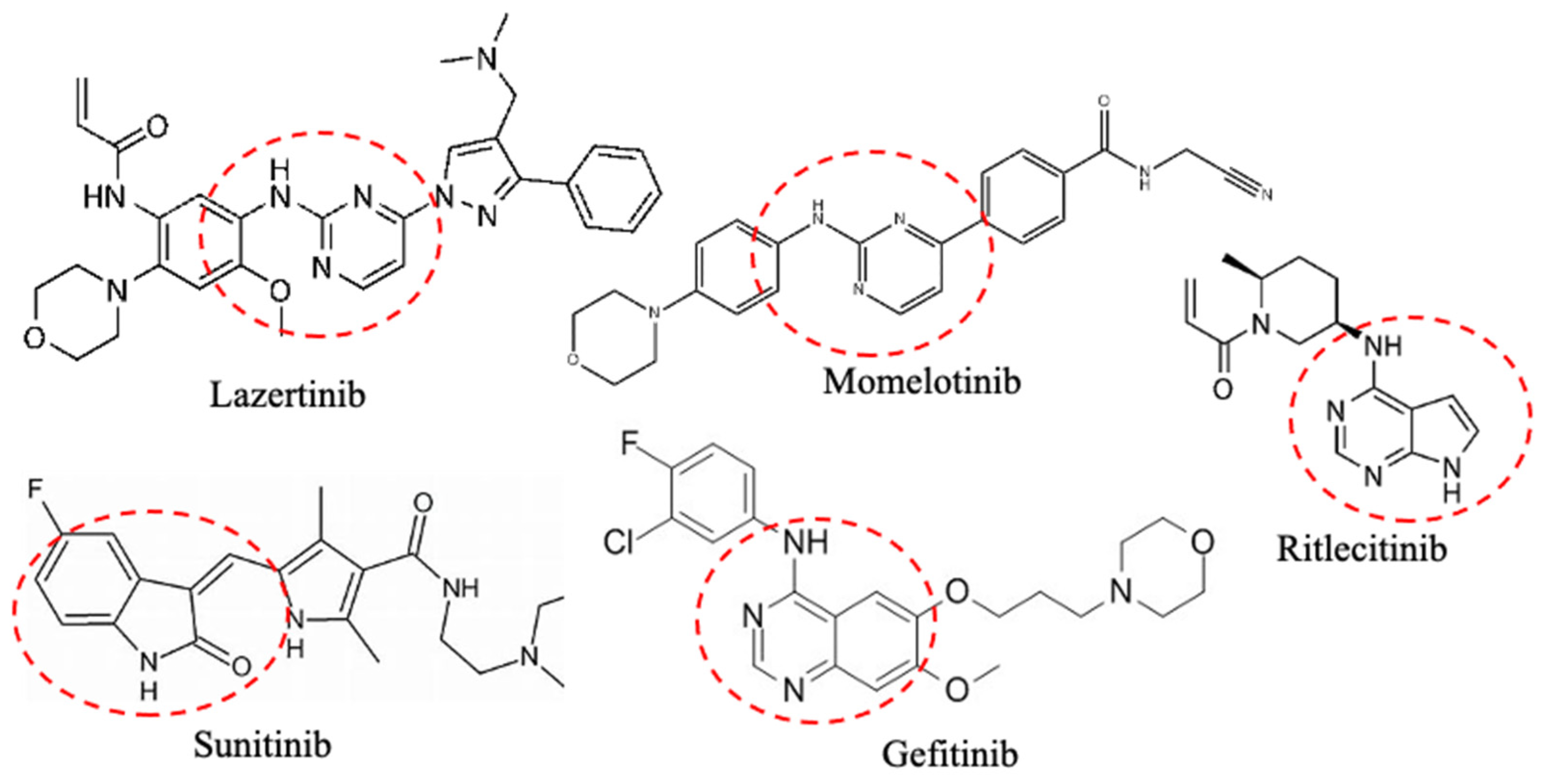
1.2. Small Protein Kinase Inhibitors Approved by FDA

Targeted therapies generally are divided into two types: small molecules (<1000 Da) and macromolecules (ranging from 1500 to 150,000 Da), including monoclonal antibodies, polypeptides, antibody–drug conjugates, and nucleic acids [32]. Compared to macromolecule medications, small-molecule targeted drugs offer advantages in pharmacokinetic properties, cost-effectiveness, patient compliance, and drug storage and transport [33].
Small-molecule kinase inhibitors are named according to the specific kinase family they target, with each family associated with a distinctive suffix. “-inib” is used for tyrosine kinase (TK) inhibitors, “-limus” for mTOR inhibitors, “-rafenib” for RAF inhibitors, “-anib” for VEGFR inhibitors, “-metinib” for MEK inhibitors, “-lisib” for PI3K inhibitors, “-clib” for CDK inhibitors, and “-mab” for monoclonal antibodies that target kinases. The mechanism of action for small-molecule protein kinase inhibitors involves reversible and irreversible interactions. Reversible inhibitors partially occupy the ATP-binding pocket and form hydrogen bonds with the hinge region linking the enzyme’s small and large lobes. This category includes types I, I½ A, I½ B, II A/B, II A, and II B. Additionally, reversible inhibitors can bind to an allosteric site in the cleft between the kinase’s small and large lobes near the ATP-binding pocket, as seen in types III and IV.
Moreover, type V inhibitors extend across two separate regions of the kinase domain, limiting kinase activity and exhibiting diverse residence times, indicating the duration they stay bound to the enzyme. Irreversible interactions occur when type VI inhibitors form covalent bonds with the kinase’s active site. Furthermore, most protein kinases contain extra domains that regulate kinase activity, oligomerization, and the recruitment of binding partners.
From 518 members of the protein kinase superfamily, FDA approved only 26 drugs with therapeutic applications that targeted PKs associated with different diseases (Table 1). Due to the conserved ATP-binding site across the human kinome, ATP-mimicking compounds often interact with multiple kinases, resulting in drugs with broad, sometimes promiscuous, activity profiles. The promiscuous ATP-binding site applies to drugs such as Abemaciclib, Palbociclib, Ribociclib, and Trilaciclib, all targeting the CDK4/6 protein. Kinases like CDK4/6, AKT, HER2, ErbB1/2/4, and FKBP12/mTOR are regarded as therapeutic targets for breast cancer treatment [34] (Figure 2; Table 1).
In contrast, some inhibitors are designed for a single target, such as the kinase inhibitor Capivasertiv, which targets the AKT protein and is considered an oncogene. The AKT protein is part of the phosphatidylinositol-3-kinase/protein kinase B/mammalian target of rapamycin (PI3K/Akt/mTOR) signaling pathway, which has become a key target in cancer, diabetes, obesity, neurodegenerative diseases, aging, and autoimmunity. That is due to its central role in controlling essential cellular processes like cell growth, proliferation, survival, and metabolism [35].
Additionally, the VEGFR protein serves as a broad-spectrum therapeutic target, with ten drugs known to inhibit it and treat various cancers (Table 1). The regulation of protein kinases is critical from a therapeutic perspective, as some protein kinase inhibitors can directly block multiple proteins or therapeutic targets, as seen with Afatinib, Lapatinib, Neratinib, and Tucatinib (Table 1). Some inhibitors are promiscuous, targeting more than two proteins, as in the case of Ponatinib and Regorafenib, each with nearly 11 targets (Table 1). Ponatinib is specifically indicated for treating a single disease (leukemia-CML or ALL).
Approximately 28 approved drugs act as multi-kinase inhibitors (Table 1), and the simultaneous inhibition of multiple protein kinases offers potential benefits, as blocking multiple targets may enhance the therapeutic effectiveness of these agents. Due to the inherent genetic alterations in cancer cells, resistance to protein kinase inhibitors nearly always develops [36]. This challenge led to the creation of second-, third-, and later-generation inhibitors designed to target the original enzyme and the associated malignancy. Gatekeeper mutations often drive the acquired drug resistance in the initial protein kinase target [37]. A prime example is the gatekeeper mutation in BCR-Abl, which spurred the development of first-, second-, and third-generation drugs that have significantly extended the life expectancy of leukemia patients. First-generation Imatinib, second-generation Bosutinib, Dasatinib, and Nilotinib are FDA-approved for frontline therapy. Ponatinib, a third-generation drug, is approved for resistant cases with the BCR::ABL fusion protein (T315I mutation) or after failure with at least two other protein–tyrosine kinase inhibitors [38].
In 2024, the FDA approved two new small-molecule protein kinase inhibitors, Lazertinib and Tovorafenib. Lazertinib is an epidermal growth factor receptor (EGFR) kinase inhibitor targeting EGFR exon 19 deletions and exon 21 L858R substitution mutations at lower concentrations than wild-type EGFR. In non-small-cell lung cancer (NSCLC) and mouse xenograft models with EGFR exon 19 deletions or EGFR L858R substitution mutations, Lazertinib showed anti-tumor effects. When combined with Amivantamab, Lazertinib demonstrated enhanced anti-tumor activity in vivo compared to either treatment alone in a mouse xenograft model of human NSCLC with an EGFR L858R mutation [39]. Tovorafenib is a Type II RAF kinase inhibitor that demonstrated anti-tumor activity in cultured cells and xenograft melanoma tumor models with BRAF V600E and V600D mutations [40].
Table 1. List of protein targets by FDA-approved small-molecule protein kinase inhibitors.
Protein TargetsDrugDiseaseReferences
CDK4/6Abemaciclib
Palbociclib
Ribociclib
Trilaciclib
Breast cancer and myelosupression[41,42,43,44]
AKTCapivasertibBreast cancer[45]
HER2Lapatinib #
Neratinib #
Tucatinib #
Breast cancer[46,47,48]
ErbB1/2/4Afatinib
Lapatinib
Neratinib
Tucatinib
Non-small-cell lung cancer (NSCLC), breast cancer, and colon cancer[47,49,50,51]
FKBP12/mTORSirolimus
Everolimus *
Breast cancer and * lymphangioleiomyomatosis[52,53]
MEK1/2Binimetinib
Cobimetinib
Selumetinib
Melanoma and neurofibromatosis[54,55,56]
BCR-AblDasatinib #
Imatinib #
Nilotinib #
Ponatinib #
Asciminib
Bosutinib #
Chronic myeloid leukemia (CML), and acute lymphocytic leukemia (ALL)[57,58,59]
Flt3Gilteritinib
Midostaurin #
Quizatinib
Leukemia[60,61,62]
BRAFDabrafenib
Encorafenib
Vemurafenib
Tovorafenib **
Melanoma[63]
JAK1/2/3Abrocitinib *
Momelotinib
Pacritinib
Baricitinib *
Fedratinib
Ritlecitinib *
Tofacitinib *
Upadacitibib *
* Atopic dermatitis, myelofibrosis, * rheumatoid arthritis, * alopecia areata, and * ulcerative colitis[64,65,66,67,68,69]
ALKLorlatinib
Alectinib
Brigatinib #
Ceritinib #
Crizotinib #
Entrectinib #
ALK-positive NSCLC, and inflammatory myofibroblastic tumor[70,71]
PDGFRMidostaurin #
Ripretinib #
Avapritinib #
Acute myeloid leukemia, and gastrointestinal stromal tumors[72,73,74]
VEGFR 1/2/3Axitinib
Cabozantinib #
Fruquintinib
Lenvatinib #
Pazopanib #
Regorafenib #
Sorafenib #
Sunitinib #
Tivozanib #
Vandetanib #
Advanced renal cell carcinoma, advanced medullary thyroid cancer, renal cell and hepatocellular carcinoma, metastatic colorectal cancer, differentiated thyroid cancer, gastrointestinal stromal, and pancreatic neuroendocrine tumors[75,76,77,78,79]
ROCK2Belumosudil ** Graft vs. host disease[80]
KitPexidartinib #
Ripretinib #
Avapritinib #
Tenosynovial giant cell tumors, and gastrointestinal stromal tumors[81,82]
METCapmatinib
Crizotinib #
Tepotinib
NSCLC, anaplastic large cell lymphoma, inflammatory myofibroblastic tumor, and MET mutant NSCLC[83]
EGFRDacomitinib #
Erlotinib
Gefitinib
Mobcertinib
Osimertinib
Lazertinib **
EGFR-mutant NSCLC, NSCLC, and pancreatic cancer[84,85]
TYK2Deucravacitinib ** Psoriasis[86]
TRKA/B/CEntrectinib
Larotrectinib
Solid tumors with NTRK fusion proteins[87]
ROS1Repotrectinib
Entrectinib #
ROS1-positive NSCLC[88]
FGFR1/2/3/4Erdafitinib
Futibatinib
Infigratinib
Nintedanib
Pemigatinib
Urothelial bladder cancer, bile duct cancer, and idiopathic pulmonary fibrosis[89,90,91,92]
BTKIbrutinib
Pirtobrutinib
Zanubrutinib
Chronic lymphocytic leukemia (CLL), and small lymphocytic lymphoma[93]
SYKFostamatinibChronic immune thrombocytopenia[94]
T970MOsimertinib #NSCLC with exon 19 deletions or exon 21 substitutions[95]
CSF1RPexidartinib #Tenosynovial giant cell tumors[96]
AxlSunitinib
Cabozantinib
Renal cell carcinoma[97,98]
* Non-oncological therapeutic applications. ** New approved drugs by FDA in 2024. # Compounds that display more than one target.
Among the 82 approved small-molecule protein kinase inhibitors, 14 target protein–serine/threonine protein kinases, 4 act on dual specificity protein kinases (MEK1/2), 20 inhibit nonreceptor protein–tyrosine kinases, and 44 block receptor protein–tyrosine kinases. While most FDA-approved kinase inhibitors are anti-neoplastic, others serve primarily as immunomodulators. Only a select group, including Abrocitinib, Baricitinib, Belumosudil, Deucravacitinib, Ritlecitinib, Tofacitinib, Upadacitinib, and Everolimus, are approved for the treatment of inflammatory conditions, such as atopic dermatitis, rheumatoid arthritis, graft vs. host disease, psoriasis, alopecia areata, ulcerative colitis, and lymphangioleiomyomatosis (non-neoplastic disease) [13].
Development efforts for inhibitors have stalled or have been abandoned for at least 11 kinase families and about 20 kinase targets, of which no agents are currently in active trials [99]. The Aurora kinase family (Aurora kinase A, Aurora kinase B, and Aurora kinase C) remains the most targeted kinase family without an approved agent despite 22 compounds having entered clinical trials. Aurora kinases, known for their role in cell division and overexpression in numerous human tumors, have long been therapeutic targets. However, more than 80% of Aurora kinase inhibitors that reached clinical trials have since been discontinued [100].

2. Cyclic Peptides as Small-Molecule Therapies Through Human Protein Kinase Inhibitors

Peptide-based medications may pose less risk, as their degradation yields only amino acids, which are harmless once they bind to their target molecules [101]. Compared to small molecules used in conventional treatments, peptides exhibit several advantages, including reduced tissue accumulation, high selectivity, exceptional potency, and specific biotarget interaction. In this sense, ultra-short peptides, defined as sequences of up to 7 amino acids, typically containing no more than 45 amino acids, stand out for their unique structural and functional properties. Ultra-short peptides present strengths and limitations; their immense potential as versatile scaffolds for diagnostics and therapeutic applications is undeniable [102]. A key strategy for enhancing the performance of these peptides is cyclization, which stabilizes their active conformation. Cyclic ultra-short peptides not only retain the advantages of their linear counterparts, as it is, exhibiting enhanced mechanical stability, efficient tissue penetration, and reduced immunogenicity, along with bio- and cytocompatibility, characterized by non-hemolytic and non-cytotoxic behavior [103]. Moreover, the cyclic ultra-short peptides in vivo improve stability, resistance to protease, and plasma degradation [103], and lead to a high bioavailability [104], excellent membrane permeability [105], strong binding affinity [106], and limited conformational flexibility [107]. These attributes make cyclic ultra-short peptides exceptionally versatile and position them as up-and-coming candidates for advancing therapeutic and diagnostic innovations. Unlike small molecules in traditional therapies, peptides offer several advantages, including minimal tissue accumulation, high selectivity, potency, and biotarget specificity [108]. Cyclic peptides (CPs) are polypeptides composed of two or more amino acids, canonical or non-canonical, linked by amide bonds in a circular sequence [104]. The ring structure can be formed by joining one end of the peptide to another through amide bonds or other chemically stable bonds like lactone, ether, thioether, or disulfide. Many cyclic peptides gain their ring structure through amide bond formation between the amino and carboxyl termini, a method called N-to-C (or head-to-tail) cyclization [109]. CPs can naturally be synthesized post-ribosomally (via post-translational modifications) or through non-ribosomal pathways, with non-ribosomal synthesis primarily involving amide bonds. In contrast, post-ribosomal synthesis commonly uses disulfide bonds [110].
Cyclic peptides are approved for various therapeutic uses, including antibiotic, antifungal, anti-cancer, and immunosuppressive activities [111]. These peptides are sourced from terrestrial and marine environments [112], animal toxins [113], plants [9], and microorganisms [114], and are even found as endogenous compounds in humans. Another way to obtain cyclic peptides is through chemical synthesis, which allows for sufficient quantities for large-scale biological testing. Although science has advanced to the point where cyclic peptides can be modified and synthesized to achieve specific biological properties, most cyclic peptides used in approved drugs are still of natural origin [115].
Compared to linear peptides, cyclic peptides identified through library screening exhibit enhanced selectivity, potency, bioavailability, and metabolic stability. Their rigid structure reduces the entropy of Gibbs free energy, enabling the stronger binding to target molecules or receptors. The absence of carboxyl and amino termini in cyclic peptides also makes them resistant to hydrolysis by exopeptidases and, in some cases, endopeptidases [116]. CPs with structures of 5 to 10 residues can bind with antibody-like specificity and affinity [106]; this structural stability in the CPs directly contributes to their bioactivity, as a longer residence time increases their capacity to interact with various pharmacological targets.
The membrane permeability of drugs often depends on their physicochemical characteristics, such as molecular weight, hydrogen bonding, and polar surface area [117]. Peptides are generally large and polar and cannot diffuse via passive transport across the cell membrane [118]. The cyclization of peptides can help increase membrane permeability by promoting intramolecular hydrogen bonds that mask the polar regions; however, permeability tends to decrease with larger molecules (over ten amino acids and >1 kDa). Synthetic peptides are usually designed to target extracellular receptors [104]. Naturally occurring CPs with a high membrane permeability often have N-methylamide bonds, ester bonds, or both in their leading chains [105,119].
The potential of CPs to mimic the binding of endogenous molecules, including receptors, transcription factors, and enzymes, is crucial for their biological effects. Kinases naturally interact with endogenous proteins and peptides, which can serve as potent inhibitors or activators. The peptide-based and cyclic structure of the CPs allows them to replicate interactions between kinases and their natural substrates, thereby influencing kinase activity, a critical factor in health and disease. Reversible protein phosphorylation, mediated by kinases and phosphatases, is vital for biological and pathological processes. By transferring the γ-phosphate group from ATP to serine (Ser), threonine (Thr), and tyrosine (Tyr) residues, protein kinases regulate core cellular functions like metabolism, cell cycle progression, and cytoskeletal organization. CPs have shown significant effectiveness in disrupting protein–protein interactions [120].

Cyclic Peptides as Therapeutic Tools: Kinase Inhibition and Biomedical Applications

Cyclic peptides can selectively target microbial proteins that differ from human proteins, making them suitable for developing antimicrobial drugs with minimal toxicity to patients [121]. Recently, the FDA approved several CPs for treating bacterial and fungal infections; for example, Telavancin and Daptomycin are effective against Gram-positive bacteria, mainly by disrupting cell membrane homeostasis through depolarization [122,123]; Polymyxin B targets Gram-negative bacteria by altering the lipid A component of lipopolysaccharide in the outer membrane [124]. Antifungal agents such as Anidulafungin inhibit fungal cell wall synthesis, leading to osmotic cell lysis [125]. Research is also underway on CPs for viral infections, as their affinity for viral fusion proteins shows antiviral potential [126]. For instance, CPs have been studied in SARS-CoV-2, where they block receptor binding in cultured cells in a dose-dependent manner [127]. A similar effect is seen in human immunodeficiency virus (HIV), where CPs interfere with the specific interaction between the virus and host proteins, preventing viral replication [128]. CPs can also function as inhibitors of RNA–protein interactions and enzymes, effectively targeting proteases, phosphatases, the proteasome, HIV integrase, and dam methyltransferase, among others [129].
Cyclic peptides emerged as promising therapeutic agents for the inhibition of kinases; deregulated kinases are often oncogenic and vital for cancer cell survival and proliferation (Table 1). The HGF-MET pathway drives essential processes like invasion, metabolic reprogramming, angiogenesis, metastasis, immune evasion, and drug resistance within the tumor microenvironment, all of which contribute to malignant transformation [130,131]. Although no selective HGF-MET inhibitors are yet to be approved as cancer therapies, the selective inhibition of MET activation is anticipated to be an effective molecularly targeted cancer treatment. HGF is synthesized and secreted as single-chain HGF (scHGF), which becomes active two-chain HGF (tcHGF) after cleavage at Arg494-Val495 [132]. Only tcHGF can activate MET, as scHGF is an inactive precursor that cannot activate the MET receptor. The cyclic peptide HiP-8 inhibits MET activation/phosphorylation in cultured human cells, binding specifically to tcHGF with a dissociation constant (KD) of 0.4 nM and a slow dissociation rate (koff = 0.4 × 10⁻³ s−1). HiP-8 exhibits an IC50 of 0.9 nM, indicating the potent inhibition of the HGF-MET interaction. Notably, HiP-8 inhibits only tcHGF and not scHGF, suggesting its potential as cancer therapy [133]. The proteolytic conversion of scHGF to its active form by serine endopeptidases like matriptase, hepsin, and the HGF activator is the rate-limiting step in HGF/MET signaling. A novel class of macrocyclic peptide inhibitors, such as compound VD2173, potently inhibits matriptase and hepsin, blocks scHGF activation in lung cancer models, halts HGF-mediated wound healing, and overcomes the resistance to EGFR- and MET-targeted treatments [134].
Inhibiting angiogenesis has become a key strategy in cancer therapy. The new pro-apoptotic peptide CIGB-300 (P15-Tat) has demonstrated antiangiogenic effects on endothelial cells by disrupting the CK2-mediated phosphorylation. CPs are widely used as scaffolds in drug design due to their notable stability and favorable biopharmaceutical properties. Two highly stable cyclic trypsin inhibitors, MCoTI-II and SFTI-1, serve as frameworks to integrate the bioactive epitope P15 into various backbone loops. This approach enhances the antiangiogenic activity and stability of P15 when integrated into cyclic trypsin inhibitor scaffolds [135].
The human epidermal growth factor receptor 2 (HER2) is overexpressed in numerous cancers, making it an important therapeutic target [136]. Trastuzumab is an FDA-approved monoclonal antibody that targets HER2 and is used to treat HER2-positive cancers [137]. The cyclic peptide Cyclo-GCGPep1 was designed in silico, based on the binding interaction between an antibody and the HER2 protein to create peptide–drug conjugates with Camptothecin. Biological assessments show that the resulting conjugate has antiproliferative solid effects on SK-BR-3 and NCI-N87 cells, retaining Camptothecin’s pro-apoptotic and Topo I inhibitory actions. Simultaneously, Cyclo-GCGPep1 enables the effective targeting of HER2-positive cells.
Compared to Camptothecin alone, the conjugated peptide shows superior permeability in tumor spheroid models. Designing CPs based on antibody–protein target binding modes is crucial for developing new therapeutic strategies. The Cyclo-GCGPep1 and Camptothecin conjugate holds promise as a treatment for HER2-positive cancers [138].
The polo-box domain (PBD) of Polo-like kinase 1 (Plk1) is a promising target for cancer therapies. PBD plays a role in substrate targeting and Plk1’s subcellular localization and it regulates kinase activity through an auto-inhibitory interaction with the kinase domain. Plk1, a key serine/threonine kinase, is essential in various mitotic processes, including mitotic entry, centrosome maturation, bipolar spindle assembly, chromosome segregation, and cytokinesis [139]. Elevated Plk1 levels and activity are associated with tumor progression and a poor prognosis [140]. These findings indicate that targeting Plk1/PBD could be an effective strategy for developing new anti-cancer drugs. Innovative phosphorylated macrocyclic peptidomimetics, engineered based on the acyclic phosphopeptide PMQSpTPL, target PBD and demonstrated anti-tumor activity both in vitro and in vivo. The novel macrocyclic peptidomimetics could be powerful templates for developing potent Plk1-PBD inhibitors [141].
Cancer cells undergo an epithelial–mesenchymal transition (EMT), transforming into invasive forms that spread to other organs. A novel treatment strategy to inhibit cancer cell migration and invasion involves targeting molecules associated with the EMT process. The cyclic-pentapeptide (-Arg-Gly-Asp-D-Phe-Lys) (cRGDfK) has been recognized as an integrin αβ3 inhibitor, functioning as a targeted peptide to enhance tumor cell sensitivity to drugs in anti-angiogenic cancer therapy. When used in conjunction with cRGDfK, Sunitinib increased its anti-EMT effects and decreased the expression levels of mesenchymal marker mRNA and protein compared to its use alone [142]. The use of cyclic pentapeptides (cRGDfK) containing the arginine-glycin-aspartic acid (RGD) motif enhances the target selectivity and drug delivery of anti-cancer medications [143,144]. Numerous studies have demonstrated the potential of RGD-containing peptides as delivery vehicles for anti-cancer drugs. Cyclic peptides, particularly cRGDfK and cRGDyK (cyclo Arg-Gly-Asp-D-Tyr-Lys), exhibit a strong affinity and selectivity for binding integrin αβ3. Thus, CPs can deliver therapeutic small molecules such as doxorubicin and paclitaxel to cancer patients [145].
The epidermal growth factor receptor (EGFR) family comprises EGFR (ErbB1), ErbB2, ErbB3, and ErbB4, all exhibiting tyrosine kinase activity. These receptors can form homodimers or heterodimers upon binding to ligands, leading to the phosphorylation of specific tyrosine residues in their intracellular domains. These phosphorylated residues serve as docking sites that can activate several intracellular signaling pathways [146]. The activation of EGFR signaling is associated with various physiological and pathological processes, including embryonic development, cell proliferation, cell survival, and cancer [147]. Small-molecule inhibitors, such as Gefitinib and Erlotinib, have been clinically approved to target the tyrosine kinase domain of EGFR (TK-EGFR). Lazertinib, a recently developed third-generation EGFR tyrosine kinase inhibitor (TKI), is used for patients with advanced EGFR T790M-positive non-small-cell lung cancer who have shown disease progression after treatment with first- and second-generation TKIs [148] (Figure 2; Table 1). Despite progress, significant concerns persist regarding the severity of side effects, off-target effects, and drug resistance. Among the CPs explored for novel drug development, NP1 has shown an inhibitory effect on TK-EGFR with an IC50 in the nanomolar range. NP1 has a binding affinity (KD) of 18.40 ± 5.50 µM for EGFR. It has been shown to inhibit phosphorylated EGFR levels upon activation in cancer cells [149]. The EGFR gene can undergo several mutations, particularly in exons 18–21, which encode part of the EGFR kinase domain associated with certain lung cancer types [150]. The most common mutation is EGFR variant III (EGFRvIII), which involves the deletion of exons 2–7 and is linked to more aggressive diseases and a worse prognosis [151]. Two cyclic peptides, c(CHVPGSYIC) and c(CVNAMQSYC), effectively bind and internalize the EGFR and EGFRvIII cell lines. Docking simulations revealed that the peptides interact with the EGFR receptor near the EGF binding site, showing substantial overlap with EGF [151]. A novel cyclic peptide, M-2-5, demonstrates the robust targeting of the extracellular domain of EGFR, binding tightly and effectively antagonizing EGF stimulation; EGFR phosphorylation and the subsequent signal transduction, representing a potential therapeutic use for cancer treatment [152]. PAD-1, a cyclic peptide self-assembled into a scaffold, recognizes the membrane protein EGFR and arrests EGFR signaling by inducing aggregation through multivalent interactions. By promoting by the endocytosis of overexpressed EGFR on cancer cell membranes, PAD-1 forms nanofibers in the lysosome, resulting in lysosomal membrane permeabilization (LMP). This mechanism disrupts EGFR homeostasis and inhibits downstream signaling transduction, presenting a promising therapeutic approach [153].
Pathological angiogenesis begins when abnormally expressed vascular endothelial growth factors (VEGFs) bind to their receptors (VEGFRs). Inhibiting the interaction between VEGF and VEGFR has been clinically validated as an effective cancer treatment. Novel cyclic peptides A-cL1, B-cL1, and P-cL1 mimic Loop 1 of VEGF-A, VEGF-B, and PlGF. These peptides primarily bind to the D3 domain of VEGFR1 and VEGFR2, successfully blocking their activation. They exhibit antiangiogenic and anti-cancer effects by inhibiting the MAPK/ERK1/2 signaling pathway. The VEGFR-targeting peptide VEGF125–136 (QR) blocked VEGF signaling, promoting tumor vascular normalization. When conjugated with a lytic peptide (KLU), it forms a peptide–drug conjugate QR-KLU, inhibiting liver cancer cell growth and downregulating VEGF expression under normoxic and hypoxic conditions [154].
Renal fibrosis is a standard and irreversible process associated with chronic kidney disease (CKD), characterized by excessive extracellular matrix deposition. It is a common endpoint for nearly all forms of CKDs. Despite significant advancements in recent years, no approved antifibrotic therapies for kidney conditions [155].
The extracellular matrix (ECM) protein CCN2 regulates the fibrotic process by activating the EGFR signaling pathway. To create compelling and durable selective inhibitors of the CCN2/EGFR interaction, new peptides targeting CCN2 have been developed. Notably, the 7-mer cyclic peptide OK2 shows intense activity in inhibiting STAT3 phosphorylation and cellular ECM protein synthesis induced by CCN2/EGFR activation. In vivo experiments also indicate that OK2 significantly reduced renal fibrosis in a mouse model of unilateral ureteral obstruction, providing a promising new strategy for treating renal fibrosis [156].
The monomeric GTPases K-Ras, H-Ras, and N-Ras play critical roles in various signaling pathways, such as cell proliferation, differentiation cytoskeletal reorganization, membrane trafficking, and apoptosis, with mutations found in about 30% of cancer cases [157,158]. Genetic studies indicate that inhibiting the interaction between Ras and its effector proteins could be a potential strategy for treating cancer patients. However, developing effective drugs has proven challenging due to the lack of distinct binding pockets for small-molecule drugs on the Ras protein surface. Wild-type Ras cycles consisting of its inactive GDP-bound state (Ras-GDP) and its active GTP-bound state (Ras-GTP), the Ras-GTP activating effector proteins like Raf, PI3K, and Ral-GDS, or the inhibitors designed to block Ras-effector protein interactions have typically exhibited deficiencies in potency, selectivity, and/or membrane permeability [159]. Cyclorasin 9A5 is an 11-residue cyclic peptide that can penetrate cells and selectively inhibits Ras-GTP. The simultaneous inhibition of the MEK and PI3K signaling pathways results in a combined decrease in cell viability and an increase in apoptosis in Ras mutant cancer cells. Cyclorasin 9A5 effectively disrupts protein–protein interactions (PPI) in H358 lung cancer cells and reduces Ras-dependent signaling in a dose-dependent manner, with an IC50 of approximately 3 mM [160]. Structure–activity relationship studies found that Cyclorasin 9A54 exhibits even more potent PPI inhibition (IC50 = 18 nM). Both peptides contain a cell-penetrating peptide (CPP)-like motif (Arg-Arg-dNal-Arg-Fpa) where dNal is d-b-naphthylalanine and Fpa is l-4-fluorophenylalanine. However, only Cyclorasin 9A5 effectively penetrates cells to demonstrate significant cell-based activity, suggesting that the conformational flexibility allowing the peptides to adopt an amphipathic structure is essential for their ability to permeate cells [161].
KS-58 is a bicyclic peptide advancing the development of new cancer therapeutics. It enters cells and blocks the interaction between K-Ras (G12D) and their effectors, restricting cell growth and showing considerable resistance to protease degradation [162]. P110α, a family member of the phosphoinositide 3-kinase (PI3K), is critical in transducing signals downstream of RAS. RAS proteins facilitate the activation of p110 α through direct interaction with its RAS binding domain (RBD), thereby promoting various cellular functions, including growth, proliferation, and survival. Cyclo-CRVLIR is a novel kinase inhibitor that binds to the p110α-RBD domain, blocking its interaction and exhibiting promising therapeutic potential in RAS-associated malignancies [163].
N-methyl-D-aspartate receptors (NMDARs), neuronal nitric oxide synthase (nNOS), and Postsynaptic Density Protein-95 (PSD-95) form a complex that is an attractive therapeutic target for the treatment of acute ischemic stroke. The complex is formed via the PDZ protein domains of PSD-95. Attempts to disrupt the complex often rely on C-terminal peptides sourced from the NMDAR; however, nNOS interacts with PSD-95 via the β-hairpin motif, presenting an alternate foundation for developing PSD-95 inhibitors. The inhibition strategy interrupts the nNOS/PSD-95 interaction using a cyclic nNOS β-hairpin mimetic peptide. Peptide macrocycles exhibit one of the highest affinities documented for a PDZ domain, providing insights for developing a treatment for acute ischemic stroke. [164]. Tropomyosin receptor kinases (Trks) are tyrosine kinases activated by neurotrophic factors called neurotrophins. TrkA engages with nerve growth factor (NGF), leading to pain induction. A macrocyclic peptide named Peptide 19 obstructs interactions between TrkA and NGF (IC50 = 21 nM), resulting in an analgesic effect in a rat model of incisional pain [165].
Momelotinib is an ATP-competitive small-molecule type I inhibitor of Janus kinases JAK1 and JAK2, approved for use in anemic patients with high- or intermediate-risk myelofibrosis (MF) (Figure 2) [166]. Cytokines act as communicative agents for the immune system, serving critical roles in various biological responses and influencing the immunological response. When cytokine production or biological activity is disrupted, the homeostatic balance of the immune response is altered, resulting in many diseases, including autoimmune and inflammatory disorders. Cytokines attach to specific receptors on cells, activating intracellular enzymes known as Janus kinases (JAKs). JAK3 modulates a limited range of γ cytokines, rendering it a potentially optimal target. Ritlecitinib is the most advanced selective irreversible JAK3 type VI inhibitor, exhibiting no activity against other JAK isoforms, and is approved for the treatment of alopecia areata (Figure 2). Z583 is a new and effective inhibitor of JAK3 with significant therapeutic potential for autoimmune illnesses, particularly in rheumatoid arthritis (RA) [167]. The activation of the Janus kinase/signal transducers and activators of the transcription (JAK/STAT) pathway is primarily marked by the building and disassembly of protein complexes, which are strongly associated with the expression of inflammatory genes. Suppressors of cytokine signaling (SOCS) proteins negatively regulate the JAK/STAT pathway. SOCS1 possesses a small kinase inhibitory region (KIR) responsible for inhibiting JAK kinases. A peptidomimetic of KIR SOCS1, designated PS5, diminishes JAK/STAT phosphorylation and the production of interferon regulatory factor-1 (IRF-1), leading to a significant decrease in inflammatory gene expression, demonstrating potential therapeutic effects in cardiovascular diseases [168].
PEP1, a cyclic peptide, efficiently transports into COS-1 cells and inhibits the phosphorylation of Gonadotropin-regulated testicular RNA helicase (GRTH) in a dose-dependent manner [169]. Rats, mice, and humans express GRTH, a constituent of the DEAD-box family of RNA helicases, in their testes, functioning as a post-transcriptional regulator of genes essential for the completion of spermatogenesis [170]. Prior research suggested that blocking the GRTH phospho-site or the interaction between GRTH and protein kinase A (PKA) might help create male contraceptives [171].
One tactic to potentially improve peptide therapies effectiveness, target selectivity, and metabolic regulation is the introduction of non-natural amino acids [172]. D/L-Hybrid peptides represent another exciting type of molecular modality due to their enhanced resistance to proteolysis and broader structural diversity compared to peptides composed only of proteinogenic L-amino acids. Macrocyclic D/L-hybrid peptides, featuring five types of D-amino acids (D-Tryptophan, D-Alanine, D-Serine, D-Histidine, and D-Tyrosine), demonstrate inhibitory activity against hEGF–hEGFR interaction and exhibited remarkable protease resistance after 24 h of incubation in human serum [173]. Moreover, the hEGF–hEGFR interaction can be inhibited by α/β3-macrocyclic peptides (in which β-homoglycine, β-homoalanine, β-homophenylglycine, and β-homoglutamine are introduced by reprogramming the genetic code). Peptides composed of β-amino acids frequently exhibit distinctive and robust folding capabilities, thereby earning the designation of foldamers [174]. Peptides composed exclusively of β-amino acids (β-peptides) and those having a combination of α- and β-amino acids (α/β-peptides) exhibit superior folding capabilities relative to α-peptides. Due to the self-folding capability of these α/β-peptides, they can achieve a favorable entropic effect relative to α-peptides, resulting in an anticipated enhanced binding affinity to target proteins. Furthermore, α/β-peptides typically exhibit enhanced proteolytic stability compared to α-peptides due to the absence or limited efficacy of naturally occurring peptidases on peptide bonds containing β-amino acids [175]. The application of β3AAs (β3-amino acids) improves the pharmacological characteristics of peptides, offering a distinctive platform for developing macrocycles targeting a specific protein [176].

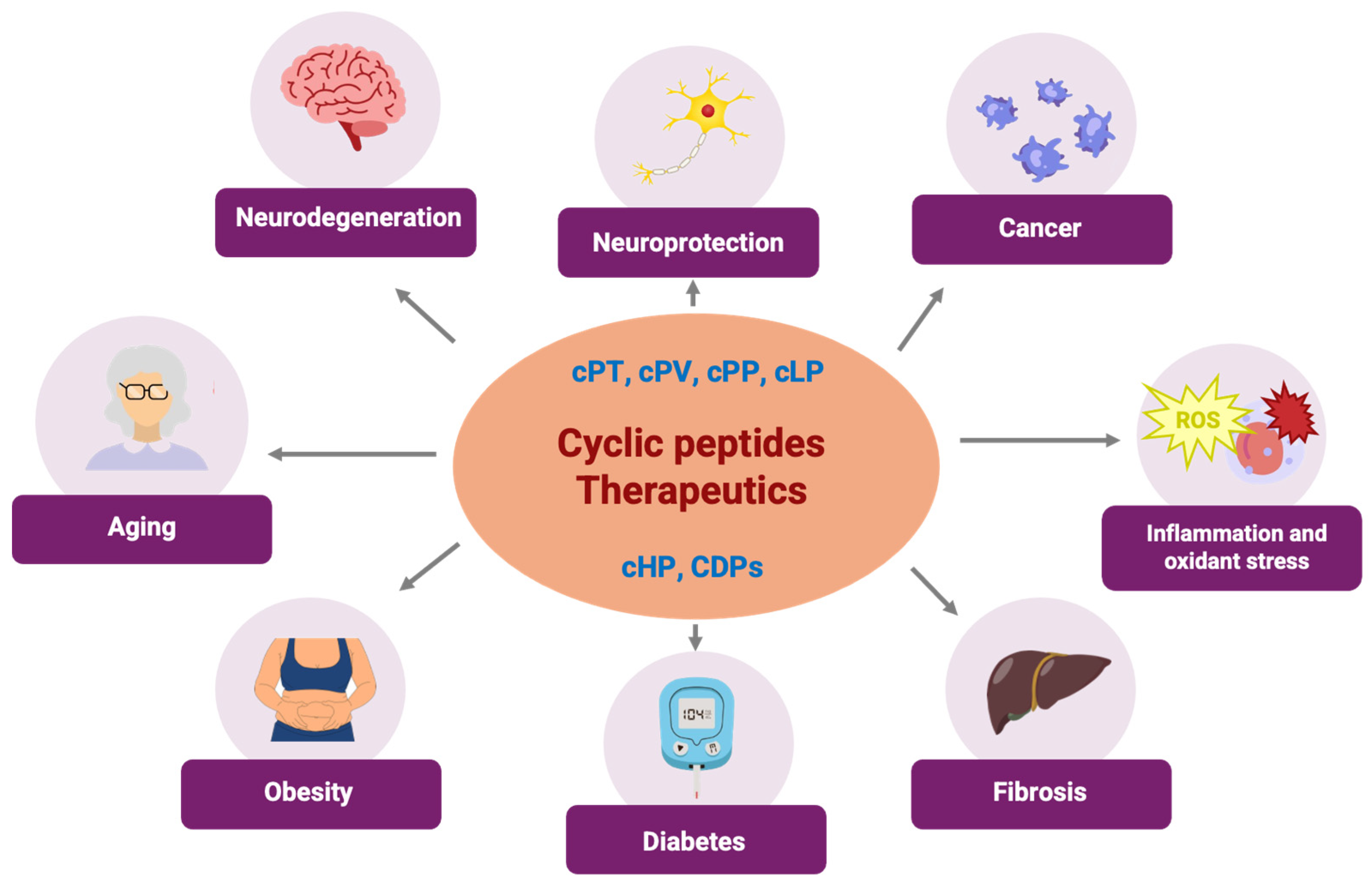
3. Cyclodipeptides, a Type of Cyclic Peptides with Potential Use in Human Diseases That Are Protein-Kinase-Associated

Cyclodipeptides (CDPs) or diketopiperazines are the simplest cyclic peptides found abundantly in nature [177] (Figure 3). CDPs that include proline exhibit distinctive characteristics such as an exceptionally rigid conformation, significant resistance, enhanced cellular permeability, and an augmented ability to bind various substrates with superior affinity. CDPs have emerged as crucial metabolic intermediates and a promising platform for therapeutic advancement. These molecules offer the structural stability and functional versatility characteristic of cyclic peptides, making them a unique class of biomolecules with vast potential [178,179]. They are often regarded as “diamonds in the rough”, presenting numerous opportunities for innovative therapies. Since their initial discovery in 1924, there has been growing interest in their applications, with multiple studies in recent years highlighting their broad-spectrum biological activities. This increasing attention underscores the potential of CDPs to play a pivotal role in future therapeutic advancements [180].
CDPs have been gaining increased attention since they are biomolecules with structural diversity, an absence of charged termini, enhanced lipophilicity facilitating rapid membrane absorption, reduced conformational freedom, and distinctive structural rigidity, which imparts more excellent resistance against enzymatic degradation, and their cyclic nature contributes to increased stability [178,179]. Moreover, CDPs have broad-spectrum bioactive properties and can be functionalized, making them promising scaffolds for a new drug design [180,181]. Due to these notable chemical features and diverse pharmacological activities, including the involvement of signal transduction pathways, where protein kinases participate [182,183,184,185,186,187], CDPs can potentially become candidates for drug development as PKI [178]. Regarding safety, initial studies indicate that CDPs have a low toxic profile in preclinical systems, particularly in cell cytotoxicity tests [188,189,190]. This makes them attractive candidates for new drug development. However, challenges still relate to the specificity of their biological activity, stability in physiological media, and possible accumulation in tissues, which could influence their long-term safety. Therefore, more extensive studies, including in vivo assays and comprehensive toxicological analyses, are needed to ensure their safe use in humans.

3.1. Cyclodipeptide and Protein Kinase Interaction in Cancer
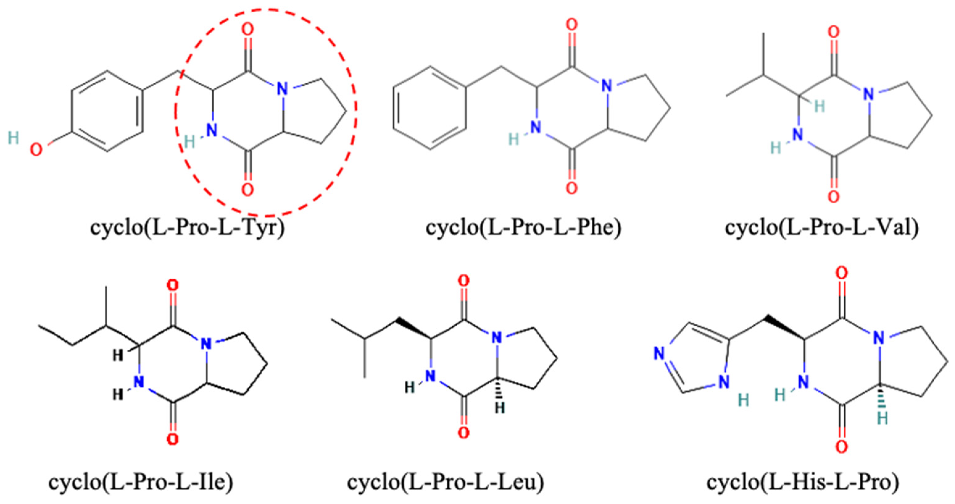
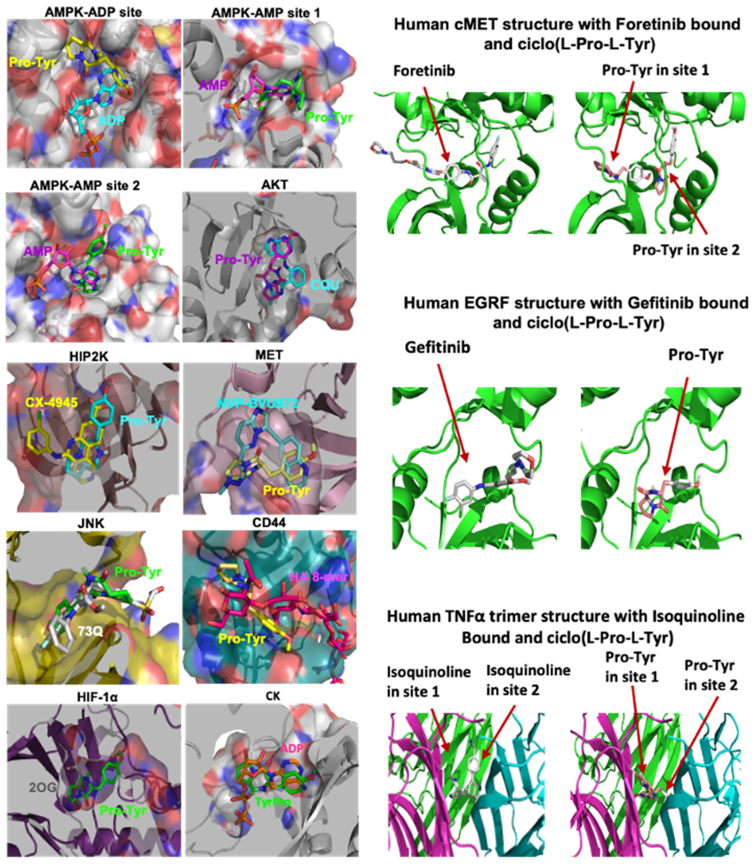
The cyclo(L-Pro-L-Tyr) (cPT), cyclo(L-Pro-L-Val) (cPV), and cyclo(L-Pro-L-Phe) (cPP) from Pseudomonas aeruginosa PAO1 (CDP-PA) [191] promoted cell death and apoptosis in a dose-dependent manner in cultures of cervical adenocarcinoma (HeLa cells), colorectal adenocarcinoma (Caco-2 cells) [189], melanoma (B16-F0 cells) [183], and human breast cancer (MCF-7 and MDA-MB-231 cells) [192] (Figure 4). The CDPs cPL, cPV, and cPP were applied in an isolated way in Hela cells and also promoted cell death with an LD50 of 61, 37, and 15 µg/mL, respectively, and induced apoptosis with an EC50 of 0.32, 0.27, and 0.50 µg/mL respectively [188]. In the B16-F0 melanoma cell line, the CDP-PA mix showed an LD50 of 10.7 µg/mL and EC50 of 24 µg/mL [183]. In MCF-7 and MDA-MB-231 cell lines, the LD50 was 50 and 10 µg/mL, respectively, while the EC50 was 50 µg/mL [192].
The xenotransplant melanoma tumor model caused a substantial reduction in both the weight and volume of the implanted tumor, with measurements of 5.5 g compared 2 g and 2100 mm3 versus 30 mm3 for untreated versus CDP-treated mice, respectively. Interestingly, mice injected with the B16-F0 tumor and concurrently treated with CDP-PA did not develop tumors in 40% of the subjects. In B16-F0 melanoma tumor cells treated with CDPs, apoptosis was associated with an exacerbated generation of ROS. Immunoblotting analysis indicated the involvement of the PI3K–Akt–mTOR signaling pathway which occurred since the phosphorylation of Akt-S473 and S6k-T389 was strongly activated in the tumor mice group, which decreased in the CDP treatment group [183]. Met C-terminal regulatory domain tail phosphorylation acts as a docking site for intracellular adaptors of molecules, activating downstream signaling cascades, such as Rac1/Cdc42, PI3k/Akt, and Erk/MAPK. In breast cancer MCF-7 cells, phosphorylation on the Met-Tyr1003, EGF-Tyr1068, and the Gab1-Tyr307 factor was significantly decreased by the CDP-PA exposure; however, in MDA-MB-231 cells, the Met-Tyr1003 phosphorylation was induced considerably by the CDP treatment [192].
The mechanism by which CDPs inhibit cell proliferation in HeLa cells includes the blockade of DNA synthesis and the arrest of cells at the G0–G1 stage. This process induces apoptosis through an intrinsic pathway reliant on caspase-9 and caspase-3 activation, affecting the mitochondrial functionality before cytochrome c release and increasing ROS generation [188]. The inhibition of cell proliferation occurred through the PI3K/Akt/mTOR, Ras/Raf/MEK/ ERK1/2, PI3K/JNK/PKA, p27Kip1/CDK1/survivin, MAPK, HIF-1, Wnt/β-catenin, HSP27, EMT, and CSCs signaling pathways and receptors, such as EGF/ErbB2/HGF/Met, since the inhibition of the phosphorylation of Stat3-Tyr705, Akt-Thr308, Akt-Ser473, AMPKα-Thr172, S6-RPSer235/236, mTOR-Ser2448, HSP27-Ser78, Bad-Ser112, p70S6k-Thr389, PRAS40-Thr246, P53-Ser15, P38-Thr180/ Tyr182, SAPK/JNK-Thr183/Tyr185, PARP-Asp214, Caspase 3-Asp175, GSK-3β-Ser9, Gab1-Tyr307, Met-Tyr1003, Met-Tyr1234/1235, Met-Tyr1349, EGF-Tyr992 and EGF-Tyr1068 with a cyclic behavior in a time- and concentration-dependent manner was shown [188,192,193]. The further implication of the PI3K/AKT/mTOR pathway was confirmed by utilizing the AZD8055 mTOR inhibitor and LY294002 PI3K inhibitor [193].
The transcriptomics analysis of untreated and CDP-PA HeLa cells exhibited 151 differentially expressed genes that were either up- or downregulated in response to CDP-PA across 15 biological processes. These processes included cellular component organization or biogenesis, cellular processes, localization, biological regulation, response to stimulus, signaling, developmental processes, rhythmic processes, multicellular organismal processes, locomotion, biological adhesion, metabolic processes, growth, cell population proliferation, and immune system processes. The biological pathways potentially targeted by CDP-PA in HeLa cells included the insulin/IGF/MAP kinase cascade, P38/MAPK, interleukins, EGF receptor, PI3K kinase, PDGF, Notch, cadherin, P53, the gonadotropin-releasing hormone receptor pathway, Hedgehog, TGF-β, FGF, the FAS signaling pathways, the Gonadotropin-releasing hormone receptor pathway, angiogenesis, cholesterol biosynthesis, and additional pathways related to inflammation, oxidative stress response, apoptosis, cytoskeleton, and T- and B-cell growth/activation [187]. This fact confirmed the role of CDP-PA in various signal transduction pathways, including PI3K-Akt, mTOR, FoxO, Wnt, MAPK, P53, and TGF-β.
A molecular docking analysis showed possible interactions between bacterial CDPs and protein kinases such as AKT, HIPK2, AMPK, MET, JNK, HIF-1α, CD44, cMET, and EGFR [183,192]; thus, the CDP-PA mechanism for inhibiting or activating protein kinases in cancer models may involve obstructing the substrate binding site of these protein factors (Figure 4). The CDP-PA mix demonstrates a negligible impact on apoptosis induction in cultures of normal human mononuclear blood cells, as well as in cellular structures from the spleen, liver, heart, kidney, lung, and skin from treated mice; thus, CDP-PA treatments were safe in those models [183,188]. The findings suggest that CDP-PA has potential as PKI anti-neoplastic agents for treating human cervical adenocarcinoma and melanoma.

3.2. Cyclo(Pro-Tyr)

Cyclo(Pro-Tyr) is also produced by sponge Callyspongia fistularis, Bacillus pumilus [194], and Bacillus sp. [195] (Figure 3). This CDP demonstrated cytotoxic and apoptotic-inducing activity against the human hepatocellular carcinoma (liver cancer) HepG2 cell line, with an IC50 value ranging from 42.98–140 µg/mL. In contrast, no toxic effects on non-cancerous cell lines were observed. Cyclo(Pro-Tyr)-treatments significantly increased the percentage of cells in the G2/M phase, induced ROS production, and caused nuclear fragmentation, chromatin condensation, and membrane blebbing. Additionally, the Bax/Bcl2 ratio protein expression was significantly amplified, as was the protein expression of cytochrome c, caspase-3, and cleaved PARP, indicating that apoptosis was promoted [194]. Hepatocellular carcinoma (HCC) generated by N-diethylnitrosamine (DEN) in male Swiss albino mice caused weight loss. Meanwhile, in cyclo(Pro-Tyr)-treated mice, the body weight of the animals was equal to that of the control group. In cyclo(Pro-Tyr)-treated mice, aspartate and alanine aminotransferase markers of liver damage showed decreased levels of both enzymes, cell proliferation was inhibited, and apoptosis was stimulated [196] (Scheme 1).
Like in the CDP-PA mix, the PI3K/AKT signaling pathway plays a vital role in cyclo(Pro-Tyr)-treated cells and animals since phosphorylated PI3K-Y607 and phosphorylated AKT-T308 levels were diminished. In contrast, the levels of PTEN (a negative pathway regulator), Bax, Caspase 3, and P53 increased [194,195,196,197]. Thus, cyclo(Pro-Tyr) has the potential to act as a PKI anti-cancer drug, suppressing HCC tumor development by targeting the PI3K/AKT pathway.

3.3. Cyclo(L-Leu-L-Pro)

The cyclo(L-Leu-L-Pro; cLP) has been isolated from Lactobacillus coryniformis BCH [198], Staphylococcus sp. MB30 has been observed to have anti-fungal, antimicrobial, and antioxidant activity. In addition, cyclo(L-Leu-L-Pro) has proven to have cytotoxic and antiproliferative activities against lung (A549) and cervical (HeLa) cancer cells [199] (Scheme 1).
In MDA-MB-231 and MDA-MB-468 Triple-Negative Breast Cancer (TNBC) cellular lines treated with cyclo(L-Leu-L-Pro) and with eribulin (a chemotherapeutic agent primarily for the treatment of metastatic breast cancer and liposarcoma), we observed cell cycle arrest at the G2-M phase, inhibited proliferation and migration, and induced DNA strand breaks, DNA damage, and apoptosis; those effects were not detected in human breast healthy epithelial cell line (MCF-12A). A bioinformatics analysis predicted the cyclo(L-Leu-L-Pro) interaction with tetraspanin protein CD151 and EGFR, a member of the ErbB family of receptor tyrosine kinases. Immunoprecipitation assays showed that cyclo(L-Leu-L-Pro) reduces the interaction of CD151 with EGFR in MDA-MB 231 and MDA-MB 468 cell lines. Therefore, in TNBC cells, the cyclo(L-Leu-L-Pro) action mechanism could be through the disruption of CD151-EGFR crosstalk and the subsequent EGFR downstream signaling [200].

3.4. Cyclodipeptide–Protein Kinase Interaction Related to Antioxidant and Anti-Inflammatory Effects

Cyclo(His-Pro) (cHP) was discovered in the 1970s as an endogenous molecule produced by the cleavage of the hypothalamic thyrotropin-releasing hormone (TRH) [201,202]. cHP therapy offered cytoprotection, reduced apoptotic cell death, and decreased tissue damage in cells exposed to reactive oxygen species (ROS), pro-inflammatory or inflammation-induced tissue damage agents via transcription factor Nrf2 [182,185,186,190]. cHP stimulates Nrf2 phosphorylation and nuclear location and enhances ROS detoxification by the induction of gene expression [182,185,186,190,202,203]. Furthermore, cHP reduces inducible oxide nitric synthase (iNOS) gene expression with a consequent decrease in reactive oxygen–nitrogen species (ROSN) and ROS. Moreover, cHP inhibits the gene expression of the transcriptional factor involved in the inflammatory process, NF-κB, which suppresses pro-inflammatory signaling pathways and reduces apoptosis activities such as caspase 3 and cytochrome c release [202] (Scheme 1).
In human LX-2 hepatic stellate cells, the cHP molecular mechanism has been associated with increased levels of cytosolic Ca2+; this fact may promote the phosphorylation of Nfr2 by protein kinase C (PKC) and the phosphorylated p38 MAPK prevented cHP-mediated Nfr2 activation [186,204].

3.5. Cyclodipeptide–Protein Kinase Interaction Related to Neuroprotective Effects

Following head trauma, metabolic changes are initiated, including alterations in ionic homeostasis, excitatory amino acids, free radicals, proteases, and inflammatory-immune responses. These changes ultimately cause neuronal cell death in adjacent areas of physical insult [205].
In paraquat-exposed hSOD-1G93A microglial cells, cyclo(His-Pro) treatment causes an early and sustained increase in glutathione (GSH), showing a protective effect against oxidant insults via nuclear translocation of Nrf2, an upregulation of Nrf2-driven genes, such as Gclc, Gclm, xCT, and Nqo1 by increased GSH. cHP inhibited ERK1/2 phosphorylation and delayed AKT inactivation. Furthermore, cHP enhances BDNF expression, which may lead to the proposal of this CDP as a neurotrophic agent [184] (Scheme 1).
The research has focused on novel drugs that target multiple injury factors. The CDP 1-ARA-35b (35b) derived from a modified thyrotropin-releasing hormone (TRH) analog showed neuroprotective characteristics in in vitro and in vivo models [205,206]. The 35b decreased cell death linked with necrosis, apoptosis, and mechanical injury in neuronal-glial cocultures. In a model of traumatic brain injury in rats, 35b improved motion recovery and led to a lower lesion volume [205].
Compounds with neuroprotective potential, CDPs related to cHP were synthesized by replacing the imidazole moiety from histidine residue with 3,5-di-tert-butyltyrosine. Two diastereomeric CDPs cyclo[(R)-30,50-di-tert-butyl-Tyr-L-Pro] and cyclo[(S)-30,50-di-tert-butyl-Tyr-L-Pro] were obtained. Both compounds showed neuroprotective activity by preventing Ca2+-induced necrosis and cell death induced by FeSO4, a free radical generator [207]. Other derived CDPs from cHP, 35b, 144, 606, and 807, also produced motion and cognitive recovery and neuroprotective activity using in vitro models of neuronal injury and after a controlled cortical impact in mice. All CDPs were evaluated using models pertinent to necrotic cell death or caspase-dependent apoptosis, including mechanical injury and glutamate toxicity models that induce necrotic cell death. In contrast, trophic withdrawal and β-amyloid models lead to caspase-dependent apoptosis [205,206]. The fact that the functional CDP, 35b, 144, 606, and 807, effects were similar to those of cHP indicates that they may share the same or similar underlying molecular mechanisms [206].

3.6. Cyclodipeptide–Protein Kinase Interaction Related to Fibrosis

Recently, the cyclo(His-Pro) was tested for its potential to prevent distinct-type fibrosis (Figure 3). Interestingly, cHP reduces fibrosis-related inflammatory molecules and immune cell infiltration, reducing inflammation [182,185,208] and attenuating steatohepatitis while reducing fibrosis and inflammation by reducing the infiltration of immune cells in liver tissue [208]. In human LX-2 hepatic stellate cells, a fibrinogenic cell type associated with fibrosis, cHP decreases fibrous extracellular matrix proteins, including COL1A [208]. The downregulation of COL1A protein was related to a reduction in ROS levels resulting from cHP treatment. In addition, by trying to silence Nrf2, the collagen-reducing effect of cHP was diminished, suggesting that the anti-fibrotic activity depends on Nrf2 signaling [186,208] (Scheme 1).
The liver transcriptomic analysis revealed that gene sets related to fibrosis, inflammation, and lipid metabolism were upregulated in mice with non-alcoholic fatty liver disease (NAFLD). Concurrently, cHP reduces the expression of essential genes involved in inflammation, fibrosis (extracellular matrix, hepatic stellate cells activation, and collagen production), lipid metabolism, oxidative stress, and apoptosis [208]. In NAFLD and CCl4-induced liver fibrosis models, cHP significantly downregulated the ERK signaling cascade. This pathway is activated in two human datasets collected from patients with advanced non-alcoholic fatty liver disease and steatohepatitis (NASH) and fibrosis, as the ERK pathway is related to inflammation and fibrosis [208]. cHP treatment diminishes the phosphorylation levels of ERK1/2 within a short duration (4 h), with levels approaching baseline after 24 h. The inhibition of ERK1 has been related to a reduced proliferation of hepatic stellate cells, diminished fibrosis, and improved lipid homeostasis.
In contrast, the activation of ERK2 influences hepatocyte proliferation and contributes to inflammation and fibrosis following liver injury [208]. The CDPs cyclo(His-Ala) and cyclo(His-Gly) also exhibit anti-fibrotic properties since they inhibit fibrin formation [209]. However, the mechanism has not yet been elucidated, and it probably involves the inhibition of the ERK pathway as cHP.

3.7. Cyclodipeptide–Protein Kinase Interaction Related to Aging

During a study of the therapeutic effect of cyclo(His-Pro) on age-related muscle decline, few mice died in the control group. In contrast, no aged mice were lost in the treated group, indicating a beneficial effect of cHP on lifespan extension, like in C. elegans nematode [182]. The cHP lifespan extension mechanism probably involves the reduction in reactive oxygen species (ROS) and inhibiting the ERK pathway, as in the cHP anti-inflammatory and anti-fibrosis effect (Scheme 1).
Advanced (glycation) end products (AGEs) result from binding a sugar molecule to a protein and accumulating throughout the body over a lifetime. AGEs can bind to the receptor of advanced glycation end products (RAGEs), triggering the release of inflammatory mediators by the NF-κB pathway [210]. AGEs are recognized as key contributors to various age-related diseases, including diabetic vascular complications, NAFLD, thyroid cancer progression, coronary artery disease, kidney pathologies, atherosclerosis, and Alzheimer’s disease [179,210]. Twelve CDPs, cyclo(Dha-L-Leu), cyclo(L-Phe-L-Ala), cyclo(L-Leu-L-Ala), cyclo(L-Tyr-L-Val), cyclo(L-Tyr-L-Tyr), cyclo(L-Tyr-L-Leu), cyclo(L-Tyr-L-Pro), cyclo(L-Pro-L-Val), cyclo(L-Pro-L-Ile), cyclo(L-Leu-L-Leu), cyclo(L-Met-L-Met), and cyclo(L-Pro-L-Pro), derived from Sphingobacterium sp. possess detoxifying activity that effectively eliminates intracellular carboxymethyl lysine (CML). This final AGE product accumulates with age in human dermal fibroblast cells [210].

3.8. Cyclodipeptide–Protein Kinase Interaction Related to Diabetes

The cyclo(His-Pro) has been identified in the human blood and gastrointestinal tract, where it can be absorbed [201]. In humans, plasma levels of cHP increase following glucose ingestion, indicating its function as a gut peptide of the entero-insular axis [201]. Blood glucose metabolism is directly associated with cHP concentration, as cHP enhances intestinal zinc absorption and muscle glucose uptake (zinc is involved in glucose uptake), insulin synthesis and secretion, and insulin utilization in insulin-requiring cells [211,212]. The cHP influences leptin-type compounds, which are involved in appetite regulation and contribute to reduced food intake [204] (Scheme 1).
In a non-obese Type II diabetes mellitus model, cyclo-His-Pro and zinc (cycloZ) enhanced blood glucose metabolism by promoting glucose utilization in muscle cells. The anti-diabetic activity of cycloZ remains unclear; nonetheless, insulin-mediated signal transduction mechanisms are likely involved in muscle [212]. A similar effect was observed in genetically obese (ob/ob) Type II diabetic mice by cHP plus zinc and L-histidine treatment. The primary cause of Type II diabetes is insulin resistance, whereas a significant contributor to obesity is leptin resistance [213]. CycloZ combined with L-histidine may improve insulin and leptin resistance since both systems participate in signal transduction mechanisms through PI3K phosphorylation [212].
In insulin resistance diabetes, cycloZ treatment resulted in improved tissue condition alongside reduced inflammation [208,214]. The global acetyl-lysine level in the liver of cycloZ-treated mice was diminished, indicating that cycloZ influences the regulation of lysine acetylation, which is related to the activity of various metabolic enzymes and transcription factors. Non-histone transcription factors with decreased acetylation include p65, LKB1, and PGC-1α, associated with mitochondrial biogenesis [215,216]. In addition to deacetylation, PGC-1α requires AMPK-mediated phosphorylation for its complete activation. CycloZ increases AMPK phosphorylation in the liver and epididymal adipose tissue. The cycloZ effect was also evaluated in a severe hyperglycemia model, yielding results consistent with those previously reported [214].

4. Future Perspective

This review demonstrates the advantages and potential of cyclic dipeptides as small-molecule protein kinase inhibitors. Cyclic peptides predominantly originate from natural sources; however, the advancements in peptide chemical synthesis have brought many advantages. These include the capacity to incorporate molecular modifications into analogs and derivatives with improved pharmacokinetic and pharmacodynamic properties. Moreover, they possess the benefit of being able to traverse the cell membrane. Recent advancements have made it possible to design cell-permeable, biologically active CPs rationally. These advances have consequently made cyclic peptides a promising therapeutic modality, perhaps offering a definitive solution to the “undruggable” intracellular PPI targets.
Nevertheless, considerable exploration remains concerning this class of compounds as emerging issues, such as gatekeeper mutations, promoting the intense research of numerous compounds, including CPs, which have been growing and gaining their space as therapeutic agents performing diverse activities. The most significant potential of CPs lies in their capacity to target intracellular proteins currently undruggable by small molecules or monoclonal antibodies. A second area of opportunity is multi-kinase inhibitor CPs, which would have a significant advantage over the current biologics regarding only one target. Many predominantly hydrophobic CPs have already exhibited favorable oral bioavailability. Due to their structural and metabolic stability properties, high binding affinity to targets, and low toxicity, CDPs show promising potential as therapeutic effects on complex diseases involving protein kinases, such as diabetes, cancer, obesity, aging, neurodegenerative diseases, and fibrosis. Although some protein targets of CDPs have already been found, such as the CLIC1 protein target of cHP, everything indicates that it has more molecular targets, so the panorama is opened to look for the mechanisms by which CDPs exert their effects at the molecular level.

Author Contributions

All authors contributed to study conception, design, and writing. L.H.-P., L.M.-A. and J.C.-G. wrote the final draft. All authors have read and agreed to the published version of the manuscript.

Funding

This study was funded by the Consejo Nacional de Humanidades, Ciencia y Tecnología (CONAHCYT) of México (grant number 256119) and by a grant from the Universidad Michoacana de San Nicolás de Hidalgo/C.I.C.2.14. LHP received the scholarship of CONAHCYT No. 74366.

Acknowledgments

We thank the Kinases and Phosphatases editorial staff and MDPI for the free APC support.

Conflicts of Interest

The authors declare no conflicts of interest.

Abbreviations

ABLAbelson kinase
AGEsadvanced glycation end products
AKTprotein kinase B
AMPKAMP-activated protein kinase
ALKanaplastic lymphoma kinase
ALLacute lymphocytic leukemia
Argarginine
ATPadenosine triphosphate
Axlaxl gene
BaxBCL2-associated X protein
Bcl2B-cell lymphoma 2 protein
BCRbreakpoint cluster region
BCR-ABLfusion protein of BCR gene and ABL gene
BDNFbrain derived neurotrophic factor
BRAFv-raf murine sarcoma viral oncogene homolog B
BTKBruton’s tyrosine kinase
CCN2cellular communication network factor 2
Cdc42cell division cycle 42
CDKCyclin-dependent kinase
CDPscyclodipeptides
CDPs-PAPseudomonas aeruginosa cPT, cPV and cPP
cHPcyclo(His-Pro)
CKcreatine kinase
CK2casein kinase 2
CKDchronic kidney disease
cLPcyclo(L-Leu-L-Pro)
CMLcarboxymethyl lysine
COL1Acollagen type I alpha 1 chain
CPPcell-penetrating peptide
cPPcyclo(L-Pro-L-Phe)
cPTcyclo(L-Pro-L-Tyr)
cPVcyclo(L-Pro-L-Val)
cRGDfKcyclic-pentapeptide (-Arg-Gly-Asp-D-Phe-Lys)
cRGDyKcyclic-pentapeptide (-Arg-Gly-Asp-D-Tyr-Lys)
CSCcancer stem cells
CSF1Rcolony-stimulating factor 1 receptor
cycloZcyclo(His-Pro) and zinc
DENN-diethylnitrosamine
DFGAsp-Phe-Gly motif
dNa1d-b-naphthylalanine
EC50half maximal effective concentration
ECMextracellular matrix
EGFepidermal growth factor
EGFRepidermal growth factor receptor
EMTepithelial-mesenchymal transition
ePKeucaryotic catalytic domain
ErbBerb-b tyrosine kinase receptor
ERK1/2extracellular signal-regulated kinase
FAStumor necrosis factor receptor superfamily member 6 (TNFRSF6)
FDAfood and drug administration
FGFFibroblast Growth Factor
FGFRfibroblast growth factor receptor
FKBP12FK506-binding protein 12
Flt3fms related tyrosine kinase 3
Fpa1-4-fluorophenylalanine
G12DK-Ras gene
Gab1GRB2 associated binding protein 1
GRTHgonadotropin-regulated testicular RNA helicase
GSHglutathione
HCChepatocellular carcinoma
HCDHigher-energy collisional dissociation
hEGF-Hegfrhuman EGF receptor bound ligand
HER2epidermal factor receptor 2
HGFhepatocyte growth factor
HIF-1 αhypoxia-inducible factor 1-alpha
HiP-8HGF-inhibitory peptide-8
HIP2Khomeodomain-interacting protein kinase 2
HIVhuman immunodeficiency virus
H-RasHarvey rat sarcoma viral oncogene homolog
IC50half-maximal inhibitory concentration
iNOSinducible oxide nitric synthase
IRF-1interferon regulatory factor-1
JAKsJanus kinases
KDdissociation constant
KIRkinase inhibitory region
KitKIT proto-oncogene
KLIFSkinase-ligand interaction fingerprint and structure
KLUlytic peptide
K-RasKirsten Ras oncogene homolog
LD50lethal dose 50
LKB1liver kinase B1
LMPlysosomal membrane permeabilization
McoTIMomordica cochinchinensis trypsin inhibitor
MEKmitogen-activated protein kinase kinase 7
METmesenchymal-epithelial transition factor
MFintermediate-risk myelofibrosis
mRNAsmessenger ribonucleic acid
mTORmammalian target of rapamycin
NAFLDnon-alcoholic fatty liver disease
NASHnon-alcoholic fatty liver disease and steatohepatitis
NF-κBnuclear factor kappa-light-chain-enhancer of activated B cells
NGFnerve growth factor
NMDARsN-methyl-D-aspartate receptors
nNOSneuronal oxide synthase
Nrf2nuclear factor erythroid-related factor 2
NSCLCnon-small cell lung cancer
OK27-mer cyclic peptide
P1GFplacental growth factor
P53Tumor Suppressor Protein p53
PARPPoly(ADP-ribose) polymerase
PBDpolo-box domain
PDGFRplatelet derived growth factor receptor
PDZbinding domain PDZ
PGC-1alfaperoxisome proliferator-activated receptor gamma coactivator 1-alpha
PI3Kphosphoinositide 3-kinase
PKAprotein kinase A
PKBprotein kinase B
PKCprotein kinase C
PKIprotein kinase inhibitors
Plk1polo like kinase
PPIprotein-protein interactions
PSD-95Postsynaptic density protein-95
PTENphosphatase and tensin homolog
QRVEGFR-targeting peptide VEGF125−136
RArheumatoid arthritis
Rac1Rac family small GTPase 1
RAFRAF kinase
RAGEreceptor of advanced glycation end products
Ral-GDSRal guanine nucleotide dissociation stimulator
RBDRAS binding domain
Ras-GDPinactive GDP–bound state
Ras-GTPactive GTP-bound state
RGDarginine-glycin-aspartic acid
ROCKRho kinase
ROSreactive oxygen species
ROS1ROS proto-oncogene 1 receptor tyrosine kinase
ROSNreactive oxygen-nitrogen species
RTKreceptor tyrosine kinase
SAPKStress-activated MAP kinases
SARS-CoV2severe acute respiratory syndrome coronavirus 2
scHGFsingle chain HGF
SFTI-1sunflower trypsin inhibitor
SH2Src homology 2
SOCSsuppressors of cytokine signaling
Srcstored response chain kinase
STATsignal transducer and activator of transcription
SYKspleen tyrosine kinase
tcHGFtwo chain HGF
TKtyrosine kinase
TK-EGFRtyrosine kinase domain of EGFR
TKItyrosine kinase inhibitor
TNBCtriple negative breast cancer
TRHhypothalamic thyrotropin-releasing hormone
TrkATropomyosin receptor kinase A
TRKstropomyosin receptor kinases
TyKstyrosin kinases
Valvaline
VEGFsvascular endothelial growth factors
VEGFRsvascular endothelial growth factor receptors
WntWingless-related integration site

References

  1. Lahiry, P.; Torkamani, A.; Schork, N.J.; Hegele, R.A. Kinase mutations in human disease: Interpreting genotype–phenotype relationships. Nat. Rev. Genet. 2010, 11, 60–74. [Google Scholar] [CrossRef] [PubMed]
  2. Zhai, Y.; Yang, J.; Zhang, J.; Yang, J.; Li, Q.; Zheng, T. Src-family protein tyrosine kinases: A promising target for treating cardiovascular diseases. Int. J. Med. Sci. 2021, 18, 1216. [Google Scholar] [CrossRef] [PubMed]
  3. Zhang, L.; Zhu, Q.; Tan, Y.; Deng, M.; Zhang, L.; Cao, Y.; Guo, X. Mitogen-activated protein kinases MPK3 and MPK6 phosphorylate receptor-like cytoplasmic kinase CDL1 to regulate soybean basal immunity. Plant Cell 2024, 36, 963–986. [Google Scholar] [CrossRef] [PubMed]
  4. Ferguson, F.M.; Gray, N.S. Kinase inhibitors: The road ahead. Nat. Rev. Drug Discov. 2018, 17, 353–377. [Google Scholar] [CrossRef]
  5. Nikolic, I.; Leiva, M.; Sabio, G. The role of stress kinases in metabolic disease. Nat. Rev. Endocrinol. 2020, 16, 697–716. [Google Scholar] [CrossRef]
  6. Knighton, D.R.; Zheng, J.; Ten Eyck, L.F.; Ashford, V.A.; Xuong, N.-H.; Taylor, S.S.; Sowadski, J.M. Crystal structure of the catalytic subunit of cyclic adenosine monophosphate-dependent protein kinase. Science 1991, 253, 407–414. [Google Scholar] [CrossRef]
  7. Manning, G.; Whyte, D.B.; Martinez, R.; Hunter, T.; Sudarsanam, S. The protein kinase complement of the human genome. Science 2002, 298, 1912–1934. [Google Scholar] [CrossRef]
  8. Cheek, S.; Zhang, H.; Grishin, N.V. Sequence and structure classification of kinases. J. Mol. Biol. 2002, 320, 855–881. [Google Scholar] [CrossRef]
  9. Daly, N.L.; Wilson, D.T. Plant derived cyclic peptides. Biochem. Soc. Trans. 2021, 49, 1279–1285. [Google Scholar] [CrossRef]
  10. Buljan, M.; Ciuffa, R.; van Drogen, A.; Vichalkovski, A.; Mehnert, M.; Rosenberger, G.; Lee, S.; Varjosalo, M.; Pernas, L.E.; Spegg, V. Kinase interaction network expands functional and disease roles of human kinases. Mol. Cell 2020, 79, 504–520. e509. [Google Scholar] [CrossRef]
  11. Edelman, A.M.; Blumenthal, D.K.; Krebs, E.G. Protein serine/threonine kinases. Annu. Rev. Biochem. 1987, 56, 567–613. [Google Scholar] [CrossRef] [PubMed]
  12. Hanks, S.K.; Quinn, A.M.; Hunter, T. The protein kinase family: Conserved features and deduced phylogeny of the catalytic domains. Science 1988, 241, 42–52. [Google Scholar] [CrossRef] [PubMed]
  13. Roskoski, R., Jr. Properties of FDA-approved small molecule protein kinase inhibitors: A 2024 update. Pharmacol. Res. 2024, 107059. [Google Scholar] [CrossRef] [PubMed]
  14. Abou-Fayçal, C.; Hatat, A.-S.; Gazzeri, S.; Eymin, B. Splice variants of the RTK family: Their role in tumour progression and response to targeted therapy. Int. J. Mol. Sci. 2017, 18, 383. [Google Scholar] [CrossRef]
  15. Sultan, M.; Schulz, M.H.; Richard, H.; Magen, A.; Klingenhoff, A.; Scherf, M.; Seifert, M.; Borodina, T.; Soldatov, A.; Parkhomchuk, D. A global view of gene activity and alternative splicing by deep sequencing of the human transcriptome. Science 2008, 321, 956–960. [Google Scholar] [CrossRef]
  16. Gan, H.K.; Cvrljevic, A.N.; Johns, T.G. The epidermal growth factor receptor variant III (EGFR v III): Where wild things are altered. FEBS J. 2013, 280, 5350–5370. [Google Scholar] [CrossRef]
  17. Johnson, J.L.; Yaron, T.M.; Huntsman, E.M.; Kerelsky, A.; Song, J.; Regev, A.; Lin, T.-Y.; Liberatore, K.; Cizin, D.M.; Cohen, B.M. An atlas of substrate specificities for the human serine/threonine kinome. Nature 2023, 613, 759–766. [Google Scholar] [CrossRef]
  18. Oliveira, M.; Rugo, H.S.; Howell, S.J.; Dalenc, F.; Cortes, J.; Gomez, H.L.; Hu, X.; Toi, M.; Jhaveri, K.; Krivorotko, P. Capivasertib and fulvestrant for patients with hormone receptor-positive, HER2-negative advanced breast cancer (CAPItello-291): Patient-reported outcomes from a phase 3, randomised, double-blind, placebo-controlled trial. Lancet Oncol. 2024, 25, 1231–1244. [Google Scholar] [CrossRef]
  19. Zhu, Y.; Hu, X. Molecular Recognition of FDA-Approved Small Molecule Protein Kinase Drugs in Protein Kinases. Molecules 2022, 27, 7124. [Google Scholar] [CrossRef]
  20. Arter, C.; Trask, L.; Ward, S.; Yeoh, S.; Bayliss, R. Structural features of the protein kinase domain and targeted binding by small-molecule inhibitors. J. Biol. Chem. 2022, 298. [Google Scholar] [CrossRef]
  21. Hubbard, S.R.; Till, J.H. Protein tyrosine kinase structure and function. Annu. Rev. Biochem. 2000, 69, 373–398. [Google Scholar] [CrossRef] [PubMed]
  22. Palmieri, L.; Rastelli, G. αC helix displacement as a general approach for allosteric modulation of protein kinases. Drug Discov. Today 2013, 18, 407–414. [Google Scholar] [CrossRef] [PubMed]
  23. Kumar, A.; Petri, E.T.; Halmos, B.; Boggon, T.J. Structure and clinical relevance of the epidermal growth factor receptor in human cancer. J. Clin. Oncol. 2008, 26, 1742–1751. [Google Scholar] [CrossRef] [PubMed] [PubMed Central]
  24. Peng, Y.-H.; Shiao, H.-Y.; Tu, C.-H.; Liu, P.-M.; Hsu, J.T.-A.; Amancha, P.K.; Wu, J.-S.; Coumar, M.S.; Chen, C.-H.; Wang, S.-Y. Protein kinase inhibitor design by targeting the Asp-Phe-Gly (DFG) motif: The role of the DFG motif in the design of epidermal growth factor receptor inhibitors. J. Med. Chem. 2013, 56, 3889–3903. [Google Scholar] [CrossRef] [PubMed]
  25. Kanev, G.K.; de Graaf, C.; Westerman, B.A.; de Esch, I.J.; Kooistra, A.J. KLIFS: An overhaul after the first 5 years of supporting kinase research. Nucleic Acids Res. 2021, 49, D562–D569. [Google Scholar] [CrossRef]
  26. Van Linden, O.P.; Kooistra, A.J.; Leurs, R.; De Esch, I.J.; De Graaf, C. KLIFS: A knowledge-based structural database to navigate kinase–ligand interaction space. J. Med. Chem. 2014, 57, 249–277. [Google Scholar] [CrossRef]
  27. Parsons, S.J.; Parsons, J.T. Src family kinases, key regulators of signal transduction. Oncogene 2004, 23, 7906–7909. [Google Scholar] [CrossRef]
  28. Heitman, J.; Movva, N.R.; Hall, M.N. Targets for cell cycle arrest by the immunosuppressant rapamycin in yeast. Science 1991, 253, 905–909. [Google Scholar] [CrossRef]
  29. Vasquez, E.M. Sirolimus: A new agent for prevention of renal allograft rejection. Am. J. Health Syst. Pharm. 2000, 57, 437–448. [Google Scholar] [CrossRef]
  30. Fabbro, D.; Ruetz, S.; Buchdunger, E.; Cowan-Jacob, S.W.; Fendrich, G.; Liebetanz, J.; Mestan, J.; O’Reilly, T.; Traxler, P.; Chaudhuri, B. Protein kinases as targets for anticancer agents: From inhibitors to useful drugs. Pharmacol. Ther. 2002, 93, 79–98. [Google Scholar] [CrossRef]
  31. Ayala-Aguilera, C.C.; Valero, T.; Lorente-Macias, A.; Baillache, D.J.; Croke, S.; Unciti-Broceta, A. Small molecule kinase inhibitor drugs (1995–2021): Medical indication, pharmacology, and synthesis. J. Med. Chem. 2021, 65, 1047–1131. [Google Scholar] [CrossRef] [PubMed]
  32. Lee, Y.T.; Tan, Y.J.; Oon, C.E. Molecular targeted therapy: Treating cancer with specificity. Eur. J. Pharmacol. 2018, 834, 188–196. [Google Scholar] [CrossRef] [PubMed]
  33. Zhong, L.; Li, Y.; Xiong, L.; Wang, W.; Wu, M.; Yuan, T.; Yang, W.; Tian, C.; Miao, Z.; Wang, T.; et al. Small molecules in targeted cancer therapy: Advances, challenges, and future perspectives. Signal Transduct. Target. Ther. 2021, 6, 201. [Google Scholar] [CrossRef] [PubMed] [PubMed Central]
  34. Lee, P.Y.; Yeoh, Y.; Low, T.Y. A recent update on small-molecule kinase inhibitors for targeted cancer therapy and their therapeutic insights from mass spectrometry-based proteomic analysis. FEBS J. 2023, 290, 2845–2864. [Google Scholar] [CrossRef]
  35. Peng, Y.; Wang, Y.; Zhou, C.; Mei, W.; Zeng, C. PI3K/Akt/mTOR Pathway and Its Role in Cancer Therapeutics: Are We Making Headway? Front. Oncol. 2022, 12, 819128. [Google Scholar] [CrossRef] [PubMed] [PubMed Central]
  36. Essegian, D.; Khurana, R.; Stathias, V.; Schürer, S.C. The clinical kinase index: A method to prioritize understudied kinases as drug targets for the treatment of cancer. Cell Rep. Med. 2020, 1. [Google Scholar] [CrossRef] [PubMed] [PubMed Central]
  37. Zhou, Y.; Xiang, S.; Yang, F.; Lu, X. Targeting gatekeeper mutations for kinase drug discovery. J. Med. Chem. 2022, 65, 15540–15558. [Google Scholar] [CrossRef]
  38. Jabbour, E.; Kantarjian, H.; Cortes, J. Use of second-and third-generation tyrosine kinase inhibitors in the treatment of chronic myeloid leukemia: An evolving treatment paradigm. Clin. Lymphoma Myeloma Leuk. 2015, 15, 323–334. [Google Scholar] [CrossRef]
  39. Cho, B.C.; Felip, E.; Hayashi, H.; Thomas, M.; Lu, S.; Besse, B.; Sun, T.; Martinez, M.; Sethi, S.N.; Shreeve, S.M. MARIPOSA: Phase 3 study of first-line amivantamab + lazertinib versus osimertinib in EGFR-mutant non-small-cell lung cancer. Future Oncol. 2022, 18, 639–647. [Google Scholar] [CrossRef]
  40. Isaak, A.J.; Clements, G.R.; Buenaventura, R.G.M.; Merlino, G.; Yu, Y. Development of Personalized Strategies for Precisely Battling Malignant Melanoma. Int. J. Mol. Sci. 2024, 25, 5023. [Google Scholar] [CrossRef]
  41. Bewersdorf, J.P.; Derkach, A.; Masarova, L.; Pemmaraju, N.; Stein, E.M.; Mauro, M.; Rampal, R.K.; Bose, P. A Phase I Study of Ruxolitinib in Combination with Abemaciclib for Patients with Primary or Post-Polycythemia Vera/Essential Thrombocythemia Myelofibrosis. Blood 2023, 142, 6440. [Google Scholar] [CrossRef]
  42. Norman, H.; Lee, K.T.; Stearns, V.; Alcorn, S.R.; Mangini, N.S. Incidence and severity of myelosuppression with palbociclib after palliative bone radiation in advanced breast cancer: A single center experience and review of literature. Clin. Breast Cancer 2022, 22, e65–e73. [Google Scholar] [CrossRef] [PubMed]
  43. Eggersmann, T.K.; Degenhardt, T.; Gluz, O.; Wuerstlein, R.; Harbeck, N. CDK4/6 inhibitors expand the therapeutic options in breast cancer: Palbociclib, ribociclib and abemaciclib. BioDrugs 2019, 33, 125–135. [Google Scholar] [CrossRef] [PubMed]
  44. Tan, A.R.; Wright, G.S.; Thummala, A.R.; Danso, M.A.; Popovic, L.; Pluard, T.J.; Han, H.S.; Vojnović, Ž.; Vasev, N.; Ma, L. Trilaciclib prior to chemotherapy in patients with metastatic triple-negative breast cancer: Final efficacy and subgroup analysis from a randomized phase II study. Clin. Cancer Res. 2022, 28, 629–636. [Google Scholar] [CrossRef]
  45. Luboff, A.J.; DeRemer, D.L. Capivasertib: A novel AKT inhibitor approved for hormone-receptor-positive, HER-2-negative metastatic breast cancer. Ann. Pharmacother. 2024, 58, 10600280241241531. [Google Scholar] [CrossRef]
  46. Yuan, Y.; Liu, X.; Cai, Y.; Li, W. Lapatinib and lapatinib plus trastuzumab therapy versus trastuzumab therapy for HER2 positive breast cancer patients: An updated systematic review and meta-analysis. Syst. Rev. 2022, 11, 264. [Google Scholar] [CrossRef]
  47. Guo, L.; Shao, W.; Zhou, C.; Yang, H.; Yang, L.; Cai, Q.; Wang, J.; Shi, Y.; Huang, L.; Zhang, J. Neratinib for HER2-positive breast cancer with an overlooked option. Mol. Med. 2023, 29, 134. [Google Scholar] [CrossRef]
  48. Sirhan, Z.; Thyagarajan, A.; Sahu, R.P. The efficacy of tucatinib-based therapeutic approaches for HER2-positive breast cancer. Mil. Med. Res. 2022, 9, 39. [Google Scholar] [CrossRef]
  49. Zubair, T.; Bandyopadhyay, D. Small molecule EGFR inhibitors as anti-cancer agents: Discovery, mechanisms of action, and opportunities. Int. J. Mol. Sci. 2023, 24, 2651. [Google Scholar] [CrossRef]
  50. Normann, L.S. HER2-Positive Breast Cancer and Drug Response in Pre-Clinical Models. Ph.D. Thesis, University of Oslo, Oslo, Norway, 2023. [Google Scholar]
  51. Ippolitov, D.; Lin, Y.-H.; Spence, J.; Glogowska, A.; Thanasupawat, T.; Beiko, J.; Del Bigio, M.R.; Xu, X.; Wang, A.; Calvo, R. Overcoming brain-derived therapeutic resistance in HER2+ breast cancer brain metastasis. bioRxiv 2024. [Google Scholar]
  52. Lee, B.J.; Boyer, J.A.; Burnett, G.L.; Thottumkara, A.P.; Tibrewal, N.; Wilson, S.L.; Hsieh, T.; Marquez, A.; Lorenzana, E.G.; Evans, J.W. Selective inhibitors of mTORC1 activate 4EBP1 and suppress tumor growth. Nat. Chem. Biol. 2021, 17, 1065–1074. [Google Scholar] [CrossRef] [PubMed]
  53. Artemenko, M.; Zhong, S.S.; To, S.K.; Wong, A.S. p70 S6 kinase as a therapeutic target in cancers: More than just an mTOR effector. Cancer Lett. 2022, 535, 215593. [Google Scholar] [CrossRef] [PubMed]
  54. Tran, B.; Cohen, M.S. The discovery and development of binimetinib for the treatment of melanoma. Expert. Opin. Drug Discov. 2020, 15, 745–754. [Google Scholar] [CrossRef] [PubMed]
  55. Schmidt, M.; Mortensen, L.S.; Loeffler-Wirth, H.; Kosnopfel, C.; Krohn, K.; Binder, H.; Kunz, M. Single-cell trajectories of melanoma cell resistance to targeted treatment. Cancer Biol. Med. 2022, 19, 56. [Google Scholar] [CrossRef]
  56. O’Sullivan Coyne, G.H.; Gross, A.M.; Dombi, E.; Tibery, C.; Carbonell, A.; Takebe, N.; Derdak, J.; Pichard, D.; Srivastava, A.K.; Herrick, W. Phase II Trial of the MEK 1/2 Inhibitor Selumetinib (AZD6244, ARRY-142886 Hydrogen Sulfate) in Adults with Neurofibromatosis Type 1 (NF1) and Inoperable Plexiform Neurofibromas (PN); American Society of Clinical Oncology: Alexandria, VA, USA, 2020. [Google Scholar]
  57. Wang, Z.; Wang, X.; Wang, Z.; Feng, Y.; Jia, Y.; Jiang, L.; Xia, Y.; Cao, J.; Liu, Y. Comparison of hepatotoxicity associated with new BCR-ABL tyrosine kinase inhibitors vs imatinib among patients with chronic myeloid leukemia: A systematic review and meta-analysis. JAMA Netw. Open 2021, 4, e2120165. [Google Scholar] [CrossRef]
  58. Choi, E.-J. Asciminib: The first-in-class allosteric inhibitor of BCR:: ABL1 kinase. Blood Res. 2023, 58, S29–S36. [Google Scholar] [CrossRef]
  59. van Outersterp, I.; Tasian, S.K.; Reichert, C.E.; Boeree, A.; de Groot-Kruseman, H.A.; Escherich, G.; Boer, J.M.; den Boer, M.L. Tyrosine kinase inhibitor response of ABL-class acute lymphoblastic leukemia: The role of kinase type and SH3 domain. Blood 2024, 143, 2178–2189. [Google Scholar] [CrossRef]
  60. Pulte, E.D.; Norsworthy, K.J.; Wang, Y.; Xu, Q.; Qosa, H.; Gudi, R.; Przepiorka, D.; Fu, W.; Okusanya, O.O.; Goldberg, K.B. FDA approval summary: Gilteritinib for relapsed or refractory acute myeloid leukemia with a FLT3 mutation. Clin. Cancer Res. 2021, 27, 3515–3521. [Google Scholar] [CrossRef]
  61. Schmalbrock, L.K.; Dolnik, A.; Cocciardi, S.; Sträng, E.; Theis, F.; Jahn, N.; Panina, E.; Blätte, T.J.; Herzig, J.; Skambraks, S. Clonal evolution of acute myeloid leukemia with FLT3-ITD mutation under treatment with midostaurin. Blood J. Am. Soc. Hematol. 2021, 137, 3093–3104. [Google Scholar] [CrossRef]
  62. Garcia-Horton, A.; Yee, K.W. Quizartinib for the treatment of acute myeloid leukemia. Expert Opin. Pharmacother. 2020, 21, 2077–2090. [Google Scholar] [CrossRef]
  63. Imani, S.; Roozitalab, G.; Emadi, M.; Moradi, A.; Behzadi, P.; Kaboli, P.J. The evolution of BRAF-targeted therapies in melanoma: Overcoming hurdles and unleashing novel strategies. Front. Oncol. 2024, 14, 1504142. [Google Scholar] [CrossRef] [PubMed]
  64. Wei, Y.; Zhang, T. Janus Kinases and Autoimmunity: Bridging Pathways to Therapy. Int. J. Drug Discov. Pharmacol. 2024, 3, 100007. [Google Scholar] [CrossRef]
  65. Duminuco, A.; Chifotides, H.T.; Giallongo, S.; Giallongo, C.; Tibullo, D.; Palumbo, G.A. ACVR1: A novel therapeutic target to treat anemia in myelofibrosis. Cancers 2023, 16, 154. [Google Scholar] [CrossRef] [PubMed]
  66. Sardana, K.; Bathula, S.; Khurana, A. Which is the Ideal JAK Inhibitor for Alopecia Areata–Baricitinib, Tofacitinib, Ritlecitinib or Ifidancitinib-Revisiting the Immunomechanisms of the JAK Pathway. Indian Dermatol. Online J. 2023, 14, 465–474. [Google Scholar] [CrossRef]
  67. Bu, Y.; Traore, M.D.M.; Zhang, L.; Wang, L.; Liu, Z.; Hu, H.; Wang, M.; Li, C.; Sun, D. A gastrointestinal locally activating Janus kinase inhibitor to treat ulcerative colitis. J. Biol. Chem. 2023, 299, 105467. [Google Scholar] [CrossRef]
  68. Talty, R.; Damsky, W.; King, B. Treatment of cutaneous sarcoidosis with tofacitinib: A case report and review of evidence for Janus kinase inhibition in sarcoidosis. JAAD Case Rep. 2021, 16, 62–64. [Google Scholar] [CrossRef]
  69. Ferreira, S.; Guttman-Yassky, E.; Torres, T. Selective JAK1 inhibitors for the treatment of atopic dermatitis: Focus on upadacitinib and abrocitinib. Am. J. Clin. Dermatol. 2020, 21, 783–798. [Google Scholar] [CrossRef]
  70. Chuang, C.-H.; Chen, H.-L.; Chang, H.-M.; Tsai, Y.-C.; Wu, K.-L.; Chen, I.-H.; Chen, K.-C.; Lee, J.-Y.; Chang, Y.-C.; Chen, C.-L. Systematic review and network meta-analysis of anaplastic lymphoma kinase (ALK) inhibitors for treatment-naïve ALK-positive lung cancer. Cancers 2021, 13, 1966. [Google Scholar] [CrossRef]
  71. Rolfo, C.; Ruiz, R.; Giovannetti, E.; Gil-Bazo, I.; Russo, A.; Passiglia, F.; Giallombardo, M.; Peeters, M.; Raez, L. Entrectinib: A potent new TRK, ROS1, and ALK inhibitor. Expert Opin. Investig. Drugs 2015, 24, 1493–1500. [Google Scholar] [CrossRef]
  72. Mathias, T.J.; Natarajan, K.; Shukla, S.; Doshi, K.A.; Singh, Z.N.; Ambudkar, S.V.; Baer, M.R. The FLT3 and PDGFR inhibitor crenolanib is a substrate of the multidrug resistance protein ABCB1 but does not inhibit transport function at pharmacologically relevant concentrations. Investig. New Drugs 2015, 33, 300–309. [Google Scholar] [CrossRef]
  73. Smith, B.D.; Kaufman, M.D.; Lu, W.-P.; Gupta, A.; Leary, C.B.; Wise, S.C.; Rutkoski, T.J.; Ahn, Y.M.; Al-Ani, G.; Bulfer, S.L. Ripretinib (DCC-2618) is a switch control kinase inhibitor of a broad spectrum of oncogenic and drug-resistant KIT and PDGFRA variants. Cancer Cell 2019, 35, 738–751. e739. [Google Scholar] [CrossRef] [PubMed]
  74. Heinrich, M.C.; Jones, R.L.; von Mehren, M.; Schöffski, P.; Serrano, C.; Kang, Y.-K.; Cassier, P.A.; Mir, O.; Eskens, F.; Tap, W.D. Avapritinib in advanced PDGFRA D842V-mutant gastrointestinal stromal tumour (NAVIGATOR): A multicentre, open-label, phase 1 trial. Lancet Oncol. 2020, 21, 935–946. [Google Scholar] [CrossRef] [PubMed]
  75. Elgawish, M.S.; Abdeldayem, E. Vascular endothelial growth factor receptors (VEGFR/PDGFR) inhibitors. In Current Molecular Targets of Heterocyclic Compounds for Cancer Therapy; Elsevier: Amsterdam, The Netherlands, 2024; pp. 443–475. [Google Scholar]
  76. Zhang, Y.; Zou, J.-Y.; Wang, Z.; Wang, Y. Fruquintinib: A novel antivascular endothelial growth factor receptor tyrosine kinase inhibitor for the treatment of metastatic colorectal cancer. Cancer Manag. Res. 2019, 11, 7787–7803. [Google Scholar] [CrossRef] [PubMed]
  77. Papadimitriou, M.; Papadimitriou, C.A. Antiangiogenic tyrosine kinase inhibitors in metastatic colorectal cancer: Focusing on regorafenib. Anticancer Res. 2021, 41, 567–582. [Google Scholar] [CrossRef] [PubMed]
  78. Fountzilas, C.; Gupta, M.; Lee, S.; Krishnamurthi, S.; Estfan, B.; Wang, K.; Attwood, K.; Wilton, J.; Bies, R.; Bshara, W. A multicentre phase 1b/2 study of tivozanib in patients with advanced inoperable hepatocellular carcinoma. Br. J. Cancer 2020, 122, 963–970. [Google Scholar] [CrossRef]
  79. Cascone, T.; Sacks, R.; Subbiah, I.; Drobnitzky, N.; Piha-Paul, S.; Hong, D.; Hess, K.; Amini, B.; Bhatt, T.; Fu, S. Safety and activity of vandetanib in combination with everolimus in patients with advanced solid tumors: A phase I study. ESMO Open 2021, 6, 100079. [Google Scholar] [CrossRef]
  80. Jagasia, M.; Lazaryan, A.; Bachier, C.R.; Salhotra, A.; Weisdorf, D.J.; Zoghi, B.; Essell, J.; Green, L.; Schueller, O.; Patel, J. ROCK2 inhibition with belumosudil (KD025) for the treatment of chronic graft-versus-host disease. J. Clin. Oncol. 2021, 39, 1888–1898. [Google Scholar] [CrossRef]
  81. Tap, W.D.; Gelderblom, H.; Palmerini, E.; Desai, J.; Bauer, S.; Blay, J.-Y.; Alcindor, T.; Ganjoo, K.; Martín-Broto, J.; Ryan, C.W. Pexidartinib versus placebo for advanced tenosynovial giant cell tumour (ENLIVEN): A randomised phase 3 trial. Lancet 2019, 394, 478–487. [Google Scholar] [CrossRef]
  82. Bauer, S.; George, S.; von Mehren, M.; Heinrich, M.C. Early and next-generation KIT/PDGFRA kinase inhibitors and the future of treatment for advanced gastrointestinal stromal tumor. Front. Oncol. 2021, 11, 672500. [Google Scholar] [CrossRef]
  83. Garon, E.B.; Brodrick, P. Targeted therapy approaches for MET abnormalities in non-small cell lung cancer. Drugs 2021, 81, 547–554. [Google Scholar] [CrossRef]
  84. Wang, J.; Lam, D.; Yang, J.; Hu, L. Discovery of mobocertinib, a new irreversible tyrosine kinase inhibitor indicated for the treatment of non-small-cell lung cancer harboring EGFR exon 20 insertion mutations. Med. Chem. Res. 2022, 31, 1647–1662. [Google Scholar] [CrossRef] [PubMed]
  85. Duggirala, K.B.; Lee, Y.; Lee, K. Chronicles of EGFR tyrosine kinase inhibitors: Targeting EGFR C797S containing triple mutations. Biomol. Ther. 2022, 30, 19. [Google Scholar] [CrossRef] [PubMed]
  86. Truong, T.M.; Pathak, G.N.; Singal, A.; Taranto, V.; Rao, B.K. Deucravacitinib: The first FDA-approved oral TYK2 inhibitor for moderate to severe plaque psoriasis. Ann. Pharmacother. 2024, 58, 416–427. [Google Scholar] [CrossRef] [PubMed]
  87. Harada, G.; Drilon, A. TRK inhibitor activity and resistance in TRK fusion-positive cancers in adults. Cancer Genet. 2022, 264, 33–39. [Google Scholar] [CrossRef]
  88. Barbato, M.I.; Bradford, D.; Ren, Y.; Aungst, S.L.; Miller, C.P.; Pan, L.; Zirkelbach, J.F.; Li, Y.; Bi, Y.; Fan, J. FDA Approval Summary: Repotrectinib for Locally Advanced or Metastatic ROS1-Positive Non–Small Cell Lung Cancer. Clin. Cancer Res. 2024, 30, 3364–3370. [Google Scholar] [CrossRef]
  89. Facchinetti, F.; Loriot, Y.; Brayé, F.; Vasseur, D.; Bahleda, R.; Bigot, L.; Barbé, R.; Nobre, C.; Combarel, D.; Michiels, S. Understanding and Overcoming Resistance to Selective FGFR inhibitors Across FGFR2-Driven Malignancies. Clin. Cancer Res. 2024, 30, 4943–4956. [Google Scholar] [CrossRef]
  90. Rizzo, A.; Ricci, A.D.; Brandi, G. Futibatinib, an investigational agent for the treatment of intrahepatic cholangiocarcinoma: Evidence to date and future perspectives. Expert Opin. Investig. Drugs 2021, 30, 317–324. [Google Scholar] [CrossRef]
  91. Repetto, M.; Crimini, E.; Giugliano, F.; Morganti, S.; Belli, C.; Curigliano, G. Selective FGFR/FGF pathway inhibitors: Inhibition strategies, clinical activities, resistance mutations, and future directions. Expert Rev. Clin. Pharmacol. 2021, 14, 1233–1252. [Google Scholar] [CrossRef]
  92. Morizumi, S.; Sato, S.; Koyama, K.; Okazaki, H.; Chen, Y.; Goto, H.; Kagawa, K.; Ogawa, H.; Nishimura, H.; Kawano, H. Blockade of pan-fibroblast growth factor receptors mediates bidirectional effects in lung fibrosis. Am. J. Respir. Cell Mol. Biol. 2020, 63, 317–326. [Google Scholar] [CrossRef]
  93. Hatashima, A.; Shadman, M. BTK inhibitors: Moving the needle on the treatment of chronic lymphocytic leukemia. Expert Rev. Hematol. 2024, 17, 687–703. [Google Scholar] [CrossRef]
  94. Connell, N.T.; Berliner, N. Fostamatinib for the treatment of chronic immune thrombocytopenia. Blood J. Am. Soc. Hematol. 2019, 133, 2027–2030. [Google Scholar] [CrossRef] [PubMed]
  95. Lamb, Y.N.; Scott, L.J. Osimertinib: A review in T790M-positive advanced non-small cell lung cancer. Target. Oncol. 2017, 12, 555–562. [Google Scholar] [CrossRef] [PubMed]
  96. Monestime, S.; Lazaridis, D. Pexidartinib (TURALIO™): The first FDA-indicated systemic treatment for tenosynovial giant cell tumor. Drugs R&D 2020, 20, 189–195. [Google Scholar]
  97. Edwards, S.J.; Wakefield, V.; Cain, P.; Karner, C.; Kew, K.; Bacelar, M.; Masento, N.; Salih, F. Axitinib, cabozantinib, everolimus, nivolumab, sunitinib and best supportive care in previously treated renal cell carcinoma: A systematic review and economic evaluation. Health Technol. Assess. 2018, 22, 1–278. [Google Scholar] [CrossRef] [PubMed] [PubMed Central]
  98. Shen, Y.; Chen, X.; He, J.; Liao, D.; Zu, X. Axl inhibitors as novel cancer therapeutic agents. Life Sci. 2018, 198, 99–111. [Google Scholar] [CrossRef]
  99. Attwood, M.M.; Fabbro, D.; Sokolov, A.V.; Knapp, S.; Schiöth, H.B. Trends in kinase drug discovery: Targets, indications and inhibitor design. Nat. Rev. Drug Discov. 2021, 20, 839–861. [Google Scholar] [CrossRef]
  100. Carmena, M.; Earnshaw, W.C.; Glover, D.M. The dawn of aurora kinase research: From fly genetics to the clinic. Front. Cell Dev. Biol. 2015, 3, 73. [Google Scholar] [CrossRef]
  101. Loffet, A. Peptides as drugs: Is there a market? J. Pept. Sci. Off. Publ. Eur. Pept. Soc. 2002, 8, 1–7. [Google Scholar] [CrossRef]
  102. Apostolopoulos, V.; Bojarska, J.; Chai, T.T.; Elnagdy, S.; Kaczmarek, K.; Matsoukas, J.; New, R.; Parang, K.; Lopez, O.P.; Parhiz, H.; et al. A Global Review on Short Peptides: Frontiers and Perspectives. Molecules 2021, 26, 430. [Google Scholar] [CrossRef] [PubMed] [PubMed Central]
  103. Chan, K.H.; Xue, B.; Robinson, R.C.; Hauser, C.A.E. Systematic Moiety Variations of Ultrashort Peptides Produce Profound Effects on Self-Assembly, Nanostructure Formation, Hydrogelation, and Phase Transition. Sci. Rep. 2017, 7, 12897. [Google Scholar] [CrossRef] [PubMed] [PubMed Central]
  104. Ji, X.; Nielsen, A.L.; Heinis, C. Cyclic peptides for drug development. Angew. Chem. Int. Ed. 2024, 63, e202308251. [Google Scholar] [CrossRef] [PubMed]
  105. White, T.R.; Renzelman, C.M.; Rand, A.C.; Rezai, T.; McEwen, C.M.; Gelev, V.M.; Turner, R.A.; Linington, R.G.; Leung, S.S.; Kalgutkar, A.S. On-resin N-methylation of cyclic peptides for discovery of orally bioavailable scaffolds. Nat. Chem. Biol. 2011, 7, 810–817. [Google Scholar] [CrossRef] [PubMed]
  106. Villar, E.A.; Beglov, D.; Chennamadhavuni, S.; Porco Jr, J.A.; Kozakov, D.; Vajda, S.; Whitty, A. How proteins bind macrocycles. Nat. Chem. Biol. 2014, 10, 723–731. [Google Scholar] [CrossRef] [PubMed]
  107. Jwad, R.; Weissberger, D.; Hunter, L. Strategies for Fine-Tuning the Conformations of Cyclic Peptides. Chem. Rev. 2020, 120, 9743–9789. [Google Scholar] [CrossRef]
  108. Akbarian, M.; Khani, A.; Eghbalpour, S.; Uversky, V.N. Bioactive peptides: Synthesis, sources, applications, and proposed mechanisms of action. Int. J. Mol. Sci. 2022, 23, 1445. [Google Scholar] [CrossRef]
  109. Joo, S.H. Cyclic peptides as therapeutic agents and biochemical tools. Biomol. Ther. 2012, 20, 19. [Google Scholar] [CrossRef]
  110. Alimbarashvili, E.; Samsonidze, N.; Grigolava, M.; Pirtskhalava, M. Small Natural Cyclic Peptides from DBAASP Database. Pharmaceuticals 2024, 17, 845. [Google Scholar] [CrossRef]
  111. Costa, L.; Sousa, E.; Fernandes, C. Cyclic peptides in pipeline: What future for these great molecules? Pharmaceuticals 2023, 16, 996. [Google Scholar] [CrossRef]
  112. Mustafa, K.; Kanwal, J.; Musaddiq, S.; Khakwani, S. Bioactive peptides and their natural sources. In Functional Foods and Nutraceuticals: Bioactive Components, Formulations and Innovations; Springer Nature: Berlin/Heidelberg, Germany, 2020; pp. 75–97. [Google Scholar]
  113. Robinson, S.D.; Undheim, E.A.; Ueberheide, B.; King, G.F. Venom peptides as therapeutics: Advances, challenges and the future of venom-peptide discovery. Expert Rev. Proteom. 2017, 14, 931–939. [Google Scholar] [CrossRef]
  114. Wang, X.; Lin, M.; Xu, D.; Lai, D.; Zhou, L. Structural diversity and biological activities of fungal cyclic peptides, excluding cyclodipeptides. Molecules 2017, 22, 2069. [Google Scholar] [CrossRef]
  115. Abdalla, M.A.; McGaw, L.J. Natural cyclic peptides as an attractive modality for therapeutics: A mini review. Molecules 2018, 23, 2080. [Google Scholar] [CrossRef] [PubMed]
  116. Merz, M.L.; Habeshian, S.; Li, B.; David, J.-A.G.; Nielsen, A.L.; Ji, X.; Il Khwildy, K.; Duany Benitez, M.M.; Phothirath, P.; Heinis, C. De novo development of small cyclic peptides that are orally bioavailable. Nat. Chem. Biol. 2024, 20, 624–633. [Google Scholar] [CrossRef] [PubMed]
  117. Hosono, Y.; Uchida, S.; Shinkai, M.; Townsend, C.E.; Kelly, C.N.; Naylor, M.R.; Lee, H.-W.; Kanamitsu, K.; Ishii, M.; Ueki, R. Amide-to-ester substitution as a stable alternative to N-methylation for increasing membrane permeability in cyclic peptides. Nat. Commun. 2023, 14, 1416. [Google Scholar] [CrossRef]
  118. Nielsen, D.S.; Shepherd, N.E.; Xu, W.; Lucke, A.J.; Stoermer, M.J.; Fairlie, D.P. Orally absorbed cyclic peptides. Chem. Rev. 2017, 117, 8094–8128. [Google Scholar] [CrossRef] [PubMed]
  119. Wang, C.K.; Northfield, S.E.; Colless, B.; Chaousis, S.; Hamernig, I.; Lohman, R.-J.; Nielsen, D.S.; Schroeder, C.I.; Liras, S.; Price, D.A. Rational design and synthesis of an orally bioavailable peptide guided by NMR amide temperature coefficients. Proc. Natl. Acad. Sci. USA 2014, 111, 17504–17509. [Google Scholar] [CrossRef]
  120. Sohrabi, C.; Foster, A.; Tavassoli, A. Methods for generating and screening libraries of genetically encoded cyclic peptides in drug discovery. Nat. Rev. Chem. 2020, 4, 90–101. [Google Scholar] [CrossRef] [PubMed]
  121. Zhang, H.; Chen, S. Cyclic peptide drugs approved in the last two decades (2001–2021). RSC Chem. Biol. 2022, 3, 18–31. [Google Scholar] [CrossRef]
  122. Plotkin, P.; Patel, K.; Uminski, A.; Marzella, N. Telavancin (vibativ), a new option for the treatment of gram-positive infections. Pharm. Ther. 2011, 36, 127. [Google Scholar]
  123. Steenbergen, J.N.; Alder, J.; Thorne, G.M.; Tally, F.P. Daptomycin: A lipopeptide antibiotic for the treatment of serious Gram-positive infections. J. Antimicrob. Chemother. 2005, 55, 283–288. [Google Scholar] [CrossRef]
  124. Brown, P.; Abdulle, O.; Boakes, S.; Duperchy, E.; Moss, S.; Simonovic, M.; Stanway, S.; Wilson, A.; Dawson, M.J. Direct modifications of the cyclic peptide Polymyxin B leading to analogues with enhanced in vitro antibacterial activity. Bioorganic Med. Chem. Lett. 2020, 30, 127163. [Google Scholar] [CrossRef]
  125. Davis, S.L.; Vazquez, J.A. Anidulafungin: An evidence-based review of its use in invasive fungal infections. Core Evid. 2007, 2, 241. [Google Scholar]
  126. Hoffmann, A.R.; Guha, S.; Wu, E.; Ghimire, J.; Wang, Y.; He, J.; Garry, R.F.; Wimley, W.C. Broad-spectrum antiviral entry inhibition by interfacially active peptides. J. Virol. 2020, 94, e01682-20. [Google Scholar] [CrossRef] [PubMed] [PubMed Central]
  127. Norman, A.; Franck, C.; Christie, M.; Hawkins, P.M.; Patel, K.; Ashhurst, A.S.; Aggarwal, A.; Low, J.K.; Siddiquee, R.; Ashley, C.L. Discovery of cyclic peptide ligands to the SARS-CoV-2 spike protein using mRNA display. ACS Cent. Sci. 2021, 7, 1001–1008. [Google Scholar] [CrossRef] [PubMed]
  128. Tavassoli, A.; Lu, Q.; Gam, J.; Pan, H.; Benkovic, S.J.; Cohen, S.N. Inhibition of HIV budding by a genetically selected cyclic peptide targeting the Gag−TSG101 interaction. ACS Chem. Biol. 2008, 3, 757–764. [Google Scholar] [CrossRef]
  129. Choi, J.-S.; Joo, S.H. Recent trends in cyclic peptides as therapeutic agents and biochemical tools. Biomol. Ther. 2020, 28, 18. [Google Scholar] [CrossRef]
  130. Trusolino, L.; Bertotti, A.; Comoglio, P.M. MET signalling: Principles and functions in development, organ regeneration and cancer. Nat. Rev. Mol. Cell Biol. 2010, 11, 834–848. [Google Scholar] [CrossRef] [PubMed]
  131. Fu, J.; Su, X.; Li, Z.; Deng, L.; Liu, X.; Feng, X.; Peng, J. HGF/c-MET pathway in cancer: From molecular characterization to clinical evidence. Oncogene 2021, 40, 4625–4651. [Google Scholar] [CrossRef] [PubMed]
  132. Matsumoto, K.; Umitsu, M.; De Silva, D.M.; Roy, A.; Bottaro, D.P. Hepatocyte growth factor/MET in cancer progression and biomarker discovery. Cancer Sci. 2017, 108, 296–307. [Google Scholar] [CrossRef] [PubMed] [PubMed Central]
  133. Sato, H.; Imamura, R.; Suga, H.; Matsumoto, K.; Sakai, K. Cyclic Peptide-Based Biologics Regulating HGF-MET. Int. J. Mol. Sci. 2020, 21, 7977. [Google Scholar] [CrossRef] [PubMed] [PubMed Central]
  134. Damalanka, V.C.; Voss, J.; Mahoney, M.W.; Primeau, T.; Li, S.; Klampfer, L.; Janetka, J.W. Macrocyclic Inhibitors of HGF-Activating Serine Proteases Overcome Resistance to Receptor Tyrosine Kinase Inhibitors and Block Lung Cancer Progression. J. Med. Chem. 2021, 64, 18158–18174. [Google Scholar] [CrossRef] [PubMed] [PubMed Central]
  135. Chan, L.Y.; Du, J.; Craik, D.J. Tuning the Anti-Angiogenic Effect of the P15 Peptide Using Cyclic Trypsin Inhibitor Scaffolds. ACS Chem. Biol. 2021, 16, 829–837. [Google Scholar] [CrossRef] [PubMed]
  136. Iqbal, N.; Iqbal, N. Human epidermal growth factor receptor 2 (HER2) in cancers: Overexpression and therapeutic implications. Mol. Biol. Int. 2014, 2014, 852748. [Google Scholar] [CrossRef] [PubMed]
  137. Kunte, S.; Abraham, J.; Montero, A.J. Novel HER2–targeted therapies for HER2–positive metastatic breast cancer. Cancer 2020, 126, 4278–4288. [Google Scholar] [CrossRef] [PubMed]
  138. Zhou, J.; Zou, Y.; Cai, Y.; Chi, F.; Huang, W.; Shi, W.; Qian, H. A designed cyclic peptide based on Trastuzumab used to construct peptide-drug conjugates for its HER2-targeting ability. Bioorg Chem. 2021, 117, 105453. [Google Scholar] [CrossRef] [PubMed]
  139. Barr, F.A.; Sillje, H.H.; Nigg, E.A. Polo-like kinases and the orchestration of cell division. Nat. Rev. Mol. Cell Biol. 2004, 5, 429–440. [Google Scholar] [CrossRef] [PubMed]
  140. Liu, Z.; Sun, Q.; Wang, X. PLK1, a potential target for cancer therapy. Transl. Oncol. 2017, 10, 22–32. [Google Scholar] [CrossRef]
  141. Ryu, S.; Park, J.E.; Ham, Y.J.; Lim, D.C.; Kwiatkowski, N.P.; Kim, D.H.; Bhunia, D.; Kim, N.D.; Yaffe, M.B.; Son, W.; et al. Novel Macrocyclic Peptidomimetics Targeting the Polo-Box Domain of Polo-Like Kinase 1. J. Med. Chem. 2022, 65, 1915–1932. [Google Scholar] [CrossRef] [PubMed] [PubMed Central]
  142. Park, K.Y.; Kim, J. Cyclic pentapeptide cRGDfK enhances the inhibitory effect of sunitinib on TGF-beta1-induced epithelial-to-mesenchymal transition in human non-small cell lung cancer cells. PLoS ONE 2020, 15, e0232917. [Google Scholar] [CrossRef] [PubMed] [PubMed Central]
  143. Morlieras, J.; Dufort, S.; Sancey, L.; Truillet, C.; Mignot, A.; Rossetti, F.; Dentamaro, M.; Laurent, S.; Vander Elst, L.; Muller, R.N. Functionalization of small rigid platforms with cyclic RGD peptides for targeting tumors overexpressing αvβ3-integrins. Bioconjugate Chem. 2013, 24, 1584–1597. [Google Scholar] [CrossRef]
  144. Kulhari, H.; Pooja, D.; Kota, R.; Reddy, T.S.; Tabor, R.F.; Shukla, R.; Adams, D.J.; Sistla, R.; Bansal, V. Cyclic RGDfK peptide functionalized polymeric nanocarriers for targeting gemcitabine to ovarian cancer cells. Mol. Pharm. 2016, 13, 1491–1500. [Google Scholar] [CrossRef]
  145. Chen, K.; Chen, X. Integrin targeted delivery of chemotherapeutics. Theranostics 2011, 1, 189. [Google Scholar] [CrossRef] [PubMed]
  146. Roskoski Jr, R. The ErbB/HER family of protein-tyrosine kinases and cancer. Pharmacol. Res. 2014, 79, 34–74. [Google Scholar] [CrossRef] [PubMed]
  147. Sheng, L.; Bayliss, G.; Zhuang, S. Epidermal Growth Factor Receptor: A Potential Therapeutic Target for Diabetic Kidney Disease. Front. Pharmacol. 2020, 11, 598910. [Google Scholar] [CrossRef] [PubMed] [PubMed Central]
  148. Jeon, H.-L.; Kwak, M.; Kim, S.; Yu, H.-Y.; Shin, J.-Y.; Jung, H.A. Comparative effectiveness of lazertinib in patients with EGFR T790M-positive non-small-cell lung cancer using a real-world external control. Sci. Rep. 2024, 14, 14659. [Google Scholar] [CrossRef]
  149. Jiwacharoenchai, N.; Tabtimmai, L.; Kiriwan, D.; Suwattanasophon, C.; Seetaha, S.; Sinthuvanich, C.; Choowongkomon, K. A novel cyclic NP1 reveals obstruction of EGFR kinase activity and attenuation of EGFR-driven cell lines. J. Cell Biochem. 2022, 123, 248–258. [Google Scholar] [CrossRef] [PubMed]
  150. Bethune, G.; Bethune, D.; Ridgway, N.; Xu, Z. Epidermal growth factor receptor (EGFR) in lung cancer: An overview and update. J. Thorac. Dis. 2010, 2, 48–51. [Google Scholar] [PubMed] [PubMed Central]
  151. Sok, J.C.; Coppelli, F.M.; Thomas, S.M.; Lango, M.N.; Xi, S.; Hunt, J.L.; Freilino, M.L.; Graner, M.W.; Wikstrand, C.J.; Bigner, D.D.; et al. Mutant epidermal growth factor receptor (EGFRvIII) contributes to head and neck cancer growth and resistance to EGFR targeting. Clin. Cancer Res. 2006, 12, 5064–5073. [Google Scholar] [CrossRef] [PubMed]
  152. Zheng, M.; Li, C.; Zhou, M.; Jia, R.; Cai, G.; She, F.; Wei, L.; Wang, S.; Yu, J.; Wang, D. Discovery of cyclic peptidomimetic ligands targeting the extracellular domain of EGFR. J. Med. Chem. 2021, 64, 11219–11228. [Google Scholar] [CrossRef]
  153. Li, Q.; Li, B.; Wang, Q.; Wang, C.; Yu, M.; Xu, T. Marine-derived EGFR inhibitors: Novel compounds targeting breast cancer growth and drug resistance. Front. Pharmacol. 2024, 15, 1396605. [Google Scholar] [CrossRef]
  154. Liu, J.; Bai, Y.; Liu, X.; Zhou, B.; Sun, P.; Wang, Y.; Ju, S.; Zhou, C.; Wang, C.; Yao, W. Enhanced efficacy of combined VEGFR peptide–drug conjugate and anti-PD-1 antibody in treating hepatocellular carcinoma. Sci. Rep. 2024, 14, 21728. [Google Scholar] [CrossRef]
  155. Yamashita, N.; Kramann, R. Mechanisms of kidney fibrosis and routes towards therapy. Trends Endocrinol. Metab. 2024, 35, 31–48. [Google Scholar] [CrossRef] [PubMed]
  156. Dong, J.; Xiao, J.; Li, J.; Yu, H.; Zhao, Q.; Tang, Q.; Chen, H.; Liu, H.; Wu, K.; Lei, J. Discovery and design of novel cyclic peptides as specific inhibitors targeting CCN2 and disrupting CCN2/EGFR interaction for kidney fibrosis treatment. J. Med. Chem. 2023, 66, 8251–8266. [Google Scholar] [CrossRef] [PubMed]
  157. Caruso, M.E.; Jenna, S.; Beaulne, S.; Lee, E.H.; Bergeron, A.; Chauve, C.; Roby, P.; Rual, J.F.; Hill, D.E.; Vidal, M.; et al. Biochemical clustering of monomeric GTPases of the Ras superfamily. Mol. Cell Proteom. 2005, 4, 936–944. [Google Scholar] [CrossRef] [PubMed]
  158. Karnoub, A.E.; Weinberg, R.A. Ras oncogenes: Split personalities. Nat. Rev. Mol. Cell Biol. 2008, 9, 517–531. [Google Scholar] [CrossRef] [PubMed] [PubMed Central]
  159. Wu, X.; Upadhyaya, P.; Villalona-Calero, M.A.; Briesewitz, R.; Pei, D. Inhibition of Ras–effector interactions by cyclic peptides. Med. Chem. Comm. 2013, 4, 378–382. [Google Scholar] [CrossRef]
  160. Upadhyaya, P.; Qian, Z.; Selner, N.G.; Clippinger, S.R.; Wu, Z.; Briesewitz, R.; Pei, D. Inhibition of Ras signaling by blocking Ras-effector interactions with cyclic peptides. Angew. Chem. Int. Ed. Engl. 2015, 54, 7602–7606. [Google Scholar] [CrossRef] [PubMed] [PubMed Central]
  161. Takeuchi, K.; Misaki, I.; Tokunaga, Y.; Fujisaki, M.; Kamoshida, H.; Takizawa, T.; Hanzawa, H.; Shimada, I. Conformational Plasticity of Cyclic Ras-Inhibitor Peptides Defines Cell Permeabilization Activity. Angew. Chem. Int. Ed. Engl. 2021, 60, 6567–6572. [Google Scholar] [CrossRef] [PubMed]
  162. Sakamoto, K.; Masutani, T.; Hirokawa, T. Generation of KS-58 as the first K-Ras (G12D)-inhibitory peptide presenting anti-cancer activity in vivo. Sci. Rep. 2020, 10, 21671. [Google Scholar] [CrossRef]
  163. Ismail, M.; Martin, S.R.; George, R.; Houghton, F.; Kelly, G.; Chaleil, R.A.; Anastasiou, P.; Wang, X.; O’Reilly, N.; Federico, S. Characterisation of a cyclic peptide that binds to the RAS binding domain of phosphoinositide 3-kinase p110α. Sci. Rep. 2023, 13, 1889. [Google Scholar] [CrossRef]
  164. Balboa, J.R.; Essig, D.J.; Ma, S.; Karer, N.; Clemmensen, L.S.; Pedersen, S.W.; Joerger, A.C.; Knapp, S.; Ostergaard, S.; Stromgaard, K. Development of a Potent Cyclic Peptide Inhibitor of the nNOS/PSD-95 Interaction. J. Med. Chem. 2023, 66, 976–990. [Google Scholar] [CrossRef] [PubMed]
  165. Yamada, T.; Mihara, K.; Ueda, T.; Yamauchi, D.; Shimizu, M.; Ando, A.; Mayumi, K.; Nakata, Z.; Mikamiyama, H. Discovery and Hit to Lead Optimization of Macrocyclic Peptides as Novel Tropomyosin Receptor Kinase A Antagonists. J. Med. Chem. 2024, 67, 11197–11208. [Google Scholar] [CrossRef] [PubMed]
  166. Tefferi, A.; Pardanani, A. Momelotinib for myelofibrosis: Our 14 years of experience with 100 clinical trial patients and recent FDA approval. Blood Cancer J. 2024, 14, 47. [Google Scholar] [CrossRef] [PubMed]
  167. Chen, C.; Yin, Y.; Shi, G.; Zhou, Y.; Shao, S.; Wei, Y.; Wu, L.; Zhang, D.; Sun, L.; Zhang, T. A highly selective JAK3 inhibitor is developed for treating rheumatoid arthritis by suppressing γc cytokine–related JAK-STAT signal. Sci. Adv. 2022, 8, eabo4363. [Google Scholar] [CrossRef] [PubMed]
  168. La Manna, S.; Lopez-Sanz, L.; Bernal, S.; Fortuna, S.; Mercurio, F.A.; Leone, M.; Gomez-Guerrero, C.; Marasco, D. Cyclic mimetics of kinase-inhibitory region of Suppressors of Cytokine Signaling 1: Progress toward novel anti-inflammatory therapeutics. Eur. J. Med. Chem. 2021, 221, 113547. [Google Scholar] [CrossRef]
  169. Raju, M.; Kavarthapu, R.; Anbazhagan, R.; Hassan, S.A.; Dufau, M.L. Blockade of GRTH/DDX25 Phosphorylation by Cyclic Peptides Provides an Avenue for Developing a Nonhormonal Male Contraceptive. J. Med. Chem. 2021, 64, 14715–14727. [Google Scholar] [CrossRef] [PubMed] [PubMed Central]
  170. Sheng, Y.; Tsai-Morris, C.-H.; Gutti, R.; Maeda, Y.; Dufau, M.L. Gonadotropin-regulated testicular RNA helicase (GRTH/Ddx25) is a transport protein involved in gene-specific mRNA export and protein translation during spermatogenesis. J. Biol. Chem. 2006, 281, 35048–35056. [Google Scholar] [CrossRef]
  171. Anbazhagan, R.; Kavarthapu, R.; Coon, S.L.; Dufau, M.L. Role of Phosphorylated Gonadotropin-Regulated Testicular RNA Helicase (GRTH/DDX25) in the Regulation of Germ Cell Specific mRNAs in Chromatoid Bodies During Spermatogenesis. Front. Cell Dev. Biol. 2020, 8, 580019. [Google Scholar] [CrossRef] [PubMed] [PubMed Central]
  172. Buckton, L.K.; Rahimi, M.N.; McAlpine, S.R. Cyclic peptides as drugs for intracellular targets: The next frontier in peptide therapeutic development. Chem. Eur. J. 2021, 27, 1487–1513. [Google Scholar] [CrossRef]
  173. Imanishi, S.; Katoh, T.; Yin, Y.; Yamada, M.; Kawai, M.; Suga, H. In Vitro Selection of Macrocyclic d/l-Hybrid Peptides against Human EGFR. J. Am. Chem. Soc. 2021, 143, 5680–5684. [Google Scholar] [CrossRef] [PubMed]
  174. Appella, D.H.; Christianson, L.A.; Karle, I.L.; Powell, D.R.; Gellman, S.H. β-Peptide foldamers: Robust helix formation in a new family of β-amino acid oligomers. J. Am. Chem. Soc. 1996, 118, 13071–13072. [Google Scholar] [CrossRef]
  175. Cabrele, C.; Martinek, T.A.; Reiser, O.; Berlicki, L. Peptides containing beta-amino acid patterns: Challenges and successes in medicinal chemistry. J. Med. Chem. 2014, 57, 9718–9739. [Google Scholar] [CrossRef] [PubMed]
  176. Wakabayashi, R.; Kawai, M.; Katoh, T.; Suga, H. In vitro selection of macrocyclic α/β3-peptides against human EGFR. J. Am. Chem. Soc. 2022, 144, 18504–18510. [Google Scholar] [CrossRef] [PubMed]
  177. Mishra, A.K.; Choi, J.; Choi, S.-J.; Baek, K.-H. Cyclodipeptides: An overview of their biosynthesis and biological activity. Molecules 2017, 22, 1796. [Google Scholar] [CrossRef] [PubMed]
  178. Widodo, W.S.; Billerbeck, S. Natural and engineered cyclodipeptides: Biosynthesis, chemical diversity, and engineering strategies for diversification and high-yield bioproduction. Eng. Microbiol. 2023, 3, 100067. [Google Scholar] [CrossRef]
  179. Mosetti, V.; Rosetti, B.; Pierri, G.; Bellotto, O.; Adorinni, S.; Bandiera, A.; Adami, G.; Tedesco, C.; Crosera, M.; Magnano, G.C. Cyclodipeptides: From their green synthesis to anti-age activity. Biomedicines 2022, 10, 2342. [Google Scholar] [CrossRef]
  180. Bojarska, J.; Mieczkowski, A.; Ziora, Z.M.; Skwarczynski, M.; Toth, I.; Shalash, A.O.; Parang, K.; El-Mowafi, S.A.; Mohammed, E.H.; Elnagdy, S. Cyclic dipeptides: The biological and structural landscape with special focus on the anti-cancer proline-based scaffold. Biomolecules 2021, 11, 1515. [Google Scholar] [CrossRef]
  181. Petrova, A.; Pancheva, M.; Kostova, K.; Zagranyarska, I.; Tavlinova-Kirilova, M.; Dimitrov, V. Stereoselective functionalization strategy of 2, 5-diketopiperazine derived from L-proline and glycine. Bulg. Chem. Commun. 2017, 49, 18–24. [Google Scholar]
  182. De Masi, A.; Zanou, N.; Strotjohann, K.; Lee, D.; Lima, T.I.; Li, X.; Jeon, J.; Place, N.; Jung, H.Y.; Auwerx, J. Cyclo His-Pro Attenuates Muscle Degeneration in Murine Myopathy Models. Adv. Sci. 2024, 2305927. [Google Scholar] [CrossRef]
  183. Durán-Maldonado, M.X.; Hernández-Padilla, L.; Gallardo-Pérez, J.C.; Díaz-Pérez, A.L.; Martínez-Alcantar, L.; Reyes De la Cruz, H.; Rodríguez-Zavala, J.S.; Pacheco-Rodríguez, G.; Moss, J.; Campos-García, J. Bacterial cyclodipeptides target signal pathways involved in malignant melanoma. Front. Oncol. 2020, 10, 1111. [Google Scholar] [CrossRef]
  184. Grottelli, S.; Bellezza, I.; Morozzi, G.; Peirce, M.J.; Marchetti, M.C.; Cacciatore, I.; Costanzi, E.; Minelli, A. Cyclo (His-Pro) Protects SOD1G93A Microglial Cells from Paraquat Induced Toxicity. J. Clin. Cell. Immunol. 2015, 6, 287. [Google Scholar] [CrossRef]
  185. Kim, J.E.; Han, D.; Kim, K.H.; Seo, A.; Moon, J.J.; Jeong, J.S.; Kim, J.H.; Kang, E.; Bae, E.; Kim, Y.C. Protective effect of Cyclo (His-Pro) on peritoneal fibrosis through regulation of HDAC3 expression. FASEB J. 2024, 38, e23819. [Google Scholar] [CrossRef] [PubMed]
  186. Ko, M.; Jung, H.-Y.; Lee, D.; Jeon, J.; Kim, J.; Baek, S.; Lee, J.Y.; Kim, J.Y.; Kwon, H.J. Inhibition of chloride intracellular channel protein 1 (CLIC1) ameliorates liver fibrosis phenotype by activating the Ca2+-dependent Nrf2 pathway. Biomed. Pharmacother. 2023, 168, 115776. [Google Scholar] [CrossRef] [PubMed]
  187. Lazaro-Mixteco, P.E.; Gonzalez-Coronel, J.M.; Hernandez-Padilla, L.; Martinez-Alcantar, L.; Martinez-Carranza, E.; Lopez-Bucio, J.S.; Guevara-Garcia, A.A.; Campos-Garcia, J. Transcriptomics reveals the mevalonate and cholesterol pathways blocking as part of the bacterial cyclodipeptides cytotoxic effects in HeLa cells of human cervix adenocarcinoma. Front. Oncol. 2022, 12, 790537. [Google Scholar] [CrossRef]
  188. Hernández-Padilla, L.; Vázquez-Rivera, D.; Sánchez-Briones, L.A.; Díaz-Pérez, A.L.; Moreno-Rodríguez, J.; Moreno-Eutimio, M.A.; Meza-Carmen, V.; Reyes-De la Cruz, H.; Campos-García, J. The antiproliferative effect of cyclodipeptides from Pseudomonas aeruginosa PAO1 on HeLa cells involves inhibition of phosphorylation of Akt and S6k kinases. Molecules 2017, 22, 1024. [Google Scholar] [CrossRef]
  189. Vázquez-Rivera, D.; González, O.; Guzmán-Rodríguez, J.; Díaz-Pérez, A.L.; Ochoa-Zarzosa, A.; López-Bucio, J.; Meza-Carmen, V.; Campos-García, J. Cytotoxicity of cyclodipeptides from Pseudomonas aeruginosa PAO1 leads to apoptosis in human cancer cell lines. BioMed Res. Int. 2015, 2015, 197608. [Google Scholar] [CrossRef]
  190. Minelli, A.; Conte, C.; Grottelli, S.; Bellezza, M.; Cacciatore, I.; Bolaños, J.P. Cyclo (His-Pro) promotes cytoprotection by activating Nrf2-mediated up-regulation of antioxidant defence. J. Cell. Mol. Med. 2009, 13, 1149–1161. [Google Scholar] [CrossRef]
  191. Ortiz-Castro, R.; Díaz-Pérez, C.; Martínez-Trujillo, M.; del Río, R.E.; Campos-García, J.; López-Bucio, J. Transkingdom signaling based on bacterial cyclodipeptides with auxin activity in plants. Proc. Natl. Acad. Sci. USA 2011, 108, 7253–7258. [Google Scholar] [CrossRef]
  192. Hernández-Padilla, L.; Durán-Maldonado, M.X.; Martínez-Alcantar, L.; Rodríguez-Zavala, J.S.; Campos-Garcia, J. The HGF/Met Receptor Mediates Cytotoxic Effect of Bacterial Cyclodipeptides in Human Cervical Cancer Cells. Curr. Cancer Drug Targets 2024, 25, 230–243. [Google Scholar] [CrossRef]
  193. Hernández-Padilla, L.; Reyes de la Cruz, H.; Campos-García, J. Antiproliferative effect of bacterial cyclodipeptides in the HeLa line of human cervical cancer reveals multiple protein kinase targeting, including mTORC1/C2 complex inhibition in a TSC1/2-dependent manner. Apoptosis 2020, 25, 632–647. [Google Scholar] [CrossRef]
  194. Karanam, G.; Arumugam, M.K.; Sirpu Natesh, N. Anticancer effect of marine sponge-associated Bacillus pumilus AMK1 derived dipeptide Cyclo (-Pro-Tyr) in human liver cancer cell line through apoptosis and G2/M phase arrest. Int. J. Pept. Res. Ther. 2020, 26, 445–457. [Google Scholar] [CrossRef]
  195. Hong, S.-W.; Moon, B.-H.; Yong, Y.-J.; Shin, S.-Y.; Lee, Y.-H.; Lim, Y.-H. Inhibitory effect against Akt by cyclic dipeptides isolated from Bacillus sp. J. Microbiol. Biotechnol. 2008, 18, 682–685. [Google Scholar] [PubMed]
  196. Karanam, G.; Arumugam, M.K. Potential anticancer effects of cyclo (-pro-tyr) against n-diethyl nitrosamine induced hepatocellular carcinoma in mouse through pi3k/akt signaling. Environ. Toxicol. 2022, 37, 256–269. [Google Scholar] [CrossRef] [PubMed]
  197. Karanam, G.; Arumugam, M.K. Reactive oxygen species generation and mitochondrial dysfunction for the initiation of apoptotic cell death in human hepatocellular carcinoma HepG2 cells by a cyclic dipeptide Cyclo (-Pro-Tyr). Mol. Biol. Rep. 2020, 47, 3347–3359. [Google Scholar] [CrossRef] [PubMed]
  198. Salman, M.; Tariq, A.; Mustafa, G.; Javed, M.R.; Naheed, S.; Qamar, S.A. Cyclo (L-Leucyl-L-Prolyl) from Lactobacillus coryniformis BCH-4 inhibits the proliferation of Aspergillus flavus: An in vitro to in silico approach. Arch. Microbiol. 2022, 204, 267. [Google Scholar] [CrossRef]
  199. Lalitha, P.; Veena, V.; Vidhyapriya, P.; Lakshmi, P.; Krishna, R.; Sakthivel, N. Anticancer potential of pyrrole (1, 2, a) pyrazine 1, 4, dione, hexahydro 3-(2-methyl propyl)(PPDHMP) extracted from a new marine bacterium, Staphylococcus sp. strain MB30. Apoptosis 2016, 21, 566–577. [Google Scholar] [CrossRef]
  200. Deepak, K.; Kumari, S.; Shailender, G.; Malla, R.R. Marine natural compound cyclo (L-leucyl-L-prolyl) peptide inhibits migration of triple negative breast cancer cells by disrupting interaction of CD151 and EGFR signaling. Chem.-Biol. Interact. 2020, 315, 108872. [Google Scholar]
  201. Minelli, A.; Bellezza, I.; Grottelli, S.; Galli, F. Focus on cyclo (His-Pro): History and perspectives as antioxidant peptide. Amino Acids 2008, 35, 283–289. [Google Scholar] [CrossRef]
  202. Minelli, A.; Grottelli, S.; Mierla, A.; Pinnen, F.; Cacciatore, I.; Bellezza, I. Cyclo (His-Pro) exerts anti-inflammatory effects by modulating NF-κB and Nrf2 signalling. Int. J. Biochem. Cell Biol. 2012, 44, 525–535. [Google Scholar] [CrossRef]
  203. Mirzaei, S.; Mohammadi, A.T.; Gholami, M.H.; Hashemi, F.; Zarrabi, A.; Zabolian, A.; Hushmandi, K.; Makvandi, P.; Samec, M.; Liskova, A. Nrf2 signaling pathway in cisplatin chemotherapy: Potential involvement in organ protection and chemoresistance. Pharmacol. Res. 2021, 167, 105575. [Google Scholar] [CrossRef]
  204. Minelli, A.; Bellezza, I.; Grottelli, S.; Pinnen, F.; Brunetti, L.; Vacca, M. Phosphoproteomic analysis of the effect of cyclo-[His-Pro] dipeptide on PC12 cells. Peptides 2006, 27, 105–113. [Google Scholar] [CrossRef]
  205. Faden, A.I.; Fox, G.B.; Di, X.; Knoblach, S.M.; Cernak, I.; Mullins, P.; Nikolaeva, M.; Kozikowski, A.P. Neuroprotective and nootropic actions of a novel cyclized dipeptide after controlled cortical impact injury in mice. J. Cereb. Blood Flow. Metab. 2003, 23, 355–363. [Google Scholar] [CrossRef] [PubMed]
  206. Faden, A.I.; Movsesyan, V.A.; Knoblach, S.M.; Ahmed, F.; Cernak, I. Neuroprotective effects of novel small peptides in vitro and after brain injury. Neuropharmacology 2005, 49, 410–424. [Google Scholar] [CrossRef] [PubMed]
  207. Prakash, K.; Tang, Y.; Kozikowski, A.P.; Flippen-Anderson, J.L.; Knoblach, S.M.; Faden, A.I. Synthesis and biological activity of novel neuroprotective diketopiperazines. Bioorganic Med. Chem. 2002, 10, 3043–3048. [Google Scholar] [CrossRef] [PubMed]
  208. De Masi, A.; Li, X.; Lee, D.; Jeon, J.; Wang, Q.; Baek, S.; Park, O.; Mottis, A.; Strotjohann, K.; Rapin, A. Cyclo (His-Pro): A further step in the management of steatohepatitis. Jhep Rep. 2023, 5, 100815. [Google Scholar] [CrossRef]
  209. Lucietto, F.; Milne, P.; Kilian, G.; Frost, C.; Van De Venter, M. The biological activity of the histidine-containing diketopiperazines cyclo (His-Ala) and cyclo (His-Gly). Peptides 2006, 27, 2706–2714. [Google Scholar] [CrossRef]
  210. André, A.; Touré, A.; Stien, D.; Eparvier, V. 2, 5-diketopiperazines mitigate the amount of advanced glycation end products accumulated with age in human dermal fibroblasts. Int. J. Cosmet. Sci. 2020, 42, 596–604. [Google Scholar] [CrossRef]
  211. Hwang, I.; Go, V.; Harris, D.; Yip, I.; Kang, K.; Song, M. Effects of cyclo (his–pro) plus zinc on glucose metabolism in genetically diabetic obese mice. Diabetes Obes. Metab. 2003, 5, 317–324. [Google Scholar] [CrossRef]
  212. Song, M.K.; Hwang, I.K.; Rosenthal, M.J.; Harris, D.M.; Yamaguchi, D.T.; Yip, I.; Go, V.L.W. Anti-hyperglycemic activity of zinc plus cyclo (his-pro) in genetically diabetic Goto-Kakizaki and aged rats. Exp. Biol. Med. 2003, 228, 1338–1345. [Google Scholar] [CrossRef]
  213. Obradovic, M.; Sudar-Milovanovic, E.; Soskic, S.; Essack, M.; Arya, S.; Stewart, A.J.; Gojobori, T.; Isenovic, E.R. Leptin and obesity: Role and clinical implication. Front. Endocrinol. 2021, 12, 585887. [Google Scholar] [CrossRef]
  214. Jeon, J.; Lee, D.; Kim, B.; Park, B.-Y.; Oh, C.J.; Kim, M.-J.; Jeon, J.-H.; Lee, I.-K.; Park, O.; Baek, S. CycloZ Improves Hyperglycemia and Lipid Metabolism by Modulating Lysine Acetylation in KK-Ay Mice. Diabetes Metab. J. 2023, 47, 653. [Google Scholar] [CrossRef]
  215. Rius-Pérez, S.; Torres-Cuevas, I.; Millán, I.; Ortega, Á.L.; Pérez, S. PGC-1α, inflammation, and oxidative stress: An integrative view in metabolism. Oxidative Med. Cell. Longev. 2020, 2020, 1452696. [Google Scholar] [CrossRef]
  216. Gan, B.; Hu, J.; Jiang, S.; Liu, Y.; Sahin, E.; Zhuang, L.; Fletcher-Sananikone, E.; Colla, S.; Wang, Y.A.; Chin, L. Lkb1 regulates quiescence and metabolic homeostasis of haematopoietic stem cells. Nature 2010, 468, 701–704. [Google Scholar] [CrossRef]
Figure 2. Molecular structures of small protein kinase inhibitors. Discontinuous circles show the adenine pocket in the therapeutic agent. Structures loaded from PubChem database (https://pubchem.ncbi.nlm.nih.gov, accessed on 22 September 2024).
Kinasesphosphatases 02 00023 g002
Figure 3. Molecular structures of bacterial cyclodipeptides (CDPs). Discontinuous circles show the adenine pocket in the therapeutic agent. Structures loaded from PubChem database (https://pubchem.ncbi.nlm.nih.gov, accessed on 22 September 2024).
Kinasesphosphatases 02 00023 g003
Figure 4. Molecular docking of human protein kinases interacting with substrates, inhibitors, and bacterial cyclodipeptides (CDPs). Docking analysis of the CDPs in the binding sites of tyrosine kinases. The protein is shown as a surface with the interior in the cartoons. Pro-Tyr, cyclo(L-Pro-L-Tyr); CQU, (N-[2-(5-methyl-4H-1,2,4-triazol-3-yl)phenyl]-7H-pyrrolo[2,3-d]pyrimidin-4-amine; CX-4945, 5-[(3-chlorophenyl)amino]benzo[c] [2,6]naphthyridine-8-carboxylic acid; NVP-BVU972,6-[[6-(1-methyl-1-H-pyrazol-4-yl)imidazo[1,2-b]pyridazin-3-yl]methyl]quinolone; 73Q/DB07218, 6-chlor-9-hydroxy-1,3-dimethyl-1,9-dihydro-4H-pyrazolo[3,4-B]quinolin-4-one; HA 8-mer, hyaluronan octamer; 2OG, 2-oxoglutarate. AMPK, AMP-activated protein kinase; AKT, protein kinase B (PKB); HIP2K, homeodomain-interacting protein kinase 2; MET, mesenchymal–epithelial transition factor; JNK, c-Jun N-terminal kinases; CD44, homing cell adhesion molecule; HIF-1 α, hypoxia-inducible factor 1-alpha; CK, creatine kinase; cMET interaction with Foretinib (inhibitor) and cyclo(L-Pro-L-Tyr) in binding sites 1 and 2; human epidermal growth factor (EGF) interaction with Gefitinib (inhibitor) and cyclo(L-Pro-L-Tyr) in the binding site. Human TNF-α interaction with isoquinoline inhibitor on sites 1 and 2 and with the cyclo(L-Pro-L-Tyr) in both binding sites. Adapted from [183,192].
Kinasesphosphatases 02 00023 g004
Scheme 1. Cyclic peptides play a pivotal role in future therapeutic advances. Due to their remarkable chemical characteristics and diverse pharmacological activities, cyclic peptides can potentially become candidates for drug development as protein kinase inhibitors, as they can interact with kinases of relevance in multiple diseases. The cyclic dipeptides cPT, cPV, cPP, and cLP (CDPs, Figure 3) modulate protein kinases to affect multiple cancer-related processes. At the same time, cHP targets protein kinases related to neurodegeneration, fibrosis, obesity, aging, diabetes, inflammation, and ROS production.
Kinasesphosphatases 02 00023 sch001
Disclaimer/Publisher’s Note: The statements, opinions and data contained in all publications are solely those of the individual author(s) and contributor(s) and not of MDPI and/or the editor(s). MDPI and/or the editor(s) disclaim responsibility for any injury to people or property resulting from any ideas, methods, instructions or products referred to in the content.

