Review

Nanotechnology-Based Delivery Systems for Enhanced Targeting of Tyrosine Kinase Inhibitors: Exploring Inorganic and Organic Nanoparticles as Targeted Carriers

1
Department of Pharmaceutical Technology and Biopharmacy, Faculty of Pharmacy, Medical University of Plovdiv, 4002 Plovdiv, Bulgaria
2
Research Institute, Medical University of Plovdiv, 4002 Plovdiv, Bulgaria
Kinases Phosphatases 2025, 3(2), 9; https://doi.org/10.3390/kinasesphosphatases3020009
Submission received: 17 March 2025 / Revised: 14 April 2025 / Accepted: 18 April 2025 / Published: 21 April 2025

Abstract

:
Kinase inhibitors are small molecules that block kinase activity and have significant applications in both therapy and diagnostics. Recent studies suggest that these inhibitors hold great potential as targets for treating a range of diseases, including autoimmune disorders, cardiovascular conditions, cancer, and inflammatory diseases like ulcerative colitis. Ongoing research focuses on developing effective carriers for tyrosine kinase inhibitors (TKIs) to enhance treatment outcomes while reducing side effects. The nano-scale drug carriers have demonstrated the ability to encapsulate a wide range of imaging and therapeutic agents, enhancing tumor diagnosis and treatment. Notably, the incorporation of drugs with poor pharmacokinetics into nanocarriers enhances their solubility and stability, offering a renewed opportunity to assess their full therapeutic potential. The entrapped agents can be released in a controlled manner to maintain a specific drug concentration within a treatment framework or triggered by specific stimuli such as time or pH to target particular tissues or cells. The multifunctionality of nanosystems offers a promising avenue for developing innovative tyrosine kinase inhibitor (TKI) delivery strategies that serve as alternative treatment options for cancer and other inflammatory diseases. This review aims to provide a comprehensive overview of innovative nano-scale delivery systems for TKIs, both as standalone treatments and in combination with other therapeutic agents or drug delivery approaches. We discuss their comparative advantages and limitations for future small-molecule TKIs research.

1. Introduction

Kinases are proteins that facilitate the transfer of phosphate groups between molecules, playing a key role in activating or deactivating other proteins or molecules [1]. They play a crucial role in cellular metabolism, regulation, endurance, and differentiation. Kinases are broadly classified into lipid kinases, carbohydrate kinases, and protein kinases. In mammalian signaling pathways, key kinase types include lipid kinases, tyrosine kinases (TKs), serine/threonine kinases, and dual-specificity kinases. Protein kinases function by transferring the terminal γ-phosphate group from adenosine triphosphate (ATP) to serine, tyrosine, or threonine residues on target proteins. Protein kinase-mediated signaling pathways have been linked to various diseases, including diabetes, inflammatory conditions such as ulcerative colitis, and cancer [2]. Research on human kinases has identified 518 protein kinases, categorized into families based on their biological roles [3]. Of these, 90 are classified as TKs. Tyrosine kinase is a phosphorylated enzyme belonging to the protein kinase family.
Among the pathways involved in cancer treatment, TKs play a crucial role in regulating cell signaling [4]. Upon activation, TKs contribute to tumor cell growth, proliferation, anti-apoptotic mechanisms, and the promotion of metastasis [5]. TKs have significantly transformed cancer treatment, leading to the development of numerous TKs inhibitors, such as erlotinib, imatinib (IMA), gefitinib, and sorafenib, which have been widely utilized in cancer therapy [6]. Tyrosine kinase inhibitors (TKIs) function by competitively inhibiting ATP at the kinase-binding site. However, each targeted compound varies in its kinase selectivity, pharmacokinetics, and associated side effects. TKIs are a vital class of chemotherapeutic agents that trigger apoptosis by disrupting cellular signaling pathways, ultimately leading to cell death. However, certain chemotherapy drugs have a narrow therapeutic index, making them both potentially harmful and unpredictable in their effects [4].
Due to their distinct molecular structures, small-molecule TKIs have emerged as promising candidates for targeted cancer therapy. These inhibitors specifically interact with intracellular tyrosine kinase domains, disrupting tyrosine phosphorylation and effectively blocking the pathways that drive malignant tumor cell proliferation [7]. Small-molecule TKIs are categorized into five main types (I–V) according to their mechanism of action (Figure 1) [8].
Type I inhibitors: These inhibitors directly compete with ATP by mimicking the heterocyclic purine ring and reversibly binding to the ATP-binding pocket of kinases. Type II inhibitors: Highly selective, these inhibitors target kinases in their inactive conformation by binding to regions with specific gatekeeper residues. Type III inhibitors: These inhibitors exert their effect allosterically by binding outside the ATP-binding site, thereby reducing kinase activity. Type IV inhibitors: Also called substrate-directed kinase inhibitors, these molecules reversibly interact at the substrate-binding domain rather than the ATP site. Type V inhibitors: These reversible inhibitors engage with two distinct regions of the kinase domain, making them bivalent in nature [8,9,10].
Current small molecules of TKIs have limitations such as poor solubility, low oral absorption, and significant side effects, restricting their clinical application. According to the Biopharmaceutical Classification System (BCS), TKIs fall into Class II or IV, with some exhibiting high permeability and others low permeability, despite their overall poor solubility [11]. Enhancing solubility is crucial for BCS Class II drugs, as their bioavailability depends on improved dissolution. TKIs have poor solubility, necessitating solubility enhancement for optimal oral bioavailability. Since TKIs are administered orally, dissolution is the first step in their absorption. Adequate solubility in gastrointestinal (GI) fluids is essential for effective absorption [12,13]. The delivery of TKIs remains challenging due to their physicochemical properties, including dissolution rate, permeability, and solubility. Additionally, the emergence of drug resistance during treatment remains a pressing issue. Currently, the majority of TKIs are administered as oral immediate-release capsules or tablets, consisting of mixtures of drug and excipient particles. A notable characteristic of many TKIs is their strong dependence on pH for solubility in gastrointestinal fluids. TKIs are weak bases and exhibit pH-dependent solubility. The GI tract has varying pH levels, with the stomach being highly acidic (pH 2), the jejunum slightly acidic (pH 5–6), and the ileum slightly alkaline (pH 7–8). TKIs are primarily absorbed in the small intestine due to its large surface area. However, their solubility in the intestine is crucial for absorption. The small intestine transit time is about 3–4 h and remains unchanged by food intake [13,14]. While they exhibit limited solubility in the stomach’s acidic environment, they become almost completely insoluble in the neutral pH of intestinal fluid [15]. Upon oral administration, TKIs encounter the acidic stomach environment, leading to ionization and increased solubility. However, as they move into the higher pH environment of the intestine, their solubility decreases. Since many TKIs exhibit pH-dependent solubility, optimizing their solubility is a key factor in designing an effective formulation [13]. The molecular weight of a drug significantly influences its ability to pass through the gastrointestinal membrane via passive diffusion. Factors such as molecular weight, lipophilicity, and surface polarity determine how well a drug can penetrate the membrane. In the case of TKIs, their size directly impacts bioavailability. According to Lipinski’s rule, drugs with a molecular weight exceeding 500 Da tend to have larger molecule sizes, which hinders passive absorption due to the concentration gradient and results in lower bioavailability due to slower absorption [16]. The molecular weight of TKIs varies, ranging from 380 Da for Upadacitinib to 584 Da for Brigatinib [13].
Conventional drug delivery systems for TKIs have shown drawbacks such as dose-dependent side effects, inconsistent bioavailability, and non-specific tissue distribution [1]. To address these challenges, researchers have explored various strategies in recent years to develop and optimize advanced delivery systems for TKIs [17]. These innovations aim to improve the efficacy of kinase inhibitors while minimizing their side effects. Adverse reactions may include digestive issues like nausea, diarrhea, and abdominal discomfort; eye-related symptoms such as dryness, blurred vision, retinal abnormalities, and fatigue; blood disorders like anemia, neutropenia, or thrombocytopenia; skin reactions such as rash, dryness, and hair loss; high blood pressure; irregular heart rhythms; kidney impairment; headaches and dizziness; nerve damage leading to peripheral neuropathy; liver dysfunction; gastrointestinal perforation; and thyroid disorders or other hormonal imbalances [18,19].
Nanotechnology offers a promising drug delivery approach, with nanoformulations presenting a lot of advantages (Table 1), which are much more significant than the disadvantages [4,7,13,17,20].
Due to their small particle size, large surface area, high surface reactivity, and excellent adsorption capacity, nanomaterials used as drug carriers have been shown to enhance drug absorption and bioavailability, improve targeted delivery, extend circulation time, and minimize harmful side effects on healthy tissues [11]. Targeted delivery using nanoparticulate systems—including organic, inorganic, and hybrid nanoparticles (NPs)—offers the potential to deliver TKIs unchanged, protected, and in sufficient quantity to the target area without any undesirable results for the patient [4]. To optimize biological distribution and prolong circulation half-life, the concentration of nanocarriers can be fine-tuned, ensuring their effective interaction with target tissues or cells [11]. Additionally, their small size enables them to bypass biological barriers and facilitates cellular uptake. These properties collectively contribute to reducing the toxicity of chemotherapeutic agents by increasing drug accumulation at the target site, ultimately leading to improved therapeutic outcomes [4]. Some studies have suggested that inhalation delivery of TKIs can be an effective anticancer strategy for lung cancer [21]. However, this approach is only feasible when TKIs are incorporated into nanocarriers. Utilizing TKI-encapsulated nanocarriers offers a promising strategy for achieving targeted drug delivery and enhancing therapeutic efficacy.

2. Nano-Scale Drug Delivery Systems

NPs can be precisely designed to selectively reach target tissues from the administration site, addressing challenges associated with conventional therapies, such as off-target organ effects and systemic toxicity, which are often worsened by frequent and prolonged dosing [22]. NPs have been extensively utilized as carriers for biologics due to their distinctive ability to target inflammation, protect encapsulated active agents, and enhance penetration through epithelial mucosa. Notably, smaller particles (<10 μm) have demonstrated interactions with macrophages and dendritic cells at inflamed sites, leading to significant accumulation within the affected tissue [23]. Additionally, leveraging nanocarriers to specifically target effector cells, particularly antigen-presenting cells, holds significant potential for enhancing cellular responses or inducing immune tolerance [22]. This is made possible by their adaptability, allowing for passive targeting through size and surface charge optimization or active targeting by functionalizing NPs with specific antibodies [24]. Moreover, NPs have been utilized to improve the permeability of bioactive compounds across colonic epithelial tissues. Growing efforts to develop more effective TKI delivery systems to mitigate their side effects have led to the advancement of TKIs nanomedicines. Colloidal carriers, including NPs and vesicles, can be effectively utilized for the intracellular delivery of TKIs derivatives. Furthermore, NPs are a successful approach to overcoming drug resistance. Nanotechnology-based nanocarriers are designed using various materials, including polymers, lipids, proteins, and biological components [21]. Therapeutic NPs can be broadly classified into two main types regarding their chemical composition: inorganic and organic (Figure 2) [17,25]:
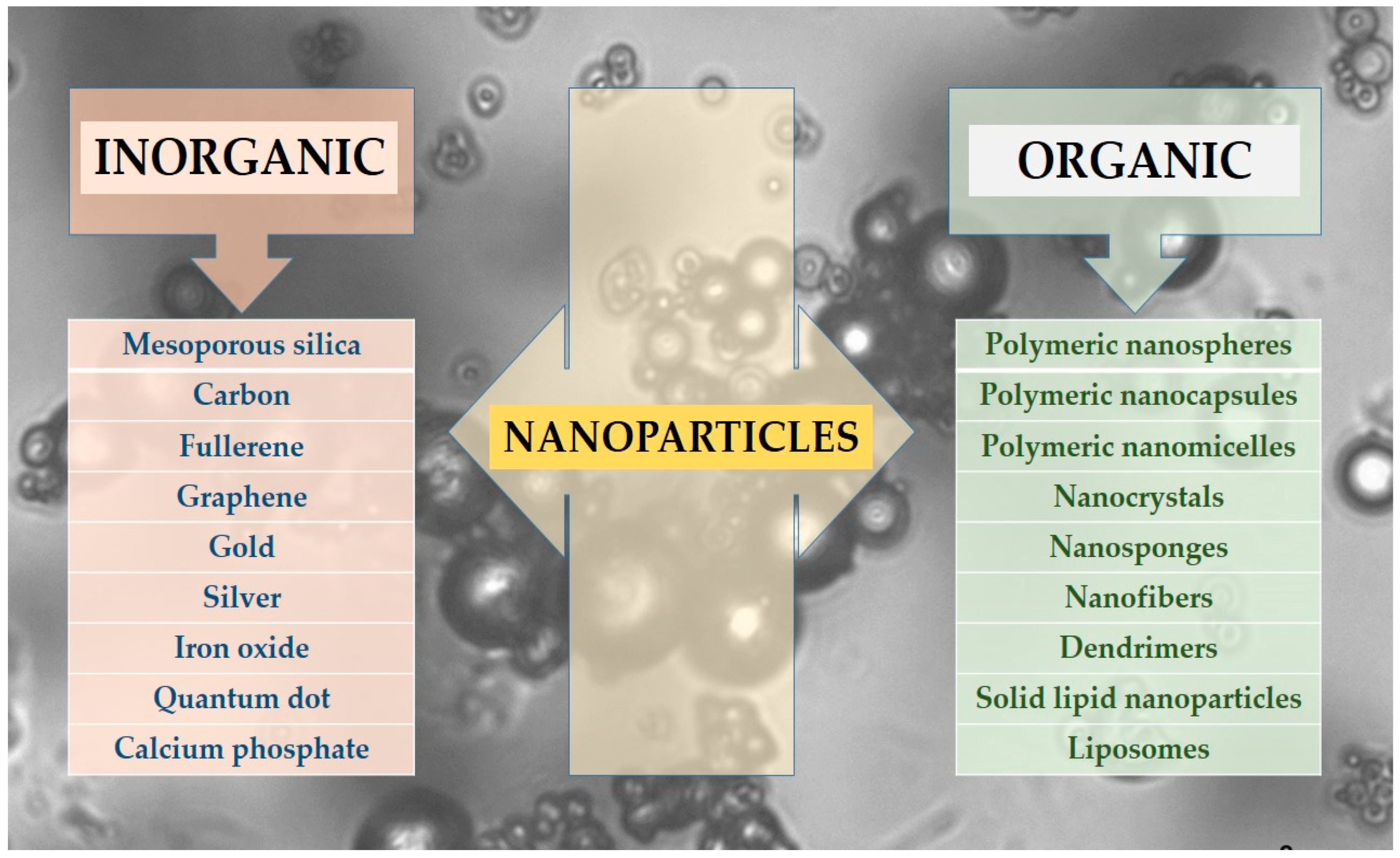
  • Inorganic NPs, including silica, carbon-based, gold, and other metallic NPs.
  • Organic NPs, which encompass polymeric NPs such as nanospheres and nanocapsules, protein-based carriers, micelles, liposomes, solid lipid nanoparticles (SLNs), dendrimers, nanotubes, nanofibers, nanocrystals, and others.

2.1. Inorganic/Metallic and Non-Metallic NPs

Nowadays, inorganic nanocarriers like gold, silver, silica, graphene, or carbon nanotubes (CNTs) have been widely explored as drug delivery systems due to their physicochemical properties. These include biocompatibility, low cytotoxicity, ease of functionalization, and natural accumulation in cancer cells, increasing intracellular drug concentrations [26].

2.1.1. Gold NPs (AuNPs)

AuNPs, ranging in size from 1 to 100 nm, are widely utilized for drug and gene delivery due to their inert nature, exceptional biocompatibility, and lower toxicity compared to other metal-based materials [26]. There has been growing scientific interest in AuNPs for biomedical applications due to their ease of functionalization, outstanding biocompatibility, and combined imaging and therapeutic potential [24]. However, it is important to note that while gold is generally well tolerated by the body, it is not biodegradable and may persist within the body for an extended period [27]. Smaller targeted NPs are more effective than larger ones, as they enhance efficiency through endocytosis and receptor binding. Among the various modification strategies, polyethylene glycol (PEG) is one of the most commonly used compounds, as it can covalently bind to the surface atoms of AuNPs, further improving their stability and performance in biomedical applications [28]. AuNPs, due to their unique physical properties, can absorb more light than other metals. Incorporating TKIs into AuNPs and applying photothermal or photodynamic therapy can create a synergistic effect for cancer treatment. However, this approach may also lead to unintended side effects on normal tissues [29].
Coelho and colleagues (2019) developed a nanoscale drug delivery system using PEGylated AuNPs for the targeted treatment of pancreatic cancer. They conjugated doxorubicin (an anthracycline) and varlitinib (a TKI) with AuNPs and evaluated their cytotoxic effects on pancreatic cancer cell lines. The nanoconjugates demonstrated a synergistic effect, significantly reducing the survival of S2-013 cancer cells while minimizing toxicity to healthy pancreatic cells. This study highlights the potential of PEGylated AuNPs as an effective platform for delivering anthracyclines and TKIs, enhancing anticancer efficacy while reducing harmful side effects. The functionalization of AuNPs presents a promising approach in cancer therapy, offering improved drug targeting and reduced systemic toxicity, which could be further explored for optimized treatment strategies [30].

2.1.2. Silver NPs (AgNPs)

Among the various nanoscale drug delivery strategies, AgNPs have emerged as one of the most extensively studied nanotechnology-based structures due to their distinct physicochemical properties [31]. In addition to their role in drug delivery, AgNPs are widely recognized for their antimicrobial, optical, electrical, and catalytic properties. Their unique characteristics, such as small size, high surface area, shape, particle morphology, composition, coating or capping, agglomeration behavior, dissolution rate, reactivity in solution, ion release efficiency, and the type of reducing agents used in their synthesis, contribute to their versatility [32]. These properties make AgNPs highly suitable as drug carriers in various biomedical applications, particularly in cancer therapy [33].
Adamo and colleagues (2023) explored the potential of AgNPs in combination with TKIs for targeting chronic lymphocytic leukemia (CLL) cells [34]. Their research demonstrated the synergistic effect of AgNPs with existing CLL treatments, such as venetoclax and ibrutinib (ITB), highlighting their potential for combination therapies. They further showed that conjugating AgNPs with the antibody rituximab in vitro. A key challenge in the clinical translation of AgNPs is their non-specific distribution, which can lead to off-target toxicity [32]. However, the study provided evidence that rituximab-conjugated AgNPs improved targeted delivery, reducing unintended cytotoxic effects while enhancing the selective elimination of CLL cells in vivo. This research also offered the first preclinical proof that AgNPs can amplify the cytotoxic effects of venetoclax, ITB, and bepridil against CLL cells. Furthermore, AgNPs conjugated with rituximab demonstrated improved efficacy, specificity, and bioavailability in CLL models. Overall, these findings highlight the potential clinical application of nanotechnology as a targeted delivery strategy for TKIs in CLL therapy [34].

2.1.3. Mesoporous Silica NPs

Silica-based carriers exist in various forms, such as silica particles, nanotubes, hollow silica particles, mesoporous silica NPs (MSNPs), and hollow mesoporous silica NPs (HMSNPs) [26]. Among these, MSNPs and HMSNPs serve as versatile nanocarriers due to their large surface area and porous structure, allowing for efficient drug loading and permeation, superior drug-loading capacity, easily modifiable surface, and excellent chemical stability. They can improve the solubility and stability of TKIs while enabling sustained drug release, which helps minimize the toxicity associated with long-term TKI therapy [29].
Torabi et al. (2023) developed MSNPs loaded with sunitinib using the sol–gel method. To enhance targeting efficiency, the MSNPs were modified with PEG 600 and functionalized with mucin 16 aptamers. The nanosystems were physicochemically characterized and tested on ovarian cancer cells. The characterization results confirmed that the MSNPs had a spherical shape, an average particle size of 56 nm, and a surface area of approximately 148 m2. Cell viability assays demonstrated increased the cytotoxicity of the targeted MSNPs in mucin 16-overexpressing OVCAR-3 cells. These findings suggest that the engineered mesoporous silica nanosystems could serve as an effective multifunctional drug delivery platform for targeting mucin 16-overexpressing cancer cells [35].

2.1.4. Carbon Nanotubes

CNTs are a cylindrical form of carbon allotropes with unique physicochemical properties. The rapid advancement of polyvalent CNTs as cancer treatment tools highlights their growing potential. Due to their ability to cross biological barriers, functionalized CNTs have shown significant promise as novel drug delivery systems. Numerous in vitro and in vivo studies have confirmed that various chemically modified CNTs are biologically compatible and can influence material behavior when introduced into living organisms [36].
Lila et al. (2023) developed a carbon nanotube-based drug delivery system incorporating the TKI neratinib for targeted breast cancer therapy. Multiwalled CNTs functionalized with carboxylic acid were non-covalently coated with a biotin–chitosan layer to enable dual-targeting capabilities—pH-responsive drug release via chitosan and biotin-receptor-mediated active targeting through biotin. The coated CNTs were then loaded with neratinib. In vitro drug release studies demonstrated a pH-sensitive release of neratinib, with higher drug release occurring under acidic conditions. Cytotoxicity assays on the SkBr3 breast cancer cell line showed dose-dependent effects, with neratinib-loaded Biotin–Chitosan-coated CNTs exhibiting significantly greater cytotoxicity compared to free neratinib or neratinib-loaded uncoated CNTs. The study further indicated that surface modification with Biotin–Chitosan enhanced the uptake of CNTs by breast cancer cells, likely through biotin receptor-mediated endocytosis. Overall, the obtained nanotubes present a promising nanocarrier system for targeted, cell-specific drug delivery in breast cancer therapy [37].

2.1.5. Magnetic NPs (MNPs)

Significant medical potential for uses such as drug delivery and bioseparation also exists for MNPs and superparamagnetic iron oxide NPs, which are biocompatible, possess a large surface area, and function as contrast agents for magnetic resonance imaging (MRI) [29]. In general, nanomaterials that induce or amplify iron-dependent programmed cell death (ferroptosis) offer a promising anticancer approach by actively and passively targeting tumor cells. Considering the complexity of tumor tissues, combining ferroptosis with TKIs can further enhance anticancer efficacy [38].
Mohaghegh and his team (2022) demonstrated the potential of MNPs in biomedical applications by designing erlotinib-loaded MNPs conjugated with anti-mucin 16 aptamers. These NPs were developed as image-guided drug delivery systems, integrating non-invasive MRI contrast agents for targeted therapy. The study found that combining MNPs with L-Asparaginase produced a synergistic effect, significantly reducing ovarian cancer cell viability. MRI and flow cytometry results showed that mucin 16-overexpressing cells exhibited significantly higher uptake of the targeted MNPs compared to non-functionalized NPs. Furthermore, MRI scans of ovarian cancer-bearing mice revealed notable signal intensity changes at the tumor site just a few hours after intravenous injection of the targeted MNPs, in contrast to non-targeted ones. These findings indicate that erlotinib-loaded MNPs offer a promising strategy for image-guided approach and targeted drug delivery system in ovarian cancer therapy [39].

2.2. Organic NPs

2.2.1. Polymeric NPs

Polymeric NPs enhance drug accumulation within solid tumor tissues due to their nanoscale size, which promotes increased permeability, retention, and cellular adsorption. Additionally, they can improve the pharmacokinetic and pharmacodynamic properties of various bioactive molecules, helping to overcome some of the limitations associated with conventional drug formulations [26]. In the synthesis of polymer NPs, polymers can be categorized into natural polymers—such as starch, albumin, sodium alginate, chitosan, gelatin, cellulose and synthetic polymers, including PEG, poly(lactic-co-glycolic acid) (PGLA), polyvinyl alcohol, polyvinyl pyrrolidone, and others. Chitosan, a natural biopolymer derived from the deacetylation of chitin, possesses remarkable physicochemical properties, including excellent biocompatibility, biodegradability, and non-toxicity [40]. However, its limited solubility in physiological aqueous solutions hinders its broader application. While hydrophilic modifications can enhance its water solubility, they may also introduce additional biological toxicity [41]. Poly (lactic acid) (PLA) is extensively utilized in the formulation of various polymer NPs due to its biodegradability, biocompatibility, compostability, and self-assembly capabilities [42]. Similarly, PLGA, a synthetic biodegradable polymer derived from lactic and glycolic acid, has been approved by the United States Food and Drug Administration for its exceptional biocompatibility, biodegradability, and drug encapsulation potential [41]. Due to its hydrophobic nature, PLGA is particularly effective in encapsulating lipophilic drugs. PLGA-based nanocarriers offer several advantages, including enhanced biocompatibility, prolonged circulation time in vivo, sustained drug release, and excellent encapsulation efficiency [43]. Polymer NPs can be classified into polymeric micelles (PMs), polymeric nanocapsules, and polymeric nanospheres [41].

2.2.2. Polymeric Nanocapsules/Nanospheres

Sankha Bhattacharya (2020) formulated chitosan-based polymeric NPs of IMA specifically for targeting colorectal cancer. The NPs were synthesized using the ionic gelation method combined with a central composite design approach. The optimized polymeric NPs of IMA exhibited a mean particle size of around 200 nm, an in vitro cumulative drug release of about 86%, and a drug entrapment efficiency of circa 68%. Fluorescence analysis revealed that epithelial colon cells demonstrated increased fluorescent NPs accumulation following intravenous (i.v.) administration. Additionally, no signs of tissue damage were observed in the final formulations, suggesting their safe administration via the i.v. route. The study concluded that the formulated chitosan-based polymeric NPs of the TKI IMA could serve as a promising strategy for colorectal cancer treatment through intravenous delivery [44].
Zhong and his team (2021) developed biodegradable triblock polymeric NPs for the co-delivery of two TKIs, sorafenib and crizotinib, to assess their impact on lung cancer in vitro and in vivo. The polymeric NPs had an average particle size of approximately 30 nm and exhibited a steady release of both drugs. The NPs were rapidly taken up by cancer cells and, compared to free drugs, led to significant tumor reduction, prolonged survival rates, and fewer side effects. Moreover, the dual-TKI-loaded NPs effectively suppressed tumor growth in vivo, extended survival, reduced tumor glucose metabolism, and caused minimal damage to vital organs. Based on these findings, the researchers concluded that the developed NPs have potential as a treatment strategy for lung cancer [45].

2.2.3. Polymeric Micelles (PMs)

PMs are an emerging drug delivery system composed of amphiphilic di- or tri-block, graft, and ionic copolymers that self-assemble in aqueous environments once their concentration surpasses the critical micelle concentration. Featuring a core–shell structure, the hydrophobic core encapsulates drugs, while the hydrophilic shell enhances stability and solubility. Crystallized polycaprolactone is commonly used to strengthen PMs, ensuring drug retention in the gastrointestinal tract. Furthermore, improving the cohesive interactions between the drug and polymeric core can enhance the physical stability of drug-loaded micelles [46]. With their nanoscale size (<100 nm), PMs can penetrate tissues without being recognized by the mononuclear phagocyte system, allowing sufficient time for accumulation at the target site. In addition to these advantages, PMs offer controlled drug release, enhanced drug loading capacity, and environmental protection for encapsulated pharmaceuticals [36]. Compared to liposomes, PMs offer greater structural stability, a smaller size range, and the ability to simultaneously encapsulate multiple therapeutic agents with diverse physicochemical properties, making them a promising platform for drug delivery [26,47].
Shih and their team (2022) researched the efficacy of PMs co-loaded with the TKI sunitinib and 7-ethyl-10-hydroxy-camptothecin (SN-38) for colorectal cancer treatment. Using the lyophilization-rehydration method, they prepared nanoscale micelles containing SN-38 or sunitinib at different drug/polymer ratios. The study assessed cell viability and cellular uptake in HCT-116 tumor cells. The results showed that SN-38/sunitinib micelles exhibited the highest tumor accumulation compared to normal organs. In antitumor efficacy studies, HCT-116 tumors in mice treated with SN-38/sunitinib micelles were the smallest for a month, significantly smaller than in other experimental groups. These findings confirm that SN-38/sunitinib co-loaded micelles can enhance the antitumor effects of TKIs and SN-38, suggesting that this PMs formulation holds potential for colorectal cancer treatment [48].

2.2.4. Solid Lipid NPs

Solid lipid NPs (SLNs) are colloidal drug delivery systems with particle sizes ranging from 50 to 1000 nm and, so, the increase in surface area enhances drug absorption [49]. They consist of a solid biodegradable and physiologically compatible lipid core, stabilized by a hydrophilic surfactant. SLNs offer reduced toxicity and lower cytotoxicity compared to polymeric NPs. Additionally, the small size facilitates the bypassing of physiological barriers in the gastrointestinal tract, allowing SLNs to improve the bioavailability of small-molecule TKIs [50]. They are particularly effective in delivering lipophilic drugs [49].
SLNs provide several advantages over liposomes and polymeric NPs, including feasibility for large-scale production, high stability, biodegradability, enhanced drug entrapment efficiency, controlled drug release, and the potential for drug targeting through surface modification. Controlled drug release occurs through degradation, erosion, or diffusion of the lipid matrix [51].
In cancer therapy, SLNs can effectively deliver TKIs with improved bioavailability and reduced resistance. Lipid-based nanocarriers like SLNs help overcome the limitations of conventional anticancer treatments by enhancing the therapeutic efficacy and functionality of potent anticancer drugs [13].
Moinuddin and his team (2023) formulated dasatinib/hesperidin-loaded SLNs for the treatment of CML. These NPs were synthesized using a high-shear homogenizer and optimized through a central composite design. The researchers developed both a dasatinib suspension and dasatinib-loaded SLNs, analyzing their pharmacokinetic profiles via HPLC. The presence of SLNs significantly enhanced dasatinib’s efficacy. A comparative analysis demonstrated that SLNs exhibited greater cytotoxicity than the free drug. Consequently, the dual-targeted SLN formulation improved cellular sensitivity to the encapsulated drug more effectively than the suspension form. In vivo efficacy data and the increased lifespan percentage suggest that dasatinib in SLN formulation provides prolonged therapeutic effects compared to a pure drug suspension. These findings highlight the advantages of SLN-based targeted drug delivery [52].

2.2.5. Liposomes

Liposomes are spherical structures made up of one or more lipid bilayers that encase an aqueous core, composed primarily of cholesterol and phospholipid molecules. Depending on the drug’s lipophilicity, it can be incorporated either within the lipid bilayer or in the internal aqueous core. The liposome’s membrane structure, which closely resembles that of cell membranes, enables the effective cellular uptake of drugs, making liposomes an ideal drug delivery system. Their unique properties, such as biocompatibility, non-immunogenicity, high drug loading capacity, and controlled release capabilities, have contributed to their widespread use in enhancing the biodistribution of therapeutic agents, especially in cancer treatment [26]. Liposome-based drug delivery systems protect encapsulated drugs by shielding them from metabolizing enzymes during circulation, thereby preventing premature catabolism. However, a major challenge in liposome formulation is achieving optimal drug loading efficiency. The incorporation of TKIs into liposomes and their controlled release is hindered by the high diffusivity of these molecules through the lipid bilayer of the vesicle. Additionally, nano-crystallization within the aqueous core of liposomes is generally not a possible approach for this class of compounds [53]. Moving forward, various specialized liposomes, including thermosensitive, pH-sensitive, ultrasound-sensitive, photosensitive, and magnetic liposomes, will be further developed to address these limitations and enhance the drug delivery efficiency of TKIs. Additionally, research on precursor liposomes offers a promising approach to overcoming the instability associated with conventional liposomes [41].
PEGylated liposomes can effectively accumulate in tumor tissues through passive targeting, leveraging the enhanced permeability and retention (EPR) effect. This is made possible by nanoparticle extravasation and the increased permeability of tumor vasculature [54,55]. Additionally, PEGylation extends the liposomes’ circulation time by increasing particle size, enhancing their stability and biocompatibility while ensuring patient safety [54]. Theoretically, encapsulating a drug within PEGylated liposomes allows for a lower dosage while improving efficacy due to these unique properties [55]. Given the toxicity of sunitinib, a targeted delivery strategy is essential to minimize systemic side effects [56].
Yueh and colleagues (2024) designed PEGylated liposomes as delivery systems to precisely transport sunitinib (lipo-sunitinib) to renal cell carcinoma tumors. To address this, the researchers developed PEGylated liposomes to enhance the tumor-specific delivery of sunitinib. By harnessing the EPR effect and the prolonged circulation time provided by PEGylation, the study demonstrated significant inhibition of renal carcinoma tumors in mouse models. This innovative strategy offers a promising solution to reduce sunitinib-associated toxicity while improving its therapeutic potential for advanced renal cell carcinoma [56].

2.2.6. Nanocrystals

Nanocrystals are drug crystals with a particle size in the range of a few hundred nanometers. These nanoscale carriers are particularly advantageous for poorly soluble drugs, with most TKIs belonging to the BCS Class II category. Nanocrystals enhance the pharmacokinetic properties of drugs while also reducing fed-fasted variability, making them a preferred formulation strategy for improving drug solubility and bioavailability [36].
Wang and colleagues (2021) developed a realgar nanocrystal-based delivery system incorporating IMA for synergistic treatment of chronic myeloid leukemia (CML). To ensure stability, bovine serum albumin was carefully selected to encapsulate the realgar (arsenide) nanocrystals, resulting in a uniform particle size of approximately 40 nm. Additionally, the nanosystem was functionalized with folic acid to enhance tumor targeting, which was confirmed through both in vitro and in vivo studies. This approach led to significant tumor growth inhibition and prolonged survival in mice without noticeable side effects. In vivo studies further demonstrated that the nanosystem extended the circulation half-life of IMA, actively accumulated in tumor sites, and achieved superior therapeutic efficacy compared to arsenide or IMA monotherapy. This study presents a synergistic nanoplatform for targeted CML treatment, overcoming realgar’s biomedical limitations and emphasizing the benefits of combining TKIs with arsenide for disease management [57].

2.2.7. Nanosponges (NSs)

Among different types of polymer-based nanocarriers, NSs have emerged as a promising strategy for the efficient delivery of a wide range of therapeutics, particularly for targeted, sustained, and prolonged drug release [58]. These spherical nanocarriers possess a highly porous surface and extensive internal cavities, allowing them to encapsulate both lipophilic and hydrophilic drugs. The fabrication of NSs involves polymers such as β-cyclodextrins cross-linked with diphenyl carbonate and ethylcellulose, with polyvinyl alcohol serving as a stabilizer [59]. The porous nature of NSs enhances their encapsulation capacity, making them ideal for delivering drugs with poor bioavailability and high dosage requirements, ensuring sustained and controlled drug release [60].
Fatima et al. (2023) developed ITB-loaded NSs to enhance cytotoxic activity against breast cancer cell lines. These NSs were prepared using emulsification solvent evaporation technology and optimized by adjusting the ratio of ethylcellulose and polyvinyl alcohol while maintaining a constant ITB dose of 50 mg. Physicochemical characterization confirmed proper drug encapsulation and compatibility with excipients. In vitro drug release studies indicated that the NSs followed the Higuchi matrix model with anomalous non-Fickian release kinetics. The ethylcellulose-based NSs provided sustained drug release for 24 h and demonstrated greater cytotoxic effects compared to free ITB. Their enhanced activity against MCF-7 cancer cells suggests that these NSs could serve as effective nanocarriers for breast cancer treatment [60].

2.2.8. Nanofibers

In the past decade, nanofibers have gained significant attention, with electrospinning being a widely used technique for their fabrication [61]. Electrospun polymeric nanofibers are excellent candidates for the delivery of TKIs due to their high surface area and customizable composition. These fibers are produced by electrospinning a viscous polymer solution, where various factors influence their properties and morphology [62]. Solution parameters such as viscosity and polymer concentration, along with operational factors like needle diameter, spinning distance, applied voltage, and flow rate, play crucial roles in fiber formation [63]. Additionally, humidity is a key environmental factor that significantly impacts the development of porous structures in nanofibers [64].
Chen and colleagues (2023) developed supramolecular peptide nanofibers (SPNs) co-loaded with dabrafenib (Da) and doxorubicin (Dox) for the targeted and synergistic therapy of differentiated thyroid carcinoma. To enhance peptide stability in vivo, they incorporated D-phenylalanine and D-tyrosine. Through multiple non-covalent interactions, SPNs/Da/Dox self-assembled into longer, denser nanofibers. A cancer-targeting ligand was employed to enhance cellular uptake of the nanofibers, improving drug delivery efficiency. The encapsulation of Da and Dox within SPNs led to decreased IC50 values for both drugs, indicating enhanced potency. The co-delivery system exhibited the strongest therapeutic effect in both in vitro and in vivo studies. Additionally, SPNs facilitated efficient drug transport while enabling a lower Dox dosage, significantly reducing its associated side effects. These findings highlight the potential of SPNs as a promising drug delivery platform for the synergistic treatment of differentiated thyroid carcinoma [65].

2.2.9. Dendrimers

A dendrimer is a nanoscale, radially symmetrical molecule characterized by a well-defined, uniform, and monodisperse structure [25]. It is a highly branched, spherical, and multivalent molecule with synthetic flexibility, making it suitable for various applications, including catalysis and drug delivery [66]. The polymeric core of a dendrimer could be obtained, for example, by polypropylene imine, PEG, and others. The surface functional groups determine whether the dendrimer exhibits an anionic, cationic, or neutral charge [67]. Some dendrimers incorporate inorganic elements like silicon or phosphorus as branching points. Notably, positively charged phosphorus dendrimers have been utilized as carriers for anticancer drugs. Their hydrophilic surface and hydrophobic backbone enable effective membrane penetration, making them valuable in drug delivery applications [17].
Aleanizy et al. (2020) designed trastuzumab (TZ)-grafted dendrimers loaded with neratinib, a TKI, for targeted breast cancer therapy. The dendrimers were also fluorescently labeled with fluorescein isothiocyanate. The study demonstrated the successful encapsulation of neratinib with a sustained release profile. In vitro comparisons showed that neratinib-TZ-loaded dendrimers exhibited greater selectivity and higher cytotoxicity against SKBR-3 cells than both neratinib alone and neratinib-loaded dendrimers. These results highlight the potential of TZ-conjugated dendrimers as targeted drug carriers, improving the delivery of cytotoxic agents like neratinib and, thereby, reducing severe side effects [68].

3. Possibilities and Limitations of Nanotechnology-Based TKIs Carriers

Nano-scale drug delivery systems represent one of the most rapidly advancing areas in pharmaceutical research. The benefits offered by nanoparticles are well-established, prompting intensive efforts toward the development of novel nanoparticle types, innovative preparation techniques, and the incorporation of new polymers to enhance structural stability.
Table 2 summarizes the advantages and limitations of various nanoparticle systems, categorized into organic and inorganic nanoparticles [69,70,71,72,73,74,75,76]. There is no “ideal” type of nanoparticle that has only positive characteristics. Some drugs are suitable for encapsulation only in a certain type of nanoparticles. A particularly intriguing aspect of current research is the successful incorporation of tyrosine kinase inhibitors across all nanoparticle classes.
The chemical composition of nanoparticles plays a pivotal role in determining their physicochemical properties. Metallic nanoparticles, for example, are highly regarded for their superior electrical and thermal conductivity, as well as their distinct magnetic behavior. Despite being among the earliest nanoparticles used historically as pigments, they have recently gained significant attention for their potential to encapsulate TKIs for biomedical applications. Lipid-based organic nanoparticles are particularly effective as carriers for TKIs, owing to their biocompatible and structurally favorable composition. Polymer-based nanoscale carriers of TKIs exhibit notable mechanical strength and chemical stability, making them suitable for a wide range of therapeutic uses. Additionally, various carbon carriers, due to their versatile bonding characteristics, can form a diverse array of nanostructures, each possessing unique and valuable properties when including TKIs [71].
The emergence of drug resistance to TKIs poses a significant obstacle in the treatment of certain diseases. Nanomedicine presents a promising strategy to overcome this issue by enabling targeted drug delivery and controlled release. These capabilities have shown encouraging results in preclinical studies. The use of nanoparticles and multifunctional nanosystems highlights their potential to enhance therapeutic outcomes, especially in cases where resistance to TKIs has developed [77]. Drug resistance significantly limits the effectiveness of chemotherapy and remains an inevitable challenge in the treatment of tumors. Whether intrinsic or acquired, drug resistance is a major contributor to treatment failure in over 90% of patients with metastatic cancer. Resistance to TKIs often develops within the first year of therapy. To improve patient outcomes and extend survival, various strategies have been explored to counteract drug resistance. Common approaches include combination therapies, the modulation of drug concentrations within tumor cells, and targeting the tumor microenvironment using nanotechnology [41]. Nano-scaled carriers aim to protect drug molecules from external environmental factors, enable targeted delivery to specific organs, facilitate controlled and sustained release, and support the emerging paradigm of personalized medicine.
While nanotechnologies exhibit a promising approach, further investigation is essential to fully understand and optimize their potential in TKI encapsulation and targeting.

4. Future Directions: Clinical Trials of Nano-Scaled TKI Carriers

Nanotechnology is gaining increasing recognition for its potential in cancer therapy. Nanoencapsulated drugs have shown enhanced pharmacokinetic and pharmacodynamic properties, contributing to more effective treatment outcomes. Due to their small size, nanoparticles can navigate through difficult-to-reach areas in the body, while their high surface-area-to-volume ratio enables the delivery of larger drug quantities directly to tumor sites. Additionally, the surface of nanoparticles can be functionalized with ligands such as small molecules, antibodies, or peptides [69]. These ligands can be tailored to target specific markers that are unique to or overexpressed on cancer cells, such as tumor-specific surface receptors. This targeted approach not only boosts the efficacy of the drug but also minimizes systemic toxicity and adverse side effects [77].
Combining TKIs with immunotherapy represents a promising strategy for cancer treatment. This approach is supported by several key observations: TKIs not only reduce tumor burden but also enhance immune recognition and destruction of cancer cells, and they influence the intestinal barrier and help modulate gut dysbiosis. Preclinical studies in various mouse models have demonstrated the potential effectiveness of this combination. The growing number of clinical trials highlights the increasing interest in this therapeutic strategy. Additionally, the wide range of cancers being addressed through such combinations suggests their broad applicability [78].
An illustrative example of such combination therapy is the Phase I study by Chien et al. (2009), which evaluated a 2-day lapatinib chemosensitization pulse followed by nanoparticle albumin-bound paclitaxel in patients with advanced solid tumors. Preclinical evidence suggests that short-term, high-dose administration of TKIs targeting the human epidermal growth factor receptor family can prime tumor vasculature, enhancing the delivery and effectiveness of chemotherapy. This clinical study explores the translational potential of that strategy [79]. Currently, a number of clinical trials are underway to incorporate TKИc into nanoparticles in combination with an antitumor agent. Some of them are in phase 1, and others have already moved to phase 4. This is a significant advance in cancer treatment and has promising benefits.
The rational design of biopolymer-based nanocarriers with optimized physicochemical properties, structural morphology, and drug release kinetics is essential for developing next-generation drug delivery platforms. These systems aim to enhance safety profiles and therapeutic outcomes, representing a central objective in the field of nanomedicine. Although numerous methods for nanoparticle synthesis have been successfully developed, there remains a need for more refined and precise techniques to address the limitations of current approaches and to facilitate their effective translation to commercial-scale applications.

5. Conclusions

TKIs are extensively utilized in both in vivo and in vitro studies for targeted drug delivery in various cancers and inflammatory diseases. Preclinical research indicates that incorporating nanocarriers with TKIs leads to improved therapeutic outcomes compared to using TKIs alone. Polymeric, metallic, and inorganic nanocarriers possess unique properties that could enhance efficacy and minimize side effects of TKIs. Targeted nanocarriers and NPs with extended blood circulation and enhanced therapeutic efficiency have gained significant research interest. Optimizing TKIs nanosystems requires surface modification and functionalization to ensure compatibility and effectiveness in biological systems. The use of NPs as targeting carriers is a promising approach, as they present diverse structures and properties that make them well suited for delivering small-molecule TKIs.

Funding

This research received no external funding.

Acknowledgments

This research is supported by the programme “Research, Innovation and Digitalisation for Smart Transformation” 2021–2027, funded by the European Union, Project BG16RFPR002-1.014-0007 “Center for Competence “PERIMED-2”.

Conflicts of Interest

The author declares no conflicts of interest.

Abbreviations

The following abbreviations are used in this manuscript:
TKsTyrosine kinases
TKITyrosine kinase inhibitor
TKIsTyrosine kinase inhibitors
ATPAdenosine triphosphate
BCSBiopharmaceutics Classification System
NPsNanoparticles
CNTsCarbon nanotubes
AuNPsGold nanoparticles
AgNPsSilver nanoparticles
CLLChronis lymphocytic leukemia
MSNPsMesoporous silica nanoparticles
HMSNPsHollow mesoporous silica nanoparticles
MNPsMagnetic nanoparticles
MRIMagnetic resonance imaging
PMsPolymeric micelles
SN-387-ethyl-10-hydroxy-camptothecin
SLNsSolid lipid nanoparticles
CMLChronic myeloid leukemia
EPREnhanced permeability and retention
IMAImatinib
NSsNanosponges
ITBIbrutinib
SPNsSupramolecular peptide nanofibers
DaDabrafenib
DOxDoxorubicin
TZTrastuzumab
i.v.Intravenous
PLAPoly(lactic) acid
PLGAPoly(lactic-co-glycolic) acid
PEGPolyethylene glycol

References

  1. Korucu Aktas, P.; Baysal, I.; Yabanoglu-Ciftci, S.; Lamprecht, A.; Arica, B. Recent Progress in Drug Delivery Systems for Tyrosine Kinase Inhibitors in the Treatment of Lung Cancer. Int. J. Pharm. 2024, 650, 123703. [Google Scholar] [CrossRef] [PubMed]
  2. Nishal, S.; Jhawat, V.; Gupta, S.; Phaugat, P. Utilization of Kinase Inhibitors as Novel Therapeutic Drug Targets: A Review. Oncol. Res. 2022, 30, 221–230. [Google Scholar] [CrossRef] [PubMed]
  3. Baier, A.; Szyszka, R. Compounds from Natural Sources as Protein Kinase Inhibitors. Biomolecules 2020, 10, 1546. [Google Scholar] [CrossRef]
  4. Foroughi-Nia, B.; Barar, J.; Memar, M.Y.; Aghanejad, A.; Davaran, S. Progresses in Polymeric Nanoparticles for Delivery of Tyrosine Kinase Inhibitors. Life Sci. 2021, 278, 119642. [Google Scholar] [CrossRef]
  5. Lu, Y.; Bian, D.; Zhang, X.; Zhang, H.; Zhu, Z. Inhibition of Bcl-2 and Bcl-xL Overcomes the Resistance to the Third-Generation EGFR Tyrosine Kinase Inhibitor Osimertinib in Non-Small Cell Lung Cancer. Mol. Med. Rep. 2021, 23, 48. [Google Scholar] [CrossRef]
  6. Caponnetto, S.; Cantale, O.; Friedlaender, A.; Gomes, F.; Daryanani, S.; Gelibter, A.; Cortellini, A.; Giuffrida, D.; Addeo, A.; Banna, G.L. A Comparison Between First-, Second- and Third-Generation Epidermal Growth Factor Receptor Tyrosine Kinase Inhibitors in Patients with Non-Small-Cell Lung Cancer and Brain Metastases. J. Mol. Pathol. 2021, 2, 1. [Google Scholar] [CrossRef]
  7. Yin, Y.; Yuan, X.; Gao, H.; Yang, Q. Nanoformulations of Small Molecule Protein Tyrosine Kinases Inhibitors Potentiate Targeted Cancer Therapy. Int. J. Pharm. 2020, 573, 118785. [Google Scholar] [CrossRef]
  8. Kumar, R.; Goel, H.; Solanki, R.; Rawat, L.; Tabasum, S.; Tanwar, P.; Pal, S.; Sabarwal, A. Recent Developments in Receptor Tyrosine Kinase Inhibitors: A Promising Mainstay in Targeted Cancer Therapy. Med. Drug Discov. 2024, 23, 100195. [Google Scholar] [CrossRef]
  9. Metibemu, D.S.; Akinloye, O.A.; Akamo, A.J.; Ojo, D.A.; Okeowo, O.T.; Omotuyi, I.O. Exploring Receptor Tyrosine Kinases-Inhibitors in Cancer Treatments. Egypt. J. Med. Hum. Genet. 2019, 20, 35. [Google Scholar] [CrossRef]
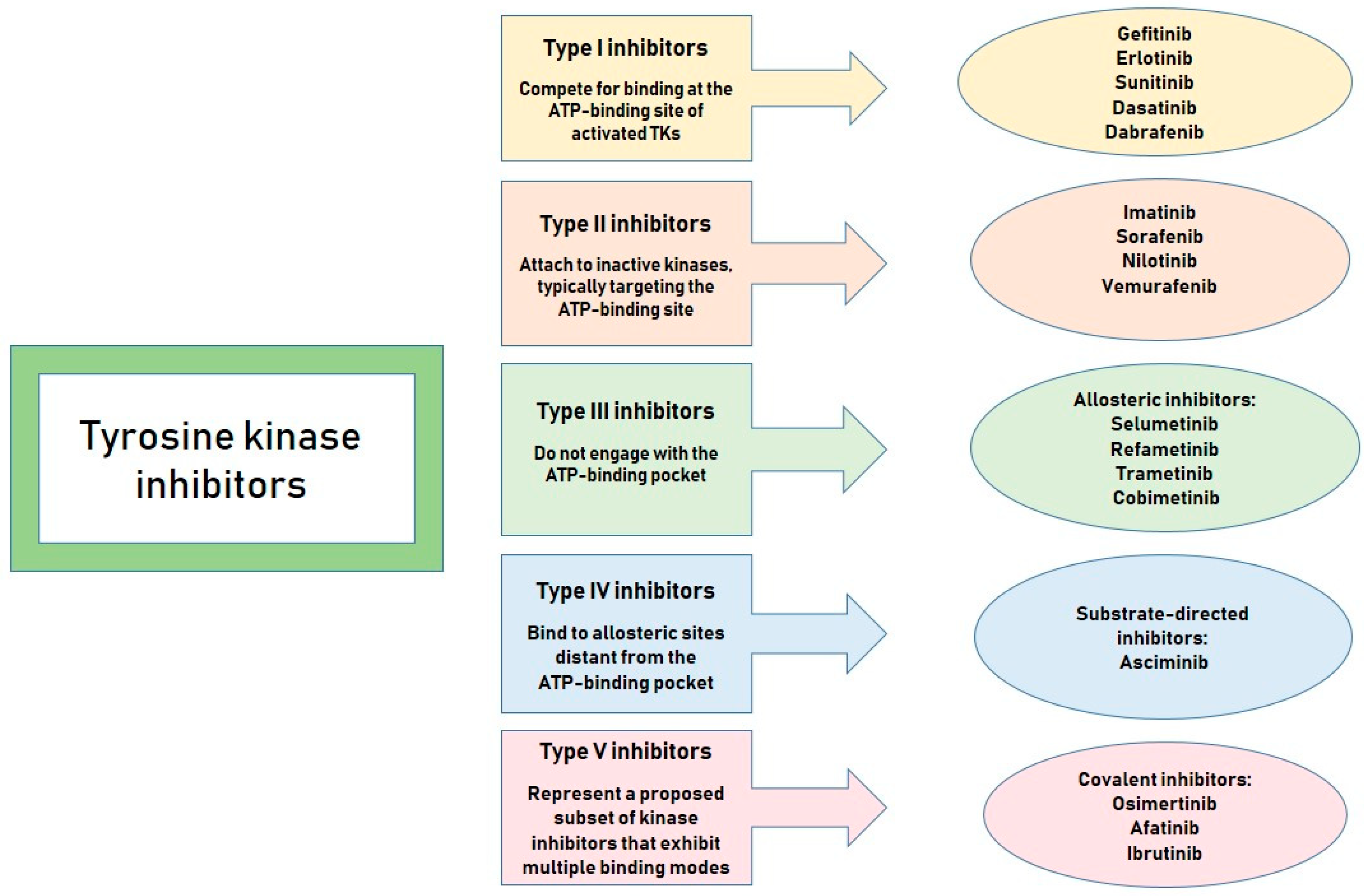
  10. Das, R.; Choithramani, A.; Shard, A. A Molecular Perspective for the Use of Type IV Tyrosine Kinase Inhibitors as Anticancer Therapeutics. Drug Discov. Today 2022, 27, 808–821. [Google Scholar] [CrossRef]
  11. Moradpour, Z.; Barghi, L. Novel Approaches for Efficient Delivery of Tyrosine Kinase Inhibitors. J. Pharm. Pharm. Sci. Publ. Can. Soc. Pharm. Sci. Soc. Can. Sci. Pharm. 2019, 22, 37–48. [Google Scholar] [CrossRef] [PubMed]
  12. Herbrink, M.; Schellens, J.H.M.; Beijnen, J.H.; Nuijen, B. Inherent Formulation Issues of Kinase Inhibitors. J. Control Release Off. J. Control Release Soc. 2016, 239, 118–127. [Google Scholar] [CrossRef]
  13. Satapathy, S.; Patro, C.S. Solid Lipid Nanoparticles for Efficient Oral Delivery of Tyrosine Kinase Inhibitors: A Nano Targeted Cancer Drug Delivery. Adv. Pharm. Bull. 2021, 12, 298. [Google Scholar] [CrossRef] [PubMed]
  14. Devadasu, V.R.; Deb, P.K.; Maheshwari, R.; Sharma, P.; Tekade, R.K. Physicochemical, Pharmaceutical, and Biological Considerations in GIT Absorption of Drugs. In Dosage Form Design Considerations; Elsevier: Amsterdam, The Netherlands, 2018; pp. 149–178. ISBN 978-0-12-814423-7. [Google Scholar]
  15. Blaesi, A.H.; Saka, N. Gastroretentive Fibrous Dosage Forms for Prolonged Delivery of Sparingly-Soluble Tyrosine Kinase Inhibitors. Part 1: Dosage Form Design, and Models of Expansion, Post-Expansion Mechanical Strength, and Drug Release. Int. J. Pharm. 2024, 2024, 124360. [Google Scholar] [CrossRef]
  16. Lipinski, C.A.; Lombardo, F.; Dominy, B.W.; Feeney, P.J. Experimental and Computational Approaches to Estimate Solubility and Permeability in Drug Discovery and Development Settings. Adv. Drug Deliv. Rev. 2001, 46, 3–26. [Google Scholar] [CrossRef]
  17. Russo, E.; Spallarossa, A.; Tasso, B.; Villa, C.; Brullo, C. Nanotechnology of Tyrosine Kinase Inhibitors in Cancer Therapy: A Perspective. Int. J. Mol. Sci. 2021, 22, 6538. [Google Scholar] [CrossRef] [PubMed]
  18. Shyam Sunder, S.; Sharma, U.C.; Pokharel, S. Adverse Effects of Tyrosine Kinase Inhibitors in Cancer Therapy: Pathophysiology, Mechanisms and Clinical Management. Signal Transduct. Target. Ther. 2023, 8, 262. [Google Scholar] [CrossRef]
  19. Sankarapandian, V.; Rajendran, R.L.; Miruka, C.O.; Sivamani, P.; Maran, B.A.V.; Krishnamoorthy, R.; Gangadaran, P.; Ahn, B.-C. A Review on Tyrosine Kinase Inhibitors for Targeted Breast Cancer Therapy. Pathol.-Res. Pract. 2024, 263, 155607. [Google Scholar] [CrossRef]
  20. Indermun, S.; Kumar, P.; Govender, M.; Choonara, Y.E. Can Nanomedicinal Approaches Provide an Edge to the Efficacy of Tyrosine Kinase Inhibitors? Curr. Med. Chem. 2023, 30, 1482–1501. [Google Scholar] [CrossRef]
  21. Kumar, A.; Vaiphei, K.K.; Gulbake, A. A Nanotechnology Driven Effectual Localized Lung Cancer Targeting Approaches Using Tyrosine Kinases Inhibitors: Recent Progress, Preclinical Assessment, Challenges, and Future Perspectives. Int. J. Pharm. 2024, 666, 124745. [Google Scholar] [CrossRef]
  22. Brusini, R.; Varna, M.; Couvreur, P. Advanced Nanomedicines for the Treatment of Inflammatory Diseases. Adv. Drug Deliv. Rev. 2020, 157, 161–178. [Google Scholar] [CrossRef]
  23. Yang, J.; Li, D.; Zhang, M.; Lin, G.; Hu, S.; Xu, H. From the Updated Landscape of the Emerging Biologics for IBDs Treatment to the New Delivery Systems. J. Control. Release 2023, 361, 568–591. [Google Scholar] [CrossRef] [PubMed]
  24. Dacoba, T.G.; Olivera, A.; Torres, D.; Crecente-Campo, J.; Alonso, M.J. Modulating the Immune System through Nanotechnology. Semin. Immunol. 2017, 34, 78–102. [Google Scholar] [CrossRef] [PubMed]
  25. Ojo, O.A.; Olayide, I.I.; Akalabu, M.C.; Ajiboye, B.O.; Ojo, A.B.; Oyinloye, B.E.; Ramalingam, M. Nanoparticles and Their Biomedical Applications. Biointerface Res. Appl. Chem. 2020, 11, 8431–8445. [Google Scholar] [CrossRef]
  26. Zhou, X.; Shi, K.; Hao, Y.; Yang, C.; Zha, R.; Yi, C.; Qian, Z. Advances in Nanotechnology-Based Delivery Systems for EGFR Tyrosine Kinases Inhibitors in Cancer Therapy. Asian J. Pharm. Sci. 2020, 15, 26–41. [Google Scholar] [CrossRef]
  27. Varna, M.; Xuan, H.V.; Fort, E. Gold Nanoparticles in Cardiovascular Imaging. WIREs Nanomed. Nanobiotechnol. 2018, 10, e1470. [Google Scholar] [CrossRef]
  28. Nicol, J.R.; Dixon, D.; Coulter, J.A. Gold Nanoparticle Surface Functionalization: A Necessary Requirement in the Development of Novel Nanotherapeutics. Nanomedicine 2015, 10, 1315–1326. [Google Scholar] [CrossRef]
  29. Tarighatnia, A.; Foroughi-Nia, B.; Nader, N.D.; Aghanejad, A. Recent Trends and Advances in Nanosystems with Tyrosine Kinase Inhibitors for Image-Guided Cancer Treatments. J. Drug Deliv. Sci. Technol. 2023, 88, 104938. [Google Scholar] [CrossRef]
  30. Coelho, S.C.; Reis, D.P.; Pereira, M.C.; Coelho, M.A.N. Doxorubicin and Varlitinib Delivery by Functionalized Gold Nanoparticles Against Human Pancreatic Adenocarcinoma. Pharmaceutics 2019, 11, 551. [Google Scholar] [CrossRef]
  31. Elmetwalli, A.; Abdel-Monem, M.O.; El-Far, A.H.; Ghaith, G.S.; Albalawi, N.A.N.; Hassan, J.; Ismail, N.F.; El-Sewedy, T.; Alnamshan, M.M.; ALaqeel, N.K.; et al. Probiotic-Derived Silver Nanoparticles Target mTOR/MMP-9/BCL-2/Dependent AMPK Activation for Hepatic Cancer Treatment. Med. Oncol. 2024, 41, 106. [Google Scholar] [CrossRef]
  32. Ferdous, Z.; Nemmar, A. Health Impact of Silver Nanoparticles: A Review of the Biodistribution and Toxicity Following Various Routes of Exposure. Int. J. Mol. Sci. 2020, 21, 2375. [Google Scholar] [CrossRef] [PubMed]
  33. Zivic, F.; Grujovic, N.; Mitrovic, S.; Ahad, I.U.; Brabazon, D. Characteristics and Applications of Silver Nanoparticles. In Commercialization of Nanotechnologies—A Case Study Approach; Brabazon, D., Pellicer, E., Zivic, F., Sort, J., Dolors Baró, M., Grujovic, N., Choy, K.-L., Eds.; Springer International Publishing: Cham, Switzerland, 2018; pp. 227–273. ISBN 978-3-319-56978-9. [Google Scholar]
  34. Adamo, F.M.; Silva Barcelos, E.C.; De Falco, F.; Dorillo, E.; Rompietti, C.; Sorcini, D.; Stella, A.; Del Papa, B.; Baldoni, S.; Esposito, A.; et al. Therapeutic Targeting Potential of Novel Silver Nanoparticles Coated with Anti-CD20 Antibody against Chronic Lymphocytic Leukemia. Cancers 2023, 15, 3618. [Google Scholar] [CrossRef]
  35. Torabi, M.; Aghanejad, A.; Savadi, P.; Barzegari, A.; Omidi, Y.; Barar, J. Fabrication of Mesoporous Silica Nanoparticles for Targeted Delivery of Sunitinib to Ovarian Cancer Cells. BioImpacts 2023, 13, 255. [Google Scholar] [CrossRef] [PubMed]
  36. Mukherjee, S.; Joshi, V.; Reddy, K.P.; Singh, N.; Das, P.; Datta, P. Biopharmaceutical and Pharmacokinetic Attributes to Drive Nanoformulations of Small Molecule Tyrosine Kinase Inhibitors. Asian J. Pharm. Sci. 2024, 19, 100980. [Google Scholar] [CrossRef]
  37. Abu Lila, A.S.; Bhattacharya, R.; Moin, A.; Al Hagbani, T.; Abdallah, M.H.; Danish Rizvi, S.M.; Khafagy, E.-S.; Hussain, T.; Gangadharappa, H.V. Dual Targeting Multiwalled Carbon Nanotubes for Improved Neratinib Delivery in Breast Cancer. RSC Adv. 2023, 13, 24309–24318. [Google Scholar] [CrossRef] [PubMed]
  38. Yin, W.; Chang, J.; Sun, J.; Zhang, T.; Zhao, Y.; Li, Y.; Dong, H. Nanomedicine-Mediated Ferroptosis Targeting Strategies for Synergistic Cancer Therapy. J. Mater. Chem. B 2023, 11, 1171–1190. [Google Scholar] [CrossRef]
  39. Mohaghegh, S.; Tarighatnia, A.; Omidi, Y.; Barar, J.; Aghanejad, A.; Adibkia, K. Multifunctional Magnetic Nanoparticles for MRI-Guided Co-Delivery of Erlotinib and L-Asparaginase to Ovarian Cancer. J. Microencapsul. 2022, 39, 394–408. [Google Scholar] [CrossRef]
  40. Jafari, H.; Mahdavinia, G.R.; Kazemi, B.; Ehrlich, H.; Joseph, Y.; Rahimi-Nasrabadi, M. Highly Efficient Sunitinib Release from pH-Responsive mHPMC@Chitosan Core-Shell Nanoparticles. Carbohydr. Polym. 2021, 258, 117719. [Google Scholar] [CrossRef]
  41. Xu, W.; Ye, C.; Qing, X.; Liu, S.; Lv, X.; Wang, W.; Dong, X.; Zhang, Y. Multi-Target Tyrosine Kinase Inhibitor Nanoparticle Delivery Systems for Cancer Therapy. Mater. Today Bio 2022, 16, 100358. [Google Scholar] [CrossRef]
  42. Nofar, M.; Sacligil, D.; Carreau, P.J.; Kamal, M.R.; Heuzey, M.-C. Poly (Lactic acid) Blends: Processing, Properties and Applications. Int. J. Biol. Macromol. 2019, 125, 307–360. [Google Scholar] [CrossRef]
  43. Chen, Y.; Li, J.-X.; Shu, N.; Zheng, S.-J.; Ma, M.; Zhao, Z.-B.; Cao, Z.-T.; Zhou, Q.; Du, J.-Z.; Wang, J. A Polymeric Nanoformulation Improves the Bioavailability and Efficacy of Sorafenib for Hepatocellular Carcinoma Therapy. Biomater. Sci. 2021, 9, 2508–2518. [Google Scholar] [CrossRef] [PubMed]
  44. Bhattacharya, S. Fabrication and Characterization of Chitosan-Based Polymeric Nanoparticles of Imatinib for Colorectal Cancer Targeting Application. Int. J. Biol. Macromol. 2020, 151, 104–115. [Google Scholar] [CrossRef]
  45. Zhong, T.; Liu, X.; Li, H.; Zhang, J. Co-Delivery of Sorafenib and Crizotinib Encapsulated with Polymeric Nanoparticles for the Treatment of in Vivo Lung Cancer Animal Model. Drug Deliv. 2021, 28, 2108–2118. [Google Scholar] [CrossRef] [PubMed]
  46. El Asmar, A.; Morandi, G.; Burel, F. Synthesis of Dual Sensitive Lipid- b -Poly(Dimethylaminoethyl Methacrylate) Copolymers, Self-Assemblies and Modulation of Cloud Point Temperatures through Physical Blends with Lipid- b -Poly(2-Isopropyl-2-Oxazoline). Macromolecules 2019, 52, 9160–9167. [Google Scholar] [CrossRef]
  47. Wang, Z.; Wen, J.; Liu, H.; Zheng, L.; Luo, H.; Liu, Z.; Chen, X.; Wang, F.; Li, D.; Pei, H.; et al. Biodegradable Interlayer-Crosslinked Polymer Micelles with Reduction Sensitivity for Non-Small Cell Lung Cancer Therapy. J. Biomed. Nanotechnol. 2018, 14, 1225–1238. [Google Scholar] [CrossRef]
  48. Shih, Y.-H.; Peng, C.-L.; Chiang, P.-F.; Shieh, M.-J. Dual-Functional Polymeric Micelles Co-Loaded with Antineoplastic Drugs and Tyrosine Kinase Inhibitor for Combination Therapy in Colorectal Cancer. Pharmaceutics 2022, 14, 768. [Google Scholar] [CrossRef]
  49. Duan, Y.; Dhar, A.; Patel, C.; Khimani, M.; Neogi, S.; Sharma, P.; Siva Kumar, N.; Vekariya, R.L. A Brief Review on Solid Lipid Nanoparticles: Part and Parcel of Contemporary Drug Delivery Systems. RSC Adv. 2020, 10, 26777–26791. [Google Scholar] [CrossRef]
  50. Salah, E.; Abouelfetouh, M.M.; Pan, Y.; Chen, D.; Xie, S. Solid Lipid Nanoparticles for Enhanced Oral Absorption: A Review. Colloids Surf. B Biointerfaces 2020, 196, 111305. [Google Scholar] [CrossRef]
  51. Ahmed, M.M.; Fatima, F.; Anwer, M.K.; Aldawsari, M.F.; Alsaidan, Y.S.M.; Alfaiz, S.A.; Haque, A.; Az, A.; Alhazzani, K. Development and Characterization of Brigatinib Loaded Solid Lipid Nanoparticles: In-Vitro Cytotoxicity against Human Carcinoma A549 Lung Cell Lines. Chem. Phys. Lipids 2020, 233, 105003. [Google Scholar] [CrossRef]
  52. Moinuddin; Neekhra, S.; Ahmad, S.; Swarnkar, S.; Gupta, D.; Khunteta, A.; Jain, P.; Jain, S. Evaluation of Oral Bioavailability and In-Vivo Anti-Leukemic Potential of Dasatinib Loaded Solid Lipid Nanoparticles. Int. J. Pharm. Qual. Assur. 2023, 14, 587–591. [Google Scholar] [CrossRef]
  53. Salmaso, S.; Mastrotto, F.; Roverso, M.; Gandin, V.; De Martin, S.; Gabbia, D.; De Franco, M.; Vaccarin, C.; Verona, M.; Chilin, A.; et al. Tyrosine Kinase Inhibitor Prodrug-Loaded Liposomes for Controlled Release at Tumor Microenvironment. J. Control. Release 2021, 340, 318–330. [Google Scholar] [CrossRef] [PubMed]
  54. Shi, Y.; Van Der Meel, R.; Chen, X.; Lammers, T. The EPR Effect and beyond: Strategies to Improve Tumor Targeting and Cancer Nanomedicine Treatment Efficacy. Theranostics 2020, 10, 7921–7924. [Google Scholar] [CrossRef]
  55. Ikeda-Imafuku, M.; Wang, L.L.-W.; Rodrigues, D.; Shaha, S.; Zhao, Z.; Mitragotri, S. Strategies to Improve the EPR Effect: A Mechanistic Perspective and Clinical Translation. J. Control. Release 2022, 345, 512–536. [Google Scholar] [CrossRef]
  56. Yueh, P.-F.; Chiang, C.-S.; Tsai, I.-J.; Tseng, Y.-L.; Chen, H.-R.; Lan, K.-L.; Hsu, F.-T. A Multifunctional PEGylated Liposomal-Encapsulated Sunitinib Enhancing Autophagy, Immunomodulation, and Safety in Renal Cell Carcinoma. J. Nanobiotechnol. 2024, 22, 459. [Google Scholar] [CrossRef] [PubMed]
  57. Wang, S.; Liu, X.; Wang, S.; Ouyang, L.; Li, H.; Ding, J.; Deng, G.; Zhou, W. Imatinib Co-Loaded Targeted Realgar Nanocrystal for Synergistic Therapy of Chronic Myeloid Leukemia. J. Control. Release 2021, 338, 190–200. [Google Scholar] [CrossRef]
  58. Iravani, S.; Varma, R.S. Nanosponges for Drug Delivery and Cancer Therapy: Recent Advances. Nanomaterials 2022, 12, 2440. [Google Scholar] [CrossRef] [PubMed]
  59. Ahmed, M.M.; Fatima, F.; Anwer, M.K.; Ibnouf, E.O.; Kalam, M.A.; Alshamsan, A.; Aldawsari, M.F.; Alalaiwe, A.; Ansari, M.J. Formulation and in Vitro Evaluation of Topical Nanosponge-Based Gel Containing Butenafine for the Treatment of Fungal Skin Infection. Saudi Pharm. J. 2021, 29, 467–477. [Google Scholar] [CrossRef]
  60. Fatima, F.; Anwer, M.K. Development and Characterization of Ibrutinib-Loaded Ethylcellulose-Based Nanosponges: Cytotoxicity Assay against MCF-7 Cell Lines. Appl. Sci. 2023, 13, 4984. [Google Scholar] [CrossRef]
  61. Pirdadeh-Beiranvand, M.; Afkhami, A.; Madrakian, T. Magnetic Molecularly Imprinted Electrospun Nanofibers for Selective Extraction of Nilotinib from Human Serum. Anal. Bioanal. Chem. 2020, 412, 1629–1637. [Google Scholar] [CrossRef]
  62. Aytac, Z.; Ipek, S.; Erol, I.; Durgun, E.; Uyar, T. Fast-Dissolving Electrospun Gelatin Nanofibers Encapsulating Ciprofloxacin/Cyclodextrin Inclusion Complex. Colloids Surf. B Biointerfaces 2019, 178, 129–136. [Google Scholar] [CrossRef]
  63. Xue, J.; Wu, T.; Dai, Y.; Xia, Y. Electrospinning and Electrospun Nanofibers: Methods, Materials, and Applications. Chem. Rev. 2019, 119, 5298–5415. [Google Scholar] [CrossRef]
  64. Kotrotsiou, O.; Kiparissides, C. Water Treatment by Molecularly Imprinted Materials. In Nanoscale Materials in Water Purification; Elsevier: Amsterdam, The Netherlands, 2019; pp. 179–230. ISBN 978-0-12-813926-4. [Google Scholar]
  65. Chen, P.; Cai, X.; Mu, G.; Duan, Y.; Jing, C.; Yang, Z.; Yang, C.; Wang, X. Supramolecular Nanofibers Co-Loaded with Dabrafenib and Doxorubicin for Targeted and Synergistic Therapy of Differentiated Thyroid Carcinoma. Theranostics 2023, 13, 2140–2153. [Google Scholar] [CrossRef] [PubMed]
  66. Karpagavinayagam, P.; Vedhi, C. Green Synthesis of Iron Oxide Nanoparticles Using Avicennia Marina Flower Extract. Vacuum 2019, 160, 286–292. [Google Scholar] [CrossRef]
  67. Iacobazzi, R.M.; Porcelli, L.; Lopedota, A.A.; Laquintana, V.; Lopalco, A.; Cutrignelli, A.; Altamura, E.; Di Fonte, R.; Azzariti, A.; Franco, M.; et al. Targeting Human Liver Cancer Cells with Lactobionic Acid-G(4)-PAMAM-FITC Sorafenib Loaded Dendrimers. Int. J. Pharm. 2017, 528, 485–497. [Google Scholar] [CrossRef]
  68. Aleanizy, F.S.; Alqahtani, F.Y.; Setó, S.; Khalil, N.; Aleshaiwi, L.; Alghamdi, M.; Alquadeib, B.; Alkahtani, H.; Aldarwesh, A.; Alqahtani, Q.H.; et al. Trastuzumab Targeted Neratinib Loaded Poly-Amidoamine Dendrimer Nanocapsules for Breast Cancer Therapy. Int. J. Nanomed. 2020, 15, 5433–5443. [Google Scholar] [CrossRef]
  69. Holder, J.E.; Ferguson, C.; Oliveira, E.; Lodeiro, C.; Trim, C.M.; Byrne, L.J.; Bertolo, E.; Wilson, C.M. The Use of Nanoparticles for Targeted Drug Delivery in Non-Small Cell Lung Cancer. Front. Oncol. 2023, 13, 1154318. [Google Scholar] [CrossRef] [PubMed]
  70. Bhardwaj, A.; Arora, R. Nanoformulations: A Novel Approach Against Hypoxia. In Management of High Altitude Pathophysiology; Elsevier: Amsterdam, The Netherlands, 2018; pp. 231–256. ISBN 978-0-12-813999-8. [Google Scholar]
  71. Khan, S.; Hossain, M.K. Classification and Properties of Nanoparticles. In Nanoparticle-Based Polymer Composites; Elsevier: Amsterdam, The Netherlands, 2022; pp. 15–54. ISBN 978-0-12-824272-8. [Google Scholar]
  72. Ghezzi, M.; Pescina, S.; Padula, C.; Santi, P.; Del Favero, E.; Cantù, L.; Nicoli, S. Polymeric Micelles in Drug Delivery: An Insight of the Techniques for Their Characterization and Assessment in Biorelevant Conditions. J. Control. Release 2021, 332, 312–336. [Google Scholar] [CrossRef]
  73. Kasina, V.; Mownn, R.J.; Bahal, R.; Sartor, G.C. Nanoparticle Delivery Systems for Substance Use Disorder. Neuropsychopharmacology 2022, 47, 1431–1439. [Google Scholar] [CrossRef]
  74. Abdi Syahputra, R.; Dalimunthe, A.; Utari, Z.D.; Halim, P.; Sukarno, M.A.; Zainalabidin, S.; Salim, E.; Gunawan, M.; Nurkolis, F.; Park, M.N.; et al. Nanotechnology and Flavonoids: Current Research and Future Perspectives on Cardiovascular Health. J. Funct. Foods 2024, 120, 106355. [Google Scholar] [CrossRef]
  75. Havelikar, U.; Ghorpade, K.B.; Kumar, A.; Patel, A.; Singh, M.; Banjare, N.; Gupta, P.N. Comprehensive Insights into Mechanism of Nanotoxicity, Assessment Methods and Regulatory Challenges of Nanomedicines. Discov. Nano 2024, 19, 165. [Google Scholar] [CrossRef]
  76. Altemimi, A.B.; Farag, H.A.M.; Salih, T.H.; Awlqadr, F.H.; Al-Manhel, A.J.A.; Vieira, I.R.S.; Conte-Junior, C.A. Application of Nanoparticles in Human Nutrition: A Review. Nutrients 2024, 16, 636. [Google Scholar] [CrossRef] [PubMed]
  77. Kole, E.; Jadhav, K.; Singh, R.; Mandpe, S.; Abhang, A.; Verma, R.K.; Naik, J. Recent Developments in Tyrosine Kinase Inhibitor-Based Nanotherapeutics for EGFR-Resistant Non-Small Cell Lung Cancer. Curr. Drug Deliv. 2025, 22, 249–260. [Google Scholar] [CrossRef] [PubMed]
  78. Petrazzuolo, A.; Maiuri, M.C.; Zitvogel, L.; Kroemer, G.; Kepp, O. Trial Watch: Combination of Tyrosine Kinase Inhibitors (TKIs) and Immunotherapy. OncoImmunology 2022, 11, 2077898. [Google Scholar] [CrossRef] [PubMed]
  79. Chien, A.J.; Illi, J.A.; Ko, A.H.; Korn, W.M.; Fong, L.; Chen, L.; Kashani-Sabet, M.; Ryan, C.J.; Rosenberg, J.E.; Dubey, S.; et al. A Phase I Study of a 2-Day Lapatinib Chemosensitization Pulse Preceding Nanoparticle Albumin-Bound Paclitaxel for Advanced Solid Malignancies. Clin. Cancer Res. 2009, 15, 5569–5575. [Google Scholar] [CrossRef]
Figure 1. Classification of tyrosine kinase inhibitors according to their action (accessed on 7 March 2025 by PowerPoint).
Kinasesphosphatases 03 00009 g001
Figure 2. Classification of nanoparticles (accessed on 10 March 2025 by PowerPoint).
Kinasesphosphatases 03 00009 g002
Table 1. Advantages and disadvantages of nanoformulations.
Nano-Scale Drug Delivery Systems
AdvantagesDisadvantages
  • Increase pharmacological efficacy and therapeutic effectiveness
  • Enhance chemical and physical stability
  • Provide controlled and sustained drug release
  • Facilitate targeted drug delivery
  • Improve drug solubility and bioavailability
  • Protect the drug against chemical, physical, and biological degradation
  • Enhance permeability and retention at the target site
  • Can encapsulate hydrophobic and hydrophilic drugs and enable their penetration across physiological barriers
  • Decrease drug toxicity
  • Reduce drug dosing
  • Reduce adverse effects associated with conventional drugs
  • Enhance tolerability within the body
  • Allow for the delivery of larger drug payloads
  • High production costs
  • Low yield efficiency
  • Challenges in scaling up manufacturing processes
  • Risk of nanoparticle inhalation leading to potential lung toxicity
  • Elevated immunogenic responses
  • Possibility of suboptimal targeting accuracy
  • Tendency to aggregate within biological environments
  • Delayed or insufficient drug release in certain scenarios
  • Potential for pseudoallergic reactions and inflammatory responses
Table 2. Advantages and disadvantages of different classes of nanoparticles.
Type of NanoparticlesAdvantagesDisadvantages
Inorganic nanoparticles, including silica, carbon-based, gold, and other metallic nanoparticles1. Possess tunable electrical, magnetic, and optical characteristics
2. Offer flexibility in size, shape, and structural design
3. Well-suited for theranostic applications combining therapy and diagnostics
4. Easier tracking
5. Exhibit catalytic properties
6. Respond to various external stimuli
7. Support diverse and adaptable surface functionalization
8. Promote increased cellular uptake through ionic interactions with the blood–brain barrier
1. Limited information on long-term toxicity and stability
2. Prone to aggregation and stability challenges
3. Involves intricate synthesis processes and high production costs
4. Raises environmental safety concerns
5. Exhibits poor solubility under certain conditions
6. Lacks biodegradability, leading to persistence in the body
7. Potential for accumulation in vital organs
8. Associated with various toxicity risks
9. Risk of toxicity due to the metal accumulation
Organic nanoparticlesPolymeric nanoparticles1. Enhanced stability under various conditions
2. Biodegradable and biocompatible
3. Broad acceptance across biomedical applications
4. Easy surface modification
5. Capable of encapsulating both hydrophilic and hydrophobic drugs
6. High capacity for drug loading
7. Enables sustained and controlled drug release
8. Versatile design adaptability
9. Allows for precise control over particle size, shape, and surface characteristics
1. Involves complex and expensive fabrication techniques
2. Susceptible to aggregation/agglomeration, necessitating surface functionalization
3. Faces significant challenges in large-scale production
4. Limited ability for real-time tracking and monitoring
5. Typically results in low production yield
6. Risk of pseudoallergic reactions
7. Uncontrolled or premature drug release into the bloodstream remains a major concern
Lipid-based nanoparticles1. Capable of interacting with cells and facilitating membrane transfer
2. Facilitates ligand attachment to enhance circulation time in the bloodstream
3. Supports encapsulation of both hydrophilic and hydrophobic drug molecules
4. Simple formulation process
5. Flexible payload capacity for diverse therapeutic agents
6. Biocompatible and biodegradable materials
7. Generally non-toxic
8. Allows for straightforward surface modification
1. Possible cytotoxic effects resulting from non-specific cellular uptake
2. Pseudoallergy and inflammation
3. Limited encapsulation efficiency
4. Inconsistencies between production batches
5. Low efficiency in incorporating certain therapeutic agents
6. Slow drug release
7. Relatively short shelf life
8. Suboptimal physical and chemical stability
Disclaimer/Publisher’s Note: The statements, opinions and data contained in all publications are solely those of the individual author(s) and contributor(s) and not of MDPI and/or the editor(s). MDPI and/or the editor(s) disclaim responsibility for any injury to people or property resulting from any ideas, methods, instructions or products referred to in the content.

