Article

Toxigenic Cyanobacteria and Microcystins in a Large Northern Oligotrophic Lake Onego, Russia

1
Karelian Research Center, Russian Academy of Sciences, 185030 Petrozavodsk, Russia
2
Faculty of Biology and Ecology, P.G. Demidov Yaroslavl State University, 150057 Yaroslavl, Russia
3
Scientific Research Centre for Ecological Safety, St. Petersburg Federal Research Center, Russian Academy of Sciences, 197110 St. Petersburg, Russia
4
Institute of Evolution, University of Haifa, Haifa 3498838, Israel
5
Papanin Institute for Biology of Inland Waters, Russian Academy of Sciences, Borok, 152742 Yaroslavl, Russia
*
Author to whom correspondence should be addressed.
Toxins 2024, 16(11), 457; https://doi.org/10.3390/toxins16110457
Submission received: 20 September 2024 / Revised: 17 October 2024 / Accepted: 24 October 2024 / Published: 25 October 2024

Abstract

:
Toxigenic cyanobacteria and microcystins in the oligotrophic pelagic zone and mesotrophic bay of Lake Onego—the second largest lake in Europe—were found for the first time. Microscopic analysis revealed that Dolichospermum lemmermannii, D. circinale and D. spiroides dominated in bloom spots in the oligotrophic zone of the lake and D. flos-aquae and Microcystis aeruginosa OKin the eutrophic bay. The abundance of cyanobacteria in bloom spots is potentially hazardous for humans and animals. PCR-analysis showed that mcyA gene involved in microcystin biosynthesis was found in cyanobacteria of the genera Dolichospermum and Microcystis. Five structural variants of intracellular microcystins were detected in a trace amount using high-performance liquid chromatography–mass-spectrometry of high resolution. The most hazardous hepatotoxin, MC-LR, was found only in the eutrophic bay. In the present study, the reasons for the low cyanotoxin content in the phytoplankton dominated by Dolichospermum are discussed. The findings of our study make a significant contribution to the accumulation of facts which state that toxigenic cyanobacterial blooms can occur in large oligotrophic lakes.
Key Contribution: For the first time toxigenic blooms were recorded in the oligotrophic Lake Onego, the second largest lake in Europe. Cyanobacterial communities in bloom spots consisted generally of Dolichospermum, and Microcystis was present in small amounts. The potential ability of Dolichospermum and Microcystis to produce microcystins has been supported by molecular—genetic analysis. The possible reasons for low intracellular microcystin content in Dolichospermum-dominated bloom spots are discussed.

1. Introduction

Cyanobacterial blooms are a well-studied natural phenomenon [1,2,3,4]. The extensive growth of cyanobacteria takes place during the seasonal succession of phytoplankton in calm weather, when the water is well-heated, and nutrients are abundant. The danger of the phenomenon is associated not only with the degradation of aquatic ecosystems (trophic links are simplified, organic matter is accumulated, and the shortage of dissolved oxygen is felt), but also with the invasion of toxic cyanobacteria. Toxin-producing species, belonging to the genera Microcystis, Cylindrospermopsis (Raphidiopsis), Dolichospermum, Aphanizomenon, Planktothrix, Nodularia, etc., are known [2,4,5,6,7]. The most common and hazardous cyanobacterial toxins are hepatotoxic microcystins (MCs) [5].
In oligotrophic, especially northern, water bodies, where the abundance of cyanobacteria is usually limited by nutrients and low water temperature, the formation of bloom spots has been a rare phenomenon. However, due to climate warming, water blooms have spread to northern lakes, where this phenomenon had not previously been observed [3,8,9,10]. Water blooming in large oligotrophic lakes with significant freshwater reserves and biological resources has been a global challenge. Microcystins can have adverse effects on humans through the consumption of poorly treated drinking water, as well as on aquatic biota, including fish [11,12,13,14]. Hepatotoxins can bioaccumulate in fish directly when released into the water, indirectly through food chains, or be absorbed through the gills [11,12,13,14]. Toxic cyanobacterial blooms have been shown to cause significant economic costs [15].
Information on cyanotoxin occurrence in the water bodies of Northwest Russia is missing. Cyanotoxin presence has been studied in the Gulf of Finland of the Baltic Sea [16], in lakes on the Karelian Isthmus [17], and in a number of lakes, such as Krivoye, Krugloye [18], Sestroretsky Razliv, Nizhneye Suzdalskoye [19], and Chudsko-Pskovskoye [20]. The phenomenon of cyanobacteria blooming in Lake Onego was documented for the first time in July 2022 [21]. This phenomenon requires study of its possible causes and an assessment of its harmfulness.
The aim of the present study was to obtain the first data on the occurrence and distribution of potentially toxic cyanobacteria and microcystins in the large oligotrophic Lake Onego.

2. Results

Bloom spots in the pelagic zone of the oligotrophic Lake Onego were observed in July 2022. The total abundance and biomass of cyanobacteria in the pelagic zone were abnormally high for these oligotrophic parts (Table 1). High concentrations of chlorophyll a in water samples (31–47 µg L−1) corresponded with the eutrophic state (Table 1). Surface bloom scum consisted almost entirely of Dolichospermum species (Table 2). Dolichospermum lemmermannii (P.G.Richt) P.Wacklin, L.Hoffm. and J.Komárek was one of the dominant species in all three stations (S_1, S_3, L_5). Dolichospermum spiroides (Kleb.) (P.Wacklin, L.Hoffm. and J.Komárek) was a co-dominant species at station S_1, and Dolichospermum circinale (Rabenh. ex Bornet and Flahault) P.Wacklin, L.Hoffm. and J.Komárek) at stations S_3 and L_5. Other cyanobacterial taxa did not contribute markedly to the formation of bloom spots in the pelagic part of the lake, except for sub-domination in abundance of Aphanothece nidularis P.G.Richt at station L_5.
Cyanobacterial abundance and biomass were lower in Kondopozhskaya Bay compared to the pelagic zone by 2–10 and 10–50 times, respectively. In Kondopozhskaya Bay, the concentration of chlorophyll a was also 3–4 times lower, which was associated with the recorded low abundance and biomass (Table 1). Dolichospermum flos-aquae (Brébisson ex Bornet and Flahault) P.Wacklin, L.Hoffm. and J.Komárek, D. spiroides and Microcystis aeruginosa Kütz.gure were dominant species in the mesotrophic Kondopozhskaya Bay (station K50). D. flos-aquae made up over half of the cyanobacterial community in abundance and biomass. D. lemmermannii, prevailing in the pelagic zone, was a sub-dominant species in the Bay (Table 2).
In our study, extracellular MCs were not detected. Intracellular MCs were recorded at two stations, K50 and S_3 (Table 3). At station K50, MC concentration was the highest (153 ng L−1), and 5 structural variants [D-Asp3]MC-LR, MC-LR, [D-Asp3]MC-RR, MC-RR, MC-YR] were identified (Figure 1 and Figure 2). The share of [D-Asp3]MC-RR of the total content was maximal (up to 82%). The share of the most toxic variant of MC-LR was only 5% of the total. At station S_3, MC concentration was lower (12 ng L−1); two structural variants (MC-YR and [D-Asp3]MC-LR) were identified (Table 3; Figure 1). For reliable identification, the fragment spectra were obtained from the detected MC congeners (Figure 3). The characteristic fragments for each congener were selected according to the data on MC fragmentation pattern obtained using the mass spectrometer LTQ Orbitrap XL (Thermo Fisher Scientific, Waltham, MA, USA), reported in our previous work [22]. They were as follows: m/z 599 [Arg-Adda-Glu+H]+ and m/z 499 [Ala-Arg-D-Asp-Arg+H]+ for [D-Asp3]MC-RR; m/z 599 [Arg-Adda-Glu+H]+ and m/z 539 [Mdha-Ala-Leu-D-Asp-Arg+H]+ for [D-Asp3]MC-LR; m/z 596 [Mdha-Ala-Arg-MeAsp-Arg+H]+ and m/z 453 [Arg-Adda+H−NH3]+ for MC-RR; m/z 599 [Arg-Adda-Glu+H]+ and m/z 553 [Mdha-Ala-Leu-MeAsp-Arg+H]+ for MC-LR; m/z 599 [Arg-Adda-Glu+H]+ and m/z [Mdha-Ala-Tyr-MeAsp-Arg+H]+ for MC-YR.
Since Dolichospermum spp. are known to produce anatoxin-a [23], screening for the presence of anatoxin-a was performed using high-resolution mass spectrometry. Signals corresponding to anatoxin-a (m/z 166.12318) were not detected in any samples studied. An example of an isolated ion mass chromatogram is shown in Figure 3. However, an intense signal for the isobaric compound phenylalanine (m/z 166.0860) was present in the mass chromatogram for each phytoplankton sample examined (Figure 3A). This signal could be misidentified as anatoxin-a. The difference between these m/z values could only be distinguished using high-resolution mass spectrometry. In the case of low-resolution, fragment spectra should be examined for reliable identification [24].
PCR-analysis performed with genus-specific primers mcyA_MF/MR revealed amplification products of about 225 bp length in eDNA from stations K50, S_1 and S_3 (Table 3, Supplementary Figure S1). The PCR product, 225 bp in length, was also amplified using the genus-specific primers mcyA_AF/AR in eDNA samples collected at stations K50 and S_3 but were not found in eDNA sample taken at station S_1 (Table 3, Supplementary Figure S2).

3. Discussion

Toxigenic cyanobacteria and microcystins in the oligotrophic pelagic zone and mesotrophic bay of Lake Onego were found for the first time, which raises serious concerns. Lake Onega is a source of drinking water for the population of three large cities. The lake’s ichthyofauna includes valuable fish species (salmon and whitefish), so industrial and amateur fishing is developed. The lake is a major aquaculture site, where trout are grown. The detection of MC-producing cyanobacteria may pose potential risks of harmful effects on human health and aquatic fauna.
Microscopic analysis showed that the surface scum consisted almost entirely of Dolichospermum species with a minor contribution of other cyanobacteria, such as M. aeruginosa. Four species of Dolichospermum were previously identified in the phytoplankton of Lake Onego, where D. lemmermannii and D. flos-aquae have been most common [25,26]. The growth level of the two other Dolichospermum spp., which inhabited Lake Onego, D. spiroides and D. circinale, was much lower; and they occurred as dominant only in the shallow littoral and skerry zones of the lake [26,27,28]. The seasonal maximum of Dolichospermum in Lake Onego usually occurs in late July–August in a period of maximal water warming. Until now, the abundance of D. flos-aquae and D. lemmermannii in the pelagic zone did not exceed 103 cells L−1 [25,26]. In our study, the recorded cyanobacteria abundance in the samples from the pelagic zone of Lake Onego exceeded 20–100 × 106 cells L−1 (Table 2). In accordance with the WHO guidelines, the recorded abundance of cyanobacteria is associated with an expected high concentration of MC, which may pose a risk to human health [29].
Analysis of the cyanobacterial composition of the bloom spots indicated Dolichospermum and Microcystis as likely producers of MCs. Molecular analysis was performed to search for the mcyA gene of MC biosynthesis. Fragments of mcyA gene specific to Microcystis were found in eDNA from three collected samples (S_1, S_3, K50). This confirmed the potential ability of Microcystis to produce MCs. Microscopic analysis revealed the presence of M. aeruginosa colonies in lake phytoplankton at stations K50 and S_3. MCs were detected in the same samples by HPLC–MSHR. At station S_1, neither M. aeruginosa nor MCs were found. The discrepancy of the results for station S_1 obtained by different methods could be explained by the high sensitivity of the PCR method [30,31]. Baker et al. (2002) [32] showed that 10 cells mL−1 is a sufficient population density of toxigenic cyanobacteria for the successful amplification of the mcyA gene from eDNA. Fragments of the mcyA gene specific to Dolichospermum were detected in eDNA extracted from the plankton samples K50 and S_3. Thus, PCR-analysis indicated that Dolichospermum species were potential producers of MCs in Lake Onego. In our study, four Dolichospermum species were identified, and three of them (D. flos-aquae, D. circinale and D. lemmermannii) are known as MC producers [33,34,35]. In sample S_1, despite D. lemmermannii being identified, Dolichospermum-specific mcyA in eDNA and cell-bound MCs were absent (Table 3). Apparently, the non-MC-producing population of D. lemmermannii was present at station S_1. At stations K50 and S_3, D. lemmermannii was present together with other possible MC producers, such as D. flos-aquae and D. circinale (Table 2). We assumed that these two co-dominated Dolichospermum species could produce MCs. In the future, for more accurate identification of MC producers at the species level, it is necessary to isolate strains.
In spite of the significant abundance of cyanobacteria and proved presence of microcystin-producing species, MCs were detected in surface bloom scum only at station S_3, and their concentration was at trace level. In mesotrophic Kondopogskaya Bay (station K50), chlorophyll a, cell density and biomass of cyanobacteria were much lower than in the pelagic zone (Table 2) and recorded intracellular MC concentration did not exceed 0.15 µg L−1. We hypothesize that bloom spots formed in the coastal zone and in the bays can subsequently be carried by the current to the pelagic zone. Moreover, it is known that Lake Onego has a system of stable and vigorous circulation currents [36]. A similar mechanism of surface scum formation in the pelagic zone has been noted in other large oligotrophic lakes. For instance, a bloom spot of Dolichospermum in Lake Superior moved over a distance of up to 8 km from the shore [37].
In general, MC concentrations in bloom spots were one order of magnitude lower than guideline values (1 and 24 µg L−1, accordingly) established by WHO for drinking and recreational waters [38]. We did not expect significant toxic risks to human health from consumption of lake fish due to the low concentrations of MC in the bloom spots. Recent studies have not found bioaccumulation of MC in fish meat that would be hazardous to humans in large eutrophic lakes with prolonged periods of cyanobacterial blooms [39,40]. Nevertheless, obtaining data on MC content in fish from Lake Onego is important, but is beyond the scope of this study.
The low intracellular MC content in surface scum dominated by Dolichospermum could be due to a low proportion of MC-producing genotypes related to total cell numbers. Earlier, a similar observation was made in some lakes in Germany and Denmark, where it has been demonstrated that the MC content of Dolichospermum–dominated samples was lower than that of Microcystis- and Planktothrix-dominated samples [1]. Probably, trace MC concentrations in bloom spots could have been provoked by a high abundance of akinetes found in the filaments and colonies of Dolichospermum species. The number of akinetes in individual species was not calculated, but they generally make up 50–60% of the total number of Dolichospermum cells in the pelagic zone and about 30% in Kondopogskaya Bay. It has been known that akinetes are used by cyanobacteria for withstanding unfavorable conditions, such as low water temperature or lack of nutrients. In particular, limited phosphate supply is recognized as a trigger of akinete differentiation in oligotrophic water bodies [41,42]. We assumed, that akinete formation of Dolichospermum in bloom spots in July 2022 seemed to have been supported by phosphorus deficit in the water of the pelagic zone, where TP concentration is lower than 5–10 µg L−1 [43]. Dolichospermum akinetes were found to contain genes responsible for the biosynthesis of neurotoxic anatoxin-a [44]. However, whether MCs and other cyanotoxins are retained in akinetes after its formation has not yet been made clear. If MCs are present in small amounts in akinetes, or if they are absent, then trace intracellular concentrations of this cyanotoxin in bloom spots in Lake Onego might be explained.
According to the published data, Dolichospermum contributes substantially to water bloom and MC occurrence in large deep oligotrophic lakes. The growth of Dolichospermum in deep oligotrophic lakes is favored by its buoyancy, the formation of heterocysts for nitrogen fixation, and survival supported by akinetes. The presence of gas vesicles and a buoyancy control mechanism enables Dolichospermum to migrate within a large photic zone in oligotrophic lakes to search for optimum photic and trophic conditions and to spread over great distances [7,37,45]. D. lemmermannii is a major contributor to harmful blooms in the near-shore zone of Lake Superior. Bloom occurred at maximum seasonal water temperatures of 19.5–23.5 °C in August, and recorded MC concentrations were less than 0.1 µg L−1 [37]. In Lake Baikal, water blooms recorded in the near-shore zone were affected by human activities. During the bloom, Dolichospermum and Microcystis dominated; and extracellular MC concentrations varied from 0.1 to 0.17 µg L−1. Intracellular MC content cannot be compared with our data, because the MC concentration in biomass was expressed in µg g−1 dry weight [46,47]. The mono-domination of D. sigmoideum (Nygaard) Wacklin, L. Hoffmann and Komárek was noted in Lake Ladoga in the Valaam Archipelago in July 2021 with an abundance of 145.6 × 10⁶ cells L−1 and a biomass of 9.53 mg L−1, but no MCs were detected. Later in August, cyanobacteria were represented by Dolichospermum and Aphanizomenon flos-aquae. The abundance of 614.0 × 10⁶ cells L−1 and a biomass of 58.37 mg L−1 were recorded, and intracellular MC concentration was 7.71 µg L−1, including 0.18 µg L−1 MC-LR (Unpublished results). In Lake Michigan, subjected to reoligotrophization, water blooms were documented in the eutrophicated Green Bay in July–August. Average MC concentrations produced by Dolichospermum and Microcystis varied from 0.9 to 5 µg L−1 [48]. Thus, we can summarize that MC concentrations are not high in large oligotrophic lakes even when cyanobacterial bloom occurs. This evidence is in good agreement with the first data on MC concentrations in Lake Onego obtained during our study.

4. Conclusions

This paper presents the first results on the occurrence of MCs and their producers in registered bloom spots in the large oligotrophic Lake Onego in July 2022. It was shown that cyanobacterial communities consisted almost entirely of Dolichospermum spp., while M. aeruginosa was present in small amounts in bloom spots in the oligotrophic pelagic zone (South Onego and Maloe Onego). Molecular–genetic analysis has supported the potential ability of Dolichospermum and Microcystis to produce MCs. The recorded abundance of cyanobacteria in bloom spots in the pelagic zone could be potentially hazardous to man and animals. However, MC concentration was at trace level in the cyanobacterial biomass from bloom spots. We believe that this could be due to either a very low abundance of MC-producing genotypes among cyanobacterial populations or an abundance of akinetes in a potentially toxigenic Dolichospermum. It should be emphasized that MC content in cyanobacterial akinetes has not yet been thoroughly studied.
The findings of our study confirmed the information in the literature on the possibility of cyanobacterial blooms in large oligotrophic lakes, which is important for understanding the mechanisms of eutrophication of large lakes and developing a theory of eutrophication.

5. Materials and Methods

5.1. General Characteristics of the Study Area

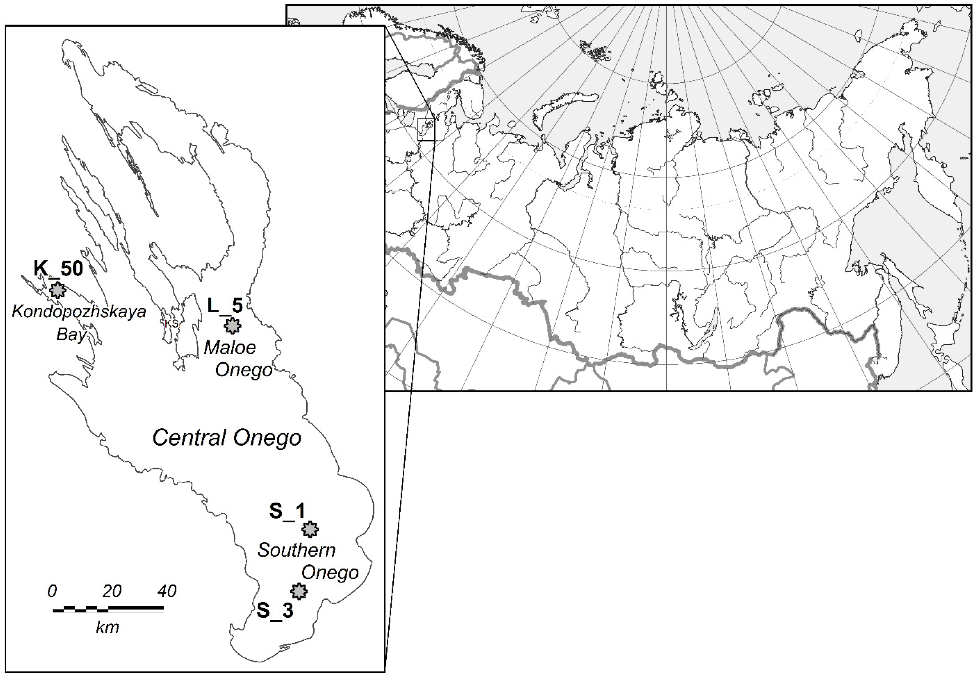
Lake Onego (61°38′34″ N 35°31′08″ W), the second largest body of water in Europe, is located in northwestern Russia on the border with the subarctic zone. The lake is of glacial origin. Its northern part is located on the Fennoscandian crystalline shield, and the southern part is on the East European Plain. The area of the lake is 9720 km2, the water volume is 295 km3, the average depth is 30 m, and the maximum is 120 m. The period of conditional water exchange is 15.6 years. The reservoir has 152 tributaries, including 52 rivers longer than 10 km. The Svir River flows out of the lake. The catchment area is 70% covered by coniferous forests, and 17% by open bogs [49]. In spring and autumn, the water is completely mixed. The average surface temperature of Lake Onego during the ice-free period (190 days) does not exceed 8 °C. The duration of biological summer with a water temperature above 10 °C is 100 days [36]. Lake Onego is a low-mineralized oligotrophic reservoir (Table 4). The polluted part of the lake is Kondopozhskaya Bay, which makes up only 2.5% of the lake area. The bay experiences a long-term load of biogenic and organic matter from wastewater from a pulp and paper mill and trout farms [50,51,52,53]. According to the concentration of total phosphorus and chlorophyll a, Kondopozhskaya Bay is characterized as mesotrophic. In coastal areas of the bay experiencing anthropogenic pressure, these characteristics reach the level of eutrophic ecosystems (Table 4).
River runoff contains a large amount of organic matter, mainly of humus origin. The influence of rivers is felt in estuarine areas, where elevated values of TOC (6.6–8.8 mg L−1), watercolor (35–64 mg Pt-Co L−1), and TP—10–19 µg L−1 are observed [41,50].

5.2. Sampling

Water samples were taken from the surface water layer from aboard the Ecologist research vessel in July 2022. The set of samples (2 samples from each station) were collected from bloom spots in the southern Onego (station S_1, depth 32.5 m and station S_3, depth 34.0 m) and the Maloe Onego (st. L_5, depth 43.0 m), where there are no known sources of water pollution (Figure 4). A water sample was also taken from Kondopogskaya Bay, subjected to biogenic and organic load from the pulp and paper mills’ wastewater and the trout farms (station K50, depth 35 m).

5.3. Phytoplankton Analysis

To study phytoplankton, a 500 mL water sample was fixed with formalin at a final concentration of 1%, and then concentrated on a membrane filter with a pore diameter of 0.8 µm (Vladipor, Russia) to a sample volume of 5 mL above the filter, according to [54]. The cell density of cyanobacteria was determined using a standard counting chamber method following Guillard (1978) [55] and LeGresley and McDermott (2010) [56]. Biomass was assessed from individual cell volumes calculated from geometric figures [57]. Species making up over 10% of total abundance and biomass were assessed as dominant [58]. The term ‘bloom spot’ was used for conditions when the abundance of cyanobacteria in water samples is over 107 cells L−1 [59]. The cyanobacterial species identification was performed according to [60,61]. The concentration of chlorophyll a in water samples was determined by spectrophotometric method [62].

5.4. HPLC–MSHR Analysis of Cyanotoxins

To distinguish water-dissolved and intracellular microcystin fractions and to separate planktonic DNA, water samples were divided into filtrate and phytoplankton biomass by filtration through Vladipor membrane filters (D pore = 0.8 µm).
The profile and quantification of MCs were assessed by the method of high-performance liquid chromatography–mass spectrometry of high-resolution (HPLC–MSHR) using LTQ Orbitrap XL (Thermo Fisher Scientific, Waltham, MA, USA) with Prominence LC-20 system (Shimadzu, Japan). Sample preparation procedures included solid-phase extraction (Oasis HLB, Waters) for filtration and treatment with 75% methanol in an ultrasonic bath for biomass on filters.
MCs were separated chromatographically using Thermo Hypersil Gold RP C18 columns (100 × 3 mm, 3 µm, Thermo Fisher Scientific) in a gradient elution regime (0.2 mL min−1) by a water–acetonitrile mixture containing 0.05% formic acid. Mass-spectrometric analysis of MCs was performed under electrospray ionization conditions in a positive ion detection mode. Target compounds were identified by high-precision measurement of ion mass [M + H]+ or [M + 2H]2+ (resolution 30,000, accuracy within 5 ppm) [19], the collected fragmentation spectrum of the ion [22] and the retention time. Quantification was performed by the external standard method. Calibration dependencies were constructed using solutions of 9 standard MCs (MC–LR, MC–RR, MC–YR (Sigma Aldrich, St. Louis, MI, USA) and MC–LY, MC–LA, MC– LW, MC–LF, [D–Asp3]MC–LR и [D–Asp3]MC-RR (Enzo Life Sciences, Inc., Farmingdale, NY, USA). The procedural (taking into account concentrating stage during the sample preparation) LOD was 0.06 ng L−1, and LOQ was 0.2 ng L−1. Qualitative analysis for the presence of anatoxin-a was performed using high-resolution mass spectrometry. Isocratic elution (5% of organic phase) in the same chromatographic system was used.

5.5. DNA Extraction and PCR Detection of mcyA Gene

Polymerase chain reaction (PCR-analysis) was used to identify MC-producing cyanobacteria [63]. DNA from planktonic samples (environmental DNA, eDNA) was isolated with Diatom DNA Prep 200 reagents (OOO Isogene Laboratory, Russia) in accordance with the producer’s instructions. PCR amplification of MC biosynthesis mcyA gene was performed using the genus-specific primers mcyA_MF/MR (Microcystis, PCR product length is 225 bp) and mcyA AF/AR (Dolichospermum, 225 bp) [64]. Fragments of mcyA gene were amplified in a CFX96 Touch thermocycler (Bio–Rad, Hercules, CA, USA) using 20 µL of GenPak PCR Core ready-made mixture of reagents (Isogene Laboratory, Moscow, Russia) in accordance with the following protocol: preliminary DNA denaturation at 95 °C for 3 min followed by 40 amplification cycles at 95 °C for 30 s, 58 °C for 30 s and 72 °C for 1 min and the last stage (elongation) at 72 °C for 10 min. DNAs from the MC-producing strain Microcystis aeruginosa PCC 7806 and field colonies of Dolichospermum lemmermannii [65] were used as positive controls. PCR products were fractionated electrophoretically in 1.5% agar gel and analyzed in UV light after staining with ethidium bromide using the Gel Doc XR+ gel-documenting system (Bio–Rad, Hercules, CA, USA).

Supplementary Materials

The following supporting information can be downloaded at: https://www.mdpi.com/article/10.3390/toxins16110457/s1, Figure S1: Amplification of the Microcystis-specific section of the mcyA gene using primers mcyA_MF/MR. M—DNA molecular mass marker (bp), K_50, S_1, S_3—samples of planktonic DNA isolated from water at the studied stations of Lake Onego, NC-—negative control, PC+—positive control. Figure S2: Amplification of the Dolichospermum-specific section of the mcyA gene using primers mcyA_AF/AR. M—DNA molecular mass marker (bp), K_50, S_1, S_3—samples of planktonic DNA isolated from water at the studied stations of Lake Onego, NC-—negative control, PC+—positive control.

Author Contributions

Conceptualization, E.T. and N.K.; methodology, S.S., E.C., A.S. and V.S.; validation, S.S., A.S. and S.B.; formal analysis, S.S. and E.C.; investigation, S.S., E.C., A.S., S.B. and V.S.; writing—original draft preparation, E.T., S.S., E.C., N.K. and V.S.; writing—review and editing, S.B.; supervision, S.B. All authors have read and agreed to the published version of the manuscript.

Funding

The study was funded by a state assignment for the Northern Water Problems Institute, Karelian Research Center of the Russian Academy of Sciences (no. 121021700117-3). The molecular–genetic study of toxigenic cyanobacteria was conducted in the scientific educational laboratory ‘Molecular genetics and biotechnology’, under YarSU’s development program framework for 2030 (no. 123042800011-6). This work was partly performed within the framework of the state budgetary theme no. 122041100086-5.

Institutional Review Board Statement

Not applicable.

Informed Consent Statement

Not applicable.

Data Availability Statement

Data supporting the presented results are available in the text of the article and in the Supplementary Materials.

Acknowledgments

The authors are grateful to the crew of the research vessel Ecologist for their comprehensive assistance and support during the cruise on Lake Onego. We are also very grateful to O.V. Derusova for her help in preparing the figures for the article.

Conflicts of Interest

The authors declare no conflicts of interest.

References

  1. Chorus, I. Cyanotoxins: Occurrence, Causes, Consequences; Springer: Berlin, Germany, 2001. [Google Scholar] [CrossRef]
  2. O’Neil, J.M.; Davis, T.W.; Burford, M.A.; Gobler, C.J. The rise of harmful cyanobacteria blooms: The potential roles of eutrophication and climate change. Harmful Algae 2012, 14, 313–334. [Google Scholar] [CrossRef]
  3. Paerl, H.W.; Huisman, J. Climate change: A catalyst for global expansion of harmful cyanobacterial blooms. Environ. Microbiol. Rep. 2009, 1, 27–37. [Google Scholar] [CrossRef] [PubMed]
  4. Sukharevich, V.I.; Polak, Y.M. Global occurrence of cyanobacteria: Causes and effects (review). Inland Water Biol. 2020, 4, 566–575. [Google Scholar] [CrossRef]
  5. Apeldoorn, M.E.; Egmond, H.P.; Speijers, G.J.A.; Bakker, G.J.I. Toxins of cyanobacteria. Mol. Nutr. Food Res. 2007, 51, 7–60. [Google Scholar] [CrossRef]
  6. Harke, M.J.; Steffen, M.M.; Gobler, C.J.; Otten, T.G.; Wilhelm, S.W.; Wood, S.A.; Paerl, H.W. A review of the global ecology, genomics, and biogeography of the toxic cyanobacterium, Microcystis spp. Harmful Algae 2016, 54, 4–20. [Google Scholar] [CrossRef]
  7. Li, X.; Dreher, T.W.; Li, R. An overview of diversity, occurrence, genetics and toxin production of bloom-forming Dolichospermum (Anabaena) species. Harmful Algae 2016, 54, 54–68. [Google Scholar] [CrossRef] [PubMed]
  8. Chorus, I.; Fastner, J.; Welker, M. Cyanobacteria and Cyanotoxins in a Changing Environment: Concepts, Controversies, Challenges. Water 2021, 13, 2463. [Google Scholar] [CrossRef]
  9. Gabyshev, V.; Sidelev, S.; Chernova, E.; Vilnet, A.; Davydov, D.; Barinova, S.; Gabysheva, O.; Zhakovskaya, Z.; Voronov, I. Year-round presence of microcystins and toxin-producing Microcystis in water column and ice cover of a eutrophic lake located in the continuous permafrost zone (Yakutia, Russia). Toxins 2023, 15, 467. [Google Scholar] [CrossRef]
  10. Przytulska, A.; Bartosiewicz, M.; Vincent, W.F. Increased risk of cyanobacterial blooms in northern high-latitude lakes through climate warming and phosphorus enrichment. Freshw. Biol. 2017, 62, 1986–1996. [Google Scholar] [CrossRef]
  11. de Magalhães, V.F.; Soares, R.M.; Azevedo, S.M. Microcystin contamination in fish from the Jacarepaguá Lagoon (Rio de Janeiro, Brazil): Ecological implication and human health risk. Toxicon 2001, 39, 1077–1085. [Google Scholar] [CrossRef]
  12. Poste, A.E.; Hecky, R.E.; Guildford, S.J. Evaluating microcystin exposure risk through fish consumption. Environ. Sci. Technol. 2011, 45, 5806–5811. [Google Scholar] [CrossRef] [PubMed]
  13. Onyango, D.M.; Orina, P.S.; Ramkat, R.C.; Kowenje, C.; Githukia, C.M.; Lusweti, D.; Lung’ayia, H.B. Review of current state of knowledge of microcystin and its impacts on fish in Lake Victoria. Lakes Reserv. Res. Manag. 2020, 25, 350–361. [Google Scholar] [CrossRef]
  14. Shahmohamadloo, R.S.; Frenken, T.; Rudman, S.M.; Ibelings, B.W.; Trainer, V.L. Diseases and disorders in fish due to harmful algal blooms. In Climate Change on Diseases and Disorders of Finfish in Cage Culture, 3rd ed.; Woo, P.T.K., Subasinghe, R.P., Eds.; CABI: Wallingford, UK, 2023; pp. 387–429. [Google Scholar] [CrossRef]
  15. Smith, R.B.; Bass, B.; Sawyer, D.; Depew, D.; Watson, S.B. Estimating the economic costs of algal blooms in the Canadian Lake Erie Basin. Harmful Algae 2019, 87, 101624. [Google Scholar] [CrossRef] [PubMed]
  16. Chernova, E.; Sidelev, S.; Russkikh, I.; Voyakina, E.; Zhakovskaya, Z. First observation of microcystin- and anatoxin-a-producing cyanobacteria in the easternmost part of the Gulf of Finland (the Baltic Sea). Toxicon 2019, 157, 18–24. [Google Scholar] [CrossRef]
  17. Voloshko, L.; Kopecky, J.; Safronova, T.; Pljusch, A.; Titova, N.; Hrouzek, P.; Drabkova, V. Toxins and other bioactive compounds produced by cyanobacteria in Lake Ladoga. Est. J. Ecol. 2008, 57, 100–110. [Google Scholar] [CrossRef]
  18. Litvinchuk, L.F.; Sharov, A.N.; Chernova, E.N.; Smirnov, V.V.; Berezina, N.A. Mutual links between microcystins-producing cyanobacteria and plankton community in clear and brow2n northern lakes. Food Webs 2023, 35, e00279. [Google Scholar] [CrossRef]
  19. Chernova, E.; Russkikh, I.; Voyakina, E.; Zhakovskaya, Z. Occurrence of microcystins and anatoxin-a in eutrophic lakes of Saint Petersburg, Northwestern Russia. Oceanol. Hydrobiol. Stud. 2016, 45, 466–484. [Google Scholar] [CrossRef]
  20. Voyakina, E.J.; Russkikh, I.V.; Chernova, E.N.; Zhakovskaya, Z.A. Toxic cyanobacteria and their metabolites in the lakes of the Russian northwest. Theor. Appl. Ecol. 2020, 1, 124–129, In Russian. [Google Scholar] [CrossRef]
  21. Tekanova, E.V.; Kalinkina, N.M.; Makarova, E.M.; Smirnova, V.S. The current trophic state and water quality of Lake Onego. Inland Water Biol. 2023, 16, 967–973. [Google Scholar] [CrossRef]
  22. Chernova, E.N.; Russkikh, Y.V.; Podolskaya, E.P.; Zhakovskaya, Z.A. Determination of microcystins and anatoxin-a using liquid chromato-mass-spectrometry of unit resolution. Nauchnoe Priborostr. 2016, 26, 11–25. Available online: http://iairas.ru/mag/2016/full1/Art2.pdf (accessed on 21 October 2024). (In Russian). [CrossRef]
  23. Rapala, J.; Sivonen, K.; Luukkainen, R.; Niemela, S.I. Anatoxin-a concentration in Anabaena and Aphanizomenon under different environmental conditions and comparison of growth by toxic and nontoxic Anabaena-strains—A laboratory study. J. Appl. Phycol. 1993, 5, 581–591. [Google Scholar] [CrossRef]
  24. Dimitrakopoulos, I.K.; Kaloudis, T.S.; Hiskia, A.E.; Thomaidis, N.S.; Koupparis, M.A. Development of a fast and selective method for the sensitive determination of anatoxin-a in lake waters using liquid chromatography–tandem mass spectrometry and phenylalanine-d5 as internal standard. Anal. Bioanal. Chem. 2010, 397, 2245–2252. [Google Scholar] [CrossRef] [PubMed]
  25. Barinova, S.; Chekryzheva, T. Phytoplankton dynamic and bioindication in the Kondopoga Bay, Lake Onego (Northern Russia). J. Limnol. 2014, 73, 282–297. [Google Scholar] [CrossRef]
  26. Smirnova, V.S.; Tekanova, E.V.; Kalinkina, N.M. Phytoplankton as an indicator of the ecosystem state of the Kondopoga Bay of Lake Onego under cage trout farming. Ecosyst. Transform. 2024, 7, 177–195. [Google Scholar] [CrossRef]
  27. Chekryzheva, T.A.; Kalinkina, N.M. Structure and seasonal dynamics of phytoplankton communities in the exposed and sheltered littoral zones of Lake Onego (Pinguba Bay, Pukhtinskaya Bay). Trans. Karelian Res. Cent. Russ. Acad. Sci. 2016, 12, 83–95. (In Russian) [Google Scholar] [CrossRef]
  28. Petrova, N.A. Fitoplankton Litoralnoj Zony Onezhskogo Ozera. In The Vegetation of the Lake Onego; Raspopov, I.M., Ed.; Nauka: Leningrad, Russia, 1975; p. 138144. Available online: https://search.rsl.ru/ru/record/01007196947 (accessed on 21 October 2024). (In Russian)
  29. Chorus, I.; Bartram, J. Toxic Cyanobacteria in Water: A Guide to Their Public Health Consequences, Monitoring and Management, 1st ed.; CRC Press: London, UK, 1999. [Google Scholar] [CrossRef]
  30. Saker, M.L.; Vale, M.; Kramer, D.; Vasconcelos, V.M. Molecular techniques for the early warning of toxic cyanobacteria blooms in freshwater lakes and rivers. Appl. Microbiol. Biotechnol. 2007, 75, 441–449. [Google Scholar] [CrossRef]
  31. Sidelev, S.I.; Korneva, L.G.; Solovyeva, V.V.; Zubishina, A.A.; Pligin, D.N. Molecular genetic identification and seasonal succession of toxigenic cyanobacteria in phytoplankton of the Rybinsk Reservoir (Russia). Inland Water Biol. 2016, 9, 368–374. [Google Scholar] [CrossRef]
  32. Baker, J.A.; Entsch, B.; Neilan, B.A.; McKay, D.B. Monitoring changing toxigenicity of a cyanobacterial bloom by molecular methods. Appl. Environ. Microbiol. 2002, 68, 6070–6076. [Google Scholar] [CrossRef]
  33. Capelli, C.; Ballot, A.; Cerasino, L.; Papini, A.; Salmaso, N. Biogeography of bloom-forming microcystin producing and non-toxigenic populations of Dolichospermum lemmermannii (Cyanobacteria). Harmful Algae 2017, 67, 1–12. [Google Scholar] [CrossRef]
  34. Harada, K.; Ogawa, K.; Kimura, Y.; Murata, H.; Suzuki, M.; Thorn, P.M.; Evans, W.R.; Carmichael, W.W. Microcystins from Anabaena flosaquae NRC 525-17. Chem. Res. Toxicol. 1991, 4, 535–540. [Google Scholar] [CrossRef]
  35. Vezie, C.; Brient, L.; Sivonen, K.; Bertru, G.; Lefeuvre, J.-C.; Salkinoja-Salonen, M. Variation of microcystin content of cyanobacterial blooms and isolated strains in Lake Grand-Lieu (France). Microb. Ecol. 1998, 35, 126–135. [Google Scholar] [CrossRef] [PubMed]
  36. Rukhovets, L.; Filatov, N. Ladoga and Onego—Great European Lakes. In Observations and Modeling; Springer: Berlin/Heidelberg, Germany, 2010; Available online: https://www.springer.com/gp/book/9783540681441 (accessed on 21 October 2024).
  37. Sterner, R.W.; Reinl, K.L.; Lafrancois, B.M.; Brovold, S.; Miller, T.R. A first assessment of cyanobacterial blooms in oligotrophic Lake Superior. Limnol. Oceanogr. 2020, 65, 2984–2998. [Google Scholar] [CrossRef]
  38. Chorus, I.; Welker, M. Toxic Cyanobacteria in Water: A Guide to Their Public Health Consequences, Monitoring and Management, 2nd ed.; CRC Press: London, UK, 2021. [Google Scholar] [CrossRef]
  39. Shahmohamadloo, R.S.; Bhavsar, S.P.; Almirall, X.O.; Marklevitz, S.A.; Rudman, S.M.; Sibley, P.K. Lake Erie fish safe to eat yet afflicted by algal hepatotoxins. Sci. Total Environ. 2023, 861, 160474. [Google Scholar] [CrossRef] [PubMed]
  40. Shahmohamadloo, R.S.; Bhavsar, S.P.; Almirall, X.O.; Marklevitz, S.A.; Rudman, S.M.; Sibley, P.K. Cyanotoxins accumulate in Lake St. Clair fish yet their fillets are safe to eat. Sci. Total Environ. 2023, 874, 162381. [Google Scholar] [CrossRef]
  41. Callieri, C.; Bertoni, R.; Contesini, M.; Bertoni, F. Lake level fluctuations boost toxic cyanobacterial “oligotrophic blooms”. PLoS ONE 2014, 9, e109526. [Google Scholar] [CrossRef]
  42. Kaplan-Levy, R.N.; Hadas, O.; Summers, M.L.; Rücker, J.; Sukenik, A. Akinetes: Dormant cells of cyanobacteria. In Dormancy and Resistance in Harsh Environments; Lubzens, E., Cerda, J., Clark, M., Eds.; Springer: Berlin, Germany, 2010; pp. 5–27. Available online: https://link.springer.com/book/10.1007/978-3-642-12422-8 (accessed on 21 October 2024).
  43. Zobkov, M.; Zobkova, M.; Galakhina, N.; Efremova, T.; Efremenko, N.; Kulik, N. Data on the chemical composition of Lake Onego water in 2019–2021. Data Brief 2022, 42, e108079. [Google Scholar] [CrossRef] [PubMed]
  44. Legrand, B.; Lamarque, A.; Sabart, M.; Latour, D. Characterization of akinetes from cyanobacterial strains and lake sediment: A study of their resistance and toxic potential. Harmful Algae 2016, 59, 42–50. [Google Scholar] [CrossRef]
  45. Reinl, K.L.; Brookes, J.D.; Carey, C.C.; Harris, T.D.; Ibelings, B.W.; Morales-Williams, A.M.; De Senerpont Domis, L.N.; Atkins, K.S.; Isles, P.D.F.; Mesman, J.P.; et al. Cyanobacterial blooms in oligotrophic lakes: Shifting the high-nutrient paradigm. Freshw. Biol. 2021, 66, 1846–1859. [Google Scholar] [CrossRef]
  46. Belykh, O.I.; Fedorova, G.A.; Kuzmin, A.V.; Tikhonova, I.V.; Timoshkin, O.A.; Sorokovikova, E.G. Microcystins in cyanobacterial biofilms from the littoral zone of Lake Baikal. Mosc. Univ. Biol. Sci. Bull. 2017, 72, 225–231. [Google Scholar] [CrossRef]
  47. Belykh, O.I.; Gladkikh, A.S.; Sorokovikova, E.G.; Tikhonova, I.V.; Butina, T.V. Identification of Toxic Cyanobacteria in Lake Baikal. Dokl. Biochem. Biophys. 2015, 463, 220–224. [Google Scholar] [CrossRef]
  48. Bartlett, S.L.; Brunner, S.L.; Val Klump, J.; Houghton, E.M.; Miller, T.R. Spatial analysis of toxic or otherwise bioactive cyanobacterial peptides in Green Bay, Lake Michigan. J. Great Lakes Res. 2018, 44, 924–933. [Google Scholar] [CrossRef]
  49. Litinsky, P.Y. Onego Lake catchment ecosystem GIS as a tool for estimating components of the water and carbon balance. Trans. Karelian Res. Cent. Russ. Acad. Sci. 2019, 9, 136–144. (In Russian) [Google Scholar] [CrossRef]
  50. Galakhina, N.; Zobkov, M.; Zobkova, M. Current chemistry of Lake Onego and its spatial and temporal changes for the last three decades with special reference to nutrient concentrations. Environ. Nanotechnol. Monit. Manag. 2022, 17, e100619. [Google Scholar] [CrossRef]
  51. Sabylina, A.V.; Lozovik, P.A.; Zobkov, M.B. Water chemistry in Onega Lake and its tributaries. Water Resour. 2010, 37, 842–853. [Google Scholar] [CrossRef]
  52. Tekanova, E.V.; Litvinova, I.A. Organic matter destruction in the Kodopoga Bay, Onego Lake, under changing anthropogenic load. Water Res. 2022, 49, 1009–1016. [Google Scholar] [CrossRef]
  53. Timakova, T.M.; Kulikova, T.P.; Litvinova, I.A.; Polyakova, T.N.; Syarki, M.T.; Tekanova, E.V.; Chekryzheva, T.A. Changes in biocenoses of Kondopoga Bay, Lake Onego, under the effect of effluents from a pulp and paper mill. Water Res. 2014, 41, 78–86. [Google Scholar] [CrossRef]
  54. Clark, W.J.; Sigler, W.F. Method of concentrating phytoplankton samples using membrane filters. Limnol. Oceanogr. 1963, 8, 127–129. [Google Scholar] [CrossRef]
  55. Guillard, R.R.L. Counting slides. In Phytoplankton Manual, Monographs on Oceanographic Methodology; Sournia, A., Ed.; UNESCO Publishing: Paris, France, 1978; Volume 6, pp. 182–189. [Google Scholar]
  56. LeGresley, M.; McDermott, G. Counting chamber methods for quantitative phytoplankton analysis—Haemocytometer, Palmer-Maloney cell and Sedgewick-Rafter cell. In Microscopic and Molecular Methods for Quantitative Phytoplankton Analysis; Karlson, B., Cusack, C., Bresnan, E., Eds.; UNESCO: Paris, France, 2010; pp. 25–30. [Google Scholar]
  57. Olenina, I.; Hajdu, S.; Edler, L.; Andersson, A.; Wasmund, N.; Busch, S.; Göbel, J.; Gromisz, S.; Huseby, S.; Huttunen, M.; et al. Biovolumes and size-classes of phytoplankton in the Baltic Sea. Helcom Balt. Sea Environ. Proc. 2006, 106, 144. Available online: https://helcom.fi/wp-content/uploads/2019/08/BSEP106.pdf (accessed on 21 October 2024).
  58. Okhapkin, A.; Sharagina, E.; Kulizin, P.; Startseva, N.; Vodeneeva, E. Phytoplankton community structure in highly mineralized small gypsum karst lake (Russia). Microorganisms 2022, 10, 386. [Google Scholar] [CrossRef]
  59. Ger, K.A.; Hansson, L.-A.; Lürling, M. Understanding cyanobacteria-zooplankton interactions in a more eutrophic world. Freshw. Biol. 2014, 59, 1783–1798. [Google Scholar] [CrossRef]
  60. Komarek, J. Cyanoprokaryota. 3. Heterocytous genera. In Süswasserflora von Mitteleuropa/Freshwater Flora of Central Europe; Büdel, B., Gartner, G., Krienitz, L., Schagerl, M., Eds.; Springer Spektrum: Berlin/Heidelberg, Germany, 2013; 1130p. [Google Scholar]
  61. Komárek, J.; Anagnostidis, K. Cyanoprokaryota. T. 1. Chroococcales; Gustav Fischer Verlag: Jena, Germany, 1998; 548p, Available online: https://www.springer.com/gp/book/9783827421111 (accessed on 21 October 2024).
  62. Report of SCOR-Unesco Working Group 17. Determination of photosynthetic pigments in sea-water. In Monographs Onocéanographie Methodology; UNESCO: Paris, France, 1966; pp. 9–18. [Google Scholar]
  63. Kurmayer, R.; Sivonen, K.; Wilmotte, A.; Salmaso, N. Molecular Tools for Detection and Quantification of Toxigenic Cyanobacteria; Wiley: Hoboken, NJ, USA, 2017. [Google Scholar]
  64. Lee, J.; Choi, J.; Fatka, M.; Swanner, E.; Ikuma, K.; Liang, X.; Leung, T.; Howe, A. Improved detection of mcyA genes and their phylogenetic origins in harmful algal blooms. Water Res. 2020, 176, e115730. [Google Scholar] [CrossRef] [PubMed]
  65. Sidelev, S.I. Detection of microcystin-producing cyanobacteria Microcystis, Planktothrix and Dolichospermum using multiprimer amplification of mcy genes. Microbiology 2022, 91, 792–795. [Google Scholar] [CrossRef]
Figure 1. Extracted ion chromatogram of high resolution (mass accuracy within 5 ppm) for MC congeners detected in phytoplankton samples of Lake Onego: standard mix solution, concentration 50 ng mL−1 (A), sample K_50 (B), sample S_3 (C) (from top to bottom): m/z 512.78235 ([M+2H]2+ [D-Asp3]MC-RR); m/z 519.79077 ([M+2H]2+ MC-RR); m/z 981.54095 ([M+H]+ [D-Asp3]MC-LR); m/z 995.55658 ([M+H]+ MC-LR); m/z 1045.53589 ([M+H]+ MC-YR).
Toxins 16 00457 g001
Figure 2. MS2 spectra of the MC congeners identified in the extract of sample K_50: [D-Asp3]MC-RR) (A); [D-Asp3]MC-LR) (B); MC-RR (C); MC-LR (D); MC-YR (E).
Toxins 16 00457 g002
Figure 3. Example of extracted ion high-resolution chromatogram (mass accuracy within 5 ppm) of phytoplankton extract (sample S_3): phenylalanine (A), anatoxin-a (B).
Toxins 16 00457 g003
Figure 4. Location of sampling stations. Lake Onego, July 2022.
Toxins 16 00457 g004
Table 1. Abundance (Ntot) and biomass (Btot) of the cyanobacterial community and chlorophyll a in water samples from Lake Onego in July 2022.
StationNtot, 106 cells L−1Btot, mg L−1Chl a, µg L−1
S_1129.2936.4231
S_384.5219.3833
L_524.326.8247
K5012.230.6911
Table 2. Species composition and structure of the cyanobacterial community in water samples from Lake Onego in July 2022.
Sampling StationSpeciesShare of the Species
in Abundance, %
Share of the Species
in Biomass, %
S_1Aphanizomenon flos-aquae0.0030.04
Dolichospermum lemmermannii5774.9
Dolichospermum spiroides4325.0
S_3Aphanizomenon flos-aquae0.050.79
Aphanothece minutissima10.04
Aphanocapsa grevillei10.06
Dolichospermum circinale4931.2
Dolichospermum lemmermannii4667.7
Microcystis aeruginosa20.12
L_5Aphanothece nidularis80.3
Dolichospermum circinale4428
Dolichospermum lemmermannii4971.7
K50Aphanizomenon flos-aquae0.00716.5
Dolichospermum flos-aquae5652.6
Dolichospermum spiroides1211.5
Dolichospermum lemmermannii48.7
Microcystis aeruginosa2610.6
Table 3. Intracellular MC concentration (ng L−1) and the results of PCR detection of the mcyA gene in samples from Lake Onego in July 2022.
MC Congeners
and Detection of mcyA Gene
S_1K50S_3L_50
[D-Asp3]MC-LR128
MC-LR8
[D-Asp3]MC-RR 125
MC-RR14
MC-YR44
Total MCs concentration15312
Dolichospermum-specific mcyA + 2+NA
Microcystis-specific mcyA+++NA
1 “‒” MCs (< 0.06 ng L−1) or the mcyA gene were not detected, 2 “+” the mcyA gene was detected in eDNA, NA—not analyzed.
Table 4. Averaged chemical variables of Onelo Lake water in 2019–2021 [34,43].
CharacteristicsPelagic zone
of Lake Onego
Kondopozhskaya Bay
Off-Shore
Kondopozhskaya Bay
In-Shore
Conductivity, μS cm−150.748.553.0
pH7.37.27.0
TOC, mg L−16.47.612.3
Color, mg Pt-Co L−131.035.651.7
TP, µg L−18.122.068.5
PO4-P, µg L−10019.0
NO3-N, mg L−10.150.160.10
NH4-N, mg L−10.010.010.08
TN, mg L−10.360.380.56
Chlorophyll a, µg L−13.24.68.8
Disclaimer/Publisher’s Note: The statements, opinions and data contained in all publications are solely those of the individual author(s) and contributor(s) and not of MDPI and/or the editor(s). MDPI and/or the editor(s) disclaim responsibility for any injury to people or property resulting from any ideas, methods, instructions or products referred to in the content.