Multi-Functional Electrospun Nanofibers from Polymer Blends for Scaffold Tissue Engineering


Abstract
1. Introduction
2. Electrospinning
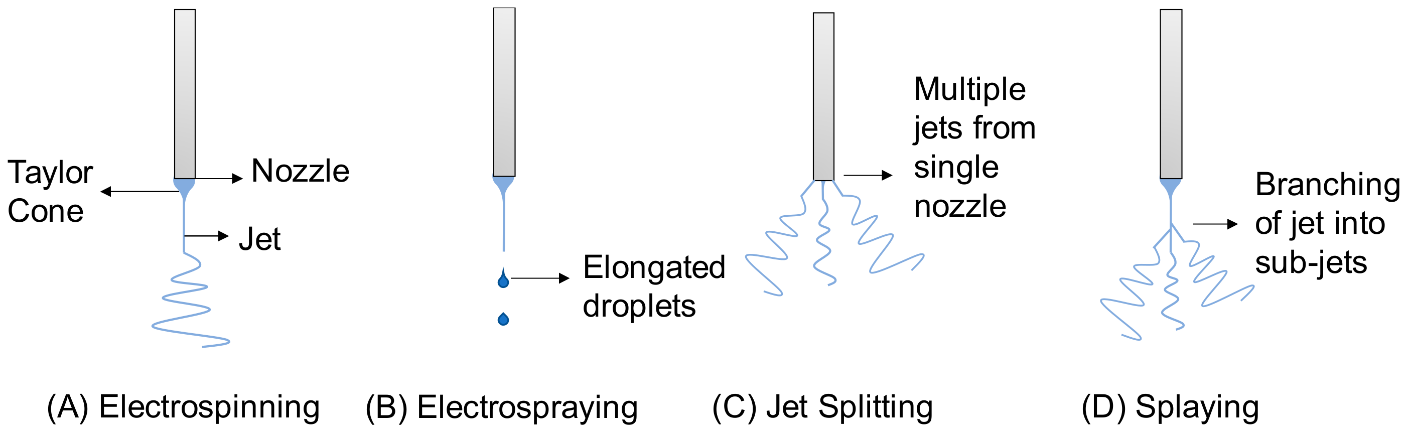
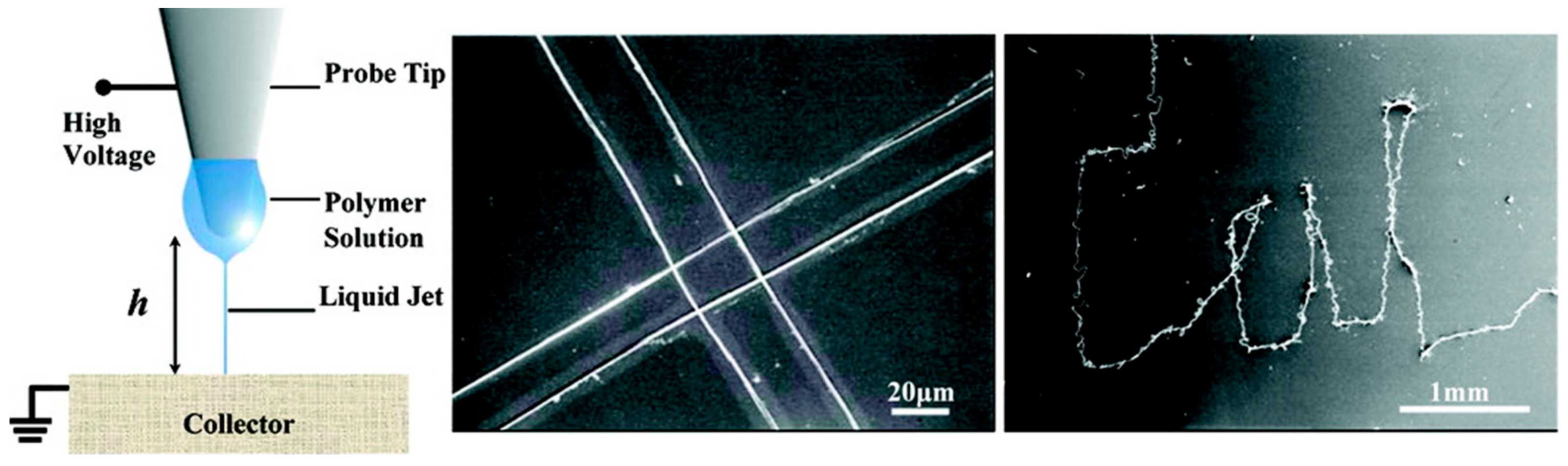
2.1. Theory
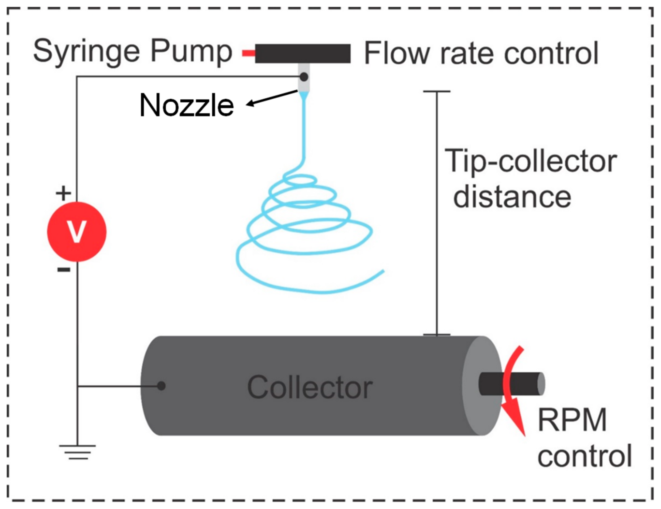
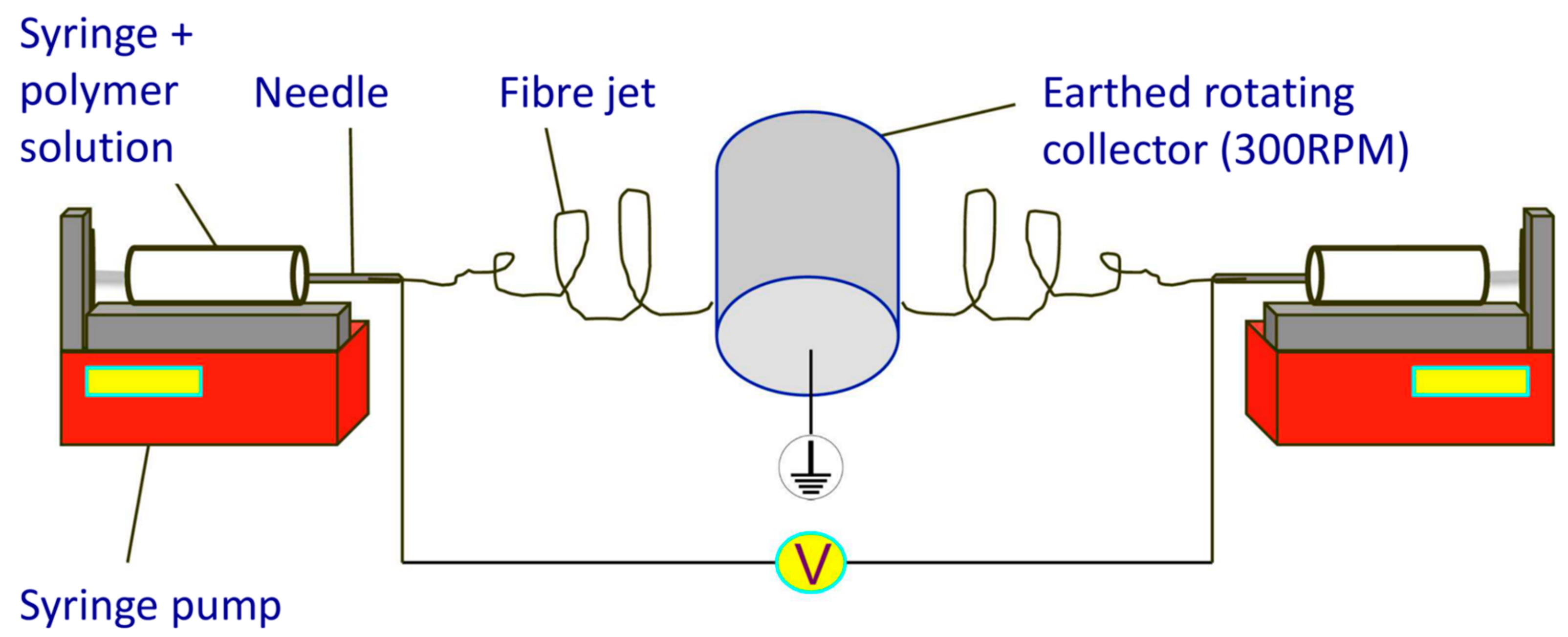
2.2. The Electrospinning Apparatus
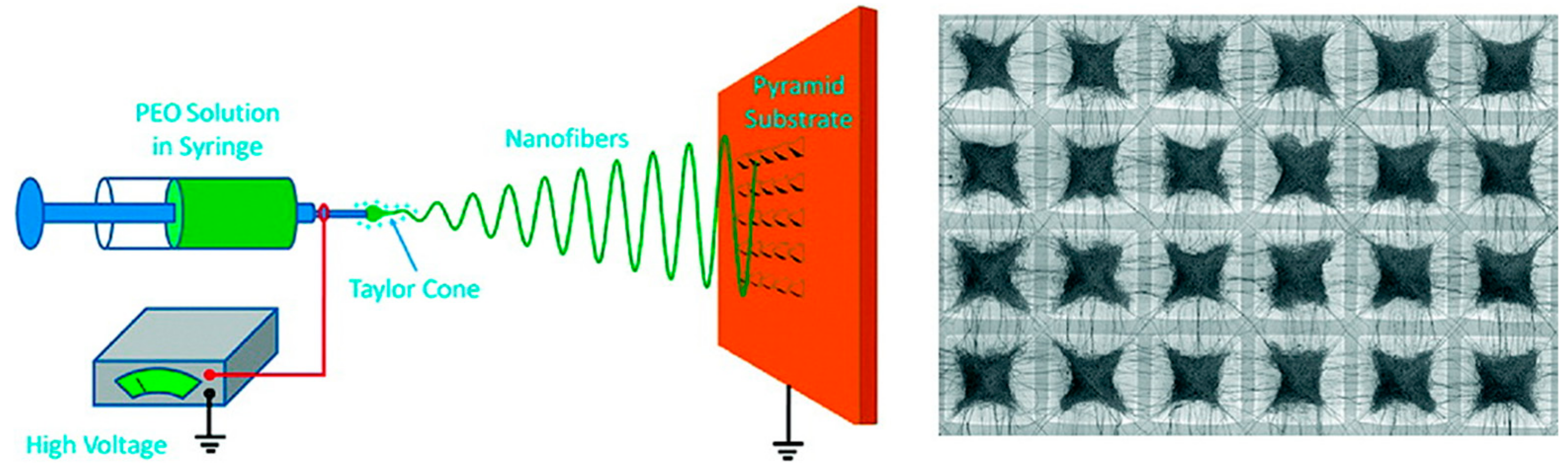
2.2.1. Changes in Collector Design
Rotating Mandrel
Patterned Collector
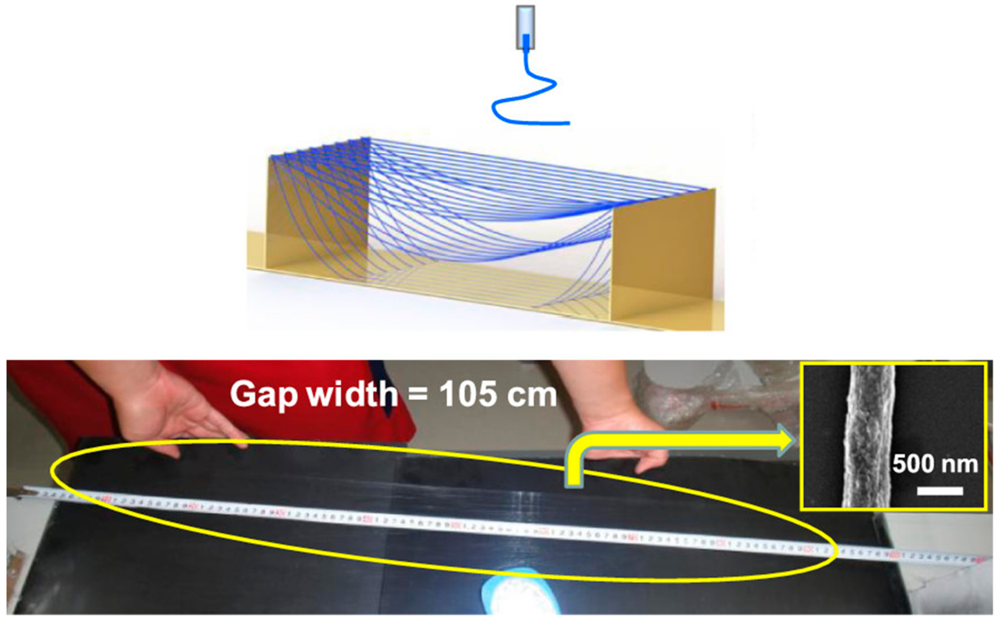
Gap Electrospinning
Magnetic Field Associated Electrospinning
Wet Spinning
2.2.2. Changes in Orientation
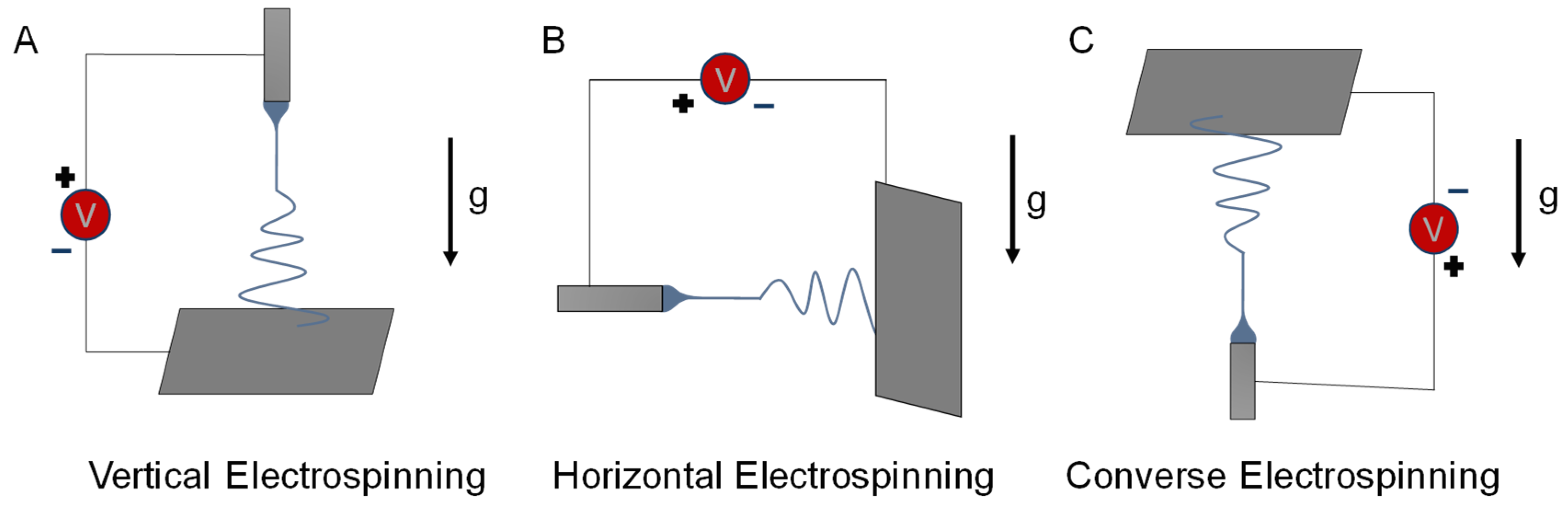
Vertical Electrospinning
Horizontal Electrospinning
Converse Electrospinning
2.2.3. Changes in Spinneret
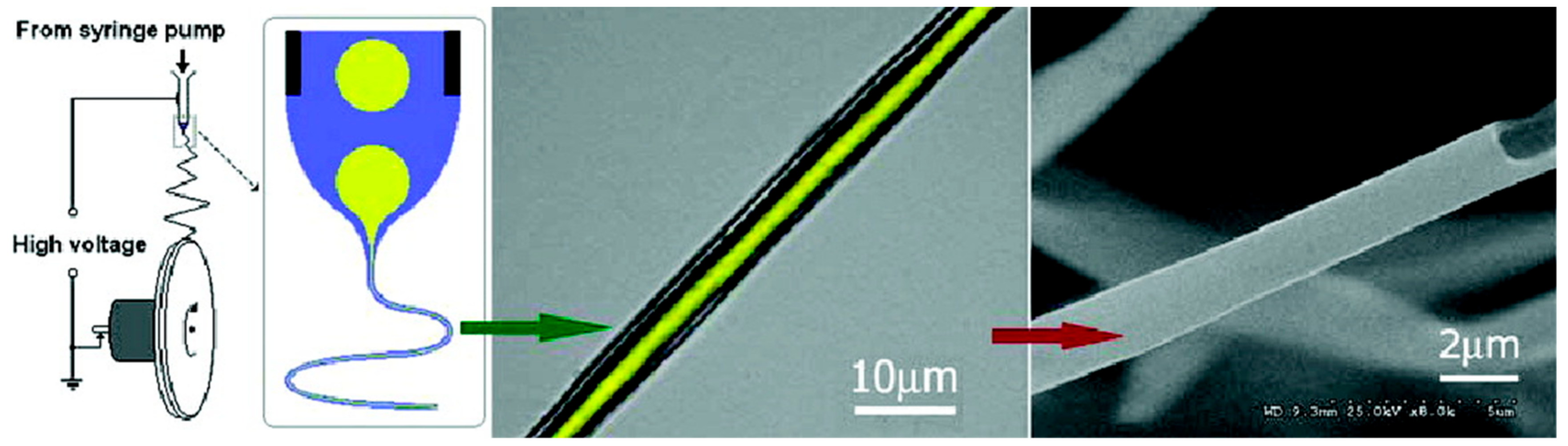
Coaxial Electrospinning
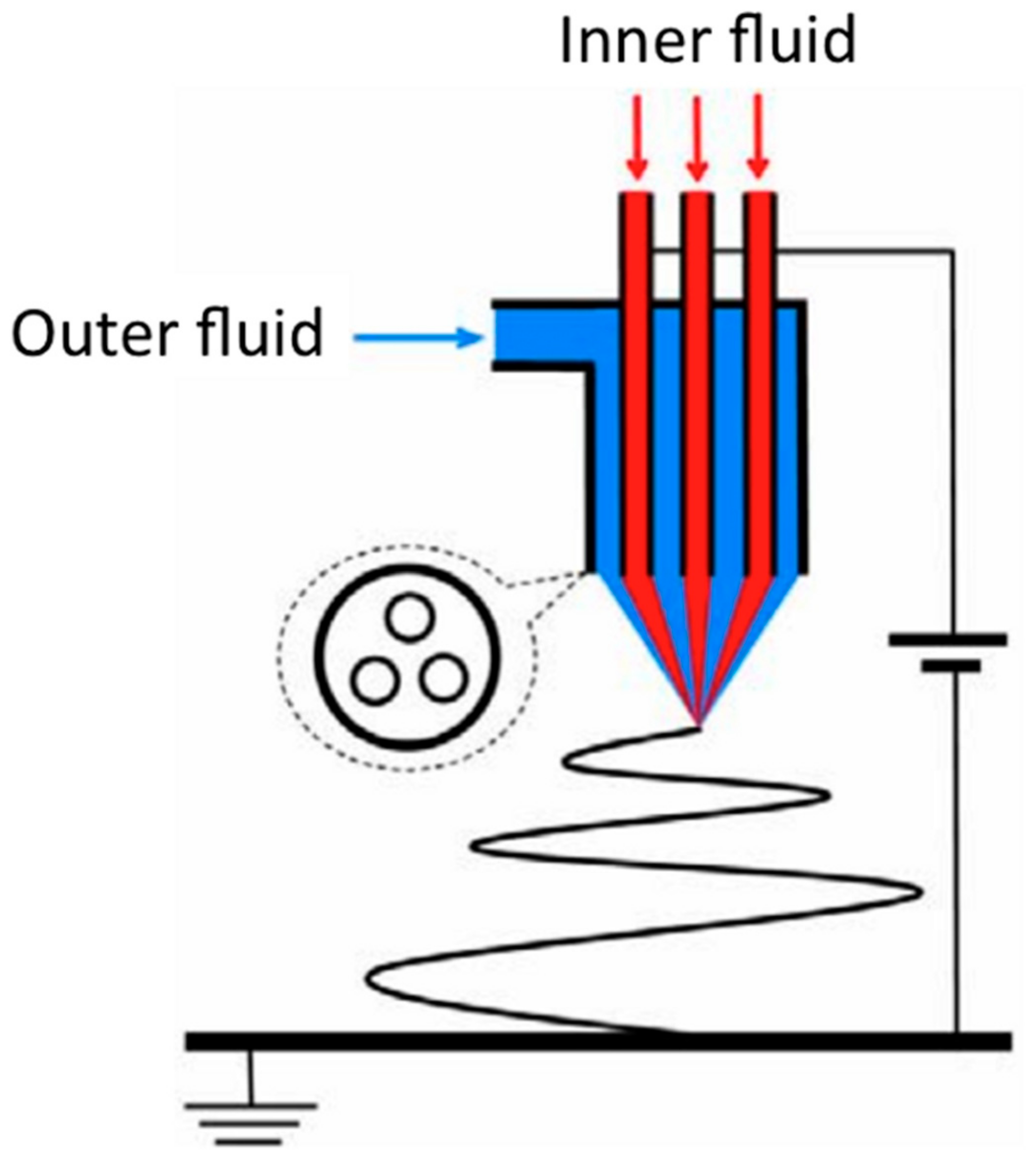
Co-Electrospinning
In-Line Polymer Blending
2.2.4. Other Modifications
Centrifugal Electrospinning
Near Field Electrospinning
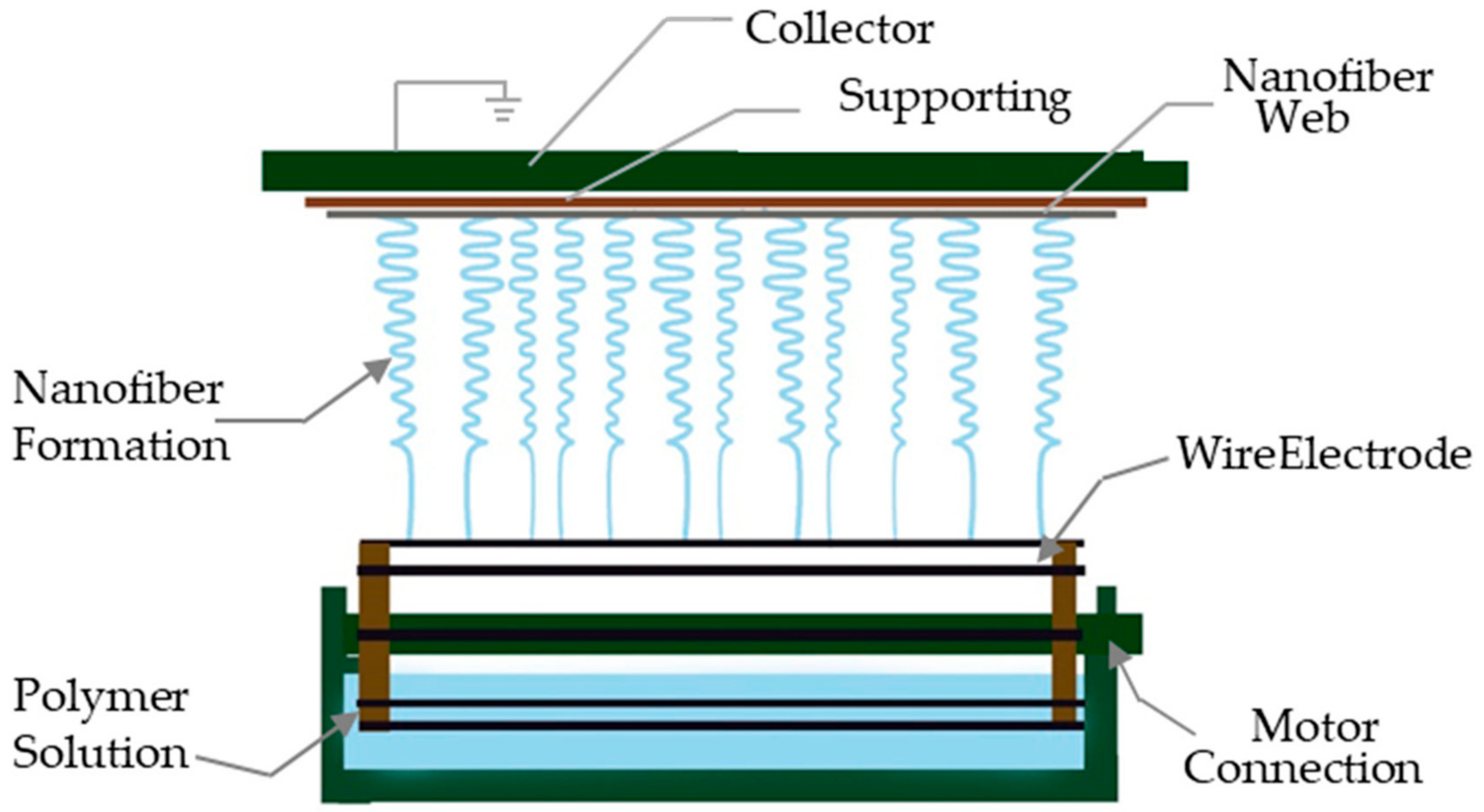
Needleless Electrospinning
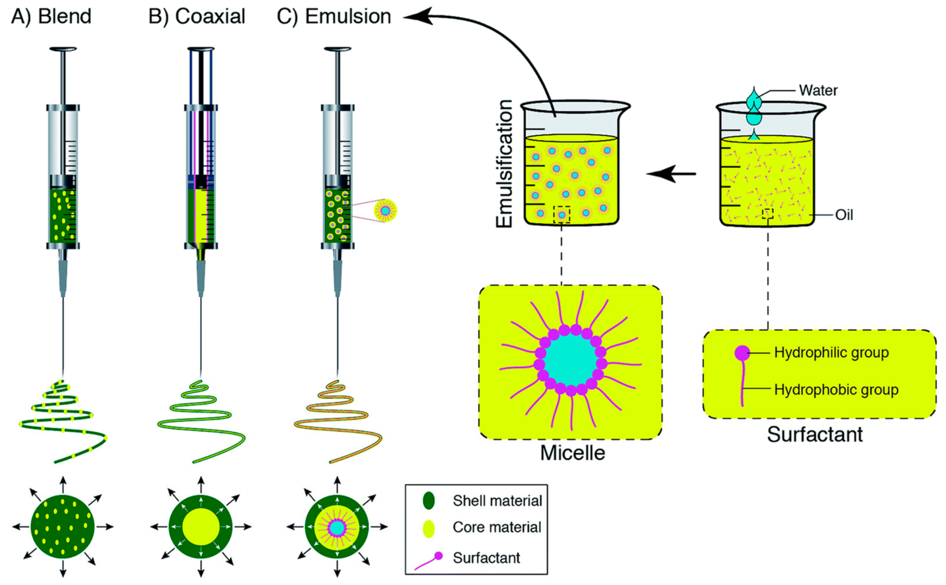
Emulsion Electrospinning
3. Polymer Blends for Tissue Scaffold Engineering
3.1. Natural Polymer Blends
3.2. Synthetic Polymer Blends
3.3. Mixed Polymer Blends
3.4. Nanofiller Polymer Blends
4. Perspectives and Conclusions
Author Contributions
Funding
Acknowledgments
Conflicts of Interest
Abbreviations
| CA | Cellulose Acetate |
| CF | Chloroform; |
| DCE | 1,2-dichloroethane |
| DCM | Dichloromethane |
| DMAC | Dimethylacetamide |
| DMEM | Dulbecco Modified Eagle’s Medium |
| DMF | N,N-dimethylformamide |
| DMSO | Dimethyl sulfoxide |
| ECM | Extracellular Matrix |
| GO | Graphene Oxide |
| HA | Hydroxyapatite; |
| HFP | 1,1,1,3,3,3-hexa-fluoro-2-propanol |
| FESEM | Field Emission Scanning Electron Microscope |
| HSA | Human Serum Albumin |
| MWCNT | multiwalled-carbon nanotubes |
| PANI | Polyaniline |
| PBAT | Poly(butylene adipate-co-terephthalate) |
| PBS | Phosphate buffered saline |
| PCL | Poly-caprolactone |
| PDMS | polydimethylsiloxane |
| PEA | Poly(ester amide) |
| PGA | Polyglycolide |
| PGS | Poly(Glycerol Sebacate) |
| PHB | Polyhydroxybutyrate |
| PHBV | Poly(hydroxybutyrate-cohydroxyvalerate) |
| PLA | Poly(lactic acid) |
| PLGA | Poly(lactic-co-glycolic acid) |
| PLLA | Poly(l-lactic acid) |
| PLLA-CL | Poly(L-lactic acid-co-e-caprolactone) |
| PMMA | Poly(methyl methacrylate) |
| PPy | Polypyrrole |
| PVA | Polyvinyl alcohol |
| PVAc | Polyvinyl acetate |
| PVDF | Polyvinylidene fluoride |
| PVP | Polyvinyl pyrrolidone |
| SF | Silk fibroin |
| TFA | Trifluoracetic acid |
| TFE | 2,2,2-Trifluoroethanol |
| TFE | 2,2,2-Trifluoroethanol |
| THF | Tetrahydrofuran |
References
- Griffith, L.G.; Naughton, G.J.S. Tissue engineering—Current challenges and expanding opportunities. Science 2002, 295, 1009–1014. [Google Scholar] [CrossRef] [PubMed]
- Ma, P.X. Scaffolds for tissue fabrication. Mater. Today 2004, 7, 30–40. [Google Scholar] [CrossRef]
- Place, E.S.; Evans, N.D.; Stevens, M.M. Complexity in biomaterials for tissue engineering. Nat. Mater. 2009, 8, 457–470. [Google Scholar] [CrossRef] [PubMed]
- Chen, F.M.; Liu, X.H. Advancing biomaterials of human origin for tissue engineering. Prog. Polym. Sci. 2016, 53, 86–168. [Google Scholar] [CrossRef] [PubMed]
- Hinderer, S.; Layland, S.L.; Schenke-Layland, K. ECM and ECM-like materials-Biomaterials for applications in regenerative medicine and cancer therapy. Adv. Drug Deliv. Rev. 2016, 97, 260–269. [Google Scholar] [CrossRef]
- Liu, J.; Sun, L.S.; Xu, W.Y.; Wang, Q.Q.; Yu, S.J.; Sun, J.Z. Current advances and future perspectives of 3D printing natural-derived biopolymers. Carbohydr. Polym. 2019, 207, 297–316. [Google Scholar] [CrossRef] [PubMed]
- Min, L.L.; Pan, H.; Chen, S.Y.; Wang, C.Y.; Wang, N.; Zhang, J.; Cao, Y.; Chen, X.Y.; Hou, X. Recent progress in bio-inspired electrospun materials. Compos. Commun. 2019, 11, 12–20. [Google Scholar] [CrossRef]
- Bose, S.; Ke, D.X.; Sahasrabudhe, H.; Bandyopadhyay, A. Additive manufacturing of biomaterials. Prog. Mater. Sci. 2018, 93, 45–111. [Google Scholar] [CrossRef]
- Moroni, L.; Boland, T.; Burdick, J.A.; De Maria, C.; Derby, B.; Forgacs, G.; Groll, J.; Li, Q.; Malda, J.; Mironov, V.A.; et al. Biofabrication: A Guide to Technology and Terminology. Trends Biotechnol. 2018, 36, 384–402. [Google Scholar] [CrossRef]
- Cooley, J.F. Improved methods of and apparatus for electrically separating the relatively volatile liquid component from the component of relatively fixed substances of composite fluids. UK Pat. 1900, 6385, 19. [Google Scholar]
- Doshi, J.; Reneker, D.H. Electrospinning process and applications of electrospun fibers. In Proceedings of the Industry Applications Society Annual Meeting, Toronto, ON, Canada, 2–8 October 1993; pp. 1698–1703. [Google Scholar]
- Doshi, J.; Reneker, D.H. Electrospinning process and applications of electrospun fibers. J. Electrost. 1995, 35, 151–160. [Google Scholar] [CrossRef]
- Small, P.A. Some factors affecting the solubility of polymers. J. Appl. Chem. 1953, 3, 71–80. [Google Scholar] [CrossRef]
- Koenhen, D.M.; Smolders, C.A. The determination of solubility parameters of solvents and polymers by means of correlations with other physical quantities. J. Appl. Polym. Sci. 1975, 19, 1163–1179. [Google Scholar] [CrossRef]
- Barton, A.F. CRC Handbook of Solubility Parameters and Other Cohesion Parameters; Routledge: London, UK, 2017. [Google Scholar]
- Sionkowska, A. Current research on the blends of natural and synthetic polymers as new biomaterials: Review. Prog. Polym. Sci. 2011, 36, 1254–1276. [Google Scholar] [CrossRef]
- Reneker, D.H.; Yarin, A.L. Electrospinning jets and polymer nanofibers. Polymer 2008, 49, 2387–2425. [Google Scholar] [CrossRef]
- Nagam Hanumantharao, S. A 3D Biomimetic Scaffold Using Electrospinning for Tissue Engineering Applications. Master’s Thesis, Michigan Technological University, Houghton, MI, USA, 2017. [Google Scholar]
- Zargham, S.; Bazgir, S.; Tavakoli, A.; Rashidi, A.S.; Damerchely, R. The Effect of Flow Rate on Morphology and Deposition Area of Electrospun Nylon 6 Nanofiber. J. Eng. Fibers Fabr. 2012, 7, 42–49. [Google Scholar] [CrossRef]
- Yarin, A.L.; Koombhongse, S.; Reneker, D.H. Taylor cone and jetting from liquid droplets in electrospinning of nanofibers. J. Appl. Phys. 2001, 90, 4836–4846. [Google Scholar] [CrossRef]
- Tan, S.H.; Inai, R.; Kotaki, M.; Ramakrishna, S. Systematic parameter study for ultra-fine fiber fabrication via electrospinning process. Polymer 2005, 46, 6128–6134. [Google Scholar] [CrossRef]
- Eda, G.; Liu, J.; Shivkumar, S. Solvent effects on jet evolution during electrospinning of semi-dilute polystyrene solutions. Eur. Polym. J. 2007, 43, 1154–1167. [Google Scholar] [CrossRef]
- Guerrero, J.; Rivero, J.; Gundabala, V.R.; Perez-Saborid, M.; Fernandez-Nieves, A. Whipping of electrified liquid jets. Proc. Natl. Acad. Sci. USA 2014, 111, 13763. [Google Scholar] [CrossRef]
- Wannatong, L.; Sirivat, A.; Supaphol, P. Effects of solvents on electrospun polymeric fibers: Preliminary study on polystyrene. Polym. Int. 2004, 53, 1851–1859. [Google Scholar] [CrossRef]
- Reneker, D.H.; Kataphinan, W.; Theron, A.; Zussman, E.; Yarin, A.L. Nanofiber garlands of polycaprolactone by electrospinning. Polymer 2002, 43, 6785–6794. [Google Scholar] [CrossRef]
- Pelipenko, J.; Kristl, J.; Janković, B.; Baumgartner, S.; Kocbek, P. The impact of relative humidity during electrospinning on the morphology and mechanical properties of nanofibers. Int. J. Pharm. 2013, 456, 125–134. [Google Scholar] [CrossRef]
- Nezarati, R.M.; Eifert, M.B.; Cosgriff-Hernandez, E. Effects of humidity and solution viscosity on electrospun fiber morphology. Tissue Eng. Part Cmethods 2013, 19, 810–819. [Google Scholar] [CrossRef]
- Li, D.; Xia, Y.N. Electrospinning of nanofibers: Reinventing the wheel? Adv. Mater. 2004, 16, 1151–1170. [Google Scholar] [CrossRef]
- Theron, S.A.; Zussman, E.; Yarin, A.L. Experimental investigation of the governing parameters in the electrospinning of polymer solutions. Polymer 2004, 45, 2017–2030. [Google Scholar] [CrossRef]
- Deitzel, J.M.; Kleinmeyer, J.; Harris, D.; Tan, N.C.B. The effect of processing variables on the morphology of electrospun nanofibers and textiles. Polymer 2001, 42, 261–272. [Google Scholar] [CrossRef]
- Kim, K.W.; Lee, K.H.; Khil, M.S.; Ho, Y.S.; Kim, H.Y. The effect of molecular weight and the linear velocity of drum surface on the properties of electrospun poly (ethylene terephthalate) nonwovens. Fibers Polym. 2004, 5, 122–127. [Google Scholar] [CrossRef]
- Baji, A.; Mai, Y.-W.; Wong, S.-C.; Abtahi, M.; Chen, P. Electrospinning of polymer nanofibers: Effects on oriented morphology, structures and tensile properties. Compos. Sci. Technol. 2010, 70, 703–718. [Google Scholar] [CrossRef]
- Persano, L.; Dagdeviren, C.; Su, Y.; Zhang, Y.; Girardo, S.; Pisignano, D.; Huang, Y.; Rogers, J.A. High performance piezoelectric devices based on aligned arrays of nanofibers of poly (vinylidenefluoride-co-trifluoroethylene). Nat. Commun. 2013, 4, 1633. [Google Scholar] [CrossRef]
- Li, D.; Ouyang, G.; McCann, J.T.; Xia, Y. Collecting Electrospun Nanofibers with Patterned Electrodes. Nano Lett. 2005, 5, 913–916. [Google Scholar] [CrossRef]
- Ding, Z.; Salim, A.; Ziaie, B. Selective Nanofiber Deposition through Field-Enhanced Electrospinning. Langmuir 2009, 25, 9648–9652. [Google Scholar] [CrossRef]
- Lei, T.; Xu, Z.; Cai, X.; Xu, L.; Sun, D. New Insight into Gap Electrospinning: Toward Meter-long Aligned Nanofibers. Langmuir 2018, 34, 13788–13793. [Google Scholar] [CrossRef]
- Liu, Y.; Zhang, X.; Xia, Y.; Yang, H. Magnetic-field-assisted electrospinning of aligned straight and wavy polymeric nanofibers. Adv. Mater. 2010, 22, 2454–2457. [Google Scholar] [CrossRef]
- Tzezana, R.; Zussman, E.; Levenberg, S. A Layered Ultra-Porous Scaffold for Tissue Engineering, Created via a Hydrospinning Method. Tissue Eng. Part C Methods 2008, 14, 281–288. [Google Scholar] [CrossRef]
- Spivak, A.F.; Dzenis, Y.A.; Reneker, D.H. A model of steady state jet in the electrospinning process. Mech. Res. Commun. 2000, 27, 37–42. [Google Scholar] [CrossRef]
- Yarin, A.L.; Koombhongse, S.; Reneker, D.H. Bending instability in electrospinning of nanofibers. J. Appl. Phys. 2001, 89, 3018–3026. [Google Scholar] [CrossRef]
- Hohman, M.M.; Shin, M.; Rutledge, G.; Brenner, M.P. Electrospinning and electrically forced jets. II. Applications. Phys. Fluids 2001, 13, 2221–2236. [Google Scholar] [CrossRef]
- Zhao, J.; Si, N.; Xu, L.; Tang, X.; Song, Y.; Sun, Z. Experimental and theoretical study on the electrospinning nanoporous fibers process. Mater. Chem. Phys. 2016, 170, 294–302. [Google Scholar] [CrossRef]
- Yang, C.; Jia, Z.; Xu, Z.; Wang, K.; Guan, Z.; Wang, L. Comparisons of fibers properties between vertical and horizontal type electrospinning systems. In Proceedings of the Electrical Insulation and Dielectric Phenomena, Virginia Beach, VA, USA, 18–21 October 2009; pp. 204–207. [Google Scholar]
- Loscertales, I.G.; Barrero, A.; Guerrero, I.; Cortijo, R.; Marquez, M.; Gañán-Calvo, A.M. Micro/Nano Encapsulation via Electrified Coaxial Liquid Jets. Science 2002, 295, 1695. [Google Scholar] [CrossRef]
- Li, F.; Zhao, Y.; Song, Y.L. Core-shell nanofibers: nano channel and capsule by coaxial electrospinning. In Nanofibers; Kumar, A., Ed.; IntechOpen: London, UK, 2010; pp. 418–438. [Google Scholar]
- Bazilevsky, A.V.; Yarin, A.L.; Megaridis, C.M. Co-electrospinning of Core−Shell Fibers Using a Single-Nozzle Technique. Langmuir 2007, 23, 2311–2314. [Google Scholar] [CrossRef]
- Yarin, A.L. Coaxial electrospinning and emulsion electrospinning of core–shell fibers. Polym. Adv. Technol. 2011, 22, 310–317. [Google Scholar] [CrossRef]
- Xu, F.; Li, L.; Cui, X. Fabrication of Aligned Side-by-Side TiO2/SnO2 Nanofibers via Dual-Opposite-Spinneret Electrospinning. J. Nanomater. 2012, 2012, 5. [Google Scholar] [CrossRef]
- Xu, W.; Ding, Y.; Huang, R.; Zhu, Z.; Fong, H.; Hou, H. High-performance polyimide nanofibers reinforced polyimide nanocomposite films fabricated by co-electrospinning followed by hot-pressing. J. Appl. Polym. Sci. 2018, 135, 46849. [Google Scholar] [CrossRef]
- Hillary, C.J.; Roman, S.; Bullock, A.J.; Green, N.H.; Chapple, C.R.; MacNeil, S. Developing Repair Materials for Stress Urinary Incontinence to Withstand Dynamic Distension. PLoS ONE 2016, 11, e0149971. [Google Scholar] [CrossRef]
- Weitz, R.T.; Harnau, L.; Rauschenbach, S.; Burghard, M.; Kern, K. Polymer nanofibers via nozzle-free centrifugal spinning. Nano Lett. 2008, 8, 1187–1191. [Google Scholar] [CrossRef]
- Sun, D.; Chang, C.; Li, S.; Lin, L. Near-Field Electrospinning. Nano Lett. 2006, 6, 839–842. [Google Scholar] [CrossRef]
- Simm, W.; Gosling, C.; Bonart, R.; Falkai, B.V. Fibre Fleece of Electrostatically Spun Fibres and Methods of Making Same. U.S. Patent 4,143,196, 6 March 1979. [Google Scholar]
- Yu, M.; Dong, R.H.; Yan, X.; Yu, G.F.; You, M.H.; Ning, X.; Long, Y.Z. Recent Advances in Needleless Electrospinning of Ultrathin Fibers: From Academia to Industrial Production. Macromol. Mater. Eng. 2017, 302, 19. [Google Scholar] [CrossRef]
- Li, T.-T.; Yan, M.; Xu, W.; Shiu, B.-C.; Lou, C.-W.; Lin, J.-H. Mass-Production and Characterizations of Polyvinyl Alcohol/Sodium Alginate/Graphene Porous Nanofiber Membranes Using Needleless Dynamic Linear Electrospinning. Polymers 2018, 10, 1167. [Google Scholar] [CrossRef]
- Xu, X.; Zhuang, X.; Chen, X.; Wang, X.; Yang, L.; Jing, X. Preparation of Core-Sheath Composite Nanofibers by Emulsion Electrospinning. Macromol. Rapid Commun. 2006, 27, 1637–1642. [Google Scholar] [CrossRef]
- Qi, H.X.; Hu, P.; Xu, J.; Wang, A.J. Encapsulation of drug reservoirs in fibers by emulsion electrospinning: Morphology characterization and preliminary release assessment. Biomacromolecules 2006, 7, 2327–2330. [Google Scholar] [CrossRef]
- Yang, Y.; Xia, T.; Zhi, W.; Wei, L.; Weng, J.; Zhang, C.; Li, X.H. Promotion of skin regeneration in diabetic rats by electrospun core-sheath fibers loaded with basic fibroblast growth factor. Biomaterials 2011, 32, 4243–4254. [Google Scholar] [CrossRef]
- Maretschek, S.; Greiner, A.; Kissel, T. Electrospun biodegradable nanofiber nonwovens for controlled release of proteins. J. Control. Release 2008, 127, 180–187. [Google Scholar] [CrossRef]
- Xu, X.L.; Yang, L.X.; Xu, X.Y.; Wang, X.; Chen, X.S.; Liang, Q.Z.; Zeng, J.; Jing, X.B. Ultrafine medicated fibers electrospun from W/O emulsions. J. Control. Release 2005, 108, 33–42. [Google Scholar] [CrossRef]
- Xu, X.L.; Chen, X.S.; Wang, Z.F.; Jing, X.B. Ultrafine PEG-PLA fibers loaded with both paclitaxel and doxorubicin hydrochloride and their In Vitro cytotoxicity. Eur. J. Pharm. Biopharm. 2009, 72, 18–25. [Google Scholar] [CrossRef]
- Zhang, H.; Jia, X.L.; Han, F.X.; Zhao, J.; Zhao, Y.H.; Fan, Y.B.; Yuan, X.Y. Dual-delivery of VEGF and PDGF by double-layered electrospun membranes for blood vessel regeneration. Biomaterials 2013, 34, 2202–2212. [Google Scholar] [CrossRef]
- Li, X.Q.; Su, Y.; Liu, S.P.; Tan, L.J.; Mo, X.M.; Ramakrishna, S. Encapsulation of proteins in poly (L-lactide-co-caprolactone) fibers by emulsion electrospinning. Colloid Surf. B-Biointerfaces 2010, 75, 418–424. [Google Scholar] [CrossRef]
- Su, Y.; Li, X.Q.; Liu, S.P.; Mo, X.M.; Ramakrishna, S. Controlled release of dual drugs from emulsion electrospun nanofibrous mats. Colloid Surf. B-Biointerfaces 2009, 73, 376–381. [Google Scholar] [CrossRef]
- Zhou, F.; Jia, X.L.; Yang, Y.; Yang, Q.M.; Gao, C.; Hu, S.L.; Zhao, Y.H.; Fan, Y.B.; Yuan, X.Y. Nanofiber-mediated microRNA-126 delivery to vascular endothelial cells for blood vessel regeneration. Acta Biomater. 2016, 43, 303–313. [Google Scholar] [CrossRef]
- Wang, Z.B.; Qian, Y.N.; Li, L.H.; Pan, L.H.; Njunge, L.W.; Dong, L.L.; Yang, L. Evaluation of emulsion electrospun polycaprolactone/hyaluronan/epidermal growth factor nanofibrous scaffolds for wound healing. J. Biomater. Appl. 2016, 30, 686–698. [Google Scholar] [CrossRef]
- Nikmaram, N.; Roohinejad, S.; Hashemi, S.; Koubaa, M.; Barba, F.J.; Abbaspourrad, A.; Greiner, R. Emulsion-based systems for fabrication of electrospun nanofibers: Food, pharmaceutical and biomedical applications. Rsc Adv. 2017, 7, 28951–28964. [Google Scholar] [CrossRef]
- Chinnappan, A.; Baskar, C.; Baskar, S.; Ratheesh, G.; Ramakrishna, S. An overview of electrospun nanofibers and their application in energy storage, sensors and wearable/flexible electronics. J. Mater. Chem. C 2017, 5, 12657–12673. [Google Scholar] [CrossRef]
- Timin, A.S.; Muslimov, A.R.; Zyuzin, M.V.; Peltek, O.O.; Karpov, T.E.; Sergeev, I.S.; Dotsenko, A.I.; Goncharenko, A.A.; Yolshin, N.D.; Sinelnik, A.; et al. Multifunctional Scaffolds with Improved Antimicrobial Properties and Osteogenicity Based on Piezoelectric Electrospun Fibers Decorated with Bioactive Composite Microcapsules. Acs Appl. Mater. Interfaces 2018, 10, 34849–34868. [Google Scholar] [CrossRef]
- Perez, R.A.; Kim, H.-W. Core–shell designed scaffolds for drug delivery and tissue engineering. Acta Biomater. 2015, 21, 2–19. [Google Scholar] [CrossRef]
- Subbiah, R.; Guldberg, R.E. Materials Science and Design Principles of Growth Factor Delivery Systems in Tissue Engineering and Regenerative Medicine. Adv. Healthc. Mater. 2019, 8, 1801000. [Google Scholar] [CrossRef]
- Whited, B.M.; Rylander, M.N. The influence of electrospun scaffold topography on endothelial cell morphology, alignment, and adhesion in response to fluid flow. Biotechnol. Bioeng. 2014, 111, 184–195. [Google Scholar] [CrossRef]
- Ravichandran, R.; Liao, S.; Ng, C.C.; Chan, C.K.; Raghunath, M.; Ramakrishna, S. Effects of nanotopography on stem cell phenotypes. World J. Stem Cells 2009, 1, 55. [Google Scholar] [CrossRef]
- Kim, D.H.; Lipke, E.A.; Kim, P.; Cheong, R.; Thompson, S.; Delannoy, M.; Suh, K.Y.; Tung, L.; Levchenko, A. Nanoscale cues regulate the structure and function of macroscopic cardiac tissue constructs. Proc. Natl. Acad. Sci. USA 2010, 107, 565–570. [Google Scholar] [CrossRef]
- Patel, S.; Kurpinski, K.; Quigley, R.; Gao, H.; Hsiao, B.S.; Poo, M.-M.; Li, S. Bioactive nanofibers: Synergistic effects of nanotopography and chemical signaling on cell guidance. Nano Lett. 2007, 7, 2122–2128. [Google Scholar] [CrossRef]
- Christopherson, G.T.; Song, H.; Mao, H.-Q. The influence of fiber diameter of electrospun substrates on neural stem cell differentiation and proliferation. Biomaterials 2009, 30, 556–564. [Google Scholar] [CrossRef]
- Chew, S.Y.; Mi, R.; Hoke, A.; Leong, K.W. The effect of the alignment of electrospun fibrous scaffolds on Schwann cell maturation. Biomaterials 2008, 29, 653–661. [Google Scholar] [CrossRef]
- Ayres, C.; Bowlin, G.L.; Henderson, S.C.; Taylor, L.; Shultz, J.; Alexander, J.; Telemeco, T.A.; Simpson, D.G. Modulation of anisotropy in electrospun tissue-engineering scaffolds: Analysis of fiber alignment by the fast Fourier transform. Biomaterials 2006, 27, 5524–5534. [Google Scholar] [CrossRef]
- Cheng, H.L.; Yang, X.Y.; Che, X.; Yang, M.S.; Zhai, G.X. Biomedical application and controlled drug release of electrospun fibrous materials. Mater. Sci. Eng. C-Mater. Biol. Appl. 2018, 90, 750–763. [Google Scholar] [CrossRef]
- Contreras-Cáceres, R.; Cabeza, L.; Perazzoli, G.; Díaz, A.; López-Romero, M.J.; Melguizo, C.; Prados, J. Electrospun Nanofibers: Recent Applications in Drug Delivery and Cancer Therapy. Nanomater. 2019, 9, 656. [Google Scholar] [CrossRef]
- Fullana, M.J.; Wnek, G.E. Electrospun collagen and its applications in regenerative medicine. Drug Deliv. Transl. Res. 2012, 2, 313–322. [Google Scholar] [CrossRef]
- Sell, S.A.; Wolfe, P.S.; Garg, K.; McCool, J.M.; Rodriguez, I.A.; Bowlin, G.L. The Use of Natural Polymers in Tissue Engineering: A Focus on Electrospun Extracellular Matrix Analogues. Polymers 2010, 2, 522–553. [Google Scholar] [CrossRef]
- Campiglio, C.E.; Marcolin, C.; Draghi, L. Electrospun ECM macromolecules as biomimetic scaffold for regenerative medicine: Challenges for preserving conformation and bioactivity. Aims Mater. Sci. 2017, 4, 638–669. [Google Scholar] [CrossRef][Green Version]
- Lee, K.Y.; Jeong, L.; Kang, Y.O.; Lee, S.J.; Park, W.H. Electrospinning of polysaccharides for regenerative medicine. Adv. Drug Deliv. Rev. 2009, 61, 1020–1032. [Google Scholar] [CrossRef]
- Sajkiewicz, P.; Kolbuk, D. Electrospinning of gelatin for tissue engineering-molecular conformation as one of the overlooked problems. J. Biomater. Sci.-Polym. Ed. 2014, 25, 2009–2022. [Google Scholar] [CrossRef]
- Qasim, S.B.; Zafar, M.S.; Najeeb, S.; Khurshid, Z.; Shah, A.H.; Husain, S.; Rehman, I.U. Electrospinning of Chitosan-Based Solutions for Tissue Engineering and Regenerative Medicine. Int. J. Mol. Sci. 2018, 19, 26. [Google Scholar] [CrossRef]
- Kalantari, K.; Afifi, A.M.; Jahangirian, H.; Webster, T.J. Biomedical applications of chitosan electrospun nanofibers as a green polymer-review. Carbohydr. Polym. 2019, 207, 588–600. [Google Scholar] [CrossRef]
- Zhang, X.H.; Reagan, M.R.; Kaplan, D.L. Electrospun silk biomaterial scaffolds for regenerative medicine. Adv. Drug Deliv. Rev. 2009, 61, 988–1006. [Google Scholar] [CrossRef]
- Zhang, J.G.; Mo, X.M. Current research on electrospinning of silk fibroin and its blends with natural and synthetic biodegradable polymers. Front. Mater. Sci. 2013, 7, 129–142. [Google Scholar] [CrossRef]
- Majidi, S.S.; Slemming-Adamsen, P.; Hanif, M.; Zhang, Z.; Wang, Z.; Chen, M. Wet electrospun alginate/gelatin hydrogel nanofibers for 3D cell culture. Int. J. Biol. Macromol. 2018, 118, 1648–1654. [Google Scholar] [CrossRef]
- Xie, J.; Peng, C.; Zhao, Q.; Wang, X.; Yuan, H.; Yang, L.; Li, K.; Lou, X.; Zhang, Y. Osteogenic differentiation and bone regeneration of iPSC-MSCs supported by a biomimetic nanofibrous scaffold. Acta Biomater. 2016, 29, 365–379. [Google Scholar] [CrossRef]
- Joy, J.; Pereira, J.; Aid-Launais, R.; Pavon-Djavid, G.; Ray, A.R.; Letourneur, D.; Meddahi-Pellé, A.; Gupta, B. Gelatin—Oxidized carboxymethyl cellulose blend based tubular electrospun scaffold for vascular tissue engineering. Int. J. Biol. Macromol. 2018, 107, 1922–1935. [Google Scholar] [CrossRef]
- Li, Z.; Tuffin, J.; Lei, I.M.; Ruggeri, F.S.; Lewis, N.S.; Gill, E.L.; Savin, T.; Huleihel, L.; Badylak, S.F.; Knowles, T.; et al. Solution fibre spinning technique for the fabrication of tuneable decellularised matrix-laden fibres and fibrous micromembranes. Acta Biomater. 2018, 78, 111–122. [Google Scholar] [CrossRef]
- Pezeshki-Modaress, M.; Zandi, M.; Rajabi, S. Tailoring the gelatin/chitosan electrospun scaffold for application in skin tissue engineering: An In Vitro study. Prog. Biomater. 2018, 7, 207–218. [Google Scholar] [CrossRef]
- Chen, H.; Xie, S.; Yang, Y.; Zhang, J.; Zhang, Z. Multiscale regeneration scaffold In Vitro and In Vivo. J. Biomed. Mater. Res. Part B Appl. Biomater. 2018, 106, 1218–1225. [Google Scholar] [CrossRef]
- Honarpardaz, A.; Irani, S.; Pezeshki-Modaress, M.; Zandi, M.; Sadeghi, A. Enhanced chondrogenic differentiation of bone marrow mesenchymal stem cells on gelatin/glycosaminoglycan electrospun nanofibers with different amount of glycosaminoglycan. J. Biomed. Mater. Res. Part A 2019, 107, 38–48. [Google Scholar] [CrossRef]
- Bian, T.; Zhao, K.; Meng, Q.; Tang, Y.; Jiao, H.; Luo, J. The construction and performance of multi-level hierarchical hydroxyapatite (HA)/collagen composite implant based on biomimetic bone Haversian motif. Mater. Des. 2019, 162, 60–69. [Google Scholar] [CrossRef]
- Ko, E.; Lee, J.S.; Kim, H.; Yang, S.Y.; Yang, D.; Yang, K.; Lee, J.; Shin, J.; Yang, H.S.; Ryu, W.; et al. Electrospun Silk Fibroin Nanofibrous Scaffolds with Two-Stage Hydroxyapatite Functionalization for Enhancing the Osteogenic Differentiation of Human Adipose-Derived Mesenchymal Stem Cells. Acs Appl. Mater. Interfaces 2018, 10, 7614–7625. [Google Scholar] [CrossRef]
- Deng, L.; Li, Y.; Feng, F.; Zhang, H. Study on wettability, mechanical property and biocompatibility of electrospun gelatin/zein nanofibers cross-linked by glucose. Food Hydrocoll. 2019, 87, 1–10. [Google Scholar] [CrossRef]
- Cipitria, A.; Skelton, A.; Dargaville, T.R.; Dalton, P.D.; Hutmacher, D.W. Design, fabrication and characterization of PCL electrospun scaffolds-a review. J. Mater. Chem. 2011, 21, 9419–9453. [Google Scholar] [CrossRef]
- Woodruff, M.A.; Hutmacher, D.W. The return of a forgotten polymer—Polycaprolactone in the 21st century. Prog. Polym. Sci. 2010, 35, 1217–1256. [Google Scholar] [CrossRef]
- Guo, B.; Ma, P.X. Conducting polymers for tissue engineering. Biomacromolecules 2018, 19, 1764–1782. [Google Scholar] [CrossRef]
- Rajabi, A.H.; Jaffe, M.; Arinzeh, T.L. Piezoelectric materials for tissue regeneration: A review. Acta Biomater. 2015, 24, 12–23. [Google Scholar] [CrossRef]
- Nagam Hanumantharao, S.; Que, C.; Rao, S. Self-assembly of 3D nanostructures in electrospun polycaprolactone-polyaniline fibers and their application as scaffolds for tissue engineering. Materialia 2019, 6, 100296. [Google Scholar] [CrossRef]
- Malikmammadov, E.; Tanir, T.E.; Kiziltay, A.; Hasirci, V.; Hasirci, N. Polymer Edition. PCL and PCL-based materials in biomedical applications. J. Biomater. Sci. Polym. Ed. 2018, 29, 863–893. [Google Scholar] [CrossRef]
- Huang, Z.B.; Yin, G.F.; Liao, X.M.; Gu, J.W. Conducting polypyrrole in tissue engineering applications. Front. Mater. Sci. 2014, 8, 39–45. [Google Scholar] [CrossRef]
- Bertuoli, P.T.; Ordoño, J.; Armelin, E.; Pérez-Amodio, S.; Baldissera, A.F.; Ferreira, C.A.; Puiggalí, J.; Engel, E.; del Valle, L.J.; Alemán, C. Electrospun Conducting and Biocompatible Uniaxial and Core–Shell Fibers Having Poly (lactic acid), Poly (ethylene glycol), and Polyaniline for Cardiac Tissue Engineering. Acs Omega 2019, 4, 3660–3672. [Google Scholar] [CrossRef]
- De Castro, J.G.; Rodrigues, B.V.M.; Ricci, R.; Costa, M.M.; Ribeiro, A.F.C.; Marciano, F.R.; Lobo, A.O. Designing a novel nanocomposite for bone tissue engineering using electrospun conductive PBAT/polypyrrole as a scaffold to direct nanohydroxyapatite electrodeposition. Rsc Adv. 2016, 6, 32615–32623. [Google Scholar] [CrossRef]
- Granato, A.E.C.; Ribeiro, A.C.; Marciano, F.R.; Rodrigues, B.V.M.; Lobo, A.O.; Porcionatto, M. Polypyrrole increases branching and neurite extension by Neuro2A cells on PBAT ultrathin fibers. Nanomed. Nanotechnol. Biol. Med. 2018, 14, 1753–1763. [Google Scholar] [CrossRef]
- Wu, Y.; Ranjan, V.D.; Zhang, Y. A Living 3D In Vitro Neuronal Network Cultured inside Hollow Electrospun Microfibers. Adv. Biosyst. 2018, 2, 1700218. [Google Scholar] [CrossRef]
- Wang, L.; Wu, Y.B.; Hu, T.L.; Guo, B.L.; Ma, P.X. Electrospun conductive nanofibrous scaffolds for engineering cardiac tissue and 3D bioactuators. Acta Biomater. 2017, 59, 68–81. [Google Scholar] [CrossRef]
- Kharazi, A.Z.; Atari, M.; Vatankhah, E.; Javanmard, S.H. A nanofibrous bilayered scaffold for tissue engineering of small-diameter blood vessels. Polym. Adv. Technol. 2018, 29, 3151–3158. [Google Scholar] [CrossRef]
- Browe, D.; Freeman, J. Optimizing C2C12 myoblast differentiation using polycaprolactone–Polypyrrole copolymer scaffolds. J. Biomed. Mater. Res. Part A 2019, 107, 220–231. [Google Scholar] [CrossRef]
- Sedláková, V.; Voráč, Z.; Jaroš, J.; Bačovská, R.; Kloučková, M.; Svoboda, M.; Streit, L.; Dumková, J.; Vašíčková, K.; Alberti, M.; et al. Enhanced bioactivity of electrospun PCL and PLLA scaffolds blended with amino-phosphazene. Mater. Lett. 2018, 228, 339–343. [Google Scholar] [CrossRef]
- Ding, Y.; Li, W.; Müller, T.; Schubert, D.W.; Boccaccini, A.R.; Yao, Q.; Roether, J.A. Electrospun Polyhydroxybutyrate/Poly (ε-caprolactone)/58S Sol–Gel Bioactive Glass Hybrid Scaffolds with Highly Improved Osteogenic Potential for Bone Tissue Engineering. Acs Appl. Mater. Interfaces 2016, 8, 17098–17108. [Google Scholar] [CrossRef]
- Hu, S.; Chen, H.; Zhou, X.; Chen, G.; Hu, K.; Cheng, Y.; Wang, L.; Zhang, F. Thermally induced self-agglomeration 3D scaffolds with BMP-2-loaded core-shell fibers for enhanced osteogenic differentiation of rat adipose-derived stem cells. Int. J. Nanomed. 2018, 13, 4145–4155. [Google Scholar] [CrossRef]
- Farkhondehnia, H.; Amani Tehran, M.; Zamani, F. Fabrication of Biocompatible PLGA/PCL/PANI Nanofibrous Scaffolds with Electrical Excitability. Fibers Polym. 2018, 19, 1813–1819. [Google Scholar] [CrossRef]
- Yao, Q.; Cosme, J.G.L.; Xu, T.; Miszuk, J.M.; Picciani, P.H.S.; Fong, H.; Sun, H. Three dimensional electrospun PCL/PLA blend nanofibrous scaffolds with significantly improved stem cells osteogenic differentiation and cranial bone formation. Biomaterials 2017, 115, 115–127. [Google Scholar] [CrossRef]
- Zhang, X.-F.; Liu, H.-X.; Ortiz, L.S.; Xiao, Z.-D.; Huang, N.-P. Laminin-modified and aligned poly (3-hydroxybutyrate-co-3-hydroxyvalerate)/polyethylene oxide nanofibrous nerve conduits promote peripheral nerve regeneration. J. Tissue Eng. Regen. Med. 2018, 12, e627–e636. [Google Scholar] [CrossRef]
- Jiang, L.; Jiang, Y.; Stiadle, J.; Wang, X.; Wang, L.; Li, Q.; Shen, C.; Thibeault, S.L.; Turng, L.-S. Electrospun nanofibrous thermoplastic polyurethane/poly (glycerol sebacate) hybrid scaffolds for vocal fold tissue engineering applications. Mater. Sci. Eng. C 2019, 94, 740–749. [Google Scholar] [CrossRef]
- Mi, H.-Y.; Jing, X.; Napiwocki, B.N.; Li, Z.-T.; Turng, L.-S.; Huang, H.-X. Fabrication of fibrous silica sponges by self-assembly electrospinning and their application in tissue engineering for three-dimensional tissue regeneration. Chem. Eng. J. 2018, 331, 652–662. [Google Scholar] [CrossRef]
- Seethalakshmi, K.; Venkatachalapathy, B.; Kaviya, M.; Mubeena, S.; Punnoose, A.M.; Sridhar, T.M. 6-O-tritylchitosan reinforced polycaprolactone nano scaffolds for bone replacement applications—a physicochemical study. Mater. Res. Express 2019, 6, 065308. [Google Scholar] [CrossRef]
- Hu, W.-W.; Lin, C.-H.; Hong, Z.-J. The enrichment of cancer stem cells using composite alginate/polycaprolactone nanofibers. Carbohydr. Polym. 2019, 206, 70–79. [Google Scholar] [CrossRef]
- Hou, J.; Wang, Y.; Xue, H.; Dou, Y. Biomimetic Growth of Hydroxyapatite on Electrospun CA/PVP Core–Shell Nanofiber Membranes. Polymers 2018, 10, 1032. [Google Scholar] [CrossRef]
- Sharifi, F.; Atyabi, S.M.; Norouzian, D.; Zandi, M.; Irani, S.; Bakhshi, H. Polycaprolactone/carboxymethyl chitosan nanofibrous scaffolds for bone tissue engineering application. Int. J. Biol. Macromol. 2018, 115, 243–248. [Google Scholar] [CrossRef]
- Shrestha, B.K.; Mousa, H.M.; Tiwari, A.P.; Ko, S.W.; Park, C.H.; Kim, C.S. Development of polyamide-6,6/chitosan electrospun hybrid nanofibrous scaffolds for tissue engineering application. Carbohydr. Polym. 2016, 148, 107–114. [Google Scholar] [CrossRef]
- Sadeghi, D.; Karbasi, S.; Razavi, S.; Mohammadi, S.; Shokrgozar, M.A.; Bonakdar, S. Electrospun poly (hydroxybutyrate)/chitosan blend fibrous scaffolds for cartilage tissue engineering. J. Appl. Polym. Sci. 2016, 133, 9. [Google Scholar] [CrossRef]
- Agrawal, P.; Pramanik, K. Chitosan-poly (vinyl alcohol) nanofibers by free surface electrospinning for tissue engineering applications. Tissue Eng. Regen. Med. 2016, 13, 485–497. [Google Scholar] [CrossRef]
- Li, H.; Ding, Q.; Chen, X.; Huang, C.; Jin, X.; Ke, Q. A facile method for fabricating nano/microfibrous three-dimensional scaffold with hierarchically porous to enhance cell infiltration. J. Appl. Polym. Sci. 2019, 136, 47046. [Google Scholar] [CrossRef]
- Fadaie, M.; Mirzaei, E.; Geramizadeh, B.; Asvar, Z. Incorporation of nanofibrillated chitosan into electrospun PCL nanofibers makes scaffolds with enhanced mechanical and biological properties. Carbohydr. Polym. 2018, 199, 628–640. [Google Scholar] [CrossRef]
- Huang, M.; Li, J.; Chen, J.; Zhou, M.; He, J. Preparation of CS/PVA Nanofibrous Membrane with Tunable Mechanical Properties for Tympanic Member Repair. Macromol. Res. 2018, 26, 892–899. [Google Scholar] [CrossRef]
- Thomas, M.S.; Pillai, P.K.S.; Faria, M.; Cordeiro, N.; Barud, H.; Thomas, S.; Pothen, L.A. Electrospun polylactic acid-chitosan composite: A bio-based alternative for inorganic composites for advanced application. J. Mater. Sci. Mater. Med. 2018, 29, 137. [Google Scholar] [CrossRef]
- Chanda, A.; Adhikari, J.; Ghosh, A.; Chowdhury, S.R.; Thomas, S.; Datta, P.; Saha, P. Electrospun chitosan/polycaprolactone-hyaluronic acid bilayered scaffold for potential wound healing applications. Int. J. Biol. Macromol. 2018, 116, 774–785. [Google Scholar] [CrossRef]
- Mohamadi, F.; Ebrahimi-Barough, S.; Reza Nourani, M.; Ali Derakhshan, M.; Goodarzi, V.; Sadegh Nazockdast, M.; Farokhi, M.; Tajerian, R.; Faridi Majidi, R.; Ai, J. Electrospun nerve guide scaffold of poly (ε-caprolactone)/collagen/nanobioglass: An In Vitro study in peripheral nerve tissue engineering. J. Biomed. Mater. Res. Part A 2017, 105, 1960–1972. [Google Scholar] [CrossRef]
- Kang, Y.; Chen, P.; Shi, X.; Zhang, G.; Wang, C. Multilevel structural stereocomplex polylactic acid/collagen membranes by pattern electrospinning for tissue engineering. Polymer 2018, 156, 250–260. [Google Scholar] [CrossRef]
- Park, H.K.; Joo, W.; Gu, B.K.; Ha, M.Y.; You, S.J.; Chun, H.J. Collagen/poly (d,l-lactic-co-glycolic acid) composite fibrous scaffold prepared by independent nozzle control multi-electrospinning apparatus for dura repair. J. Ind. Eng. Chem. 2018, 66, 430–437. [Google Scholar] [CrossRef]
- Kim, J.I.; Kim, C.S. Harnessing nanotopography of PCL/collagen nanocomposite membrane and changes in cell morphology coordinated with wound healing activity. Mater. Sci. Eng. C 2018, 91, 824–837. [Google Scholar] [CrossRef]
- Middleton, R.; Li, X.; Shepherd, J.; Li, Z.; Wang, W.; Best, S.M.; Cameron, R.E.; Huang, Y.Y.S. Near-Field Electrospinning Patterning Polycaprolactone and Polycaprolactone/Collagen Interconnected Fiber Membrane. Macromol. Mater. Eng. 2018, 303, 1700463. [Google Scholar] [CrossRef]
- Gao, S.; Chen, M.; Wang, P.; Li, Y.; Yuan, Z.; Guo, W.; Zhang, Z.; Zhang, X.; Jing, X.; Li, X.; et al. An electrospun fiber reinforced scaffold promotes total meniscus regeneration in rabbit meniscectomy model. Acta Biomater. 2018, 73, 127–140. [Google Scholar] [CrossRef]
- Gao, S.; Guo, W.; Chen, M.; Yuan, Z.; Wang, M.; Zhang, Y.; Liu, S.; Xi, T.; Guo, Q. Fabrication and characterization of electrospun nanofibers composed of decellularized meniscus extracellular matrix and polycaprolactone for meniscus tissue engineering. J. Mater. Chem. B 2017, 5, 2273–2285. [Google Scholar] [CrossRef]
- Chen, Y.; Ye, S.-H.; Sato, H.; Zhu, Y.; Shanov, V.; Tiasha, T.; D’Amore, A.; Luketich, S.; Wan, G.; Wagner, W.R. Hybrid scaffolds of Mg alloy mesh reinforced polymer/extracellular matrix composite for critical-sized calvarial defect reconstruction. J. Tissue Eng. Regen. Med. 2018, 12, 1374–1388. [Google Scholar] [CrossRef]
- Saadatkish, N.; Nouri Khorasani, S.; Morshed, M.; Allafchian, A.-R.; Beigi, M.-H.; Masoudi Rad, M.; Esmaeely Neisiany, R.; Nasr-Esfahani, M.-H. A ternary nanofibrous scaffold potential for central nerve system tissue engineering. J. Biomed. Mater. Res. Part A 2018, 106, 2394–2401. [Google Scholar] [CrossRef]
- Hu, J.; Kai, D.; Ye, H.; Tian, L.; Ding, X.; Ramakrishna, S.; Loh, X.J. Electrospinning of poly (glycerol sebacate)-based nanofibers for nerve tissue engineering. Mater. Sci. Eng. C 2017, 70, 1089–1094. [Google Scholar] [CrossRef] [PubMed]
- Jiang, Y.-C.; Jiang, L.; Huang, A.; Wang, X.-F.; Li, Q.; Turng, L.-S. Electrospun polycaprolactone/gelatin composites with enhanced cell–matrix interactions as blood vessel endothelial layer scaffolds. Mater. Sci. Eng. C 2017, 71, 901–908. [Google Scholar] [CrossRef] [PubMed]
- Kook, Y.-M.; Kim, H.; Kim, S.; Heo, C.Y.; Park, M.H.; Lee, K.; Koh, W.-G. Promotion of Vascular Morphogenesis of Endothelial Cells Co-Cultured with Human Adipose-Derived Mesenchymal Stem Cells Using Polycaprolactone/Gelatin Nanofibrous Scaffolds. Nanomaterials 2018, 8, 117. [Google Scholar] [CrossRef] [PubMed]
- Yu, K.; Zhou, X.; Zhu, T.; Wu, T.; Wang, J.; Fang, J.; El-Aassar, M.R.; El-Hamshary, H.; El-Newehy, M.; Mo, X. Fabrication of poly (ester-urethane)urea elastomer/gelatin electrospun nanofibrous membranes for potential applications in skin tissue engineering. Rsc Adv. 2016, 6, 73636–73644. [Google Scholar] [CrossRef]
- Ma, J.; He, Y.; Liu, X.; Chen, W.; Wang, A.; Lin, C.-Y.; Mo, X.; Ye, X. A novel electrospun-aligned nanoyarn/three-dimensional porous nanofibrous hybrid scaffold for annulus fibrosus tissue engineering. Int. J. Nanomed. 2018, 13, 1553–1567. [Google Scholar] [CrossRef] [PubMed]
- Baghersad, S.; Hajir Bahrami, S.; Mohammadi, M.R.; Mojtahedi, M.R.M.; Milan, P.B. Development of biodegradable electrospun gelatin/aloe-vera/poly (ε-caprolactone) hybrid nanofibrous scaffold for application as skin substitutes. Mater. Sci. Eng. C 2018, 93, 367–379. [Google Scholar] [CrossRef]
- Saravani, S.; Ebrahimian-Hosseinabadi, M.; Mohebbi-Kalhori, D. Polyglycerol sebacate/chitosan/gelatin nano-composite scaffolds for engineering neural construct. Mater. Chem. Phys. 2019, 222, 147–151. [Google Scholar] [CrossRef]
- Sadeghi, A.; Pezeshki-Modaress, M.; Zandi, M. Electrospun polyvinyl alcohol/gelatin/chondroitin sulfate nanofibrous scaffold: Fabrication and In Vitro evaluation. Int. J. Biol. Macromol. 2018, 114, 1248–1256. [Google Scholar] [CrossRef]
- Ye, K.; Liu, D.; Kuang, H.; Cai, J.; Chen, W.; Sun, B.; Xia, L.; Fang, B.; Morsi, Y.; Mo, X. Three-dimensional electrospun nanofibrous scaffolds displaying bone morphogenetic protein-2-derived peptides for the promotion of osteogenic differentiation of stem cells and bone regeneration. J. Colloid Interface Sci. 2019, 534, 625–636. [Google Scholar] [CrossRef]
- Chen, W.; Chen, S.; Morsi, Y.; El-Hamshary, H.; El-Newhy, M.; Fan, C.; Mo, X. Superabsorbent 3D Scaffold Based on Electrospun Nanofibers for Cartilage Tissue Engineering. Acs Appl. Mater. Interfaces 2016, 8, 24415–24425. [Google Scholar] [CrossRef]
- Zhao, Q.; Cui, H.; Wang, J.; Chen, H.; Wang, Y.; Zhang, L.; Du, X.; Wang, M. Regulation Effects of Biomimetic Hybrid Scaffolds on Vascular Endothelium Remodeling. Acs Appl. Mater. Interfaces 2018, 10, 23583–23594. [Google Scholar] [CrossRef]
- Zhao, Q.; Wang, J.; Cui, H.; Chen, H.; Wang, Y.; Du, X. Programmed Shape-Morphing Scaffolds Enabling Facile 3D Endothelialization. Adv. Funct. Mater. 2018, 28, 1801027. [Google Scholar] [CrossRef]
- Garcia Garcia, A.; Hébraud, A.; Duval, J.-L.; Wittmer, C.R.; Gaut, L.; Duprez, D.; Egles, C.; Bedoui, F.; Schlatter, G.; Legallais, C. Poly (ε-caprolactone)/Hydroxyapatite 3D Honeycomb Scaffolds for a Cellular Microenvironment Adapted to Maxillofacial Bone Reconstruction. Acs Biomater. Sci. Eng. 2018, 4, 3317–3326. [Google Scholar] [CrossRef]
- Prabha, R.D.; Kraft, D.C.E.; Harkness, L.; Melsen, B.; Varma, H.; Nair, P.D.; Kjems, J.; Kassem, M. Bioactive nano-fibrous scaffold for vascularized craniofacial bone regeneration. J. Tissue Eng. Regen. Med. 2018, 12, e1537–e1548. [Google Scholar] [CrossRef]
- Tiwari, A.P.; Joshi, M.K.; Kim, J.I.; Unnithan, A.R.; Lee, J.; Park, C.H.; Kim, C.S. Bimodal fibrous structures for tissue engineering: Fabrication, characterization and In Vitro biocompatibility. J. Colloid Interface Sci. 2016, 476, 29–34. [Google Scholar] [CrossRef]
- Grant, R.; Hallett, J.; Forbes, S.; Hay, D.; Callanan, A. Blended electrospinning with human liver extracellular matrix for engineering new hepatic microenvironments. Sci. Rep. 2019, 9, 6293. [Google Scholar] [CrossRef]
- Kim, J.Y.; Kim, J.I.; Park, C.H.; Kim, C.S. Design of a modified electrospinning for the in-situ fabrication of 3D cotton-like collagen fiber bundle mimetic scaffold. Mater. Lett. 2019, 236, 521–525. [Google Scholar] [CrossRef]
- Song, S.J.; Shin, Y.C.; Kim, S.E.; Kwon, I.K.; Lee, J.H.; Hyon, S.H.; Han, D.W.; Kim, B. Aligned laminin core-polydioxanone/collagen shell fiber matrices effective for neuritogenesis. Sci. Rep. 2018, 8, 11. [Google Scholar] [CrossRef]
- Liu, X.; Zhou, L.; Heng, P.; Xiao, J.; Lv, J.; Zhang, Q.; Hickey, M.E.; Tu, Q.; Wang, J. Lecithin doped electrospun poly(lactic acid)-thermoplastic polyurethane fibers for hepatocyte viability improvement. Colloids Surf. B Biointerfaces 2019, 175, 264–271. [Google Scholar] [CrossRef]
- Wang, J.; Tian, L.; Luo, B.; Ramakrishna, S.; Kai, D.; Loh, X.J.; Yang, I.H.; Deen, G.R.; Mo, X. Engineering PCL/lignin nanofibers as an antioxidant scaffold for the growth of neuron and Schwann cell. Colloids Surf. B Biointerfaces 2018, 169, 356–365. [Google Scholar] [CrossRef]
- Jaganathan, S.K.; Mani, M.P.; Palaniappan, S.K.; Rathanasamy, R. Fabrication and characterisation of nanofibrous polyurethane scaffold incorporated with corn and neem oil using single stage electrospinning technique for bone tissue engineering applications. J. Polym. Res. 2018, 25, 146. [Google Scholar] [CrossRef]
- Didekhani, R.; Sohrabi, M.R.; Seyedjafari, E.; Soleimani, M.; Hanaee-Ahvaz, H. Electrospun composite PLLA/Oyster shell scaffold enhances proliferation and osteogenic differentiation of stem cells. Biologicals 2018, 54, 33–38. [Google Scholar] [CrossRef]
- Du, J.; Zhu, T.; Yu, H.; Zhu, J.; Sun, C.; Wang, J.; Chen, S.; Wang, J.; Guo, X. Potential applications of three-dimensional structure of silk fibroin/poly (ester-urethane) urea nanofibrous scaffold in heart valve tissue engineering. Appl. Surf. Sci. 2018, 447, 269–278. [Google Scholar] [CrossRef]
- Zhu, C.H.; Zhu, J.H.; Wang, C.W.; Chen, R.H.; Sun, L.N.; Ru, C.H. Wrinkle-Free, Sandwich, Electrospun PLGA/SF Nanofibrous Scaffold for Skin Tissue Engineering. Ieee Trans. Nanotechnol. 2018, 17, 675–679. [Google Scholar] [CrossRef]
- Yi, B.; Zhang, H.; Yu, Z.; Yuan, H.; Wang, X.; Zhang, Y. Fabrication of high performance silk fibroin fibers via stable jet electrospinning for potential use in anisotropic tissue regeneration. J. Mater. Chem. B 2018, 6, 3934–3945. [Google Scholar] [CrossRef]
- Luo, J.; Zhang, H.; Zhu, J.; Cui, X.; Gao, J.; Wang, X.; Xiong, J. 3-D mineralized silk fibroin/polycaprolactone composite scaffold modified with polyglutamate conjugated with BMP-2 peptide for bone tissue engineering. Colloids Surf. B Biointerfaces 2018, 163, 369–378. [Google Scholar] [CrossRef]
- Cai, J.; Wang, J.; Ye, K.; Li, D.; Ai, C.; Sheng, D.; Jin, W.; Liu, X.; Zhi, Y.; Jiang, J.; et al. Dual-layer aligned-random nanofibrous scaffolds for improving gradient microstructure of tendon-to-bone healing in a rabbit extra-articular model. Int. J. Nanomed. 2018, 13, 3481–3492. [Google Scholar] [CrossRef]
- Yao, Q.K.; Hu, Y.; Yu, F.; Zhang, W.J.; Fu, Y. A novel application of electrospun silk fibroin/poly (l-lactic acid-co-epsilon-caprolactone) scaffolds for conjunctiva reconstruction. RSC Adv. 2018, 8, 18372–18380. [Google Scholar] [CrossRef]
- Serôdio, R.; Schickert, S.L.; Costa-Pinto, A.R.; Dias, J.R.; Granja, P.L.; Yang, F.; Oliveira, A.L. Ultrasound sonication prior to electrospinning tailors silk fibroin/PEO membranes for periodontal regeneration. Mater. Sci. Eng. C 2019, 98, 969–981. [Google Scholar] [CrossRef]
- Cheng, G.; Ma, X.; Li, J.; Cheng, Y.; Cao, Y.; Wang, Z.; Shi, X.; Du, Y.; Deng, H.; Li, Z. Incorporating platelet-rich plasma into coaxial electrospun nanofibers for bone tissue engineering. Int. J. Pharm. 2018, 547, 656–666. [Google Scholar] [CrossRef]
- Waghmare, V.S.; Wadke, P.R.; Dyawanapelly, S.; Deshpande, A.; Jain, R.; Dandekar, P. Starch based nanofibrous scaffolds for wound healing applications. Bioact. Mater. 2018, 3, 255–266. [Google Scholar] [CrossRef]
- Jaganathan, S.K.; Mani, M.P.; Nageswaran, G.; Krishnasamy, N.P.; Ayyar, M. Single stage electrospun multicomponent scaffold for bone tissue engineering application. Polym. Test. 2018, 70, 244–254. [Google Scholar] [CrossRef]
- He, J.; Zhou, Y.; Qi, K.; Wang, L.; Li, P.; Cui, S. Continuous twisted nanofiber yarns fabricated by double conjugate electrospinning. Fibers Polym. 2013, 14, 1857–1863. [Google Scholar] [CrossRef]
- Gao, Y.; Shao, W.; Qian, W.; He, J.; Zhou, Y.; Qi, K.; Wang, L.; Cui, S.; Wang, R. Biomineralized poly (l-lactic-co-glycolic acid)-tussah silk fibroin nanofiber fabric with hierarchical architecture as a scaffold for bone tissue engineering. Mater. Sci. Eng. C 2018, 84, 195–207. [Google Scholar] [CrossRef]
- Jaganathan, S.K.; Fauzi Ismail, A. Production and hemocompatibility assessment of novel electrospun polyurethane nanofibers loaded with dietary virgin coconut oil for vascular graft applications. J. Bioact. Compat. Polym. 2017, 33, 210–223. [Google Scholar] [CrossRef]
- Pedram Rad, Z.; Mokhtari, J.; Abbasi, M. Fabrication and characterization of PCL/zein/gum arabic electrospun nanocomposite scaffold for skin tissue engineering. Mater. Sci. Eng. C 2018, 93, 356–366. [Google Scholar] [CrossRef]
- Zakaria, S.M.; Sharif, S.H.; Othman, M.R.; Yang, F.; Jansen, J.A. Nanophase Hydroxyapatite as a Biomaterial in Advanced Hard Tissue Engineering: A Review. Tissue Eng. Part B-Rev. 2013, 19, 431–441. [Google Scholar] [CrossRef]
- Shin, S.R.; Li, Y.C.; Jang, H.L.; Khoshakhlagh, P.; Akbari, M.; Nasajpour, A.; Zhang, Y.S.; Tamayol, A.; Khademhosseini, A. Graphene-based materials for tissue engineering. Adv. Drug Deliv. Rev. 2016, 105, 255–274. [Google Scholar] [CrossRef]
- Ito, A.; Kamihira, M. Tissue Engineering Using Magnetite Nanoparticles. In Nanoparticles in Translational Science and Medicine; Villaverde, A., Ed.; Elsevier Academic Press Inc.: San Diego, CA, USA, 2011; Volume 104, pp. 355–395. [Google Scholar]
- Cardoso, V.F.; Francesko, A.; Ribeiro, C.; Banobre-Lopez, M.; Martins, P.; Lanceros-Mendez, S. Advances in Magnetic Nanoparticles for Biomedical Applications. Adv. Healthc. Mater. 2018, 7, 35. [Google Scholar] [CrossRef]
- Sedghi, R.; Sayyari, N.; Shaabani, A.; Niknejad, H.; Tayebi, T. Novel biocompatible zinc-curcumin loaded coaxial nanofibers for bone tissue engineering application. Polymer 2018, 142, 244–255. [Google Scholar] [CrossRef]
- Xu, Z.; Zhao, R.; Huang, X.; Wang, X.; Tang, S. Fabrication and biocompatibility of agarose acetate nanofibrous membrane by electrospinning. Carbohydr. Polym. 2018, 197, 237–245. [Google Scholar] [CrossRef]
- Golafshan, N.; Kharaziha, M.; Fathi, M. Tough and conductive hybrid graphene-PVA: Alginate fibrous scaffolds for engineering neural construct. Carbon 2017, 111, 752–763. [Google Scholar] [CrossRef]
- Shrestha, S.; Shrestha, B.K.; Kim, J.I.; Won Ko, S.; Park, C.H.; Kim, C.S. Electrodeless coating polypyrrole on chitosan grafted polyurethane with functionalized multiwall carbon nanotubes electrospun scaffold for nerve tissue engineering. Carbon 2018, 136, 430–443. [Google Scholar] [CrossRef]
- Mahmoudi, N.; Simchi, A. On the biological performance of graphene oxide-modified chitosan/polyvinyl pyrrolidone nanocomposite membranes: In Vitro and In Vivo effects of graphene oxide. Mater. Sci. Eng. C 2017, 70, 121–131. [Google Scholar] [CrossRef]
- Nasajpour, A.; Ansari, S.; Rinoldi, C.; Rad, A.S.; Aghaloo, T.; Shin, S.R.; Mishra, Y.K.; Adelung, R.; Swieszkowski, W.; Annabi, N.; et al. A Multifunctional Polymeric Periodontal Membrane with Osteogenic and Antibacterial Characteristics. Adv. Funct. Mater. 2018, 28, 1703437. [Google Scholar] [CrossRef]
- Li, H.; Huang, C.; Jin, X.; Ke, Q. An electrospun poly (ε-caprolactone) nanocomposite fibrous mat with a high content of hydroxyapatite to promote cell infiltration. RSC Adv. 2018, 8, 25228–25235. [Google Scholar] [CrossRef]
- Wu, G.; Deng, X.; Song, J.; Chen, F. Enhanced biological properties of biomimetic apatite fabricated polycaprolactone/chitosan nanofibrous bio-composite for tendon and ligament regeneration. J. Photochem. Photobiol. B Biol. 2018, 178, 27–32. [Google Scholar] [CrossRef]
- Pavliňáková, V.; Fohlerová, Z.; Pavliňák, D.; Khunová, V.; Vojtová, L. Effect of halloysite nanotube structure on physical, chemical, structural and biological properties of elastic polycaprolactone/gelatin nanofibers for wound healing applications. Mater. Sci. Eng. C 2018, 91, 94–102. [Google Scholar] [CrossRef]
- Guo, F.; Zhang, H.; Qiu, G.; Zuo, H.; Chen, G.; Lou, Y.; Min, D.; Guo, G. Fabrication of LaCl3-containing nanofiber scaffolds and their application in skin wound healing. J. Appl. Polym. Sci. 2018, 135, 46672. [Google Scholar] [CrossRef]
- Ezati, M.; Safavipour, H.; Houshmand, B.; Faghihi, S. Development of a PCL/gelatin/chitosan/β-TCP electrospun composite for guided bone regeneration. Prog. Biomater. 2018, 7, 225–237. [Google Scholar] [CrossRef]
- Thompson, Z.; Rahman, S.; Yarmolenko, S.; Sankar, J.; Kumar, D.; Bhattarai, N. Fabrication and Characterization of Magnesium Ferrite-Based PCL/Aloe Vera Nanofibers. Materials 2017, 10, 937. [Google Scholar] [CrossRef]
- Rijal, N.P.; Adhikari, U.; Khanal, S.; Pai, D.; Sankar, J.; Bhattarai, N. Magnesium oxide-poly (ε-caprolactone)-chitosan-based composite nanofiber for tissue engineering applications. Mater. Sci. Eng. B 2018, 228, 18–27. [Google Scholar] [CrossRef]
- Stone, H.; Lin, S.; Mequanint, K. Preparation and characterization of electrospun rGO-poly (ester amide) conductive scaffolds. Mater. Sci. Eng. C 2019, 98, 324–332. [Google Scholar] [CrossRef]
- Gorodzha, S.N.; Muslimov, A.R.; Syromotina, D.S.; Timin, A.S.; Tcvetkov, N.Y.; Lepik, K.V.; Petrova, A.V.; Surmeneva, M.A.; Gorin, D.A.; Sukhorukov, G.B.; et al. A comparison study between electrospun polycaprolactone and piezoelectric poly (3-hydroxybutyrate-co-3-hydroxyvalerate) scaffolds for bone tissue engineering. Colloids Surf. B Biointerfaces 2017, 160, 48–59. [Google Scholar] [CrossRef]
- Ramesh, S.; Lungaro, L.; Tsikritsis, D.; Weflen, E.; Rivero, I.V.; Elfick, A.P.D. Fabrication and evaluation of poly (lactic acid), chitosan, and tricalcium phosphate biocomposites for guided bone regeneration. J. Appl. Polym. Sci. 2018, 135, 46692. [Google Scholar] [CrossRef]
- Sabzi, M.; Ranjbar-Mohammadi, M.; Zhang, Q.; Kargozar, S.; Leng, J.; Akhtari, T.; Abbasi, R. Designing triple-shape memory polymers from a miscible polymer pair through dual-electrospinning technique. J. Appl. Polym. Sci. 2019, 136, 47471. [Google Scholar] [CrossRef]
- Shin, Y.C.; Lee, J.H.; Jin, L.; Kim, M.J.; Kim, Y.-J.; Hyun, J.K.; Jung, T.-G.; Hong, S.W.; Han, D.-W. Stimulated myoblast differentiation on graphene oxide-impregnated PLGA-collagen hybrid fibre matrices. J. Nanobiotechnology 2015, 13, 21. [Google Scholar] [CrossRef]
- Yang, X.; Li, Y.Y.; Liu, X.J.; Huang, Q.L.; Zhang, R.R.; Feng, Q.L. Incorporation of silica nanoparticles to PLGA electrospun fibers for osteogenic differentiation of human osteoblast-like cells. Regen. Biomater. 2018, 5, 229–238. [Google Scholar] [CrossRef]
- Lai, W.-Y.; Feng, S.-W.; Chan, Y.-H.; Chang, W.-J.; Wang, H.-T.; Huang, H.-M. In Vivo Investigation into Effectiveness of Fe3O4/PLLA Nanofibers for Bone Tissue Engineering Applications. Polymers 2018, 10, 804. [Google Scholar] [CrossRef]
- Lee, S.; Joshi, M.K.; Tiwari, A.P.; Maharjan, B.; Kim, K.S.; Yun, Y.-H.; Park, C.H.; Kim, C.S. Lactic acid assisted fabrication of bioactive three-dimensional PLLA/β-TCP fibrous scaffold for biomedical application. Chem. Eng. J. 2018, 347, 771–781. [Google Scholar] [CrossRef]
- Zhou, T.; Li, G.; Lin, S.; Tian, T.; Ma, Q.; Zhang, Q.; Shi, S.; Xue, C.; Ma, W.; Cai, X.; et al. Electrospun Poly (3-hydroxybutyrate-co-4-hydroxybutyrate)/Graphene Oxide Scaffold: Enhanced Properties and Promoted in Vivo Bone Repair in Rats. Acs Appl. Mater. Interfaces 2017, 9, 42589–42600. [Google Scholar] [CrossRef]
- Jaganathan, S.K.; Mani, M.P. Single-stage synthesis of electrospun polyurethane scaffold impregnated with zinc nitrate nanofibers for wound healing applications. J. Appl. Polym. Sci. 2019, 136, 46942. [Google Scholar] [CrossRef]
- Drupitha, M.P.; Das, B.; Parameswaran, R.; Dhara, S.; Nando, G.B.; Naskar, K. Hybrid electrospun fibers based on TPU-PDMS and spherical nanohydroxyapatite for bone tissue engineering. Mater. Today Commun. 2018, 16, 264–273. [Google Scholar] [CrossRef]
- Enayati, M.S.; Behzad, T.; Sajkiewicz, P.; Rafienia, M.; Bagheri, R.; Ghasemi-Mobarakeh, L.; Kolbuk, D.; Pahlevanneshan, Z.; Bonakdar, S.H. Development of electrospun poly (vinyl alcohol)-based bionanocomposite scaffolds for bone tissue engineering. J. Biomed. Mater. Res. Part A 2018, 106, 1111–1120. [Google Scholar] [CrossRef]
- Ngadiman, H.N.; Yusof, M.N.; Idris, A.; Fallahiarezoudar, E.; Kurniawan, D. Novel Processing Technique to Produce Three Dimensional Polyvinyl Alcohol/Maghemite Nanofiber Scaffold Suitable for Hard Tissues. Polymers 2018, 10, 353. [Google Scholar] [CrossRef]
- Kim, J.I.; Lee, J.C.; Kim, M.J.; Park, C.H.; Kim, C.S. The impact of humidity on the generation and morphology of the 3D cotton-like nanofibrous piezoelectric scaffold via an electrospinning method. Mater. Lett. 2019, 236, 510–513. [Google Scholar] [CrossRef]
- Saburi, E.; Islami, M.; Hosseinzadeh, S.; Moghadam, A.S.; Mansour, R.N.; Azadian, E.; Joneidi, Z.; Nikpoor, A.R.; Ghadiani, M.H.; Khodaii, Z.; et al. In Vitro osteogenic differentiation potential of the human induced pluripotent stem cells augments when grown on Graphene oxide-modified nanofibers. Gene 2019, 696, 72–79. [Google Scholar] [CrossRef]
- Nalvuran, H.; Elçin, A.E.; Elçin, Y.M. Nanofibrous silk fibroin/reduced graphene oxide scaffolds for tissue engineering and cell culture applications. Int. J. Biol. Macromol. 2018, 114, 77–84. [Google Scholar] [CrossRef]
- Brito-Pereira, R.; Correia, D.M.; Ribeiro, C.; Francesko, A.; Etxebarria, I.; Pérez-Álvarez, L.; Vilas, J.L.; Martins, P.; Lanceros-Mendez, S. Silk fibroin-magnetic hybrid composite electrospun fibers for tissue engineering applications. Compos. Part B Eng. 2018, 141, 70–75. [Google Scholar] [CrossRef]
- Wang, S.-D.; Ma, Q.; Wang, K.; Chen, H.-W. Improving Antibacterial Activity and Biocompatibility of Bioinspired Electrospinning Silk Fibroin Nanofibers Modified by Graphene Oxide. ACS Omega 2018, 3, 406–413. [Google Scholar] [CrossRef]
- Koppes, A.N.; Zaccor, N.W.; Rivet, C.J.; Williams, L.A.; Piselli, J.M.; Gilbert, R.J.; Thompson, D.M. Neurite outgrowth on electrospun PLLA fibers is enhanced by exogenous electrical stimulation. J. Neural Eng. 2014, 11, 046002. [Google Scholar] [CrossRef]
- Fuh, Y.-K.; Wu, Y.-C.; He, Z.-Y.; Huang, Z.-M.; Hu, W.-W. The control of cell orientation using biodegradable alginate fibers fabricated by near-field electrospinning. Mater. Sci. Eng. 2016, 62, 879–887. [Google Scholar] [CrossRef]


| Properties | Design Considerations |
|---|---|
| Biocompatibility | Ensure scaffolds are compatible with the cells and do not elicit an immune response. An essential requirement of all scaffolds. |
| Biodegradable/Non-biodegradable | Based on the application, the scaffolds need to be biodegradable or non-biodegradable. Biodegradable scaffolds degrade through enzymatic or hydrolytic action in a controlled manner. |
| Electrical Conductivity | Electrical signals form an integral part of the cell signaling cascade. Scaffolds that are conductive can be used to manipulate cell behavior accordingly. |
| Morphology | Cell–scaffold behavior is influenced by the morphology of the scaffold. Porosity is one of the properties that ensures appropriate nutrient transfer to different layers of cells in the scaffold and cellular infiltration. Cellular alignment and migration are also dependent on morphology. |
| Mechanical Characteristics | Mechanical properties, like the stiffness, Young’s modulus, elasticity, and relaxation modulus, directly affect cell behavior. Mimicking the properties of the scaffold as closely as possible to the natural microenvironment is essential for an ideal scaffold. |
| Magnetic | Magnetic stimulation in electroactive tissues, like cardiac, nerve, and bone tissues, has shown increased cellular proliferation, differentiation, and cell alignment along the direction of the magnetic field lines. The magnetic field can be applied externally or by using scaffolds which exhibit magnetism. |
| Bioactivity | Bioactive scaffolds have surface ligands, like Arg-Gly-Asp (RGD) binding sequences, that can be recognized by the host. They elicit a response from the host due to the binding of surface receptors or peptides, or due to the release of degradation products from the scaffold. |
| Ease of manufacturing | Cost of raw materials, manufacturing process, storage, etc. are some of the factors that influence the effectiveness of a scaffold in tissue engineering applications on a wide scale. |
| Polymers Used | Solvents Used | Type of Electrospinning | Type of Tissue Engineering | Comments | Ref. |
|---|---|---|---|---|---|
| Alginate/Gelatin (PEO: sacrificial template; Pluronic® F-127: Surfactant) | PBS and water | Wet Electrospinning (Ethanol) | Cardiac Tissue Engineering | The method of polymer blending, and choice of electrospinning helped in the formation of a microporous network. The alginate/gelatin hydrogel scaffolds provide a 3D microenvironment which help in maturation of human iPSC-derived ventricular cardiomyocyte. | [90] |
| Collagen/Chitosan/HA (PEO: Sacrificial template) | Acetic acid, DMSO and water | Vertical Electrospinning | Bone Tissue Engineering | The scaffolds demonstrated osteogenic differentiation and bone regeneration in animal models. | [91] |
| Gelatin and oxidized carboxymethyl cellulose | Acetic acid and water | Rotating collector coated with PEG | Vascular Tissue Engineering | Scaffolds with tunable mechanical properties and pore sizes were fabricated. The tubular constructs (scaffolds) had a homogenous distribution of fibers. | [92] |
| Gelatin and Urinary Bladder Matrix | Acetic acid, water and ethyl acetate (Crosslinker: 3 wt% glyoxal) | Low Voltage Electrospinning | - | Biofunctional ECM fibers were fabricated with tunable biochemical, mechanical, and topographical properties. | [93] |
| Gelatin/Chitosan | TFA and DCM (v/v 7:3) | Vertical Electrospinning | Skin Tissue Engineering | Fibrous scaffolds with improved mechanical properties helped in attachment, migration, and proliferation of cells in vitro. | [94] |
| Gelatin/Sodium Alginate | Water (CaCl2: crosslinker) | Patterned electrospinning | - | 3D printing, freeze drying, and electrospinning were used to manufacture the porous scaffold. Long term in vivo studies demonstrated the ability of cells to vascularize on the scaffolds. | [95] |
| Gelatin/Glycosaminoglycan | TFE and water | Rotating collector | Cartilage Tissue Engineering | A nanofibrous scaffold was fabricated and tested with stem cells, 15% glycosaminoglycan in gelatin matrix showed the best results. | [96] |
| HA and collagen (PVP: sacrificial template) | Ethanol | Vertical Electrospinning | Bone Tissue Engineering | Bottom-up method was used to fabricate bone Haversian microstructure scaffold. | [97] |
| SF and HA (PEO: Sacrificial template) | Water | Wet Electrospinning | Bone Tissue Engineering | Mussel inspired polydopamine was used as an adhesive to coat another layer of HA on the fibers post-electrospinning. The scaffolds promoted cellular differentiation in vitro. | [98] |
| Zein and Gelatin | Acetic acid/water (v/v 4:1) and 5% w/v glucose | Vertical Electrospinning | - | Maillard reaction was used to crosslink glucose with the proteins. Scaffolds with variable mechanical and surface properties were obtained. | [99] |
| Polymers Used | Solvents Used | Type of Electrospinning | Type of Tissue Engineering | Comments | Ref. |
|---|---|---|---|---|---|
| PANI, PEG, and PLA | Chloroform, acetone and water | Coaxial and uniaxial electrospinning | Cardiac tissue engineering | Presence of doped PANI and PEG helps in increasing electrical conductivity and affects thermal properties. Use of PLA helps in reducing the toxicity caused by PANI. | [107] |
| PBAT and PPy | DMF and CF | Climate controlled electrospinning | Bone tissue engineering | The fabricated scaffolds provided a surface for depositing nanohydroxyapatite (nHAp). The scaffolds were bioactive and helped in the differentiation of cells. | [108] |
| PBAT/PPy | DMF and CF | Vertical Electrospinning | Neural Tissue Engineering | Scaffolds composed of a conductive polymer (PPy) and biodegradable commercial polymer (PBAT) were fabricated through polymer blending and electrospinning. The scaffolds supported neuronal differentiation and spreading. | [109] |
| PCL (PVA: sacrificial template) | PCL: CF PVA: PC12 cell culture medium | Liquid–liquid coflowing electrospinning method | Neural Tissue Engineering | Fibers with PCL sheath and PVA/PC12 cell cores were fabricated. Cells were grown inside the hollow fibers after dissolving the PVA. The scaffold provides a route to make nerve connections. | [110] |
| PCL and PANI | HFIP | Vertical Electrospinning | Cardiac Tissue engineering | The fabricated scaffolds provide a conductive 3D environment that showed potential as bio actuators. | [111] |
| PCL and PANI | Chloroform | Rotating collector | Skin Tissue Engineering | Honeycomb patterns of varying dimensions were fabricated through self-assembly by altering the voltage applied during electrospinning. | [104] |
| PCL and PGS | CF and acetone | Sequential Electrospinning | Vascular tissue engineering | Tubular scaffolds were fabricated from PGS and PCL. The PGS (inner layer) is a fast degrading polymer that provides a non-thrombogenic surface while PCL (outer layer) provides mechanical stability and controls the degradation rate. | [112] |
| PCL and PPy | DCM/DMF (v/v 1:1) | Rotating collector | Muscle tissue engineering | Copolymer of PCL-PPy was initially prepared before electrospinning. The scaffolds were conductive and composed of aligned fibers. It was found that conductivity did not play a major role in cellular differentiation. | [113] |
| PCL or PLLA and hexaaminocyclotriphosphazene (HACTP) | PCL: Formic acid and Acetic acidP LLA: TFA | Needle-less Electrospinning | - | Two different types of scaffolds were fabricated. The addition of HACTP increased the cell spreading, metabolism, proliferation, and bioactivity of the scaffolds. | [114] |
| PCL/PHB/58S bioactive glass | CF/DMF (v/v 8:2) and ethanol | Horizontal electrospinning | Skeletal tissue engineering | The fabricated fibers exhibited high stiffness of PHB, flexibility of PCL, and bioactivity of 58S bioactive glass. | [115] |
| PCL/PLGA and BMP-2 | PLGA and PCL: TFE BMP-2: BSA and water | Coaxial electrospinning | Bone Tissue Engineering | 3D scaffolds were prepared using TISA post-electrospinning. The scaffolds promoted osteogenic differentiation and proliferation. | [116] |
| PCL/PLGA/PANI | CF/DMF (v/v 3:2) | Rotating collector | Neural Tissue Engineering | Electrically conductive scaffolds were fabricated. The scaffolds when electrically stimulated resulted in neurite outgrowth and cell proliferation in vitro. | [117] |
| PCL-PLA (4:1) | DCM/DMF (v/v 3:2) | Rotating collector | Bone tissue engineering | Thermally induced nanofiber self-agglomeration (TISA) was used to create 3D nanofibrous scaffolds after the fabrication of electrospun nanofibers. PCL/PLA-3D scaffolds facilitated new bone formation in a cranial bone defect mouse model. | [118] |
| PHBV/PEO | TFE | Rotating collector electrospinning | Neural Tissue Engineering | The aligned PHBV/PEO fibers after electrospinning were coated with laminin after treatment with plasma. The scaffolds provided topographic cues for the cellular alignment and orientation. In vivo studies demonstrated the effectiveness of the scaffold for peripheral nerve regeneration. | [119] |
| PU and PGS | Two types of solvent systems were used. CF/DMF (v/v 3:2). HFIP, TFE and acetic acid | Vertical Electrospinning | Vocal fold tissue engineering | Two different solvent systems were used to obtain scaffolds composed of PU/PGS. The morphology and mechanical properties were different when the solvent system was changed. Scaffolds mimicking mechanical properties of vocal folds were fabricated. | [120] |
| PVA and tetraethyl orthosilicate | Water | Vertical Electrospinning | - | A 3D silica sponge was fabricated using self-assembly. The scaffolds have high porosity, low density, and demonstrated high cell vitality and proliferation rates. | [121] |
| Natural Polymers | Synthetic Polymers | Solvents Used | Type of Electrospinning | Type of Tissue Engineering | Comments | Ref. |
|---|---|---|---|---|---|---|
| 6-O-Tritylchitosan (Chitosan derivative) | PCL | DMF | Vertical Electrospinning | Bone Tissue Engineering | The use of chitosan derivative along with PCL helped in increasing the biocompatibility and mechanical properties of the scaffold. | [122] |
| Alginate/PEO | PCL/PEO | DMSO | Co-electrospinning | Cancer research | Scaffolds with tunable properties were obtained, which interacted with cancer cells differently. | [123] |
| CA | PVP | Acetone and water | Vertical Electrospinning | Bone Tissue Engineering | Polymer blending and electrospinning was used to create coaxial nanofibers of CA/PVP. | [124] |
| Carboxymethyl chitosan | PCL | Acetic acid/formic acid (v/v 2:3) | Vertical Electrospinning | Bone Tissue Engineering | Carboxymethyl chitosan was used in place of chitosan to ensure the fabrication of scaffolds with a uniform morphology. The scaffolds promoted cellular proliferation when compared with chitosan/PCL scaffold. | [125] |
| Chitosan | Polyamide 6,6 | Acetic acid and HFIP | Vertical Electrospinning | Bone tissue engineering | The increase in concentration of chitosan showed enhanced suitability as scaffolds by increasing the bioactivity. | [126] |
| Chitosan | PHB | TFA | Vertical Electrospinning | Cartilage tissue engineering | The blend was prepared to increase the hydrophilicity of the scaffolds. | [127] |
| Chitosan | PVA | Acetic acid | Needle-less Electrospinning | - | The scaffolds have a controlled degradation rate and mechanical properties. | [128] |
| Chitosan | PCL | DCM and DMF (v/v 7:3) | Vertical Electrospinning | - | Formation of a 3D scaffold through post-processing of electrospun mats using a needle machine and laminating multiple layers | [129] |
| Chitosan | PCL | DMF and CF | Rotating collector | - | Nano fibrillated chitosan was blended with PCL to electrospin the scaffolds, resulting in improved mechanical and surface properties compared to PCL. | [130] |
| Chitosan | PVA | Acetic acid and water | Vertical Electrospinning | - | The nanofibers were crosslinked with glutaraldehyde post electrospinning. The mechanical properties of the scaffolds could be varied by changing the crosslinking time. | [131] |
| Chitosan | PLA | PLA: CF Chitosan: acetic acid | Vertical Electrospinning | - | A porous nanofiber network of fibers was fabricated using a binary solvent system. | [132] |
| Chitosan and hyaluronic acid | PCL (PEO: Sacrificial Template) | Water, Formic Acid, Acetones | Vertical Electrospinning | Skin Tissue Engineering | A 3D bilayered scaffold composed of chitosan/PCL-hyaluronic acid was fabricated. The scaffold showed good mechanical and surface properties as well as facilitated cell proliferation and nutrient transfer in comparison to PCL and chitosan/PCL. | [133] |
| Collagen | PCL and nano bioglass | Acetic acid | Vertical Electrospinning | Nerve Tissue engineering | Bioactive with tunable biodegradation rates were fabricated. | [134] |
| Collagen | PLA | HFIP and Water (v/v 8:2) | Patterned Electrospinning | Skin Tissue Engineering | Multi-level architecture scaffolds were obtained by using a patterned collector. Collagen helped in improving the mechanical and surface properties of the scaffold. | [135] |
| Collagen | PLGA | HFIP | Co-electrospinning | Neural Tissue Engineering | The fabricated scaffolds had the advantages of collagen and PLGA. The scaffolds were tested using TBI models on animals and were found to be successful. | [136] |
| Collagen | PCL | HFIP | Modified Electrospinning setup | Wound healing applications | Manipulation of the collector during fabrication was used to fabricate a nanotopographical patterned scaffold with control over the porosity and pore size. | [137] |
| Collagen | PCL | HFIP | Near Field Electrospinning | - | Near-field electrospinning was used to create interconnected fiber junctions and fiber overlays. | [138] |
| Decellularized meniscus extracellular matrix | PCL | HFIP (Crosslinker: 1-ethyl-3-3-dimethylaminopropyl carbodiimide) | Horizontal Electrospinning | Meniscus tissue engineering | In a series of studies, the fabrication of decellularized meniscus extracellular matrix/PCL and their use as scaffolds for meniscus repair were discussed. The scaffold had the surface receptors from the decellularized meniscus extracellular matrix and the tensile strength from PCL. Post-electrospinning, freeze drying, and crosslinking was done to ensure the scaffold mimicked the natural meniscus microenvironment. | [139,140] |
| Demineralized Bone Matrix and HA | PLGA | PLGA: HFIP; Demineralized Bone Matrix and Sodium hyaluronate: Water | Multi-jet electrospinning with rotating collector | Calvarial defect reconstruction | The scaffolds were fabricated by alternating between electrospinning PLGA and electrospraying demineralized bone matrix and HA on a Mg alloy mesh. The scaffolds provided an attractive treatment option for calvarial defect reconstruction without the use of additional growth factors. | [141] |
| Fibrinogen and Gelatin | PCL | HFIP and DMEM | Vertical Electrospinning | Neural Tissue Engineering | PCL improved the mechanical properties of the scaffold while gelatin and fibrinogen increased the bioactivity and surface properties of the scaffold. Optimal concentrations of the components in polymer blend were necessary to fabricate the scaffold. | [142] |
| Gelatin | PGS-PMMA | HFP | Vertical Electrospinning | Nerve tissue engineering | Uniform nanofibers obtained from PGS-PMMA/gelatin blends, which were biocompatible. The PGS-PMMA blend has tunable molecular weights and thermal properties. | [143] |
| Gelatin | PCL | TFE and acetic acid | Vertical Electrospinning | Endothelium regeneration | Addition of gelatin increased hydrophilicity but decreased mechanical properties. A balance between the two was shown to act as a superior scaffold | [144] |
| Gelatin | PCL | TFE | Vertical Electrospinning | Vascular Tissue Engineering | Human umbilical vein endothelial cells and adipose-derived mesenchymal stem cells were co-cultured on the PCL/gelatin scaffolds to form blood vessels. | [145] |
| Gelatin | Poly(ester-urethane) urea | HFIP (Crosslinker: Glutaraldehyde) | Conjugated electrospinning | Skin Tissue Engineering | Nanoyarns were formed using the modified technique of electrospinning. Gelatin helped in increased the wettability of the scaffolds. | [146] |
| Gelatin | PLLA-CL | HFIP | Conjugated electrospinning with rotating collector | Annulus Fibrosus Tissue Engineering | Aligned nanoyarn scaffolds were fabricated, which have a fibrous 3D morphology and allowed cellular infiltration and proliferation in vivo. | [147] |
| Gelatin and Aloe Vera extract | PCL | Acetic acid | Co-Electrospinning | Skin Tissue Engineering | The addition of aloe vera extract to the polymer blend during electrospinning helped in increasing fibroblast proliferation compared to PCL and PCL/gelatin scaffolds. | [148] |
| Gelatin and Chitosan | PGS | Acetic acid | Vertical Electrospinning | Nerve tissue engineering | PGS/chitosan/gelatin (1:1:2) was used to produce nanofibers at the 80 nm scale. Gelatin was incorporated to make the blend homogenous. | [149] |
| Gelatin and Chondroitin sulfate | PVA | Acetic acid and water | Rotating collector | - | Ternary blend consisting of gelatin, chondroitin sulfate, and PVA was used to fabricate a bead free nanofibrous scaffold. | [150] |
| Gelatin and HA | PLLA | HFIP | Vertical Electrospinning | Bone Tissue Engineering | Post-processing of electrospun scaffolds was done by homogenizing, freeze-drying, and thermal crosslinking techniques to obtain a 3D scaffold. | [151] |
| Gelatin and Hyaluronic acid | PLA | HFIP | Vertical Electrospinning | Cartilage Tissue Engineering | A 3D scaffold composed of gelatin/PLA crosslinked with hyaluronic acid was fabricated, which demonstrated enhanced repair of cartilage defects in rabbits. | [152] |
| Gelatin methacrylamide | PCL | HFIP | Horizontal Electrospinning | Vascular Tissue Engineering | An optimized concentration of polymers in the blend was obtained for appropriate mechanical and surface properties. The scaffolds supported the endothelial cell remodeling by providing the required biological cues and mechanotransduction. | [153] |
| Gelatin methacrylamide | PCL | HFIP (Ethanol and 2-Hydroxy-4′-(2-hydroxyethoxy)-2-methylpropiophenone used for photocrosslinking) | Co-Electrospinning | Vascular Tissue Engineering | A shape morphing scaffold was manufactured by post-processing the gelatin methacrylamide/PCL fibers and combining it with a shape memory polymer. The scaffold was rolled into 3D tube structures at physiological temperatures. The scaffolds provided an adequate microenvironment for inducing endothelization. | [154] |
| HA | PCL | DCM/DMF (v/v 3:2) | Patterned electrospinning | Bone Tissue Engineering | Alternating electrospinning and electro spraying, and a honeycomb patterned collector were used to obtain a scaffold composed of multiple layers of honeycomb patterned PCL nanofibers with HA nanoparticles. In vitro analysis revealed the scaffold promoted osteocompatibility and osteoconduction. | [155] |
| HA bioceramic | PVA and PCL | PCL and HA bioceramic: CF and Methanol PVA: Water | Co-Electrospinning | Bone Tissue Engineering | The favorable properties of all three components helped in the fabrication of a scaffold that supported the growth of stromal stem cells. | [156] |
| HSA | PCL | HFIP and water | Electronetting | - | Bimodal structures were obtained in the shape of webs, which help in cell attachment. | [157] |
| Human Liver ECM proteins | PLLA | HFIP and Acetic acid | Vertical Electrospinning | Liver Tissue Engineering | A translatable niche for hepatocytes was obtained by providing the biochemical cues from the ECM proteins and structural support from PLLA. | [158] |
| Lactic acid | PCL | DCM/DMF (9:1 ratio by weight). | Multiple pins rotator electrospinning | Connective tissues | Scaffolds mimicking aligned collagen fibrils were fabricated. | [159] |
| Laminin and Collagen | Polydioxanone | HFIP and water | Magnetic field-assisted electrospinning with coaxial spinneret | Neural Tissue Engineering | Aligned laminin-polydioxanone/collagen core-shell fibers were fabricated. Laminin was systematically released from the fibers. The scaffolds promoted the hippocampal cell behaviors in vitro. | [160] |
| Lecithin | PLA and PU | THF/DMF mixture | Horizontal electrospinning with rotating collector | Liver Tissue Engineering | The use of PU and lecithin helped in increasing the flexibility, hydrophilicity, and bioactivity. The fibers also had higher hydrophilicity and biocompatibility than the tissue culture plate. | [161] |
| Lignin | PCL | CF | Vertical Electrospinning | Neural Tissue Engineering | Lignin-PCL copolymers were prepared and blended with PCL and electrospun. The scaffolds displayed free radical scavenging properties and promoted neurite outgrowth and myelin protein expression in Schwann cells. | [162] |
| Neem oil and Corn oil | PU | DMF | Vertical Electrospinning | Bone Tissue Engineering | Neem oil and corn oil were integrated into the PU matrix to fabricate biocompatible scaffolds with a higher tensile strength and hydrophilicity in relation to PU/corn oil and PU scaffolds. | [163] |
| Oyster shell | PLLA | CF/DMF (v/v 3:1) | Rotating collector | Bone Tissue Engineering | The scaffolds were composed of aligned fibers. The scaffolds promoted cellular adhesion and differentiation in vivo. | [164] |
| SF | PU | HFIP | Vertical Electrospinning | Cardiac Tissue Engineering | The scaffolds had variable degradation rates and mechanical properties, which could be controlled by modifying the ratio of SF in the blend. The scaffolds were viable candidates for heart valve tissue engineering | [165] |
| SF | PLGA | PLGA: THF and DMF SF: Formic Acid | Multilayer electrospinning | Skin Tissue Engineering | A novel method of electrospinning was used to prepare a sandwich of PLGA between SF. The scaffold fabricated helped in the proliferation of skin cells. | [166] |
| SF | PEO (sacrificial template) | Ethanol, Water, Formic acid, Calcium chloride | Jet Electrospinning | - | Highly aligned fibers were produced using stable jet electrospinning to form a scaffold with high anisotropy. | [167] |
| SF | PCL | Formic acid | Wet Electrospinning | Bone Tissue Engineering | Post-processing of the SF/PCL scaffolds was done by functionalizing with polyglutamate acid conjugated with BMP-2 peptide. Wet electrospinning helped in the formation of 3D scaffolds. The functionalized scaffolds enhanced cellular differentiation in comparison with the SF/PCL scaffold. | [168] |
| SF | PLLA-CL | HFIP | Rotating collector | Bone Tissue Engineering | A dual layered scaffold composed of random and aligned fibers was fabricated. It was found to be a suitable model for tendon to bone healing from in vivo experiments. | [169] |
| SF | PLLA-CL | HFIP | Vertical Electrospinning | Conjunctiva Reconstruction | Transparent scaffolds were fabricated, which are hydrophilic and porous. Conjunctival epithelial cells were seeded on the scaffolds. The cells seeded on scaffolds were able to form stratified conjunctival epithelium, including goblet cells | [170] |
| SF | PEO | Water | Vertical Electrospinning | Periodontal tissue regeneration | Ultrasonication was used as a parameter to alter the viscosity of the sol-gel prior to electrospinning. The amount of polymer in the final scaffold could be varied using this technique. | [171] |
| SF and Platelet-rich plasma | PCL and PVA | HFIP and water | Co-electrospinning | Bone Tissue Engineering | Platelet rich plasma was incorporated into the scaffolds by making a suitable blend with PVA. Co-electrospinning was used to fabricate scaffolds with SF, PCL, PVA, and platelet rich plasma. The scaffolds exhibited a sustained release of platelet rich plasma and promoted cellular differentiation, proliferation, and migration. | [172] |
| Starch | PVA | Ethanol | Vertical Electrospinning | Wound healing applications | Crosslinking using glutaraldehyde post-electrospinning helped in improving the mechanical properties of the scaffold. | [173] |
| Sunflower oil and Neem oil | PU | DMF | Vertical Electrospinning | Bone Tissue Engineering | Plant oils were successfully integrated into the polymer matrix to enhance the mechanical properties and bioactivity of the scaffolds. | [174] |
| Tussah SF | PLA | HFIP | Double conjugate electrospinning [175] | Bone Tissue Engineering | A novel method of electrospinning was used to fabricate scaffolds with high mechanical strength. | [176] |
| Virgin coconut oil | PU | DMF | Vertical Electrospinning | Vascular Tissue Engineering | The presence of virgin coconut oil in the polymer matrix helped in increasing antithrombogenicity, surface activity, and mechanical properties of the scaffold. | [177] |
| Zein and Gum Arabic | PCL | Formic acid and glacial acetic acid | Vertical Electrospinning | Skin Tissue Engineering | PCL helped in improving the mechanical properties, zein helped in moderating the degradation while gum arabic helped in improving the surface properties. | [178] |
| Polymers Used | Filler | Solvents Used | Type of Tissue Engineering | Comments | Ref. |
|---|---|---|---|---|---|
| 4-arm PCL-(Zn-curcumin complex) and PVA-carboxymethyl chitosan | GO | DMF and DCM | Bone Tissue Engineering | Core shell nanofibers were fabricated composed of PCL-(Zn-curcumin complex core and GO-PVA-carboxymethyl chitosan sheath. The scaffolds showed enhanced osteogenic capability and antibacterial activity. | [183] |
| Agarose acetate | β-tricalcium phosphate | Acetic acid and DMAC | Bone Tissue Engineering | The addition of β-tricalcium phosphate helped in increasing cellular differentiation and proliferation in comparison to the scaffold without the filler. | [184] |
| Alginic acid sodium salt/ PVA | Graphene sheets | Water | Neural Tissue Engineering | Electrically conductive scaffolds with high mechanical strength were fabricated. The use of filler helped in increasing the mechanical strength by forming strong bridges with the matrix. | [185] |
| Chitosan and PU, PPy | Functionalized MWCNT | TFA | Neural Tissue Engineering | Nerve conduit was fabricated using aligned fibers. Post processing of chitosan/PU/MWCNT fibers was done by sheathing with PPy. | [186] |
| Chitosan/PVP | GO | Acetic acid and distilled water | Skin Tissue engineering | The preparation of chitosan-based blends and addition of GO increased the mechanical properties of the scaffold. | [187] |
| PCL | ZnO | HFIP | Periodontal tissue engineering | In vivo testing of the scaffolds demonstrated the antibacterial and osteoconductive properties of the fibrous scaffold. | [188] |
| PCL | Nano HA particles | TFE | Bone Tissue Engineering | A polymer blending method to increase the quantity of nano HA particles were used to fabricate scaffolds. | [189] |
| PCL and Chitosan (PEO: Sacrificial template) | HA | Acetic acid and DMSO | Tendon and Ligament Regeneration | The HA particles were integrated into the polymer matrix for the fabrication of scaffolds, which are suitable for tendon and ligament regeneration. The scaffolds mimic the mechanical properties closely. | [190] |
| PCL and Gelatin | halloysite nanotubes | Acetic acid | Wound healing applications | Needle-less and free liquid surface electrospinning was used to fabricate uniform mats. Addition of halloysite nanotubes helped in increasing the mechanical properties of the scaffold. | [191] |
| PCL/Gelatin | Lanthanum chloride (LaCl3) | PCL: DCE and ethanol Gelatin: Formic acid and ethanol | Wound Healing applications | Co-electrospinning using a rotating collector was used for the fabrication of the scaffolds. The scaffolds showed comparable mechanical properties to skin and showed good bioactivity. | [192] |
| PCL/Gelatin/Chitosan | β-tricalcium phosphate | Acetic acid and Formic acid. | Bone Tissue Engineering | A functional scaffold for guided bone regeneration was fabricated from an immiscible blend. The mechanical and surface properties increased with the increasing concentration of the filler. | [193] |
| PCL-Aloe Vera | Mg-Ferrite nanoparticles | TFE | - | Magnetic nanofibers were prepared, and in vitro viability was tested on fibroblasts. | [194] |
| PCL-Chitosan | MgO | TFE and water | - | Fibrous scaffolds with tunable physical properties were fabricated. | [195] |
| PEA | rGO | CF and DMF | Cardiac Tissue Engineering | The nanofiller decreased the voltage required for electrospinning and increased the electrical conductivity of the scaffolds. | [196] |
| PHBV | Silicate containing HA | CF | Bone Tissue Engineering | The piezoelectric activity of PHBV and bioactivity of silicate containing HA helped in cellular differentiation, alignment, and proliferation of cells when compared to PHBV scaffolds and PCL scaffolds. | [197] |
| PLA and Chitosan | Tricalcium Phosphate | TFE | - | Cryomilling was used to prepare a fine powder of the polymers and filler before dissolution. The scaffolds are a suitable candidate for bone tissue engineering application. | [198] |
| PLA and PVAc | GO | DMF, Chloroform and Acetic acid | Bone Tissue Engineering | The dual-electrospinning technique was used to fabricate triple shaped memory polymers. Addition of GO helped to improve the properties. | [199] |
| PLGA | GO | HFIP | Skeletal tissue engineering | PLGA and GO (wt. ratio 20:3) were used to create 3D scaffolds with increased hydrophilicity. | [200] |
| PLGA | Silica Nanoparticles | HFIP | Bone Tissue Engineering | The scaffolds fabricated promoted cellular differentiation, migration, and proliferation. The mechanical properties of the scaffolds increased as the silica nanoparticles helped to reinforce the fibers. | [201] |
| PLLA | Fe3O4 | DCM/DMF (4:1 v/v) | Bone Tissue Engineering | The scaffolds with a filler helped in the better healing of bone defects in animal studies in comparison with neat PLLA grafts. | [202] |
| PLLA/Lactic acid | β-tricalcium phosphate | DCM/Acetone | Bone Tissue Engineering | Low density fluffy fibrous scaffolds were fabricated. The lactic acid was bleached out post-electrospinning from the scaffolds. The scaffolds promoted cellular infiltration because of their morphology and bioactive filler molecules. | [203] |
| Poly(3-hydroxybutyrate-co-4-hydroxybutyrate) | GO | CF | Bone Tissue Engineering | The GO in the scaffold helped in modifying the diameter of fibers, positively affecting the mechanical and surface properties of the scaffolds and enhancing cellular differentiation and proliferation in comparison with scaffolds without the filler molecules. | [204] |
| PU | Zinc Nitrate hexahydrate | DMF | Wound healing applications | The incorporation of zinc nitrate in the PU scaffolds helped in increasing the bioavailability and blood compatibility. | [205] |
| PU and PDMS | HA nanoparticles | THF | Bone Tissue Engineering | Scaffolds composed of an interconnected pore network were fabricated. The composition of the HA nanoparticles was optimized to ensure maximum cell proliferation and vitality. | [206] |
| PVA | Nanohydroxy apatite and cellulose nanofibers | Water | Bone Tissue Engineering | The fillers were used to improve the mechanical properties of the scaffold, reduce the degradation rate, and increase cellular activity in relation to PVA/nanohydroxy apatite and PVA fibers. | [207] |
| PVA | γ-Fe2O3 | Water | - | The fabrication process involved 3D printing and thermal inversion phase separation for fabrication of the collector and electrospinning of the polymer blend with filler for obtaining the scaffold. The scaffold had milli, micro, and microporous layers because of the fabrication process. The filler helped in increasing the mechanical properties of the scaffold in relation with PVA. | [208] |
| PVA and Alginate | Graphene (1% PVP dispersing agent) | Water | - | Needle-less electrospinning was used for the fabrication of conductive scaffolds with a high surface area. The inclusion of a filler improved the properties of the scaffold greatly. | [55] |
| PVDF | Barium Titanate and multiwalled-carbon nanotubes | DMF and Acetone | - | A fluffy 3D fibrous piezoelectric scaffold was fabricated by controlling the relative humidity during electrospinning | [209] |
| PVDF | GO | DMAC and Acetone | Bone Tissue Engineering | The PVDF containing GO exhibited good osteoconductive properties and can be used as a bioimplant. | [210] |
| SF | Reduced GO | Formic acid | - | The incorporation of reduced GO in the SF matrix helped improve the mechanical and thermal properties of the scaffold. The scaffolds also promoted osteogenic differentiation in vitro. | [211] |
| SF | CoFe2O4 and Fe3O4 | Formic acid | - | Magnetic fillers were used to prepare scaffolds, which are magnetically responsive and biodegradable. | [212] |
| SF | GO | Formic acid | Wound dressing applications | Scaffolds exhibiting antibacterial activity and high porosity were fabricated. GO was integrated into the polymer matrix and this increased the number of oxygen containing groups. | [213] |
© 2019 by the authors. Licensee MDPI, Basel, Switzerland. This article is an open access article distributed under the terms and conditions of the Creative Commons Attribution (CC BY) license (http://creativecommons.org/licenses/by/4.0/).
Share and Cite
Nagam Hanumantharao, S.; Rao, S. Multi-Functional Electrospun Nanofibers from Polymer Blends for Scaffold Tissue Engineering. Fibers 2019, 7, 66. https://doi.org/10.3390/fib7070066
Nagam Hanumantharao S, Rao S. Multi-Functional Electrospun Nanofibers from Polymer Blends for Scaffold Tissue Engineering. Fibers. 2019; 7(7):66. https://doi.org/10.3390/fib7070066
Chicago/Turabian StyleNagam Hanumantharao, Samerender, and Smitha Rao. 2019. "Multi-Functional Electrospun Nanofibers from Polymer Blends for Scaffold Tissue Engineering" Fibers 7, no. 7: 66. https://doi.org/10.3390/fib7070066
APA StyleNagam Hanumantharao, S., & Rao, S. (2019). Multi-Functional Electrospun Nanofibers from Polymer Blends for Scaffold Tissue Engineering. Fibers, 7(7), 66. https://doi.org/10.3390/fib7070066