Review

Advances in Regenerative Dentistry: A Systematic Review of Harnessing Wnt/β-Catenin in Dentin-Pulp Regeneration

1
Department of Oral Biology, Institute of Basic Medical Sciences, Khyber Medical University, Peshawar 25000, Pakistan
2
Department of Marine Biopharmacology, College of Food Science and Technology, Shanghai Ocean University, Shanghai 201306, China
3
Department of Dental Materials Institute of Basic Medical Sciences, Khyber Medical University, Peshawar 25000, Pakistan
4
Center of Molecular Medicine and Diagnostics (COMManD), Department of Biochemistry, Saveetha Dental College and Hospitals, Saveetha Institute of Medical and Technical Sciences, Saveetha University, Chennai 600077, India
5
Department of Biomaterials Engineering, Faculty of Health Sciences, UCAM—Universidad Católica San Antonio de Murcia, Guadalupe, 30107 Murcia, Spain
*
Authors to whom correspondence should be addressed.
Cells 2024, 13(13), 1153; https://doi.org/10.3390/cells13131153
Submission received: 22 May 2024 / Revised: 25 June 2024 / Accepted: 1 July 2024 / Published: 6 July 2024
(This article belongs to the Special Issue Molecular Mechanism of Bone Disease)

Abstract

:
Dentin pulp has a complex function as a major unit in maintaining the vitality of teeth. In this sense, the Wnt/β-Catenin pathway has a vital part in tooth development, maintenance, repair, and regeneration by controlling physiological activities such as growth, differentiation, and migration. This pathway consists of a network of proteins, such as Wnt signaling molecules, which interact with receptors of targeted cells and play a role in development and adult tissue homeostasis. The Wnt signals are specific spatiotemporally, suggesting its intricate mechanism in development, regulation, repair, and regeneration by the formation of tertiary dentin. This review provides an overview of the recent advances in the Wnt/β-Catenin signaling pathway in dentin and pulp regeneration, how different proteins, molecules, and ligands influence this pathway, either upregulating or silencing it, and how it may be used in the future for clinical dentistry, in vital pulp therapy as an effective treatment for dental caries, as an alternative approach for root canal therapy, and to provide a path for therapeutic and regenerative dentistry.

1. Introduction

The tooth is an intricate soft connective tissue, which is surrounded by a combination of hard structures such as enamel, dentin, and cementum. Worldwide, tooth loss and damage are considered to be a global health problem that may be caused by caries, trauma, or iatrogenic factors [1]. The prevalence of traumatic dental injuries in permanent and primary dentition was registered to be 15.2% and 22.7% worldwide, respectively, indicating the high prevalence of dental injury [2]. The dental pulp contributes to the maintenance of tooth function by participating in homeostasis, repair, and regeneration in response to trauma or infection. This process involves the formation of odontoblast-like cells, which deposit a matrix to create reparative dentin [3]. When inflamed, the pulp expresses an intrinsic anti-inflammatory response, with elevated dentin regeneration capacity observed at all ages [4]. Stem cells of pulpal origin are multipotent mesenchymal cells with regenerative abilities [5,6]. They are present in both deciduous and permanent teeth pulp, where they initiate regenerative signals [7,8]. The importance of these cells as regulators of the pulp repair process is perceived by their movement and differentiation at the injury site to form reparative dentin and it can be used in stem cell-based medicinal regeneration and research [9,10].
The Wnt/β-catenin pathway acts as a modulator of repair in cases where the pulp is exposed in the tooth (Figure 1). This pathway activates the local stem cells, thus expressing an early response to injury in various tissues, including the dental pulp [11,12,13]. When activated, it instrumentally participates in Dental Pulp Stem Cell (DPSC) proliferation and differentiation, augmenting the tissue repair and regeneration process [2]. The significance of Wnt signaling can be perceived by its role in the regulation of pulp cells and odontoblast function, regulation of homeostasis, tooth embryogenesis in various stages, tissue maintenance, and wound repair [14].
This pathway is intricated and comprises 19 ligands, the Wnt modulator in surface ectoderm (WISE), Wnts, 10 Frizzled receptors, the co-receptors Frz and Lrp5/6, coactivators such as Norrin and R-Spondins, antagonists such as Wnt Inhibitory Factors (WIFs), Notum, the Dkks, sFRPs and sclerostin [15,16,17]. The Wnt signaling consists of two pathways, the canonical and non-canonical. The soluble Wnt ligands bind with Frz receptors, thus initiating its effects, resulting in β-catenin accumulation and its transfer into the nucleus, promoting downstream transcription in the canonical pathway [18]. Wnt-PCP and Wnt-Ca2+ pathways are included in non-canonical signaling, and they engage in bioprocesses, yet the fundamental mechanisms are still unknown [19].
Wnt proteins are lipid-modified, glycosylated, cysteine-rich secreted proteins weighing about 40,000 that control migration, differentiation, and cell division during the embryonic phase [20,21]. Recent studies on this pathway have revealed its function in the process of pulp repair [22,23]. The extracellular binding of Wnt ligands to odontogenic progenitor receptors leads to the intracellular transduction of signals. This, in turn, affects the division, differentiation, and migration of cells through either dependent or independent pathways of β-catenin [24,25,26]. In Human Dental Pulp Cells (HDPCs), Wnt5a plays a role in increasing cell movement [26,27]. Wnt3a and Wnt6 promote the division of odontoblasts precursors, whereas Wnt6 also aids in the migration of HDPCs [22,28].
In various cell types, Wnt7b expresses the Wnt/β-catenin and β-catenin independent pathways and is involved in the proliferation of cells [29,30]. Mechanically, Wnt10a participates with cells to synchronize murine fibroblasts to differentiate. This decline causes inhibition of proliferation and impaired dentin formation of murine mesenchymal cells [31,32]. In dentin formation, Wnt10a acts as a signaling molecule in different studies, but its biological effects on DPSCs have not been rigorously demonstrated [33].
In the pulp repair process, TGFβ1 induces several biological effects by Wnt/β-catenin pathway stimulation [34]. Wnt-responsive genes such as Axin2 play a role in maturation and tooth differentiation (Figure 2 and Figure 3). Inhibitors of GSK3β promote tertiary dentine production in vivo [23]. Cells expressing Axin2 are an autocrine Wnt source, which differentiate and give rise to odontoblast-like cells that modulate tertiary dentinogenesis in response to injury [13]. In one study the regeneration as well as repair of teeth was stimulated by Lithium Chloride (LiCl) which activated the Wnt/β-catenin pathway. In injured pulp tissue, LiCl is applied to stimulate reparative dentinogenesis [35]. In another study, small molecule drugs such as tidiglusib, having anti-kinase activity were used to induce odontoblast differentiation [36,37]. Tideglusib, when incorporated in nanofibrous scaffolds is released in a controlled manner, stimulating DPSCs to proliferate and differentiate resulting in odontogenic differentiation and dentin regeneration [36,37]. This review highlights the recent advances in the Wnt/β-catenin signaling pathway in the oral cavity, how various factors and substances influence it, and its role in dentin repair and pulp regeneration.

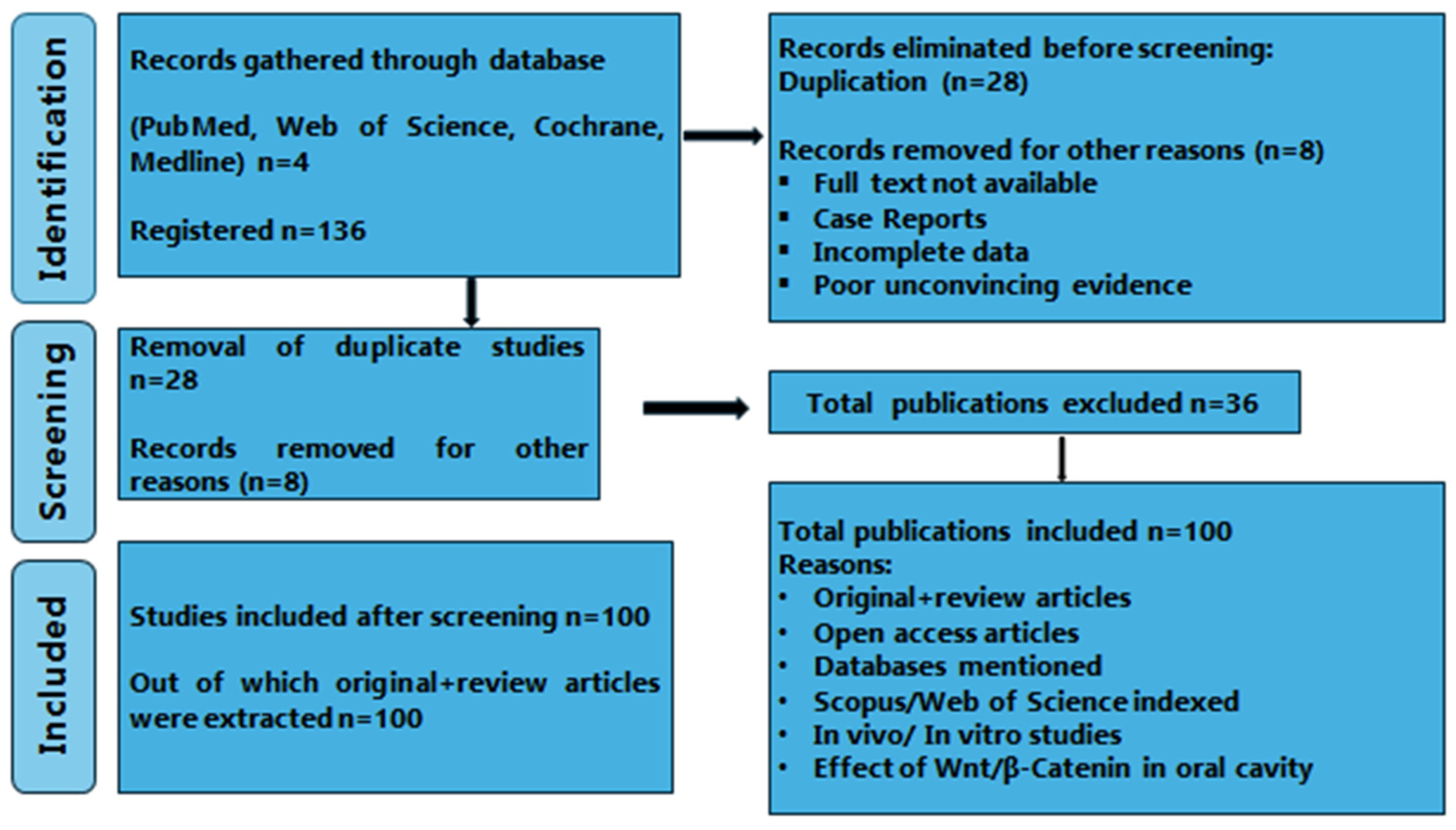
2. Materials and Methods

2.1. Source of Data

The literature in this review is based on the PRISMA-DTA guidelines, as intimated in Figure 4. Different sources such as Scopus (https://www.scopus.com/home.uri), Medline (https://www.nlm.nih.gov/medline/medline_home.html), Cochrane (https://www.cochranelibrary.com/), and Pubmed Central (https://www.ncbi.nlm.nih.gov/pmc/?db=PMC) were used for this literature review. The selection of articles is based on a combination of mesh terms and keywords, as shown in Figure 4, based on publications from the last two decades (i.e., 2002 to 2023).

2.2. Search Method

Different keywords such as Wnt signals, dental pulp cells, β-Catenin, etc., and databases were searched for relevant publications. The included articles covered topics such as the Wnt signaling pathway, Dental Pulp Stem Cells (DPSC), the significance of Wnt and stem cells in pulp regeneration, β-catenin in the oral environment, and recent advancements in dentin and pulp regeneration. The total number of articles included was 136; out of that, 28 were removed because of duplication, so the total number was reduced to 108. Furthermore, 8 publications were omitted because of the unavailability of the full text, case reports, and partial data, as indicated in Figure 4. After careful selection, the articles were reduced to 100 following deduction. This systematic review relies on original studies conducted in vivo and in vitro, as well as multiple review articles, as depicted in Figure 4. The search strategies for the specific databases were customized and executed by merging MeSH terms, keywords, and phrases to obtain potential studies containing predictive mathematical equations. For instance, a search strategy for PubMed could be (“Dental Pulp Diseases”[Mesh] OR “Dental Pulp Calcification”[Mesh]) AND (“Dental Pulp/abnormalities”[Mesh] OR “Dental Pulp/chemistry”[Mesh] OR “Dental Pulp/cytology”[Mesh] OR “Dental Pulp/drug effects”[Mesh] OR “Dental Pulp/growth and development”[Mesh] OR “Dental Pulp/immunology”[Mesh] OR “Dental Pulp/injuries”[Mesh] OR “Dental Pulp/pathology”[Mesh]) AND (“Dental Pulp Diseases/drug therapy”[Mesh] OR “Dental Pulp Diseases/epidemiology” OR “Dental Pulp Diseases/immunology”) AND (“Wnt Proteins”[Mesh] OR “beta Catenin”[Mesh]) OR (“Wnt Proteins/adverse effects”[Mesh] OR “Wnt Proteins/pharmacokinetics”[Mesh] OR “Wnt Proteins/therapeutic use”[Mesh]) AND (“β-Catenin/pharmacology”[Mesh]). The following MeSH terms, keywords, and phrases were used in Scopus and Web of Science search: (“Dental tissue”) AND (“Wnt signal” OR “β-catenin signal”) AND (“Dental Pulp Regeneration”) AND (“Regulatory signals” OR “Dentistry”) AND (“Bioactive materials”) AND (“Wnt Signals” OR “β-catenin signals”).
Inclusion Criteria
  • Related studies in Wnt/β-catenin signals
  • Human (in vitro) and animal (in vivo) studies.
  • Wnt/β-catenin in oral cavity-based publications.
  • Bioactive materials on dental pulp cells via Wnt signals
Exclude and elimination criteria
  • Unconvincing or poor evidence.
  • Case report studies
  • Incomplete text publications
  • Incomplete data
The meticulously examined articles unveiled the Wnt/β-Catenin signaling pathway in dentin and pulp regeneration. Specifically, they shed light on the impact of various proteins, molecules, and ligands on this pathway, whether it be enhancing or inhibiting its activity.

2.3. PICO Framework

Population, P: Population of interest is in vivo and in vitro studies targeting the Wnt/β-catenin signaling pathway in dentin and pulp repair.
Interest/Exposure, I: Interest is in observing the role of the Wnt/β-catenin signaling pathway in regenerative dentistry.
Comparator, C: There is no comparator.
Outcome, O: It is the association between the Wnt/β-catenin signaling pathway and various molecules, ligands, and proteins, its activation and upregulation promoting dentin repair and pulp regeneration.
The review was designed to answer this PICO question: How does the Wnt/β-Catenin pathway play a vital part in tooth development, maintenance, repair, and regeneration? P: Articles with in vitro and in vivo studies of dental regeneration were investigated; I: Intervention, Effect of Wnt/β-catenin in the oral cavity along with bio-(molecules)materials; C: The different results of bio-(molecules)materials in the Wnt/β-catenin signaling pathways to support the dental tissues were compared; O: Observation, in-vivo and in-vitro signaling experiments for the dental regeneration by controlling physiological activities such as growth, differentiation, and migration were observed.

3. Results

This systematic review includes mainly original articles, certain review articles, and in vivo and in vitro research publications that consist of a total of 100 articles, as shown in Figure 4. The evidence-based table in this systematic review is based on different articles in Table 1. Various researches emphasize the mechanism of the Wnt/β-catenin pathway and its significance in the growth and repair of dentin and pulp tissues, along with the impact of various factors on this pathway in the formation of reparative dentin and regeneration of pulp, as illustrated in Table 1. The articles published in the last two decades were selected in this review, as shown in Figure 4.
Canonical Wnt signals are specific spatiotemporally; their expression measured via Wnt10a and DKK1 is not constant but changes as the pulp regenerates. Wnt10a and DKK1 are inversely related, suggesting the intricate control mechanism of Wnt signals in the regenerated pulp. It may act as a biomarker for regenerated pulp [63]. The repair and regeneration of tissues is modulated by the Wnt/β-catenin pathway. Reparative dentin is secreted by odontoblast-like cells, which are differentiated from the Axin2-expressing cells. In tertiary dentinogenesis, Axin2 stimulates and modulates the Wnt/β-catenin pathway as a response to the injured tooth, providing a source of Wnt for regeneration [13]. Stathmin, in combination with Wnt5a in hDPSCs by the Wnt pathway, regulates the division and odontoblastic/osteogenic differentiation, thus playing a role in tooth and pulp regeneration [64].
In addition to its function in tooth development, this pathway promotes the growth of reparative dentin. In this study, GSK3β antagonists support the normal mechanisms of reparative dentin production in rat models to entirely regenerate dentin. Wnt signaling-related medicines are now perfectly suited for clinical therapy in dentistry because of this innovative technique [19]. Wnt3a promotes tertiary dentin formation by enhancing OSX, BMP2, and DMP1 expression while reducing SHEDS colony formation resulting in increased mineralization. In Wnt3a-treated defects, a noticeable increase in dentin/bone volume is seen as dentin bridge formation; thus, it can be utilized in vital pulp therapy [60]. Small molecule inhibitors of GSKβ, such as c-MET inhibitors, activate the Wnt pathway, resulting in the repair of dentin and pulp regeneration in the exposed tooth [37].
In this research, the activation of Wnt signaling was achieved through the use of Wnt3a in the damaged tooth pulp. This led to the differentiation of pulp cells into secretory odontoblasts, which produced more tertiary dentin and ultimately improved the vitality of the pulp. This approach shows promise in the treatment of dental caries [28]. The migration of HDPCs and their ability to differentiate into odontoblasts were found to be enhanced by Wnt7b. This was achieved by stimulating the Wnt/β-catenin pathway and upregulating the JNK pathway. Therefore, Wnt7b could potentially be used as a therapeutic agent to improve the treatment of injured tooth pulp [42]. Furthermore, the expression of Wnt10a by odontoblasts was found to increase as the pulp regenerated, leading to dentin differentiation. This indicates that Wnt10a plays a crucial role in the regeneration of both dentin and pulp [62]. The positive regulation of DPSC proliferation and odontoblast differentiation through the Wnt10a pathway contributes to the repair process and is mediated by the Wnt/β-catenin pathway [2]. LiCl, by inhibition of GSKβ, activates the Wnt/β-catenin signaling pathway. LiCl augments the expression of Axin2 and Wnt responsive cells, resulting in the differentiation of odontoblasts and increases the reparative formation of dentin when applied to dental pulp [55]. In TCF/Lef: H2BGFP reporter mice under the injury site, the odontoblasts and pulp cells responded to Wnt signaling, and the addition of tideglusib caused an increase in the Wnt signaling at the damaged pulp horn site. The findings demonstrated that injured dentin can be penetrated by small-molecule medications such as tideglusib, which then have an impact on odontoblasts and pulp cells, resulting in dentin and pulp regeneration [65].
Berberine in DPSCs promotes odontogenic differentiation by augmenting the nuclear β-catenin in the Wnt/β-catenin pathway; thus, it can be used to guide the treatment of dental caries [45]. Baicalin, a bioactive flavone acts as an anti-inflammatory, antioxidant, anti-tumor, and anti-viral agent. It inhibits NF-Kb and Wnt/β-catenin pathways, thus causing an increase in odonto/osteogenic differentiation of iDPSCs, resulting in pulp regeneration [61]. () Neuropilin 1, a multifunctional protein by stimulation of the Wnt pathway in DPSCs, plays a vital role in odontoblast differentiation and its mineralization, resulting in increased wound healing, tooth formation, and repair [66]. In hDPSCs, the R-spondin2, by activation and regulation of the Wnt/β-catenin pathway, encourages the proliferation together with odontogenic differentiation. The odontogenic activity of hDPSCs is impaired by Rspo2 knockout. There is a synergistic effect of Wnt3a with Rspo2 on Wnt/β-catenin signaling, thus providing an approach for reparative dentin formation [46].
Treated dentin matrix (TDM) in hDPSCs influences odontogenic differentiation by canonical Wnt/β-catenin pathway stimulation via GSKβ, resulting in dentin regeneration. TDM may be used as a base for regenerative tooth engineering [56]. Dentin secretion is modulated by the Wnt signaling pathway. Augmentation of Wnt signaling in pre-odontoblasts results in increased reparative dentin/osteodentin formation, thus contributing to vital pulp therapy by the formation of biologically based pulp capping material [44]. When MTA, calcium hydroxide, and Biodentine TM are applied directly to pulp, reparative dentin formation occurs at the exposure sites, thus promoting dentin bridge formation. Biodentine TM, when used in direct pulp capping, activates the Wnt/β-catenin pathway, resulting in increased reparative dentin formation [48].
Angelica sinensis polysaccharide (ASP) in human dental pulp stem cells (hDPSCs) through the Wnt/β-catenin pathway enhances cell proliferation and osteogenic differentiation, leading to increased dentin formation and pulp regeneration. Potential treatments for regenerative pulp diseases may involve ASP [67]. Treatment of DPSCs with lithium and Wnt1, followed by exposure to TEGDMA, triggers Wnt signaling, ultimately boosting pulp regeneration post-injury [41]. Wnt3a, Wnt10a, and β-catenin play a role in promoting reparative dentin formation from dental pulp cells and odontoblasts, facilitating the formation of dentin bridge [52]. Elevated blood glucose levels can induce cellular senescence and reduce pulp cell proliferation by upregulating β-catenin and Wnt1 expression [57]. Activation of β-catenin by Runx2 encourages dental pulp cells (DPCs) to differentiate into odontoblasts and deposit reparative dentin [40]. Exposure of pulp cells to Wnt3a leads to an increase in pre-odontoblasts and odontoblasts through the stimulation of the Wnt/β-catenin pathway, which is potentially useful in vital pulp therapy and dentin regeneration [68].

4. Discussion

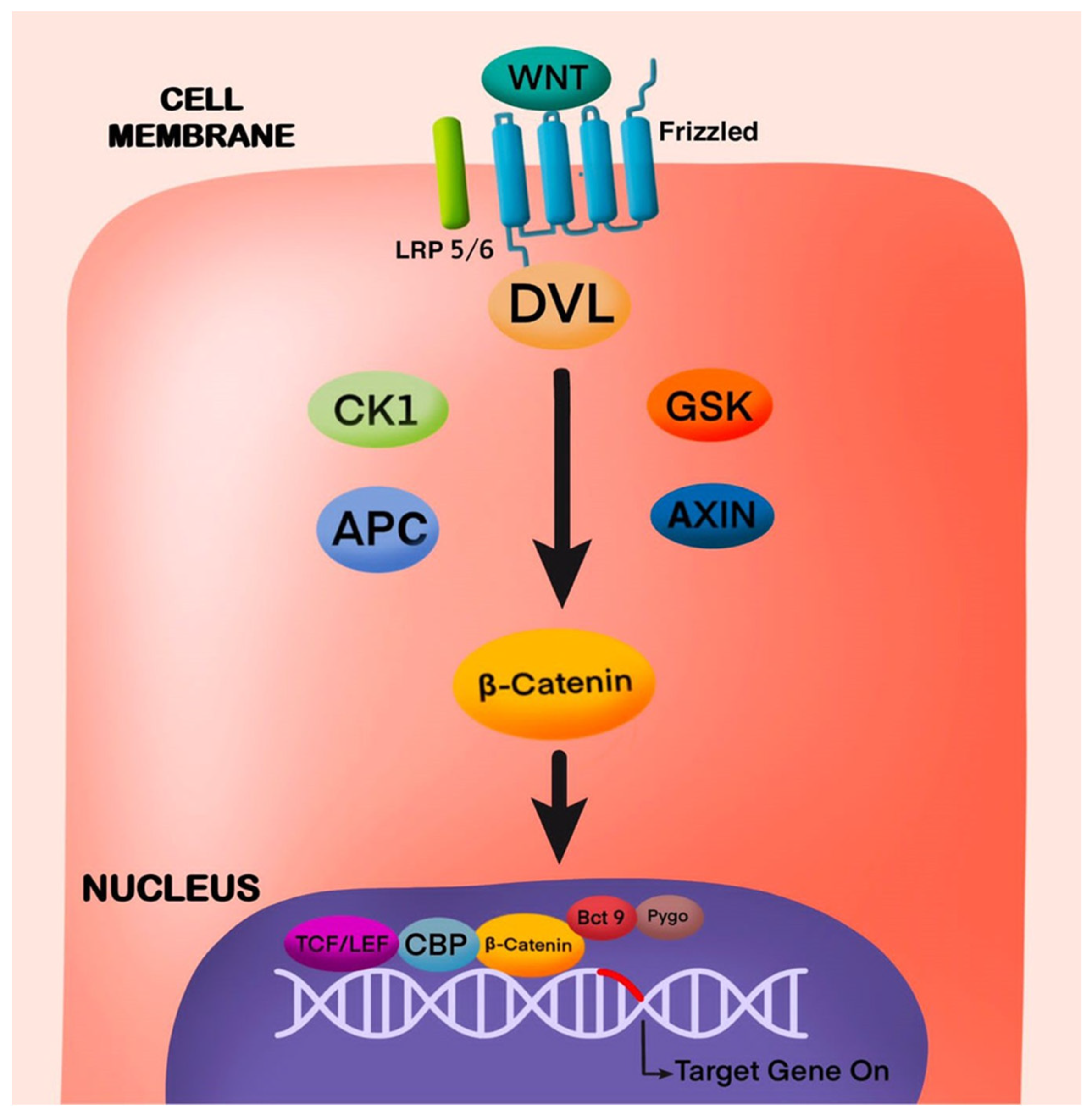
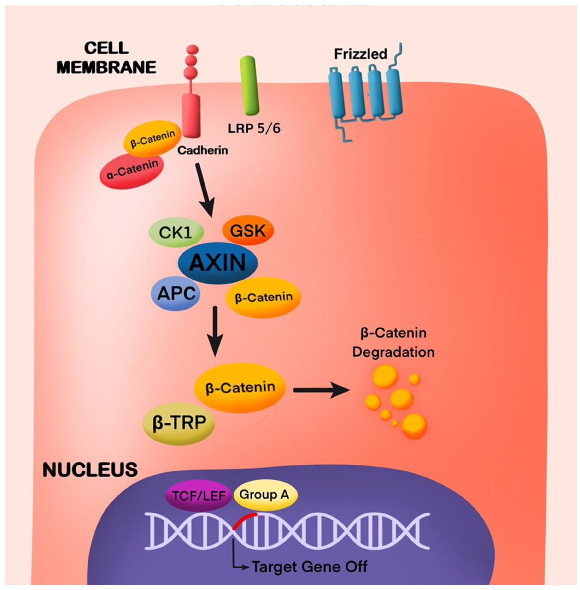
4.1. Wnt/β-Catenin Pathway in Odontogenesis

In tooth and root odontogenesis, the role of the Wnt/β-catenin pathway is essential; thus, several studies have been conducted [19,40,46,53,56,59]. In one study, the β-catenin in odontoblast and cementoblast was removed [40]; this severely disrupted the molar root shape and formation and root analog of incisors. First, in vivo evidence was given in odontoblasts; this pathway is important in tooth root development, the differentiation and proliferation of cells, and the formation of root dentin. In OC-Cre; Ctnnb1fl/f mice, β-catenin withdrawal from pre-odontoblasts resulted in the silencing of differentiation of odontoblasts and reduced proliferation cellularly, resulting in the loss of tooth root due to arrested root dentin synthesis (Figure 5). In crown odontoblasts, which reach maturation in the embryo, the silencing of β-catenin had no visual impact on the maintenance and maturation. These data indicate the significance of Wnt/β-catenin signaling in root development by modulating the mesenchymal differentiation into odontoblasts, as shown in Table 1 [39].
As shown in Table 1, the Wnt/β-catenin signaling pathway has an important role in root formation, as demonstrated by the conditional β-catenin inactivation in immature odontoblasts [28,40,44,62]. This alteration results in the development of molars without roots [35]. It is imperative to exert precise control over Wnt signaling during root formation. In this study, areas were identified exhibiting Wnt/β-catenin signaling activity during the initial postnatal stages in the underlying mesenchyme and in the enamel knot. This may be due to the Wnt ligands and β-catenin expression postnatally in the coronal epithelium [69]. Notably, postnatally, when epithelial β-catenin was removed, ectopic incisors characterized by their short and malformed enamel were seen in the experimental mice. During the transition stage of root formation, heightened Wnt/β-catenin signaling in periodontal ligament cells, odontoblast-lineage cells, and HERS cells was perceived [70]. The Wnt/β-catenin signals were also detected in PN7 from the apical papilla and dental sac [54].
Odontoblast function is regulated essentially by the Wnt/β-catenin pathway [28,71]. In pulp, as age progresses, there is a decrease in Wnt signal and Wnt responsive cells because of a decrease in dentin secretion. Primary odontoblasts and their secretion of copious amounts of dentin have a huge role in maintaining the vitality of the pulp. However, the repair response reduces with age and results in chronic inflammation, leading to pulpal necrosis. Many endodontic cases are due to chronic trauma to the pulp [44]. A strategy was proposed to enhance the Wnt signal by delivery of a Wnt protein liposomal formulation to stimulate the repair potential of adult pulp [72] and dentin deposition (Table 1) [44].

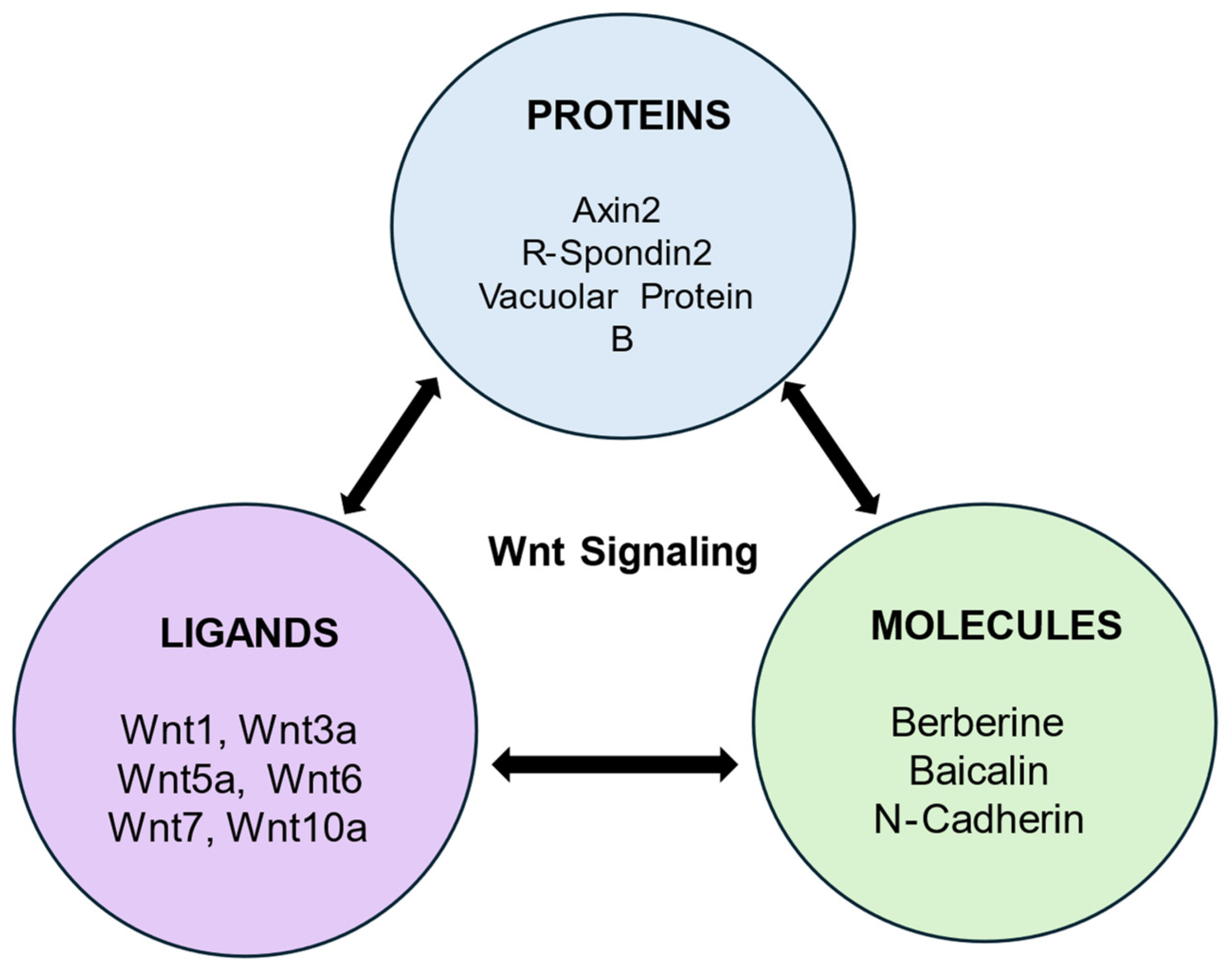
4.2. Wnt Ligands and Their Role in Dentin and Pulp Regeneration

4.2.1. Wnt1

As shown in Table 1, the Wnt/β-catenin pathway modulates DPSC differentiation via miR-140-5p. Moreover, the Wnt1 gene in miR-140-5p was validated in this study. An increase in β-catenin levels in the inhibitor group and mimic group, a decrease was observed, thus showing the involvement of miR-140-5p in activating β-catenin. This study confirmed that miR-140-5p modulated the DPSC odontoblastic differentiation via the Wnt1/β-catenin signaling pathway. In DPSCs, the miR-140-5p expression decreases during the differentiation of odontoblasts as shown in (Figure 6A). This study indicates odontoblastic differentiation is supported by miR-140-5p inhibitors in DPSCs in vitro [47].

4.2.2. Wnt3a

According to this study, when exposed pulp was treated with Wnt3a, there was an increase in the formation of tertiary dentin in comparison with the deionized water (DI) and phosphate-buffered saline (PBS) treated control groups in a rat model. This result confirms that Wnt3a can be used as a bioactive capping material for pulp to enhance reparative dentine formation. Lithium chloride-treated teeth also demonstrated dentine formation and a higher dentine/bone proportion than the total tissue proportion in comparison with the control group; however, no statistical difference was observed as shown in (Figure 6B) [55]. BMP2 was identified as an inducing factor responsible for mRNA expression of odonto/osteogenic differentiation of SHEDS via the canonical Wnt signaling pathway. A significant increase in BMP2 and mRNA levels was observed when treated with Wnt3a, and in osteogenic medium, calcium deposition of SHEDs was also observed. Thus, Wnt3a can be used as a pulp capping agent as it enables reparative dentine formation [60].

4.2.3. Wnt3a and Wnt10a in Dentin Bridge Formation

As shown in Table 1, Wnt3a and Wnt10a are the ligands of Wnt, which, together with β-catenin, stimulate the canonical Wnt signaling [33,60,62]. Near the pulp capping area, on the 4th day of direct pulp capping, Wnt3a and Wnt10a were perceived. On the 4th, 7th, and 14th days, β-catenin was expressed strongly in the odontoblast cell nucleus. On the 28th day in the nucleus, a low positive signal was observed. The results of this study state that by autocrine process, Wnt3a and Wnt10a initiate the differentiation of reparative odontoblasts to produce dentin matrix. This supports the findings that odontoblasts and DPCs during dentin bridge formation secrete Wnt3a and Wnt10a [52].

4.2.4. Wnt10a

This study shows the role of Wnt10a in dentin repair, where its overexpression in DPCs promotes cell proliferation and prevents odontoblastic differentiation. Immunostaining and reverse transcription PCR analysis showed the presence of Wnt10a in the cytoplasm of the odontoblast. Its activity, along with other Wnt molecules, was seen in cultured DPCs [33]. Wnt10a and LEF1 were expressed before DSPP at the early stage of dentin formation [31,73]. The expression of Wnt10a enhances the differentiation and self-repair ability of stem cells at the early stage of dentin repair, as shown in Table 1 [33].

4.2.5. Wnt10a, a Non-Cellular Agent for Dentin Pulp Regeneration

As shown in Table 1, an agent that can induce dentin formation by regulation of odontoblasts for dental pulp regeneration was developed. In this study, the hDPSCs were induced to differentiate into dentin. The expression of DSPP, Wnt10a, and DKK1 on days 3 and 7 of induction were analyzed. On day 7, there was an increase in Wnt10a and DSPP, whereas DKK1, an antagonist of Wnt10a, decreased. On day 3 of transplantation, blood vessels, along with other fibrous tissues, regenerated in the root canal on day 3 after transplantation. There was no change in pulp content at days 3 and 7 post-transplantation, but odontoblasts increased, suggesting the maturation of dental pulp several days after transplantation. A consequential increment of Wnt10a-positive cells was seen along with DSPP. The increase in DSPP-positive cells from day 3 to 7 states the involvement of Wnt10a in the differentiation of odontoblasts rather than directly in pulp regeneration. In conclusion, the repair is augmented in pulp with an increase in Wnt10a, which acts as a non-cellular inducing agent (Table 1) [62].

4.2.6. Wnt7b

This study shows the role of Wnt7b in the treatment of pulp injury by stimulating the HDPCs to proliferate and migrate via the Wnt/β-catenin and JNK signaling pathways [42]. In this study, an inhibitor of the JNK pathway SP600125 is used to counteract the upregulation of HDPCs induced by Wnt7b, which shows the role of the JNK pathway. According to unpublished data, the levels of mineralization in 17IIA11 odontoblast-like cells (A11) increased by Wnt7b than in HDPCs. A11 differentiates into odontoblast-like cells, which secrete dentin matrix and are easily found in the late stage of odontogenic differentiation. This shows that Wnt7b is more efficient at this stage, which results in an increase in DSPP, which is a late odontogenic marker. As Wnt7b stimulates the proliferation and migration of HDPCs, it has an important role in the treatment of tooth injuries, such as dental trauma and dental caries, as well as in the reservation of vital pulp [42].

4.2.7. Axin2

As illustrated in Table 1 [13], the utilization of Axin2LacZ and TCF/Lef: H2B-GFP reporter mice, coupled with real-time qPCR analysis for the Axin2 expression in response to injury, disclosed a swift upregulation of Wnt/β-catenin signaling. The results from genetic tracing unveiled that cells expressing Axin2 undergo a proliferative expansion after damage, with some of these cells differentiating into odontoblast-like cells. Axin2 is expressed by post-mitotic primary odontoblasts, and an increase in Wnt/β-catenin signaling in Axin2LacZ/LacZ mice enhances reparative dentine production. The specific inhibition of Axin2-expressing cell’s ability to release Wnt had a profound impact on the formation of reparative dentin, emphasizing the crucial role of Axin2-expressing cells as the internal source of Wnt. This suggests that reparative dentin formation is orchestrated by autocrine Wnt/β-catenin signaling within Axin2-expressing cells themselves as shown in (Figure 7A). The identification of Axin2 expression in odontoblast-like cells further supports the notion that these cells serve as the origin of their proliferative signals in the context of reparative dentinogenesis, as depicted in Table 1 [13].

4.2.8. R-Spondin 2

As shown in Table 1, this study demonstrates the molecular mechanism of odontoblastic differentiation mediated by Rspo2 by the activation of the Wnt signaling pathway. Rspo2, in combination with Wnt3a, augmented this pathway by expression of β-catenin. This study concludes that Rspo2 operates synergistically with Wnt3a in the activation of this pathway for odontogenic differentiation and the formation of reparative dentin. The levels of Wnt and Rspo2-related receptors of shRspo2 and shNC cells were compared in this study. Rspo2 binds to the LGR4 receptor for the regulation of the Wnt/β-catenin pathway. The decreased expression of LRP5 and LRP6 in shRspo2 activated through different Wnt ligands results in the decline in the Wnt/β catenin pathway activation [46].

4.3. Bioactive Molecules and Their Role in Dentin and Pulp Regeneration

4.3.1. Baicalin

In this study, inflammatory dental pulp stem cells were isolated from inflamed pulp. Both inflammatory (iDPSCs) and normal (nDPSCs) stained positively for vimentin. In iDPSCs, TNF-α and IL-1β increased, while in baicalin-treated iDPSCs they decreased (Figure 7B). A concentration of 20 µM of baicalin facilitated iDPSCs to differentiate osteo/odontogenically with no change in proliferation. In DPSCs, the proliferation increased, and the differentiation decreased with inflammation. Therefore, a specific concentration of baicalin has an anti-inflammatory action in the pulp. In iDPSCs, β-catenin increases while CaMKII and NLK decrease. In the baicalin-treated group, there was a decrease in proteins augmented by DKK1. In baicalin-treated iDPSCs, the odonto/osteogenic differentiation was enhanced by inhibition of the Wnt/β-catenin pathway; thus, it may prove to be effective in repair in early pulpitis as shown in Table 1 [61].

4.3.2. Berberine

Berberine is an important molecule in medicinal chemistry and pharmacology; it is a non-basic, quaternary benzylisoquinoline alkaloid [74]. The results showed that berberine increased the odontoblast differentiation marker proteins through the classical Wnt/β-Catenin signaling pathway and accelerated DPSC to differentiate odontoblastically. DPSCs were cultured from caries-free teeth and subjected to odontoblast differentiation. On day 14, the odontoblast differentiation of DPSCs was stained with alizarin red S and ALP, revealing that DPSCs were treated with berberine with DKK-1 during the process of odontoblast differentiation. The nuclear β-catenin suppressed the DKK-1 induced by berberine, while berberine caused the nuclear β-catenin to increase. Additionally, Runx2 and DMP-1, which are downstream proteins of Wnt/β-catenin signaling, were activated at higher levels by berberine. In summary, Table 1 illustrates that berberine may be a novel medication for the treatment of dental defects [45].

4.3.3. S-PRG Fillers as Dental Pulp Capping Agents

Lithium ions were added to S-PRG fillers to increase their effectiveness as dental capping agents, as Table 1 illustrates. S-PRG fillers were paired with a low concentration of LiCl in the pulp capping studies, and complete dentin production resulted. When compared with the other groups, it was discovered that from S-PRG/Li-100 mM samples, eluates could considerably augment cell migration, differentiation, and mineralization. In this investigation, at 7 and 14 days after the teeth were sealed with S-PRG cement without lithium in undamaged teeth, Axin2 was significantly expressed in the S-PRG/Li-100 mM pulp-capped groups. The result of this study demonstrates that β-catenin expression was considerably elevated in the initial phases and precipitously decreased as Axin2 continued to express itself [43].

4.3.4. Vacuolar Protein 4B and Its Effect on the Wnt/β-Catenin Pathway

This study demonstrated the total cellular level of β-catenin was reduced by the decrease in vacuolar protein 4B (VPS4B). The nuclear translocation of β-catenin was negatively affected, while there was no change in the cytoplasmic concentration. LiCl, a GSK3β inhibitor by silencing VPS4B, partly resulted in odontogenic differentiation and proliferation, thereby causing the cellular β-catenin levels to rise. The Wnt/β-catenin signaling pathway promotes osteogenic proliferation and hDPSC differentiation. VPS4B silencing results in the retardation of hDPSC migration. Further research should be carried out to investigate the relationship between VPS4B and β-catenin signaling that further regulates the proliferation and differentiation of hDPSCs, as shown in Table 1 [49].

4.3.5. N-Cadherin

As indicated in Table 1, an intentional reduction in N-cadherin in DPSCs resulted in a significant upregulation in β-catenin signaling, leading to increased odontogenic differentiation in vitro. This effect was nullified when cells were pre-treated with XAV939, a β-catenin inhibitor. Moreover, the silencing of N-cadherin facilitated the development of odontoblast-like cells, and a collagenous matrix was formed in β-TCP/DPSC composites transplanted into mice. Notably, the investigation revealed that the silencing of N-cadherin promoted the accumulation and translocation of β-catenin in the nucleus. Conversely, the inhibition of β-catenin by XAV939 hindered the odontogenic differentiation process in DPSCs. The proposed mechanism suggests that N-cadherin silencing induces the translocation and accumulation of β-catenin in the nucleus. Subsequently, this promotes the binding of β-catenin to the Runx2 promoter, facilitating Runx2 and DSPP expression. Consequently, this sequence of events stimulates DPSCs to differentiate odontogenically. A downregulation of Runx2 is required to achieve complete odontoblast differentiation in the context of dentinogenesis [53].

4.4. Lithium Chloride and Wnt/β-Catenin Pathway

As depicted in Table 1, a primary pulp culture derived from various reporter mice was utilized; this investigation delved into the mechanisms governing the effect of LiCl on the regulation of the differentiation of odontoblasts and osteoblasts. This study further demonstrated that the alterations induced by LiCl in the expression of DSPP, DMP1, and BSP in these early progenitors were effectively reversed by DKK1 [55]. These findings strongly suggest the positive influences of LiCl on the differentiation of osteo/odontoblasts from αSMA+ progenitors via the Wnt/β-catenin signaling pathway [68,75]. This study indicated that prolonged and late exposure of dental pulp cells to LiCl resulted in an elevated expression of odontoblast markers facilitated by Wnt/β-catenin signaling. This exposure further increased the number of odontoblasts expressing DMP1-Cherry and DSPP-Cerulean transgenes. It is noteworthy that the inhibitory effects observed with prolonged exposure to LiCl differed from the stimulatory effects elicited by Wnt3a on pulp cells [55].
In another study, cells from tooth apical papillae (SCAP) with specific markers CD24 STRO-1 and CD146 were taken. The viability of SCAP increases when exposed to lower concentrations of LiCl (5 and 10 mM). Higher concentrations of LiCl (20 and 30 mM) inhibited cell viability by causing G2/M cell cycle arrest. After induction of mineralization differentiation (days 4–14), an increase is seen in DSPP, ALP, and OCN, i.e., the mineralized matrix had dentine matrix characteristics. A decrease in Wnt signaling and block of odonto/osteogenic differentiation is seen with DKK1 addition, thereby preventing any Wnt1-mediated odontogenic events. Data support the significant promotion of SCAP differentiation and proliferation by Wnt/β-catenin, as shown in Table 1 [38].

4.5. Treated Dentin Matrix and Its Effect on hDPSCs via Wnt/β Catenin Pathway

The TDM (treated dentin matrix) extract is recognized for its richness in growth factors, including TGFβ, DMP-1, and DSPP, which possess the capability to induce the differentiation of stem cells odontogenically. In the TDM-induced odontogenic induction in human dental pulp stem cells (hDPSCs), various concentrations of TDM (10%, 20%, 40%, 60%, 80%, and 100%) were tested. The outcomes revealed a substantial increase in the odontogenic protein levels and mRNA in GSK3β-RNAi combined hDPSCs following TDM extract induction, as compared with the control group. Conversely, overexpressed GSK3β combined with hDPSCs, after TDM extract induction, resulted in reduced expression levels of RUNX2, ALP, DSPP, and DMP-1. These findings underscore the pivotal role of the Wnt/β-catenin signaling pathway in GSK3β-mediated odontogenic differentiation of hDPSCs [56]. The activation of the canonical Wnt/β-catenin pathway hinges on the stabilization and protection of β-catenin, achieved by inhibiting GSK3β [76]. This process facilitates the accumulation of β-catenin in the nucleus, where it collaborates with lymphoid enhancement factors and T cell factor (LEF/TCF) to exert its effects [77]. Additionally, a decrease in GSK3β expression during the odontogenesis of hDPSCs induced by TDM was observed, further emphasizing the involvement of the Wnt/β-catenin signaling pathway in this GSK3β-mediated odontogenic process as shown in Table 1 [56].

4.6. Role of Wnt Signalling in Progression of Diabetic-Induced Cellular Aging

As shown in Table 1, the effect of high glucose was investigated in the pulp in vitro model. To induce a diabetic state, solutions of concentrations 20 mM and 30 mM were prepared. The control was taken as 5 mM in a cell culture medium. After 2–3 weeks, the results showed that the glucose-enriched culture medium had an inhibitory effect on the proliferation rate. Compared with the control, there is an increase in beta galactosidase-positive cells in 20 mM and 30 mM groups. CDK inhibitor p21 expression increases, which is a cellular senescence marker. This study confirmed that chronic exposure to hyperglycemic environments induced senescence and cell cycle arrest [57]. In this study, PNU74564 was applied, which inhibited Wnt signaling [78]. The results showed that β-catenin inhibition resulted in a decrease in senescent cells; however, LiCl increased senescence due to glucose. Therefore, Wnt signaling is a potential goal for the inhibition of senescence in hyperglycemic conditions, which suggests that bioactive materials should be developed specifically for pulp capping in diabetic patients [57].

4.7. Role of Wnt Pathway in Dentin and Pulp Repair

As shown in Table 1, repair in adult pulp may be stimulated by Wnt signaling. This is dependent on odontoblasts maintaining Wnt signaling in adulthood. Two approaches were used to prove adulthood Wnt responsive status in odontoblasts and pulp cells. First, analysis of frozen tissue section from adult Axin2LacZ/+ Wnt reporter mice was performed in which X-galþve. pulp cells and polarized odontoblasts were found. Second, Axin2CreERT2/+; R26RmTmG/+ Wnt reporter mice showed GFPþve polarized odontoblasts. In L-PBS-treated samples, porous osteodentin is formed. In L-Wnt3a cases, DSPþve secretory odontoblasts formed reparative dentin, thus protecting the pulp. Based on the pulp cell’s reliance on Wnt signaling, a new technique for root canal therapy was evaluated. The liposomal Wnt3a protein improved the healing of pulp, thus giving an approach to achieving pulp regeneration in humans [28].
Materials such as calcium hydroxide and MTA are used to treat pulp exposure because of the treatment of caries. The evaluation revealed that tertiary dentin was similar to natural dentin. To induce reparative dentinogenesis, a controlled therapeutic amount of material was applied locally to prevent the negative effect of these small molecular Wnt agonists. GSK3β, an inhibitor, gives rise to reparative dentine at defect sizes translatable to minor human lesions, but it is not clear how to fully “regenerate” the lost tissue [50]. Reparative dentine is produced to heal the wound after contact with exposed pulp and pulp capping material. Pulp capping materials such as MTA and Biodentine TM have shown high clinical success rates compared with calcium hydroxide. Biodentine TM treatment showed better results when compared with MTA. On histological analysis, there was complete bridge formation with no inflammation after Biodentine TM capping. The study results also suggest the stimulation of the Wnt/β-catenin pathway by Biodentine TM. In pulp inflammation, SDF1 causes hDPSC migration. These data suggested all capping materials, calcium hydroxide, MTA, and Biodentine TM stimulate cell cycle progression [79,80,81]. Wnt signaling plays a role in Biodentine TM-induced reparative dentine formation as β-catenin was expressed in the Biodentine TM-treated group, as shown in Table 1 [48].
In another study, the endogenous canonical Wnt signaling pathway was activated by alternate mechanisms in DPSCs. For indirect Wnt activation, either lithium (LiCl) is used [82], and for direct, human recombinant (hr) Wnt-1 is used [83]. LiCl concentration causes a stimulatory and inhibitory effect on cells, indicating that increased GSK3β inhibition may be toxic to cells [84]. DPSCs, when exposed to monomer released by TEGDMA, activated the Wnt/β catenin signaling. During the repair of pulp, stem cells have a vital role in tissue engineering as well as the development of new bioactive materials that can cause repair via Wnt signaling [41].
Wnt signaling plays a crucial role in controlling the epithelial–mesenchymal transition (EMT) process through various mechanisms. Notably, the inhibition of GSK3β via multiple pathways, including the Wnt cascade, leads to the stabilization of β-catenin and Snail. This stabilization, in turn, triggers cell migration, proliferation, and the development of cancerous growth. Consequently, GSK3β emerges as a pivotal regulator of epithelial organization and function, presenting itself as a potential molecular target for therapeutic interventions.

5. Conclusions

The Wnt/β-catenin signaling pathway, encompassing both canonical and non-canonical components, is crucial for the growth, maintenance, restoration, and rejuvenation of teeth. It facilitates the development of teeth, cementum, dentin production, and the formation of periodontal tissues. Abnormalities in tooth development arise from either excessive or insufficient activity of the Wnt signaling pathway. The activation of the Wnt/β-catenin signaling pathway leads to the maturation of odontoblasts that produce tertiary dentin, aiding in the repair of damaged dental pulp and enhancing tooth vitality. Calcium hydroxide, MTA, and Biodentine TM promote pulp healing by inducing reparative dentin formation. Biodentin TM showed β-catenin expression, thus suggesting Wnt signaling pathways part in the repair of dentin and pulp. Pulp repair and regeneration are further improved by the introduction of various Wnt ligands and molecules, which may enhance the effect of the Wnt/β-catenin signaling pathway. Increased understanding of the Wnt/β-catenin signaling pathway in dentin and pulp repair and regeneration has led to the discovery that it plays a role in promoting pulp vitality, treating dental caries, and providing an alternative approach for root canal treatment. The major limitation is the inadequate comprehension of numerous natural bioactive substances when it comes to effectively regulating Wnt signals in dental applications. Further research on the Wnt/β-catenin signaling pathway may provide a path for therapeutic and regenerative dentistry.

Author Contributions

Conceptualization, S.U.R. and J.E.; methodology, M.A., L.J. and M.B.; software, S.H.F.; validation, M.K., S.I. and F.N.; data curation, S.F.; writing—original draft preparation, M.A. and L.J.; writing—review and editing, J.E.M.S.d.V. and W.W.; visualization, J.E.; supervision, S.U.R.; project administration, S.U.R.; funding acquisition, J.E. All authors have read and agreed to the published version of the manuscript.

Funding

This work is financially supported by Internal funding from UCAM- Universidad Católica San Antonio de Murcia (Grant No. PMAFI-27/21), Murcia, Spain, and Shanghai Frontiers Research Center of the Hadal Biosphere, the National Natural Science Foundation of China (No. 82173731), and the Scientific Research Project of Shanghai International Science and Technology Cooperation Fund Project (No. 17490742500).

Conflicts of Interest

The authors declare no conflicts of interest.

Abbreviations

Wnt—Wingless-related integration site; DPSC—Dental pulp stem cells; iDPSC—Inflammatory Dental pulp stem cells; DKK-1—Dickkopf-1; TGFβ1—transforming growth factor beta-1; SCAP—Stem Cells From the Apical Papilla; S-PRG—surface pre-reacted glass ionomer; VPS4B—Vacuolar Protein Sorting 4 Homolog B; MMP13—Matrix Metallopeptidase 13; SHED—stem cells from human exfoliated deciduous teeth; OSX-Osterix; cMET—Mesenchymal Epithelial Transition Factor; ASP-Angelica Sinensis Polysaccharide; Lef1—Lymphoid Enhancer Binding Factor; CaMK2—Calcium Calmodulin Dependent Protein Kinase-1; NLK-Nemo like Kinase; OCN-Osteocalcin; TNF-α—Tumor Necrosis Factor; BMP1—Bone Morphogenetic Protein-1; FRZ—Frizzled Receptors; nDPSC—Normal Dental Pulp Stem cells; HDPC-Human Dental Pulp Cells; GSK3β—Glycogen synthetase kinase 3 beta; JNK—c-Jun N-terminal Kinase; Rspo-2—R-Spondin-2; TDM—Treated Dentin Matrix; SNHG1—Small Nucleolar RNA Hose Gene 1; NF-fB pathway—Nuclear Factor pathway; DMP1—Dentin Matric Phosphoprotein 1; MTA—Mineral Trioxide Aggregate; TEGDMA—Triethylene Glycol Dimethacrylate; DSPP-Dentin Sialophosphoprotein; ALP-Alkaline Phosphatase; BSP-Bone Sialoprotein; CDK—Cyclin Dependent Kinase; IL-1 β—Interleukin 1 beta.

References

  1. Angelova Volponi, A.; Zaugg, L.K.; Neves, V.; Liu, Y.; Sharpe, P.T. Tooth Repair and Regeneration. Curr. Oral Health Rep. 2018, 5, 295–303. [Google Scholar] [CrossRef] [PubMed]
  2. Li, Y.; Wu, M.; Xing, X.; Li, X.; Shi, C. Effect of Wnt/β-Catenin Signaling Pathway on Repair and Regeneration after Dentin-Pulp Injury. Res. Sq. 2023. preprint. [Google Scholar] [CrossRef]
  3. Prakash, S.; Swaminathan, U. β-Catenin in Health: A Review. J. Oral Maxillofac. Pathol. 2015, 19, 230. [Google Scholar] [CrossRef] [PubMed]
  4. Giraud, T.; Jeanneau, C.; Rombouts, C.; Bakhtiar, H.; Laurent, P.; About, I. Pulp Capping Materials Modulate the Balance between Inflammation and Regeneration. Dent. Mater. 2019, 35, 24–35. [Google Scholar] [CrossRef] [PubMed]
  5. Gronthos, S.; Mankani, M.; Brahim, J.; Robey, P.G.; Shi, S. Postnatal Human Dental Pulp Stem Cells (DPSCs) In Vitro and In Vivo. Proc. Natl. Acad. Sci. USA 2000, 97, 13625–13630. [Google Scholar] [CrossRef]
  6. Gronthos, S.; Brahim, J.; Li, W.; Fisher, L.W.; Cherman, N.; Boyde, A.; DenBesten, P.; Robey, P.G.; Shi, S. Stem Cell Properties of Human Dental Pulp Stem Cells. J. Dent. Res. 2002, 81, 531–535. [Google Scholar] [CrossRef] [PubMed]
  7. Xia, K.; Chen, Z.; Chen, J.; Xu, H.; Xu, Y.; Yang, T.; Zhang, Q. RGD-and VEGF-Mimetic Peptide Epitope-Functionalized Self-Assembling Peptide Hydrogels Promote Dentin-Pulp Complex Regeneration. Int. J. Nanomed. 2020, 15, 6631–6647. [Google Scholar] [CrossRef]
  8. Inostroza, C.; Vega-Letter, A.M.; Brizuela, C.; Castrillón, L.; Saint Jean, N.; Duran, C.M.; Carrión, F. Mesenchymal Stem Cells Derived from Human Inflamed Dental Pulp Exhibit Impaired Immunomodulatory Capacity In Vitro. J. Endod. 2020, 46, 1091–1098.e2. [Google Scholar] [CrossRef]
  9. About, I. Dentin–Pulp Regeneration: The Primordial Role of the Microenvironment and Its Modification by Traumatic Injuries and Bioactive Materials. Endod. Top. 2013, 28, 61–89. [Google Scholar] [CrossRef]
  10. Tsutsui, T. Dental Pulp Stem Cells: Advances to Applications. Stem Cells Cloning Adv. Appl. 2020, 13, 33–42. [Google Scholar] [CrossRef]
  11. Clevers, H.; Loh, K.M.; Nusse, R. An Integral Program for Tissue Renewal and Regeneration: Wnt Signaling and Stem Cell Control. Science 2014, 346, 1248012. [Google Scholar] [CrossRef] [PubMed]
  12. Whyte, J.L.; Smith, A.A.; Helms, J.A. Wnt Signaling and Injury Repair. Cold Spring Harb. Perspect. Biol. 2012, 4, a008078. [Google Scholar] [CrossRef] [PubMed]
  13. Babb, R.; Chandrasekaran, D.; Carvalho Moreno Neves, V.; Sharpe, P.T. Axin2-Expressing Cells Differentiate into Reparative Odontoblasts via Autocrine Wnt/β-Catenin Signaling in Response to Tooth Damage. Sci. Rep. 2017, 7, 3102. [Google Scholar] [CrossRef]
  14. Birjandi, A.A.; Sharpe, P. Wnt Signalling in Regenerative Dentistry. Front. Dent. Med. 2021, 2, 725468. [Google Scholar] [CrossRef]
  15. Tamura, M.; Nemoto, E. Role of the Wnt Signaling Molecules in the Tooth. Jpn. Dent. Sci. Rev. 2016, 52, 75–83. [Google Scholar] [CrossRef] [PubMed]
  16. Kakugawa, S.; Langton, P.F.; Zebisch, M.; Howell, S.A.; Chang, T.-H.; Liu, Y.; Feizi, T.; Bineva, G.; O’Reilly, N.; Snijders, A.P.; et al. Notum Deacylates Wnt Proteins to Suppress Signalling Activity. Nature 2015, 519, 187–192. [Google Scholar] [CrossRef] [PubMed]
  17. Zhang, Z.; Nör, F.; Oh, M.; Cucco, C.; Shi, S.; Nör, J.E. Wnt/β-Catenin Signaling Determines the Vasculogenic Fate of Postnatal Mesenchymal Stem Cells. Stem Cells 2016, 34, 1576–1587. [Google Scholar] [CrossRef]
  18. Duan, P.; Bonewald, L.F. The Role of the Wnt/β-Catenin Signaling Pathway in Formation and Maintenance of Bone and Teeth. Int. J. Biochem. Cell Biol. 2016, 77, 23–29. [Google Scholar] [CrossRef]
  19. Lu, X.; Yang, J.; Zhao, S.; Liu, S. Advances of Wnt Signalling Pathway in Dental Development and Potential Clinical Application. Organogenesis 2019, 15, 101–110. [Google Scholar] [CrossRef]
  20. Clevers, H.; Nusse, R. Wnt/β-Catenin Signaling and Disease. Cell 2012, 149, 1192–1205. [Google Scholar] [CrossRef]
  21. Ke, J.; Xu, H.E.; Williams, B.O. Lipid Modification in Wnt Structure and Function. Curr. Opin. Lipidol. 2013, 24, 129–133. [Google Scholar] [CrossRef] [PubMed]
  22. Li, R.; Wang, C.; Tong, J.; Su, Y.; Lin, Y.; Zhou, X.; Ye, L. WNT6 Promotes the Migration and Differentiation of Human Dental Pulp Cells Partly through C-Jun N-Terminal Kinase Signaling Pathway. J. Endod. 2014, 40, 943–948. [Google Scholar] [CrossRef] [PubMed]
  23. Koizumi, Y.; Kawashima, N.; Yamamoto, M.; Takimoto, K.; Zhou, M.; Suzuki, N.; Saito, M.; Harada, H.; Suda, H. Wnt11 Expression in Rat Dental Pulp and Promotional Effects of W Nt Signaling on Odontoblast Differentiation. Congenit. Anom. 2013, 53, 101–108. [Google Scholar] [CrossRef]
  24. Peng, L.; Ren, L.B.; Dong, G.; Wang, C.L.; Xu, P.; Ye, L.; Zhou, X.D. Wnt5a Promotes Differentiation of Human Dental Papilla Cells. Int. Endod. J. 2010, 43, 404–412. [Google Scholar] [CrossRef]
  25. Wang, C.; Ren, L.; Peng, L.; Xu, P.; Dong, G.; Ye, L. Effect of Wnt6 on Human Dental Papilla Cells In Vitro. J. Endod. 2010, 36, 238–243. [Google Scholar] [CrossRef] [PubMed]
  26. Wang, C.; Zhao, Y.; Su, Y.; Li, R.; Lin, Y.; Zhou, X.; Ye, L. C-Jun N-Terminal Kinase (JNK) Mediates Wnt5a-Induced Cell Motility Dependent or Independent of RhoA Pathway in Human Dental Papilla Cells. PLoS ONE 2013, 8, e69440. [Google Scholar] [CrossRef] [PubMed]
  27. Rahman, S.U.; Kim, W.-J.; Chung, S.H.; Woo, K.M. Nanofibrous topography-driven altered responsiveness to Wnt5a mediates the three-dimensional polarization of odontoblasts. Mater. Today Bio. 2022, 17, 100479. [Google Scholar] [CrossRef] [PubMed]
  28. Hunter, D.J.; Bardet, C.; Mouraret, S.; Liu, B.; Singh, G.; Sadoine, J.; Dhamdhere, G.; Smith, A.; Tran, X.V.; Joy, A.; et al. Wnt Acts as a Prosurvival Signal to Enhance Dentin Regeneration. J. Bone Miner. Res. 2015, 30, 1150–1159. [Google Scholar] [CrossRef]
  29. Chen, J.; Tu, X.; Esen, E.; Joeng, K.S.; Lin, C.; Arbeit, J.M.; Rüegg, M.A.; Hall, M.N.; Ma, L.; Long, F. WNT7B Promotes Bone Formation in Part through mTORC1. PLoS Genet. 2014, 10, e1004145. [Google Scholar] [CrossRef]
  30. Wang, Z.; Shu, W.; Lu, M.M.; Morrisey, E.E. Wnt7b Activates Canonical Signaling in Epithelial and Vascular Smooth Muscle Cells through Interactions with Fzd1, Fzd10, and LRP5. Mol. Cell. Biol. 2005, 25, 5022–5030. [Google Scholar] [CrossRef]
  31. Yamashiro, T.; Zheng, L.; Shitaku, Y.; Saito, M.; Tsubakimoto, T.; Takada, K.; Takano-Yamamoto, T.; Thesleff, I. Wnt10a Regulates Dentin Sialophosphoprotein mRNA Expression and Possibly Links Odontoblast Differentiation and Tooth Morphogenesis. Differentiation 2007, 75, 452–462. [Google Scholar] [CrossRef] [PubMed]
  32. Liu, Y.; Han, D.; Wang, L.; Feng, H. Down-Regulation of Wnt10a Affects Odontogenesis and Proliferation in Mesenchymal Cells. Biochem. Biophys. Res. Commun. 2013, 434, 717–721. [Google Scholar] [CrossRef] [PubMed]
  33. Zhang, Z.; Guo, Q.; Tian, H.; Lv, P.; Zhou, C.; Gao, X. Effects of WNT10A on Proliferation and Differentiation of Human Dental Pulp Cells. J. Endod. 2014, 40, 1593–1599. [Google Scholar] [CrossRef] [PubMed]
  34. Shao, M.; Cheng, R.; Wang, F.; Yang, H.; Cheng, L.; Hu, T. β-Catenin and Rho GTPases as Downstream Targets of TGF-Β1 during Pulp Repair. Cell Biol. Int. 2011, 35, 105–109. [Google Scholar] [CrossRef] [PubMed]
  35. Kim, T.H.; Bae, C.H.; Lee, J.C.; Ko, S.O.; Yang, X.; Jiang, R.; Cho, E.S. β-Catenin Is Required in Odontoblasts for Tooth Root Formation. J. Dent. Res. 2013, 92, 215–221. [Google Scholar] [CrossRef] [PubMed]
  36. Tabassum, N.; Khalid, S.; Ghafoor, S.; Woo, K.M.; Lee, E.H.; Samie, M.; Konain, K.; Ponnusamy, S.; Arany, P.; Rahman, S.U. Tideglusib-Incorporated Nanofibrous Scaffolds Potently Induce Odontogenic Differentiation. J. Biomater. Appl. 2023, 38, 280–291. [Google Scholar] [CrossRef] [PubMed]
  37. Birjandi, A.A.; Suzano, F.R.; Sharpe, P.T. Drug Repurposing in Dentistry: Towards Application of Small Molecules in Dentin Repair. Int. J. Mol. Sci. 2020, 21, 6394. [Google Scholar] [CrossRef]
  38. Wang, J.; Liu, B.; Gu, S.; Liang, J. Effects of Wnt/Β-catenin Signalling on Proliferation and Differentiation of Apical Papilla Stem Cells. Cell Prolif. 2012, 45, 121–131. [Google Scholar] [CrossRef]
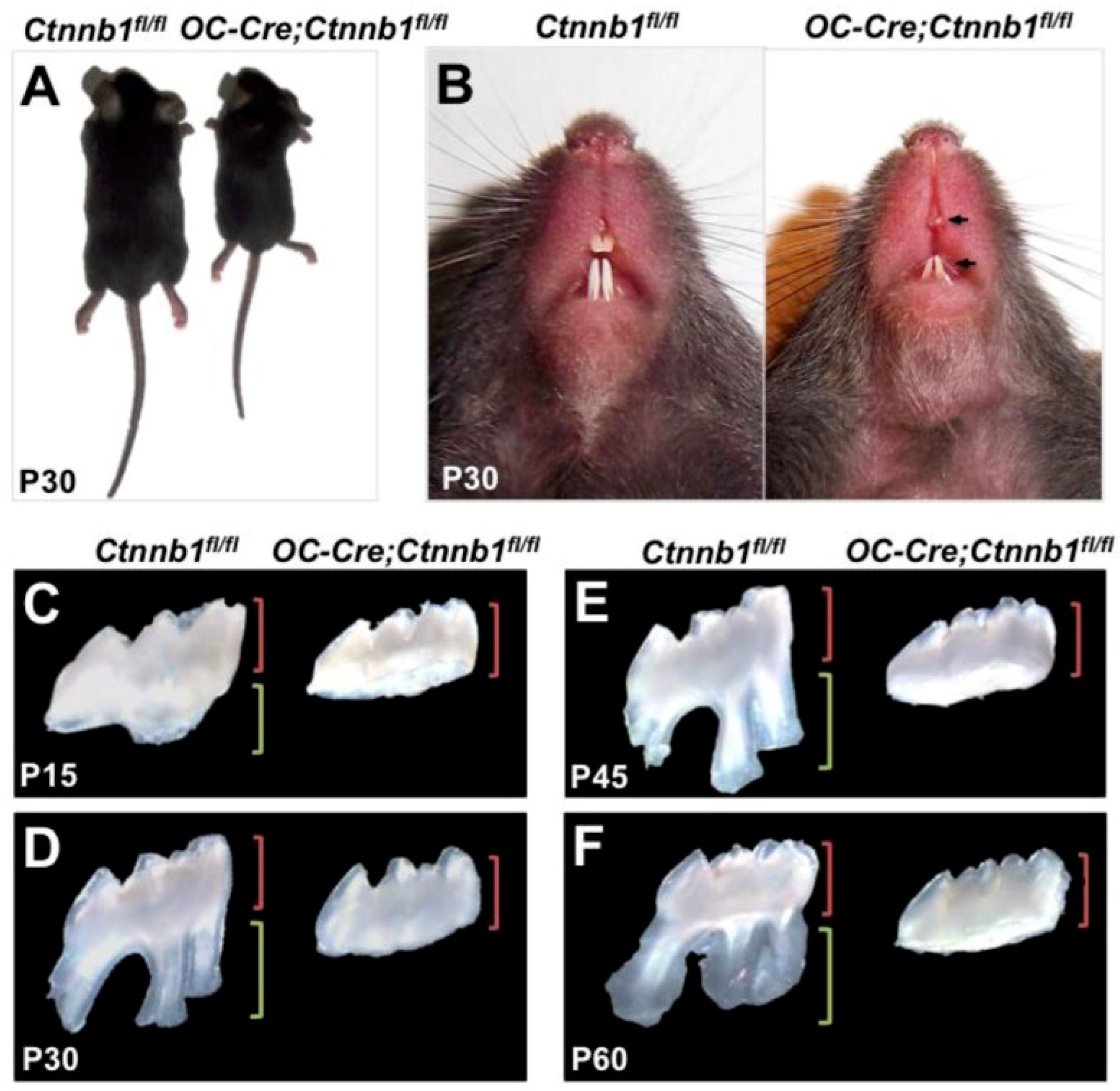
  39. Zhang, R.; Yang, G.; Wu, X.; Xie, J.; Yang, X.; Li, T. Disruption of Wnt/β-Catenin Signaling in Odontoblasts and Cementoblasts Arrests Tooth Root Development in Postnatal Mouse Teeth. Int. J. Biol. Sci. 2013, 9, 228–236. [Google Scholar] [CrossRef]
  40. Han, N.; Zheng, Y.; Li, R.; Li, X.; Zhou, M.; Niu, Y.; Zhang, Q. β-Catenin Enhances Odontoblastic Differentiation of Dental Pulp Cells through Activation of Runx2. PLoS ONE 2014, 9, e88890. [Google Scholar] [CrossRef]
  41. Bakopoulou, A.; Leyhausen, G.; Volk, J.; Papachristou, E.; Koidis, P.; Geurtsen, W. Wnt/β-Catenin Signaling Regulates Dental Pulp Stem Cells’ Responses to Pulp Injury by Resinous Monomers. Dent. Mater. 2015, 31, 542–555. [Google Scholar] [CrossRef] [PubMed]
  42. Lv, H.; Yang, J.; Wang, C.; Yu, F.; Huang, D.; Ye, L. The WNT7B Protein Promotes the Migration and Differentiation of Human Dental Pulp Cells Partly through WNT/Beta-Catenin and c-Jun N-Terminal Kinase Signalling Pathways. Arch. Oral Biol. 2018, 87, 54–61. [Google Scholar] [CrossRef]
  43. Ali, M.; Okamoto, M.; Komichi, S.; Watanabe, M.; Huang, H.; Takahashi, Y.; Hayashi, M. Lithium-Containing Surface Pre-Reacted Glass Fillers Enhance hDPSC Functions and Induce Reparative Dentin Formation in a Rat Pulp Capping Model through Activation of Wnt/β-Catenin Signaling. Acta Biomater. 2019, 96, 594–604. [Google Scholar] [CrossRef] [PubMed]
  44. Zhao, Y.; Yuan, X.; Bellido, T.; Helms, J.A. A Correlation between Wnt/Beta-Catenin Signaling and the Rate of Dentin Secretion. J. Endod. 2019, 45, 1357–1364.e1. [Google Scholar] [CrossRef] [PubMed]
  45. Wu, A.; Bao, Y.; Yu, H.; Zhou, Y.; Lu, Q. Berberine Accelerates Odontoblast Differentiation by Wnt/β-Catenin Activation. Cell. Reprogramming 2019, 21, 108–114. [Google Scholar] [CrossRef] [PubMed]
  46. Gong, Y.; Yuan, S.; Sun, J.; Wang, Y.; Liu, S.; Guo, R.; Dong, W.; Li, R. R-Spondin 2 Induces Odontogenic Differentiation of Dental Pulp Stem/Progenitor Cells via Regulation of Wnt/β-Catenin Signaling. Front. Physiol. 2020, 11, 918. [Google Scholar] [CrossRef] [PubMed]
  47. Lu, X.; Chen, X.; Xing, J.; Lian, M.; Huang, D.; Lu, Y.; Feng, G.; Feng, X. miR-140-5p Regulates the Odontoblastic Differentiation of Dental Pulp Stem Cells via the Wnt1/β-Catenin Signaling Pathway. Stem. Cell. Res. Ther. 2019, 10, 226. [Google Scholar] [CrossRef]
  48. Yaemkleebbua, K.; Osathanon, T.; Nowwarote, N.; Limjeerajarus, C.N.; Sukarawan, W. Analysis of Hard Tissue Regeneration and Wnt Signalling in Dental Pulp Tissues after Direct Pulp Capping with Different Materials. Int. Endod. J. 2019, 52, 1605–1616. [Google Scholar] [CrossRef]
  49. Pan, Y.; Lu, T.; Peng, L.; Chen, Z.; Li, M.; Zhang, K.; Xiong, F.; Wu, B. Vacuolar Protein Sorting 4B Regulates the Proliferation and Odontoblastic Differentiation of Human Dental Pulp Stem Cells through the Wnt-β-Catenin Signalling Pathway. Artif. Cells Nanomed. Biotechnol. 2019, 47, 2575–2584. [Google Scholar] [CrossRef]
  50. Zaugg, L.K.; Banu, A.; Walther, A.R.; Chandrasekaran, D.; Babb, R.C.; Salzlechner, C.; Hedegaard, M.A.B.; Gentleman, E.; Sharpe, P.T. Translation Approach for Dentine Regeneration Using GSK-3 Antagonists. J. Dent. Res. 2020, 99, 544–551. [Google Scholar] [CrossRef]
  51. Tokavanich, N.; Wein, M.N.; English, J.D.; Ono, N.; Ono, W. The Role of Wnt Signaling in Postnatal Tooth Root Development. Front. Dent. Med. 2021, 2, 769134. [Google Scholar] [CrossRef] [PubMed]
  52. Hara, M.; Horibe, K.; Mori, H.; Nakamura, H. The Role of Canonical Wnt Signaling in Dentin Bridge Formation. J. Oral Biosci. 2021, 63, 199–209. [Google Scholar] [CrossRef] [PubMed]
  53. Deng, Z.; Yan, W.; Dai, X.; Chen, M.; Qu, Q.; Wu, B.; Zhao, W. N-cadherin regulates the odontogenic differentiation of dental pulp stem cells via β-catenin activity. Front. Cell Dev. Biol. 2021, 9, 661116. [Google Scholar] [CrossRef]
  54. Wang, J.; Ran, S.; Liu, B.; Gu, S. Monitoring of Canonical BMP and Wnt Activities during Postnatal Stages of Mouse First Molar Root Formation. J. Appl. Oral Sci. 2021, 29, e20210281. [Google Scholar] [CrossRef] [PubMed]
  55. Vijaykumar, A.; Mina, M. Lithium Chloride Exerts Differential Effects on Dentinogenesis and Osteogenesis in Primary Pulp Cultures. Front. Dent. Med. 2021, 2, 649500. [Google Scholar] [CrossRef]
  56. Liu, S.; Sun, J.; Yuan, S.; Yang, Y.; Gong, Y.; Wang, Y.; Guo, R.; Zhang, X.; Liu, Y.; Mi, H.; et al. Treated Dentin Matrix Induces Odontogenic Differentiation of Dental Pulp Stem Cells via Regulation of Wnt/β-Catenin Signaling. Bioact. Mater. 2022, 7, 85–97. [Google Scholar] [CrossRef]
  57. Asghari, M.; Nasoohi, N.; Hodjat, M. High Glucose Promotes the Aging of Human Dental Pulp Cells through Wnt/Beta-Catenin Signaling. Dent. Med. Probl. 2021, 58, 39–46. [Google Scholar] [CrossRef] [PubMed]
  58. Duncan, H.F.; Kobayashi, Y.; Yamauchi, Y.; Quispe-Salcedo, A.; Chao Feng, Z.; Huang, J.; Partridge, N.C.; Nakatani, T.; D’Armiento, J.; Shimizu, E. The Critical Role of MMP13 in Regulating Tooth Development and Reactionary Dentinogenesis Repair through the Wnt Signaling Pathway. Front. Cell Dev. Biol. 2022, 10, 883266. [Google Scholar] [CrossRef]
  59. Fu, T.; Liu, Y.; Huang, X.; Guo, Y.; Shen, J.; Shen, H. lncRNA SNHG1 Regulates Odontogenic Differentiation of Human Dental Pulp Stem Cells via miR-328-3p/Wnt/β-Catenin Pathway. Stem. Cell. Res. Ther. 2022, 13, 311. [Google Scholar] [CrossRef]
  60. Sukarawan, W.; Rattanawarawipa, P.; Yaemkleebbua, K.; Nowwarote, N.; Pavasant, P.; Limjeerajarus, C.N.; Osathanon, T. Wnt3a Promotes Odonto/Osteogenic Differentiation in Vitro and Tertiary Dentin Formation in a Rat Model. Int. Endod. J. 2023, 56, 514–529. [Google Scholar] [CrossRef]
  61. Li, M.; Wang, Y.; Xue, J.; Xu, Q.; Zhang, Y.; Liu, J.; Xu, H.; Guan, Z.; Bian, C.; Zhang, G.; et al. Baicalin Can Enhance Odonto/Osteogenic Differentiation of Inflammatory Dental Pulp Stem Cells by Inhibiting the NF-κB and β-Catenin/Wnt Signaling Pathways. Mol. Biol. Rep. 2023, 50, 4435–4446. [Google Scholar] [CrossRef] [PubMed]
  62. Sakatoku, S.; Hayashi, Y.; Futenma, T.; Ishizaka, R.; Gemba, C.; Nawa, H. Wnt10a Is a Candidate as a Non-Cellular Agent for Induction of Dental Pulp Regeneration with Dentine-Inducing Capacity. J. Hard Tissue Biol. 2023, 32, 41–48. [Google Scholar] [CrossRef]
  63. Hayashi, Y.; Sakatoku, S.; Sugita, Y.; Futenma, T.; Iida, N.; Nakamura, K.; Nawa, H. A Potential Biomarker of Dental Pulp Regeneration: Wnt10a. J. Hard Tissue Biol. 2023, 32, 197–202. [Google Scholar] [CrossRef]
  64. Zhang, X.; Ning, T.; Wang, H.; Xu, S.; Yu, H.; Luo, X.; Hao, C.; Wu, B.; Ma, D. Stathmin Regulates the Proliferation and Odontoblastic/Osteogenic Differentiation of Human Dental Pulp Stem Cells through Wnt/β-Catenin Signaling Pathway. J. Proteom. 2019, 202, 103364. [Google Scholar] [CrossRef] [PubMed]
  65. Neves, V.C.M.; Yianni, V.; Sharpe, P.T. Macrophage Modulation of Dental Pulp Stem Cell Activity during Tertiary Dentinogenesis. Sci. Rep. 2020, 10, 20216. [Google Scholar] [CrossRef] [PubMed]
  66. Song, Y.; Liu, X.; Feng, X.; Gu, Z.; Gu, Y.; Lian, M.; Xiao, J.; Cao, P.; Zheng, K.; Gu, X.; et al. NRP1 Accelerates Odontoblast Differentiation of Dental Pulp Stem Cells Through Classical Wnt/β-Catenin Signaling. Cell. Reprogramming 2017, 19, 324–330. [Google Scholar] [CrossRef] [PubMed]
  67. Mao, T.; Peng, Y.; Peng, R.; Wei, X. Angelica Sinensis Polysaccharide Promotes the Proliferation and Osteogenic Differentiation of Human Dental Pulp Stem Cells (hDPSCs) by Activating the Wnt/β-Catenin Pathway. Qual. Assur. Saf. Crop. Foods 2021, 13, 31–37. [Google Scholar] [CrossRef]
  68. Vijaykumar, A.; Root, S.H.; Mina, M. Wnt/β-Catenin Signaling Promotes the Formation of Preodontoblasts In Vitro. J. Dent. Res. 2021, 100, 387–396. [Google Scholar] [CrossRef]
  69. Cate, A.T. The Role of Epithelium in the Development, Structure and Function of the Tissues of Tooth Support. Oral Dis. 1996, 2, 55–62. [Google Scholar] [CrossRef]
  70. Bae, C.-H.; Kim, T.-H.; Chu, J.-Y.; Cho, E.-S. New Population of Odontoblasts Responsible for Tooth Root Formation. Gene Expr. Patterns 2013, 13, 197–202. [Google Scholar] [CrossRef]
  71. Aurrekoetxea, M.; Irastorza, I.; García-Gallastegui, P.; Jiménez-Rojo, L.; Nakamura, T.; Yamada, Y.; Ibarretxe, G.; Unda, F.J. Wnt/β-Catenin Regulates the Activity of Epiprofin/Sp6, SHH, FGF, and BMP to Coordinate the Stages of Odontogenesis. Front. Cell Dev. Biol. 2016, 4, 25. [Google Scholar] [CrossRef] [PubMed]
  72. Zhao, Y.; Yuan, X.; Liu, B.; Tulu, U.S.; Helms, J.A. Wnt-Responsive Odontoblasts Secrete New Dentin after Superficial Tooth Injury. J. Dent. Res. 2018, 97, 1047–1054. [Google Scholar] [CrossRef] [PubMed]
  73. Harichane, Y.; Hirata, A.; Dimitrova-Nakov, S.; Granja, I.; Goldberg, A.; Kellermann, O.; Poliard, A. Pulpal Progenitors and Dentin Repair. Adv. Dent. Res. 2011, 23, 307–312. [Google Scholar] [CrossRef] [PubMed]
  74. Neag, M.A.; Mocan, A.; Echeverría, J.; Pop, R.M.; Bocsan, C.I.; Crişan, G.; Buzoianu, A.D. Berberine: Botanical Occurrence, Traditional Uses, Extraction Methods, and Relevance in Cardiovascular, Metabolic, Hepatic, and Renal Disorders. Front. Pharmacol. 2018, 9, 557. [Google Scholar] [CrossRef] [PubMed]
  75. Akhtar, M.; Woo, K.M.; Tahir, M.; Wu, W.; Elango, J.; Mirza, M.R.; Khan, M.; Shamim, S.; Arany, P.R.; Rahman, S.U. Enhancing osteoblast differentiation through small molecule-incorporated engineered nanofibrous scaffold. Clin. Oral Investig. 2022, 26, 2607–2618. [Google Scholar] [CrossRef] [PubMed]
  76. Deng, L.; Hong, H.; Zhang, X.; Chen, D.; Chen, Z.; Ling, J.; Wu, L. Down-Regulated lncRNA MEG3 Promotes Osteogenic Differentiation of Human Dental Follicle Stem Cells by Epigenetically Regulating Wnt Pathway. Biochem. Biophys. Res. Commun. 2018, 503, 2061–2067. [Google Scholar] [CrossRef]
  77. Yang, Y.; Ge, Y.; Chen, G.; Yan, Z.; Yu, M.; Feng, L.; Jiang, Z.; Guo, W.; Tian, W. Hertwig’s Epithelial Root Sheath Cells Regulate Osteogenic Differentiation of Dental Follicle Cells through the Wnt Pathway. Bone 2014, 63, 158–165. [Google Scholar] [CrossRef]
  78. Trosset, J.; Dalvit, C.; Knapp, S.; Fasolini, M.; Veronesi, M.; Mantegani, S.; Gianellini, L.M.; Catana, C.; Sundström, M.; Stouten, P.F.W.; et al. Inhibition of Protein–Protein Interactions: The Discovery of Druglike Β-catenin Inhibitors by Combining Virtual and Biophysical Screening. Proteins 2006, 64, 60–67. [Google Scholar] [CrossRef]
  79. Paula, A.B.; Laranjo, M.; Marto, C.-M.; Paulo, S.; Abrantes, A.M.; Casalta-Lopes, J.; Marques-Ferreira, M.; Botelho, M.F.; Carrilho, E. Direct Pulp Capping: What Is the Most Effective Therapy?—Systematic Review and Meta-Analysis. J. Evid. Based Dent. Pract. 2018, 18, 298–314. [Google Scholar] [CrossRef]
  80. Paula, A.; Laranjo, M.; Marto, C.M.; Abrantes, A.M.; Casalta-Lopes, J.; Gonçalves, A.C.; Sarmento-Ribeiro, A.B.; Ferreira, M.M.; Botelho, M.F.; Carrilho, E. BiodentineTM Boosts, WhiteProRoot®MTA Increases and Life® Suppresses Odontoblast Activity. Materials 2019, 12, 1184. [Google Scholar] [CrossRef]
  81. Paula, A.B.; Laranjo, M.; Marto, C.-M.; Paulo, S.; Abrantes, A.M.; Fernandes, B.; Casalta-Lopes, J.; Marques-Ferreira, M.; Botelho, M.F.; Carrilho, E. Evaluation of Dentinogenesis Inducer Biomaterials: An in Vivo Study. J. Appl. Oral Sci. 2020, 28, e20190023. [Google Scholar] [CrossRef] [PubMed]
  82. Klein, P.S.; Melton, D.A. A Molecular Mechanism for the Effect of Lithium on Development. Proc. Natl. Acad. Sci. USA 1996, 93, 8455–8459. [Google Scholar] [CrossRef] [PubMed]
  83. Nusse, R. Wnt Signaling in Disease and in Development. Cell Res. 2005, 15, 28–32. [Google Scholar] [CrossRef] [PubMed]
  84. Spagnuolo, G.; Galler, K.; Schmalz, G.; Cosentino, C.; Rengo, S.; Schweikl, H. Inhibition of Phosphatidylinositol 3-Kinase Amplifies TEGDMA-Induced Apoptosis in Primary Human Pulp Cells. J. Dent. Res. 2004, 83, 703–707. [Google Scholar] [CrossRef] [PubMed]
Figure 1. Active Canonical Wnt/β-Catenin Signaling Pathway. Wnt signal triggers Frizzled protein and activates LRP 5/6, Dishevelled (Dvl), Glycogen Synthase Kinase 3β (GSK), Casein kinase 1 (CK1), adenomatous polyposis coli (APC).
Cells 13 01153 g001
Figure 2. Inactive Canonical Wnt/β-Catenin Signaling Pathway. Cadherin activates AXIN mediated with the help of α and β-catenin and triggers gene expression.
Cells 13 01153 g002
Figure 3. Different types of proteins, molecules, and ligands in Wnt signaling. The molecules activate Wnt signaling by modulating ligands (Wnt) and signaling proteins.
Cells 13 01153 g003
Figure 4. Prisma flowchart of recent advances in Wnt/β-Catenin pathway in dentin and pulp regeneration. Identification of articles through 4 platforms, followed by screening (duplicate and other reasons such as non-English articles) and then finalized 100 articles after screening for assessment.
Cells 13 01153 g004
Figure 5. Growth delay and anomalies in the root of OC-Cre, Ctnnb1 fl/f mice (A) 1 month-old OC-Cre, Ctnnb1 fl/f mice are smaller than (Ctnnb1 fl/fl) the wild type mice. (B) Incisors at P30 show hypoplastic teeth with semi-transparent dentin (Gross appearance) (CF). Maxillary 1 molars at P15, P30, P45, and P60 with lengths of crown and root are shown by green and red brackets, respectively [39]. Reproduced with permission from Zhang R et al. [39], published by International Journal of Biological Sciences, 2013.
Cells 13 01153 g005
Figure 6. (A). Graph showing the decreased expression of miR-140-5p by qRT-PCR in differentiation groups on day 1, day 3, day 7, and day 14 vs the control group, p < 0.05. n = 6 whereby * represents significance of result [47]. Reproduced with permission from Lu Xiaohui et al. [47]., published by Stem Cell Research and Therapy (2019). (B). Tertiary dentin formation is promoted by Wnt3a in vivo in Wistar rats; molar injury as shown by white arrows in molars treated with distilled water (DI: (a,b)), phosphate-buffered saline (PBS: (c,d)), Lithium chloride (LiCl: (e,f)), and recombinant human Wnt3a (Wnt3a: (g,h)), (i) After 4 weeks analysis of specimens was performed. The dentin/bone volume to total volume was calculated as shown in the graph, with bars showing a significant difference [60]. Reproduced with permission from Sukarawan W et al. [60]., Published by International Endodontic Journal 2023.
Cells 13 01153 g006
Figure 7. (A) The Wnt/β-catenin pathway regulates reparative dentinogenesis, which is modulated by the autocrine release of Wnt by Axin2. The rapid proliferation of pulp cells is perceived after tooth damage, with a peak at day 3 and a return to baseline on day 14, with dentin bridge formation. (B) The Wnt/β-Catenin signaling pathway is inhibited by Baicailin. (A,B) β-Catenin Protein expression of Baicailin treated iDPSC at each time point (C), The expression of NLK, CaMK2, β-catenin, and GSK3β of baicailin treated iDPSCs by western blot whereby the * and ** represent the significance of the result [61]. Reproduced with permission from Mengyuan Li et al. [61], published by Springer Nature 2023.
Cells 13 01153 g007
Table 1. Evidence-based table of Recent Advances in Wnt/β-Catenin Signaling Pathway in Dentin and Pulp Regeneration.
SYear AuthorType of StudySample
Size
ObjectiveConclusionRecommendation/Limitation
12012J Wang et al. [38]In vitron = 8
3 molars
18–20 age
Role of Wnt/β-catenin signaling pathway in SCAP proliferation and differentiation.Proliferation and odonto/osteogenic differentiation of SCAP are promoted by the Wnt/β-catenin signaling pathway.Further study in SCAP mineralization and proliferation may bring improvement in dental tissue engineering.
22013Ran Zhang et al. [39] In vivo Wnt signaling and its importance in cementogenesis and odontogenesis.Wnt signaling encourages the differentiation of odontoblasts from mesenchyme.Influence of Wnt signaling on tooth development.
32014Zichun Zhang et al. [33]In vivo9Response of Wnt10a on DPSCs.Proliferation of DPCS is promoted, and their odontoblastic differentiation is negatively regulated by Wnt10a.Further research is vital to understand the biological effect of Wnt10a on DPCS.
42014Nana Han et al. [40] In vivo β-catenin during the activation of Runx2 in DPCs augments odontoblastic differentiation.Odontoblastic differentiation in tertiary dentin modulated by β-catenin.The mechanism of reparative dentin formation remains unclear.
52015Danial J Hunter et al. [28] In vivo72 miceResponse of injured pulp to amplified Wnt signaling resulting in superior pulp healing.Amplified Wnt signaling caused pulp cells to differentiate into secretory odontoblasts, thus improving pulp vitality. The role of amplified Wnt signaling on pulp cells plays a role in promoting pulp vitality, treating dental caries, and alternative approaches for root canal treatment.
62015Athina Bakopoulou et al. [41] In vitro3 molars age 18–24.
DPSCs cultured from 3 donors.
The function of the Wnt/β-catenin pathway in the healing of pulpal injury resulted from resinous monomers via DPSC mediation.DPSCs, under the effect of Wnt/β-catenin, participate in pulp healing after injury.The analysis of stem cells is important for pulp regenerative therapy and bioactive molecule planning via tissue engineering through Wnt activation.
72016Masato Tamura et al. [15] Review Article Determination of function of signaling molecules of Wnt in tooth.Tooth development, maintenance, and turnover are regulated by both canonical and non-canonical Wnt pathway.The Wnt signaling pathway and its progress in future therapeutics for tooth regeneration.
82017Hongyang LV et al. [42] In vivo/In vitroHDPCs cultured from 3 molars from 19 to 24 years of age.Function of Wnt7b protein in HDPCs relocation and differentiation.Wnt/β-catenin and JNK pathways partly have a role in the differentiation of HDPCs and are promoted by the Wnt7b protein.Wnt7b can be used in the potential treatment of dental caries and tooth trauma.
Wnt7b plays a role in maintaining the vitality of the tooth.
92017Rebecca Babb et al. [13]In vivo The role of Axin 2 via Wnt/β-catenin signaling in tertiary dentinogenesis.As a response to tooth injury, new odontoblast-like cells are formed via the Wnt signaling pathway, and Axin 2 cells provide a signal in reparative dentinogenesis.Further study is required for stem cell-based tooth repair via the Wnt/β-catenin signaling pathway in secondary and tertiary dentinogenesis
102018Manahil Ali et al. [43] In vivo/
In vitro
21To check the enhancement of S-PRG cement through the addition of LiCl and its effect on hDPSCs.S-PRG cement through the Wnt/β-catenin pathway enhances reparative dentin formation in rat teeth and promotes hDPSC profiles.Inclination of reparative dentinogenesis in clinical trials needs to be further assessed.
The interaction of Li ions with S-PRG fillers is not fully understood.
112019Yuan Zhao et al. [44] In vivo2 strains of Wnt reporter miceThe function of Wnt signaling in age-linked variations in pulp and its ability to respond to subacute traumas.Amplification of Wnt signaling results in reparative/osteodentin organization, as the secretion of dentin from odontoblasts is regulated by this pathway.Wnt signaling and its role in dentin secretion can be used to form biological pulp-capping material.
122019Xi Lu et al. [19]Review
Article
Review of Wnt signaling pathway, its modulators, and their function in odontogenesis and therapeutic potential.Tooth development is closely regulated by the canonical Wnt pathway.The tooth regeneration method may be provided by a detailed study in Wnt signaling.
132019 Anqian Wu et al. [45] In vitro(N)
n = 20 pulp
Ages 18–25
Impact of berberine on DPSCs and their odontoblast differentiation.By the stimulation of the Wnt pathway, berberine encourages odontoblast differentiation.Berberine might be used as a new drug for the treatment of dental defects.
142019Yuping Gong et al. [46] In vivo20Role of Rspo2 in promoting hDPSCs to differentiate odontogenically through the Wnt/β-catenin pathway.hDPSCs stimulated by Rspo2, both positive and negative, by enhancer and silencer agents, thus enhancing their proliferation and differentiation.Future work of
R-spondins and Wnt ligands could be on therapeutic use clinically.
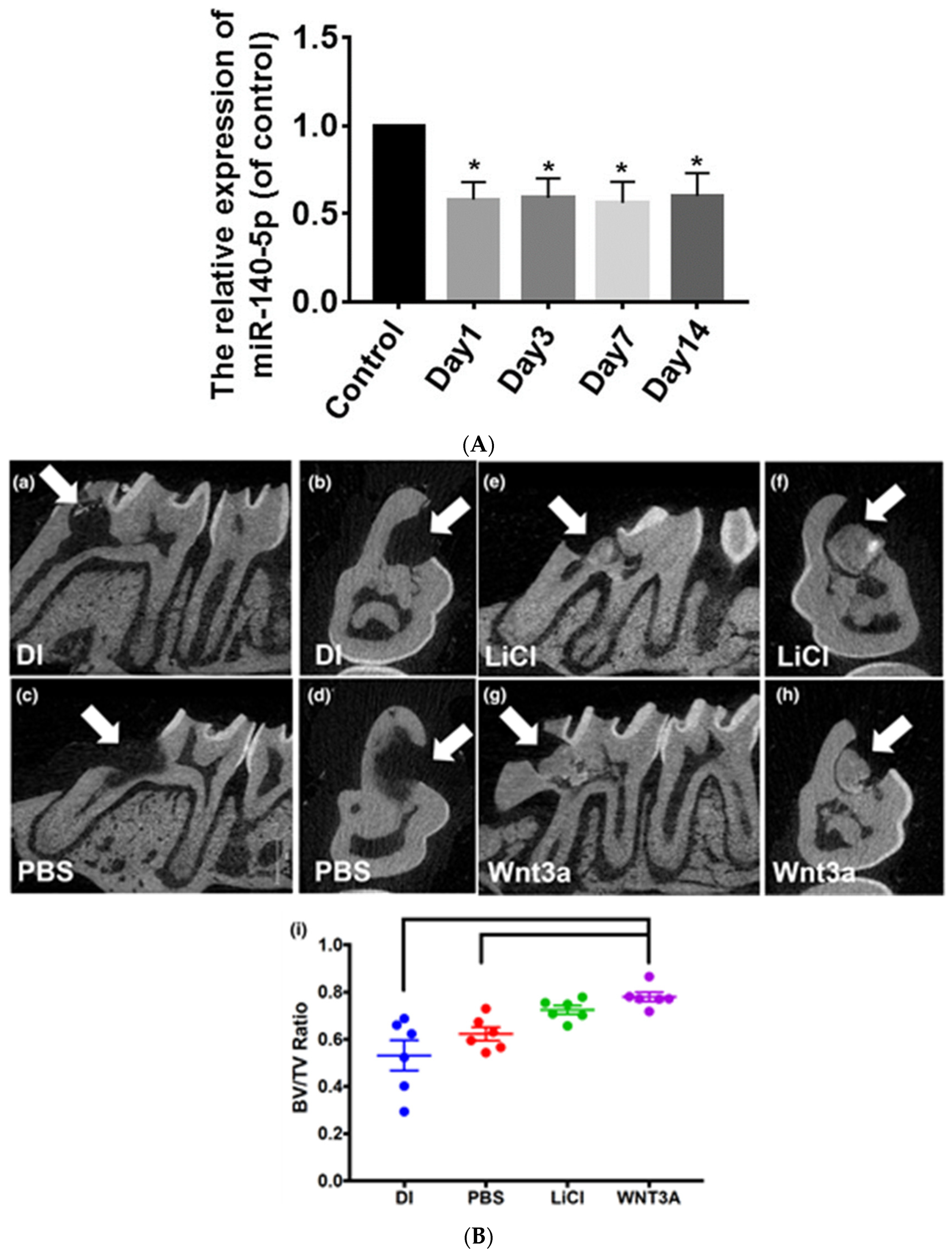
152019Xiaohui Lu et al. [47] In vitron = 10
3 molars age 14–22 yrs
miR-140-5p impact on the odontoblastic differentiation of DPSC.The odontoblastic differentiation of DPSCs is regulated by lowering miR-140-5p by targeting the Wnt/β-catenin pathway.miR-140-5p can be used as a medicinal agent that could control odontoblastic differentiation in dental medicine.
162019 K. Yaemkleebua et al. [48] In vivo32 rat molar pulps were used.Hard tissue formation after application of pulp capping materials on pinpoint exposure and the role of Wnt signaling and cell cycle regulation.Reparative dentinogenesis occurred with the application of pulp capping materials, and cyclin D1 expression was seen.The association between Wnt signaling and reparative dentin formation might be used to comprehend the response of normal pulp in injured conditions.
172019Yuhua Pan et al.
[49]
In vivo
In vitro
DPSCs culture from 3 molars of 18–20 yr
Donors
The role of VPS4B in DPSCs via the Wnt/β-catenin signaling pathway.DPSC differentiation and proliferation are regulated by VPS4B via the Wnt/β-catenin pathway.Dentin dysplasia type1 could be treated via potential medicinal therapies by the formation of dentin and reparative endodontic therapy.
182020LK Zaugg et al. [50] In vivo/
In vitro
Adult male Wistar rats (7 wk old) and CD1 mice (6 wk old) The Wnt/β-catenin signaling and its function in reparative dentinogenesis and how it is affected by the introduction of GSK3β inhibitor drugs.GSK3β inhibitor results in the tertiary dentin formation at the defect region having the size of a human lesion; however, this is not true regeneration.Augmentation of reparative dentinogenesis can be used potentially as a treatment in direct pulp capping.
192021Nicha
Tokavanich et al.
[51]
Review Article Wnt signaling and its effect on postnatal tooth development.Wnt signaling may play a vital role as a target for root formation and tooth eruption disorders.In-depth study of the Wnt signaling pathway may provide a path in the future for regenerative dentistry.
202021Miroku Hara et al. [52] In vivo321 miceDentin bridge formation at pinpoint exposure and how the Wnt signaling and its molecules affect it.The injured dental pulp is repaired by the stimulation of canonical Wnt signaling.Wnt pathway activation by the dental pulp capping materials may be a new approach for vital pulp therapy.
212021Zilong Deng et al. [53]In vitro/In vivo3 molars
Pulp
18–25 yr
n = 5 mice
N-cadherin effect on DPSCs, in-vitro and in-vivo, and its function in odontogenic differentiation.N-cadherin negatively regulates β-catenin activity in DPSCs.Further study is required on how neurogenic differentiation of DPSCs is regulated by N-cadherin
222021Jia Wang et al. [54] In vivo2 transgenic miceRole of Wnt signaling and distribution of BMP during postnatal root development.Tooth root development is regulated by BMP and Wnt signaling pathway.Further study is required for Wnt/β-catenin pathway regulation for matrix secretion and differentiation in postnatal tooth development.
232021Anushree Vijaykumar et al. [55] In vitroPulp culture from 1 and 2 molar 5–7 d miceDentin pulp complex and role of LiCl in its reparation and regeneration via Wnt/β-catenin pathway by inhibiting GSK3β.LiCl results in reparative dentin formation, which has an improved structure.GSK3β controls several transcription factors and various pathways; thus, the influence of LiCl on tertiary dentin formation may not be deemed exclusive to the Wnt/β-catenin pathway.
242021Sirui Liu et al. [56] In vitro
In vivo
DPSCs culture donor’s teeth pulp
18–25 yr
Wistar mice 26
Wnt/β-catenin signaling role in hDPSCs and its odontogenic differentiation via TDM extract.TDM via GSK3β activates the Wnt/β-catenin pathway in hDPSC and stimulates its odontogenic differentiation.The mechanism for TDM-induced differentiation requires further study.
252021Mona Asghari et al. [57] In vitro4000 cellsEffect of hyperglycemia and function of Wnt pathway in senescence of pulp cells.Hyperglycemia causes the aging of pulp cells.
Cellular aging is induced via beta-catenin.
Further study is required to determine the role of beta-catenin and pulp regeneration in diabetic patients.
262022Henry F Duncan et al. [58] In vivo
In vitro
MMP13 deficient mice and WT control mice were used for DPCs.The effect and interaction of MMP13 with the Wnt signaling pathway. Its effect on tooth development, repair, and dentin pulp regeneration.MMP13 controls the organization and regulation of tooth development in addition to dentin pulp regeneration.This study found targets for new therapy for traumatized pulp by increasing repair
response.
272022Tingting Fu et al. [59] In vitro DPSCs
Culture
Donar teeth
≤16 yr
Role of SNHG1 in hDPSCs in odontogenic differentiation via Wnt/β-catenin signaling.The Wnt/β-catenin pathway is silenced by IncRNA SNHG1 via miR-328-3p, resulting in the differentiation of hDPSCs.Useful in regenerative endodontics.
282023Waleerat Sukarawan et al. [60] In vivo6 miceTo check in SHEDs the influence of Wnt3a on the odonto/osteogenic differentiation and reparative dentin production.Wnt3a causes an increase in osteogenic differentiation, silencing of proliferation in SHEDs, advances tertiary dentin formation, and can be used as a biological molecule in vital pulp therapy.An in-depth study is required to identify the function and role of Wnt3a in tissue engineering.
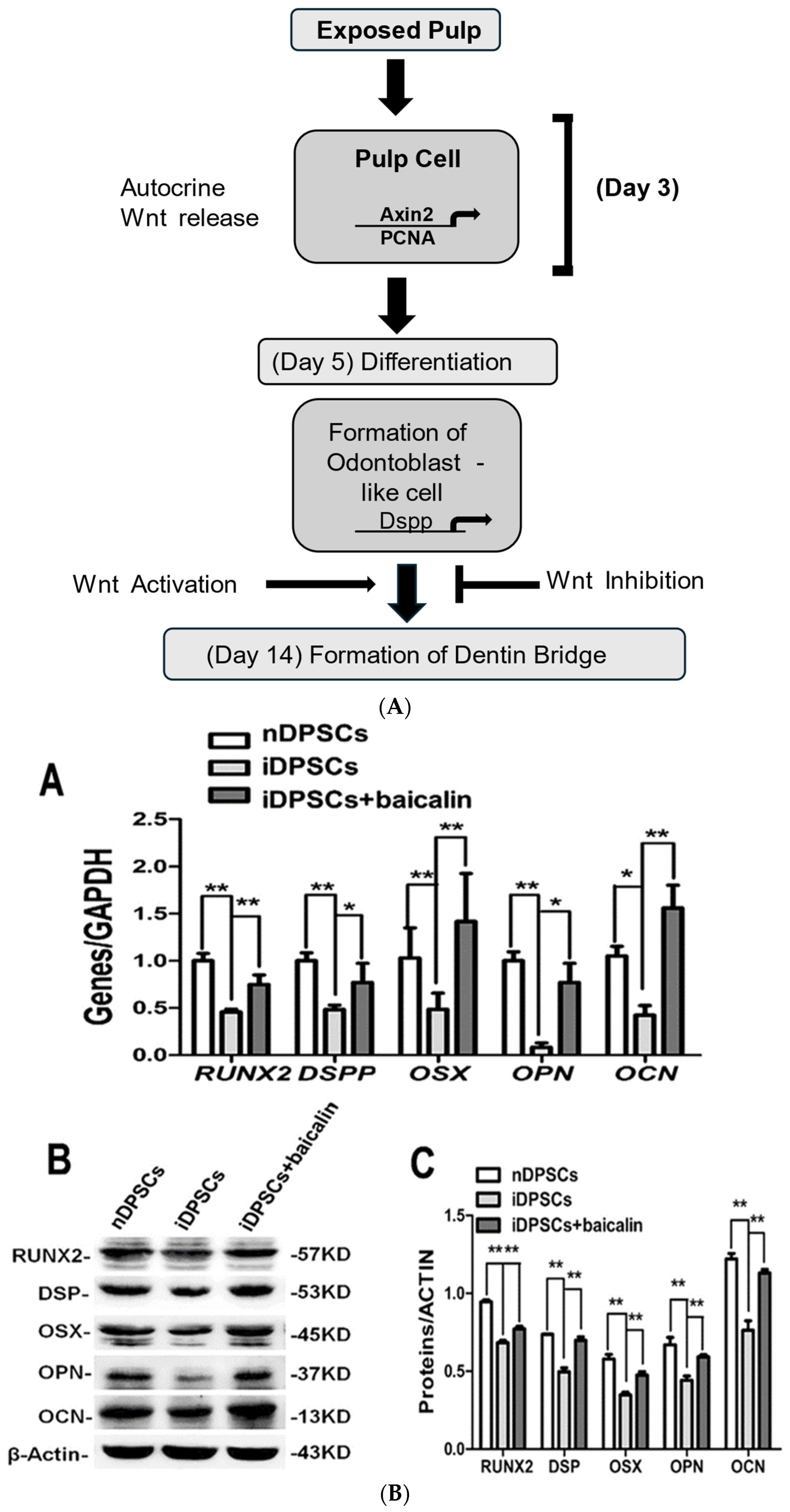
292023Mengyuan Li et al.
[61]
In vitroDPSC
In 6 well plates
Effect of Baicalin on DPSC differentiation via Wnt/β/NF-kB pathway.Baicalin impedes both NF-Kb and Wnt/β-catenin pathways, thus promoting the differentiation of DPSCs, causing repair of pulp with early irreversible pulpitis.Further investigation and research are required to apply it in the long-term
302023Shintaro Sakatoku et al. [62] In vitroDPSC in 24 well platesRole of Wnt10a and odontoblasts in regenerative dental pulp. Odontoblasts expressing Wnt10a play an increased role in pulp regeneration,
with dentin-inducing capacity.
A detailed study after transplantation is required to check the long-term effect of Wnt10a along with DKK1 levels in regenerated dental pulp.
Disclaimer/Publisher’s Note: The statements, opinions and data contained in all publications are solely those of the individual author(s) and contributor(s) and not of MDPI and/or the editor(s). MDPI and/or the editor(s) disclaim responsibility for any injury to people or property resulting from any ideas, methods, instructions or products referred to in the content.

