Journal Description
Diagnostics
Diagnostics
is an international, peer-reviewed, open access journal on medical diagnosis published semimonthly online by MDPI. The British Neuro-Oncology Society (BNOS), the International Society for Infectious Diseases in Obstetrics and Gynaecology (ISIDOG) and the Swiss Union of Laboratory Medicine (SULM) are affiliated with Diagnostics and their members receive a discount on the article processing charges.
- Open Access— free for readers, with article processing charges (APC) paid by authors or their institutions.
- High Visibility: indexed within Scopus, SCIE (Web of Science), PubMed, PMC, Embase, Inspec, CAPlus / SciFinder, and other databases.
- Journal Rank: JCR - Q2 (Medicine, General & Internal)
- Rapid Publication: manuscripts are peer-reviewed and a first decision is provided to authors approximately 20.7 days after submission; acceptance to publication is undertaken in 2.8 days (median values for papers published in this journal in the second half of 2023).
- Recognition of Reviewers: reviewers who provide timely, thorough peer-review reports receive vouchers entitling them to a discount on the APC of their next publication in any MDPI journal, in appreciation of the work done.
- Companion journal: LabMed.
Impact Factor:
3.6 (2022);
5-Year Impact Factor:
3.7 (2022)
Latest Articles
Diffusion-Weighted Imaging for Skin Pathologies of the Breast—A Feasibility Study
Diagnostics 2024, 14(9), 934; https://doi.org/10.3390/diagnostics14090934 (registering DOI) - 29 Apr 2024
Abstract
Several breast pathologies can affect the skin, and clinical pathways might differ significantly depending on the underlying diagnosis. This study investigates the feasibility of using diffusion-weighted imaging (DWI) to differentiate skin pathologies in breast MRIs. This retrospective study included 88 female patients who
[...] Read more.
Several breast pathologies can affect the skin, and clinical pathways might differ significantly depending on the underlying diagnosis. This study investigates the feasibility of using diffusion-weighted imaging (DWI) to differentiate skin pathologies in breast MRIs. This retrospective study included 88 female patients who underwent diagnostic breast MRI (1.5 or 3T), including DWI. Skin areas were manually segmented, and the apparent diffusion coefficients (ADCs) were compared between different pathologies: inflammatory breast cancer (IBC; n = 5), benign skin inflammation (BSI; n = 11), Paget’s disease (PD; n = 3), and skin-involved breast cancer (SIBC; n = 11). Fifty-eight women had healthy skin (H; n = 58). The SIBC group had a significantly lower mean ADC than the BSI and IBC groups. These differences persisted for the first-order features of the ADC (mean, median, maximum, and minimum) only between the SIBC and BSI groups. The mean ADC did not differ significantly between the BSI and IBC groups. Quantitative DWI assessments demonstrated differences between various skin-affecting pathologies, but did not distinguish clearly between all of them. More extensive studies are needed to assess the utility of quantitative DWI in supplementing the diagnostic assessment of skin pathologies in breast imaging.
Full article
(This article belongs to the Special Issue Recent Advances in Breast Imaging)
Open AccessArticle
Influence of Achalasia on the Spirometry Flow–Volume Curve and Peak Expiratory Flow
by
Jelena Jankovic, Branislava Milenkovic, Aleksandar Simic, Ognjan Skrobic, Arschang Valipour, Nenad Ivanovic, Ivana Buha, Jelena Milin-Lazovic, Natasa Djurdjevic, Aleksandar Jandric, Nikola Colic, Stefan Stojkovic and Mihailo Stjepanovic
Diagnostics 2024, 14(9), 933; https://doi.org/10.3390/diagnostics14090933 (registering DOI) - 29 Apr 2024
Abstract
Background: Achalasia is an esophageal motor disorder characterized by aperistalsis and the failure of the relaxation of the lower esophageal sphincter. We want to find out whether external compression or recurrent micro-aspiration of undigested food has a functional effect on the airway. Methods:
[...] Read more.
Background: Achalasia is an esophageal motor disorder characterized by aperistalsis and the failure of the relaxation of the lower esophageal sphincter. We want to find out whether external compression or recurrent micro-aspiration of undigested food has a functional effect on the airway. Methods: The aim of this research was to analyze the influence of achalasia on the peak expiratory flow and flow–volume curve. All of the 110 patients performed spirometry. Results: The mean diameter of the esophagus was 5.4 ± 2.1 cm, and nine of the patients had mega-esophagus. Seven patients had a plateau in the inspiratory part of the flow–volume curve, which coincides with the patients who had mega-esophagus. The rest of the patients had a plateau in the expiration part of the curve. The existence of a plateau in the diameter of the esophagus of more than 5 cm was significant (p 0.003). Statistical significance between the existence of a plateau and a lowered PEF (PEF < 80) has been proven (p 0.001). Also, a statistical significance between the subtype and diameter of more than 4 cm has been proved. There was no significant improvement in the PEF values after operation. In total, 20.9% of patients had a spirometry abnormality finding. The frequency of the improvement in the spirometry values after surgery did not differ significantly by achalasia subtype. The improvement in FEV1 was statistically significant compared to the FVC values. Conclusions: Awareness of the influence of achalasia on the pulmonary parameters is important because low values of PEF with a plateau on the spirometry loop can lead to misdiagnosis. The recognition of various patterns of the spirometry loop may help in identifying airway obstruction caused by another non-pulmonary disease such as achalasia.
Full article
(This article belongs to the Special Issue Diagnosis and Management of Liver Diseases and Inflammatory Bowel Diseases)
Open AccessInteresting Images
The Riddle of the Double Vision—A Rare Case of Intracranial Tumor: When Imaging Resolves the Mystery
by
Radina Kirkova, Svetla Dineva, Elisa Stradiotto, Ivan Tanev and Alessandra Di Maria
Diagnostics 2024, 14(9), 932; https://doi.org/10.3390/diagnostics14090932 (registering DOI) - 29 Apr 2024
Abstract
A 77-year-old-man with arterial hypertension, diabetes mellitus type II presented at our clinic for a routine ophthalmological exam. He complained of intermittent double vision. The ophthalmic examination revealed paralysis of III (n. oculomotorius) and VI (n. abducens) cranial nerves with ptosis, deficit in
[...] Read more.
A 77-year-old-man with arterial hypertension, diabetes mellitus type II presented at our clinic for a routine ophthalmological exam. He complained of intermittent double vision. The ophthalmic examination revealed paralysis of III (n. oculomotorius) and VI (n. abducens) cranial nerves with ptosis, deficit in elevation and abduction of the left eye. The patient underwent urgent MRI imaging of the brain/orbits and paranasal sinuses, and urgent neurological assessment. MRI revealed a volume-occupying process, starting from the posterior wall of the left maxillary sinus with perineural diffusion and involvement of the homolateral trigeminal nerve, intracranial spread in the medial cranial fossa and involvement of the cavernous, sphenoidal sinuses and the orbital apex on the left side. Biopsy was performed, and the histology resulted in sinonasal squamous cell carcinoma with intracranial spread.
Full article
(This article belongs to the Collection Advances in Cancer Imaging)
Open AccessReview
Present and Future Applications of Digital PCR in Infectious Diseases Diagnosis
by
Laura Sancha Dominguez, Ana Cotos Suárez, María Sánchez Ledesma and Juan Luis Muñoz Bellido
Diagnostics 2024, 14(9), 931; https://doi.org/10.3390/diagnostics14090931 (registering DOI) - 29 Apr 2024
Abstract
Infectious diseases account for about 3 million deaths per year. The advent of molecular techniques has led to an enormous improvement in their diagnosis, both in terms of sensitivity and specificity and in terms of the speed with which a clinically useful result
[...] Read more.
Infectious diseases account for about 3 million deaths per year. The advent of molecular techniques has led to an enormous improvement in their diagnosis, both in terms of sensitivity and specificity and in terms of the speed with which a clinically useful result can be obtained. Digital PCR, or 3rd generation PCR, is based on a series of technical modifications that result in more sensitive techniques, more resistant to the action of inhibitors and capable of direct quantification without the need for standard curves. This review presents the main applications that have been developed for the diagnosis of viral, bacterial, and parasitic infections and the potential prospects for the clinical use of this technology.
Full article
(This article belongs to the Section Point-of-Care Diagnostics and Devices)
Open AccessArticle
Individualizing Surveillance after Endovascular Aortic Repair Using a Modular Imaging Algorithm
by
Amun Georg Hofmann, Irene Mlekusch, Georg Wickenhauser, Corinna Walter, Fadi Taher and Afshin Assadian
Diagnostics 2024, 14(9), 930; https://doi.org/10.3390/diagnostics14090930 (registering DOI) - 29 Apr 2024
Abstract
Objectives: Surveillance after endovascular aortic repair (EVAR) and fenestrated EVAR (FEVAR) is mainly directed by one-size-fits-all approaches instead of personalized decision making, even though treatment strategies and often endografts themselves are tailor-made to adjust for individual patients. We propose a modular imaging algorithm
[...] Read more.
Objectives: Surveillance after endovascular aortic repair (EVAR) and fenestrated EVAR (FEVAR) is mainly directed by one-size-fits-all approaches instead of personalized decision making, even though treatment strategies and often endografts themselves are tailor-made to adjust for individual patients. We propose a modular imaging algorithm that escalates surveillance imaging based on invasiveness and need. Materials and Methods: In this retrospective observational study of single-center data, results of a modular imaging algorithm were analyzed. The algorithm is characterized by initiating the examination with standard B-mode then transitioning to Duplex ultrasound, B-Flow, and CEUS. Additional CT(A) studies are conducted where required. The study population included both patients receiving EVAR or FEVAR. A comparative analysis was conducted regarding endoleak detection. Results: The study population included 28 patients receiving EVAR and 40 patients receiving FEVAR. They accounted for 101 follow-up visits, which led to 431 distinct imaging studies. CEUS has the highest endoleak detection rate, followed by CTA and B-Flow. Duplex ultrasound and B-Flow resulted in 0 and 1 false positive cases, respectively, considering CEUS the reference standard. In a select group of six patients, CEUS was omitted after endoleaks were displayed by Duplex ultrasound or B-Flow, leading to a successful type II coiling and no aneurysm-related adverse events. Conclusions: The proposed modular algorithm showed great potential to incorporate principles of personalized medicine in surveillance after endovascular aortic treatment. Since Duplex ultrasound and B-Flow rarely cause false positive endoleaks, more resource-intensive and invasive imaging studies such as CEUS and CTA can be omitted after positive identification.
Full article
(This article belongs to the Special Issue Current Challenges and Perspectives of Ultrasound)
Open AccessArticle
Novel Variants Linked to the Prodromal Stage of Parkinson’s Disease (PD) Patients
by
Marwa T. Badawy, Aya A. Salama and Mohamed Salama
Diagnostics 2024, 14(9), 929; https://doi.org/10.3390/diagnostics14090929 (registering DOI) - 29 Apr 2024
Abstract
Background and objective: The symptoms of most neurodegenerative diseases, including Parkinson’s disease (PD), usually do not occur until substantial neuronal loss occurs. This makes the process of early diagnosis very challenging. Hence, this research used variant call format (VCF) analysis to detect variants
[...] Read more.
Background and objective: The symptoms of most neurodegenerative diseases, including Parkinson’s disease (PD), usually do not occur until substantial neuronal loss occurs. This makes the process of early diagnosis very challenging. Hence, this research used variant call format (VCF) analysis to detect variants and novel genes that could be used as prognostic indicators in the early diagnosis of prodromal PD. Materials and Methods: Data were obtained from the Parkinson’s Progression Markers Initiative (PPMI), and we analyzed prodromal patients with gVCF data collected in the 2021 cohort. A total of 304 participants were included, including 100 healthy controls, 146 prodromal genetic individuals, 21 prodromal hyposmia individuals, and 37 prodromal individuals with RBD. A pipeline was developed to process the samples from gVCF to reach variant annotation and pathway and disease association analysis. Results: Novel variant percentages were detected in the analyzed prodromal subgroups. The prodromal subgroup analysis revealed novel variations of 1.0%, 1.2%, 0.6%, 0.3%, 0.5%, and 0.4% for the genetic male, genetic female, hyposmia male, hyposmia female, RBD male, and RBD female groups, respectively. Interestingly, 12 potentially novel loci (MTF2, PIK3CA, ADD1, SYBU, IRS2, USP8, PIGL, FASN, MYLK2, USP25, EP300, and PPP6R2) that were recently detected in PD patients were detected in the prodromal stage of PD. Conclusions: Genetic biomarkers are crucial for the early detection of Parkinson’s disease and its prodromal stage. The novel PD genes detected in prodromal patients could aid in the use of gene biomarkers for early diagnosis of the prodromal stage without relying only on phenotypic traits.
Full article
(This article belongs to the Section Pathology and Molecular Diagnostics)
Open AccessArticle
A Multi-Stage Approach for Cardiovascular Risk Assessment from Retinal Images Using an Amalgamation of Deep Learning and Computer Vision Techniques
by
Deepthi K. Prasad, Madhura Prakash Manjunath, Meghna S. Kulkarni, Spoorthi Kullambettu, Venkatakrishnan Srinivasan, Madhulika Chakravarthi and Anusha Ramesh
Diagnostics 2024, 14(9), 928; https://doi.org/10.3390/diagnostics14090928 (registering DOI) - 29 Apr 2024
Abstract
Cardiovascular diseases (CVDs) are a leading cause of mortality worldwide. Early detection and effective risk assessment are crucial for implementing preventive measures and improving patient outcomes for CVDs. This work presents a novel approach to CVD risk assessment using fundus images, leveraging the
[...] Read more.
Cardiovascular diseases (CVDs) are a leading cause of mortality worldwide. Early detection and effective risk assessment are crucial for implementing preventive measures and improving patient outcomes for CVDs. This work presents a novel approach to CVD risk assessment using fundus images, leveraging the inherent connection between retinal microvascular changes and systemic vascular health. This study aims to develop a predictive model for the early detection of CVDs by evaluating retinal vascular parameters. This methodology integrates both handcrafted features derived through mathematical computation and retinal vascular patterns extracted by artificial intelligence (AI) models. By combining these approaches, we seek to enhance the accuracy and reliability of CVD risk prediction in individuals. The methodology integrates state-of-the-art computer vision algorithms and AI techniques in a multi-stage architecture to extract relevant features from retinal fundus images. These features encompass a range of vascular parameters, including vessel caliber, tortuosity, and branching patterns. Additionally, a deep learning (DL)-based binary classification model is incorporated to enhance predictive accuracy. A dataset comprising fundus images and comprehensive metadata from the clinical trials conducted is utilized for training and validation. The proposed approach demonstrates promising results in the early prediction of CVD risk factors. The interpretability of the approach is enhanced through visualization techniques that highlight the regions of interest within the fundus images that are contributing to the risk predictions. Furthermore, the validation conducted in the clinical trials and the performance analysis of the proposed approach shows the potential to provide early and accurate predictions. The proposed system not only aids in risk stratification but also serves as a valuable tool for identifying vascular abnormalities that may precede overt cardiovascular events. The approach has achieved an accuracy of 85% and the findings of this study underscore the feasibility and efficacy of leveraging fundus images for cardiovascular risk assessment. As a non-invasive and cost-effective modality, fundus image analysis presents a scalable solution for population-wide screening programs. This research contributes to the evolving landscape of precision medicine by providing an innovative tool for proactive cardiovascular health management. Future work will focus on refining the solution’s robustness, exploring additional risk factors, and validating its performance in additional and diverse clinical settings.
Full article
(This article belongs to the Special Issue Classifications of Diseases Using Machine Learning Algorithms)
►▼
Show Figures
Figure 1
Open AccessArticle
The Contribution of Vessel Wall Magnetic Resonance Imaging to the Diagnosis of Primary and Secondary Central Nervous System Vasculitis
by
Serena D’Aniello, Arianna Rustici, Laura Ludovica Gramegna, Claudia Godi, Laura Piccolo, Mauro Gentile, Andrea Zini, Alessandro Carrozzi, Raffaele Lodi, Caterina Tonon, Massimo Dall’Olio, Luigi Simonetti, Raffaella Chieffo, Nicoletta Anzalone and Luigi Cirillo
Diagnostics 2024, 14(9), 927; https://doi.org/10.3390/diagnostics14090927 (registering DOI) - 29 Apr 2024
Abstract
Background: To describe high-resolution brain vessel wall MRI (VW-MRI) patterns and morphological brain findings in central nervous system (CNS) vasculitis patients. Methods: Fourteen patients with confirmed CNS Vasculitis from two tertiary centers underwent VW-MRI using a 3T scanner. The images were reviewed by
[...] Read more.
Background: To describe high-resolution brain vessel wall MRI (VW-MRI) patterns and morphological brain findings in central nervous system (CNS) vasculitis patients. Methods: Fourteen patients with confirmed CNS Vasculitis from two tertiary centers underwent VW-MRI using a 3T scanner. The images were reviewed by two neuroradiologists to assess vessel wall enhancement characteristics and locations. Results: Fourteen patients were included (six females; average age 48 ± 19 years). Diagnoses included primary CNS vasculitis (PCNSV) in six patients and secondary CNS vasculitis (SCNSV) in eight, half of which were infection-related. Thirteen patients showed vessel wall enhancement, which was intense in eleven patients (84.6%) and concentric in twelve (92.3%), affecting the anterior circulation in nine patients (69.2%), posterior in two patients (15.4%), and both circulations in two patients (15.4%). The enhancement patterns were similar across different CNS vasculitis types. DWI changes corresponded with areas of vessel wall enhancement in 77% of patients. Conclusions: CNS vasculitis is often associated with intense, concentric vessel wall enhancement in VW-MRI, especially in the anterior circulation. The consistent presence of DWI alterations in affected territories suggests a possible link to microembolization or hypoperfusion. These imaging findings complement parenchymal brain MRI and MRA/DSA data, potentially increasing the possibility of a clinical diagnosis of CNS vasculitis.
Full article
(This article belongs to the Section Medical Imaging and Theranostics)
►▼
Show Figures
Figure 1
Open AccessArticle
Examination of Cardiac Activity with ECG Monitoring Using Heart Rate Variability Methods
by
Galya Georgieva-Tsaneva, Evgeniya Gospodinova and Krasimir Cheshmedzhiev
Diagnostics 2024, 14(9), 926; https://doi.org/10.3390/diagnostics14090926 (registering DOI) - 29 Apr 2024
Abstract
The paper presents a system for analyzing cardiac activity with the possibility of continuous and remote monitoring. The created sensor mobile device monitors heart activity by means of the convenient and imperceptible registration of cardiac signals. At the same time, the behavior of
[...] Read more.
The paper presents a system for analyzing cardiac activity with the possibility of continuous and remote monitoring. The created sensor mobile device monitors heart activity by means of the convenient and imperceptible registration of cardiac signals. At the same time, the behavior of the human body is also monitored through the accelerometer and gyroscope built into the device, thanks to which it is possible to signal in the event of loss of consciousness or fall (in patients with syncope). Conducting real-time cardio monitoring and the analysis of recordings using various mathematical methods (linear, non-linear, and graphical) enables the research, accurate diagnosis, timely assistance, and correct treatment of cardiovascular diseases. The paper examines the recordings of patients diagnosed with arrhythmia and syncope recorded by electrocardiography (ECG) sensors in real conditions. The obtained results are subjected to statistical analysis to determine the accuracy and significance of the obtained results. The studies show significant deviations in the patients with arrhythmia and syncope regarding the obtained values of the studied parameters of heart rate variability (HRV) from the accepted normal values (for example, the root mean square of successive differences between normal heartbeats (RMSSD) in healthy individuals is 24.02 ms, while, in patients with arrhythmia (6.09 ms) and syncope (5.21 ms), it is much lower). The obtained quantitative and graphic results identify some possible abnormalities and demonstrate disorders regarding the activity of the autonomic nervous system, which is directly related to the work of the heart.
Full article
(This article belongs to the Section Pathology and Molecular Diagnostics)
►▼
Show Figures
Figure 1
Open AccessArticle
MRI Assessed Placental Location as a Diagnostic Tool of Placental Invasiveness and Maternal Peripartum Morbidity
by
Charis Bourgioti, Marianna Konidari, Makarios Eleftheriades, Marianna Theodora, Maria Evangelia Nikolaidou, Konstantina Zafeiropoulou, Chara Tzavara, Stavros Fotopoulos, George Daskalakis and Lia Angela Moulopoulos
Diagnostics 2024, 14(9), 925; https://doi.org/10.3390/diagnostics14090925 (registering DOI) - 29 Apr 2024
Abstract
Placenta accreta spectrum (PAS) disorder is one of the leading causes of peripartum maternal morbidity and mortality; its early identification during pregnancy is of utmost importance to ensure the optimal clinical outcome. The aim of the present study is to investigate the possible
[...] Read more.
Placenta accreta spectrum (PAS) disorder is one of the leading causes of peripartum maternal morbidity and mortality; its early identification during pregnancy is of utmost importance to ensure the optimal clinical outcome. The aim of the present study is to investigate the possible association of the presence and type/location of placenta previa on MRI with PAS and maternal peripartum outcome. One hundred eighty-nine pregnant women (mean age: 35 years; mean gestational age: 32 weeks) at high risk for PAS underwent a dedicated placental MRI. All women underwent a C-section within 6 weeks from the MRI. All MRIs were evaluated by two experienced genitourinary radiologists for presence, type (complete/partial vs. marginal/low lying), and location (anterior vs. anterior-posterior vs. posterior) of placenta previa. Statistical analysis was performed for possible association of type/location of previa with placental invasiveness and peripartum outcomes. Intraoperative information was used as a reference standard. Complete/partial previa was detected in 143/189 (75.6%) and marginal/low lying previa in 33/189 (17.5%) women; in 88/189 (46.6%) women, the placenta had anterior–posterior, in 54/189 (28.6%) anterior and in 41/189 (21.7%) posterior. Complete/partial previa had an at least 3-fold probability of invasiveness and was more frequently associated with unfavorable peripartum events, including massive intraoperative blood loss or hysterectomy, compared to low-lying/marginal placenta. Posterior placental location was significantly associated with lower rates of PAS and better clinical outcomes. In conclusion, the type and location of placenta previa shown with MRI seems to be associated with severity of complications during delivery and should be carefully studied.
Full article
(This article belongs to the Special Issue Imaging of Fetal and Maternal Diseases in Pregnancy 2.0)
►▼
Show Figures
Figure 1
Open AccessArticle
Deciphering the Prognostic Efficacy of MRI Radiomics in Nasopharyngeal Carcinoma: A Comprehensive Meta-Analysis
by
Chih-Keng Wang, Ting-Wei Wang, Chia-Fung Lu, Yu-Te Wu and Man-Wei Hua
Diagnostics 2024, 14(9), 924; https://doi.org/10.3390/diagnostics14090924 (registering DOI) - 29 Apr 2024
Abstract
This meta-analysis investigates the prognostic value of MRI-based radiomics in nasopharyngeal carcinoma treatment outcomes, specifically focusing on overall survival (OS) variability. The study protocol was registered with INPLASY (INPLASY202420101). Initially, a systematic review identified 15 relevant studies involving 6243 patients through a comprehensive
[...] Read more.
This meta-analysis investigates the prognostic value of MRI-based radiomics in nasopharyngeal carcinoma treatment outcomes, specifically focusing on overall survival (OS) variability. The study protocol was registered with INPLASY (INPLASY202420101). Initially, a systematic review identified 15 relevant studies involving 6243 patients through a comprehensive search across PubMed, Embase, and Web of Science, adhering to PRISMA guidelines. The methodological quality was assessed using the Quality in Prognosis Studies (QUIPS) tool and the Radiomics Quality Score (RQS), highlighting a low risk of bias in most domains. Our analysis revealed a significant average concordance index (c-index) of 72% across studies, indicating the potential of radiomics in clinical prognostication. However, moderate heterogeneity was observed, particularly in OS predictions. Subgroup analyses and meta-regression identified validation methods and radiomics software as significant heterogeneity moderators. Notably, the number of features in the prognosis model correlated positively with its performance. These findings suggest radiomics’ promising role in enhancing cancer treatment strategies, though the observed heterogeneity and potential biases call for cautious interpretation and standardization in future research.
Full article
(This article belongs to the Section Machine Learning and Artificial Intelligence in Diagnostics)
►▼
Show Figures
Figure 1
Open AccessArticle
Quantitative 3D Analysis of Levator Ani Muscle Subdivisions in Nulliparous Women: MRI Feasibility Study
by
Nathalie Moser, Stephan Skawran, Klaus Steigmiller, Barbara Röhrnbauer, Thomas Winklehner, Cäcilia S. Reiner and Cornelia Betschart
Diagnostics 2024, 14(9), 923; https://doi.org/10.3390/diagnostics14090923 (registering DOI) - 29 Apr 2024
Abstract
Background: The levator ani muscle (LAM) is crucial for pelvic floor stability, yet its quantitative MRI assessment is only a recent focus. Our study aims to standardize the quantitative analysis of the LAM morphology within the 3D Pelvic Inclination Correction System (3D-PICS). Methods:
[...] Read more.
Background: The levator ani muscle (LAM) is crucial for pelvic floor stability, yet its quantitative MRI assessment is only a recent focus. Our study aims to standardize the quantitative analysis of the LAM morphology within the 3D Pelvic Inclination Correction System (3D-PICS). Methods: We analyzed 35 static MR datasets from nulliparous women examining the pubovisceral (PVM), iliococcygeal (ICM), coccygeal (COC), and puborectal muscle (PRM). The PVM consists of three origin-insertion pairs, namely the puboanal (PAM), puboperineal (PPM) and pubovaginal muscle (PVaM). The analysis included a quantitative examination of the morphology of LAM, focusing on the median location (x/y/z) (x: anterior–posterior, y: superior–inferior, z: left–right) of the origin and insertion points (a), angles (b) and lengths (c) of LAM. Inter-rater reliability was calculated. Results: Interindividual variations in 3D coordinates among muscle subdivisions were shown. In all, 93% of all origin and insertion points were found within an SD of <8 mm. Angles to the xz-plane range between −15.4° (right PRM) and 40.7° (left PAM). The PRM is the largest pelvic muscle in static MRI. The ICC indicated moderate-to-good agreement between raters. Conclusions: The accurate morphometry of the LAM and its subdivisions, along with reliable inter-rater agreement, was demonstrated, enhancing the understanding of normal pelvic anatomy in young nulliparous women.
Full article
(This article belongs to the Special Issue Diagnostic Imaging of the Female Pelvis)
►▼
Show Figures
Figure 1
Open AccessCommunication
Saliva as a Biological Fluid in SARS-CoV-2 Detection
by
Emily Thalia Teixeira Silva, Fabiana Barcelos Furtado, Rosana Antunes da Silveira, Karen Ingrid Tasca, Cristiane Nonato Silva, Amanda Thais Godoy, Leonardo Nazario de Moraes, Michelle Venancio Hong, Camila Gonçalves Alves, Rafael Plana Simões, Agatha Mayume Silva Kubo, Carlos Magno Castelo Branco Fortaleza, Maria Cristina Pereira-Lima, Guilherme Targino Valente and Rejane Maria Tommasini Grotto
Diagnostics 2024, 14(9), 922; https://doi.org/10.3390/diagnostics14090922 (registering DOI) - 29 Apr 2024
Abstract
Background: The polymerase chain reaction of upper respiratory tract swab samples was established as the gold standard procedure for diagnosing SARS-CoV-2 during the COVID pandemic. However, saliva collection has attracted attention as an alternative diagnostic collection method. The goal of this study was
[...] Read more.
Background: The polymerase chain reaction of upper respiratory tract swab samples was established as the gold standard procedure for diagnosing SARS-CoV-2 during the COVID pandemic. However, saliva collection has attracted attention as an alternative diagnostic collection method. The goal of this study was to compare the use of saliva and nasopharyngeal swab (NPS) samples for the detection of SARS-CoV-2. Methods: Ninety-nine paired samples were evaluated for the detection of SARS-CoV-2 by saliva and swab for a qualitative diagnosis and quantitative comparison of viral particles. Furthermore, the detection limits for each sample collection technique were determined. The cycle threshold (CT) values of the saliva samples, the vaccination status, and the financial costs associated with each collection technique were compared. Results: The results showed qualitative equivalence in diagnosis (96.96%) comparing saliva and swab collection, although there was low quantitative agreement. Furthermore, the detection limit test demonstrated equivalence for both collection methods. We did not observe a statistically significant association between CT values and vaccination status, indicating that the vaccine had no influence on viral load at diagnosis. Finally, we observed that the use of saliva incurs lower financial costs and requires less use of plastic materials, making it more sustainable. Conclusions: These findings support the adoption of saliva collection as a feasible and sustainable alternative to the diagnosis of COVID-19.
Full article
(This article belongs to the Section Diagnostic Microbiology and Infectious Disease)
►▼
Show Figures
Figure 1
Open AccessArticle
Advanced Computational Methods for Radiation Dose Optimization in CT
by
Shreekripa Rao, Krishna Sharan, Srinidhi Gururajarao Chandraguthi, Rechal Nisha Dsouza, Leena R. David, Sneha Ravichandran, Mubarak Taiwo Mustapha, Dilip Shettigar, Berna Uzun, Rajagopal Kadavigere, Suresh Sukumar and Dilber Uzun Ozsahin
Diagnostics 2024, 14(9), 921; https://doi.org/10.3390/diagnostics14090921 (registering DOI) - 29 Apr 2024
Abstract
Background: In planning radiotherapy treatments, computed tomography (CT) has become a crucial tool. CT scans involve exposure to ionizing radiation, which can increase the risk of cancer and other adverse health effects in patients. Ionizing radiation doses for medical exposure must be kept
[...] Read more.
Background: In planning radiotherapy treatments, computed tomography (CT) has become a crucial tool. CT scans involve exposure to ionizing radiation, which can increase the risk of cancer and other adverse health effects in patients. Ionizing radiation doses for medical exposure must be kept “As Low As Reasonably Achievable”. Very few articles on guidelines for radiotherapy-computed tomography scans are available. This paper reviews the current literature on radiation dose optimization based on the effective dose and diagnostic reference level (DRL) for head, neck, and pelvic CT procedures used in radiation therapy planning. This paper explores the strategies used to optimize radiation doses, and high-quality images for diagnosis and treatment planning. Methods: A cross-sectional study was conducted on 300 patients with head, neck, and pelvic region cancer in our institution. The DRL, effective dose, volumetric CT dose index (CTDIvol), and dose-length product (DLP) for the present and optimized protocol were calculated. DRLs were proposed for the DLP using the 75th percentile of the distribution. The DLP is a measure of the radiation dose received by a patient during a CT scan and is calculated by multiplying the CT dose index (CTDI) by the scan length. To calculate a DRL from a DLP, a large dataset of DLP values obtained from a specific imaging procedure must be collected and can be used to determine the median or 75th-percentile DLP value for each imaging procedure. Results: Significant variations were found in the DLP, CTDIvol, and effective dose when we compared both the standard protocol and the optimized protocol. Also, the optimized protocol was compared with other diagnostic and radiotherapy CT scan studies conducted by other centers. As a result, we found that our institution’s DRL was significantly low. The optimized dose protocol showed a reduction in the CTDIvol (70% and 63%), DLP (60% and 61%), and effective dose (67% and 62%) for both head, neck, and pelvic scans. Conclusions: Optimized protocol DRLs were proposed for comparison purposes.
Full article
(This article belongs to the Special Issue Intelligent Imaging in Nuclear Medicine—2nd Edition)
Open AccessArticle
Innovative Machine Learning Strategies for Early Detection and Prevention of Pregnancy Loss: The Vitamin D Connection and Gestational Health
by
Md Abu Sufian, Wahiba Hamzi, Boumediene Hamzi, A. S. M. Sharifuzzaman Sagar, Mustafizur Rahman, Jayasree Varadarajan, Mahesh Hanumanthu and Md Abul Kalam Azad
Diagnostics 2024, 14(9), 920; https://doi.org/10.3390/diagnostics14090920 (registering DOI) - 28 Apr 2024
Abstract
Early pregnancy loss (EPL) is a prevalent health concern with significant implications globally for gestational health. This research leverages machine learning to enhance the prediction of EPL and to differentiate between typical pregnancies and those at elevated risk during the initial trimester. We
[...] Read more.
Early pregnancy loss (EPL) is a prevalent health concern with significant implications globally for gestational health. This research leverages machine learning to enhance the prediction of EPL and to differentiate between typical pregnancies and those at elevated risk during the initial trimester. We employed different machine learning methodologies, from conventional models to more advanced ones such as deep learning and multilayer perceptron models. Results from both classical and advanced machine learning models were evaluated using confusion matrices, cross-validation techniques, and analysis of feature significance to obtain correct decisions among algorithmic strategies on early pregnancy loss and the vitamin D serum connection in gestational health. The results demonstrated that machine learning is a powerful tool for accurately predicting EPL, with advanced models such as deep learning and multilayer perceptron outperforming classical ones. Linear discriminant analysis and quadratic discriminant analysis algorithms were shown to have 98 % accuracy in predicting pregnancy loss outcomes. Key determinants of EPL were identified, including levels of maternal serum vitamin D. In addition, prior pregnancy outcomes and maternal age are crucial factors in gestational health. This study’s findings highlight the potential of machine learning in enhancing predictions related to EPL that can contribute to improved gestational health outcomes for mothers and infants.
Full article
(This article belongs to the Special Issue Machine Learning in Obstetrics and Gynecology Diagnosis)
►▼
Show Figures
Figure 1
Open AccessReview
Primary Cardiac Intimal Sarcoma: Multi-Layered Strategy and Core Role of MDM2 Amplification/Co-Amplification and MDM2 Immunostaining
by
Claudiu Nistor, Mara Carsote, Anca-Pati Cucu, Mihaela Stanciu, Florina Ligia Popa, Adrian Ciuche and Mihai-Lucian Ciobica
Diagnostics 2024, 14(9), 919; https://doi.org/10.3390/diagnostics14090919 (registering DOI) - 28 Apr 2024
Abstract
Primary cardiac tumours are relatively uncommon (75% are benign). Across the other 25%, representing malignant neoplasia, sarcomas account for 75–95%, and primary cardiac intimal sarcoma (PCIS) is one of the rarest findings. We aimed to present a comprehensive review and practical considerations from
[...] Read more.
Primary cardiac tumours are relatively uncommon (75% are benign). Across the other 25%, representing malignant neoplasia, sarcomas account for 75–95%, and primary cardiac intimal sarcoma (PCIS) is one of the rarest findings. We aimed to present a comprehensive review and practical considerations from a multidisciplinary perspective with regard to the most recent published data in the specific domain of PCIS. We covered the issues of awareness amid daily practice clinical presentation to ultra-qualified management in order to achieve an adequate diagnosis and prompt intervention, also emphasizing the core role of MDM2 immunostaining and MDM2 genetic analysis. An additional base for practical points was provided by a novel on-point clinical vignette with MDM2-positive status. According to our methods (PubMed database search of full-length, English publications from January 2021 to March 2023), we identified three studies and 23 single case reports represented by 22 adults (male-to-female ratio of 1.2; male population with an average age of 53.75 years, range: 35–81; woman mean age of 55.5 years, range: 34–70) and a 4-year-old child. The tumour-related clinical picture was recognized in a matter of one day to ten months on first admission. These non-specific data (with a very low index of suspicion) included heart failure at least NYHA class II, mitral regurgitation and pulmonary hypertension, acute myocardial infarction, ischemic stroke, obstructive shock, and paroxysmal atrial fibrillation. Awareness might come from other complaints such as (most common) dyspnoea, palpitation, chest pressure, cough, asthenia, sudden fatigue, weakness, malaise, anorexia, weight loss, headache, hyperhidrosis, night sweats, and epigastric pain. Two individuals were initially misdiagnosed as having endocarditis. A history of prior treated non-cardiac malignancy was registered in 3/23 subjects. Distant metastasis as the first step of detection (
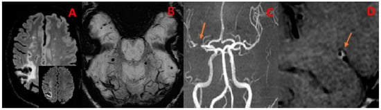
n = 2/23; specifically, brain and intestinal) or during follow-up (n = 6/23; namely, intestinal, brain and bone, in two cases for each, and adrenal) required additional imagery tools (26% of the patients had distant metastasis). Transoesophageal echocardiography, computed tomography (CT), magnetic resonance imagery, and even 
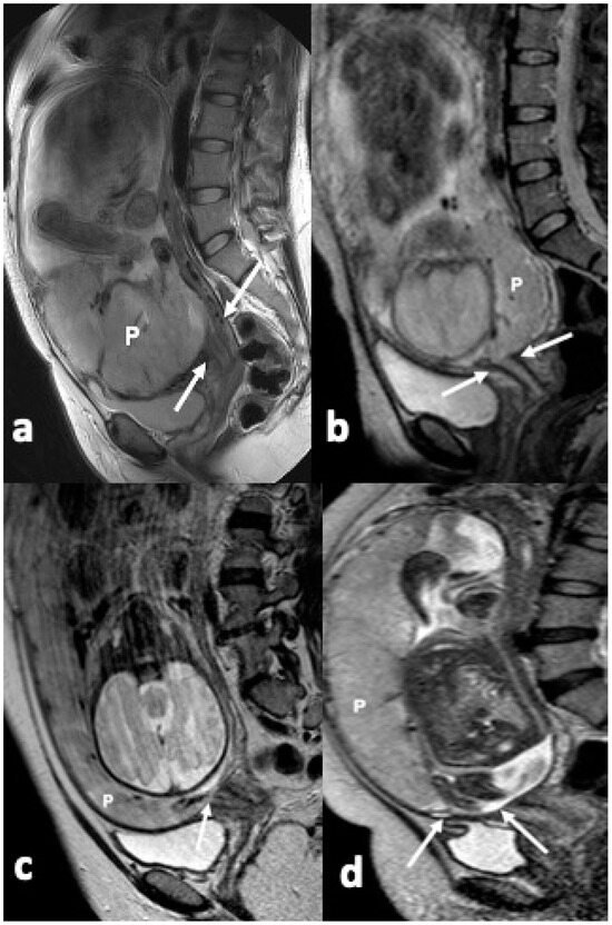
18F-FDG positronic emission tomography-CT (which shows hypermetabolic lesions in PCIS) represent the basis of multimodal tools of investigation. Tumour size varied from 3 cm to ≥9 cm (average largest diameter of 5.5 cm). The most frequent sites were the left atrium followed by the right ventricle and the right atrium. Post-operatory histological confirmation was provided in 20/23 cases and, upon tumour biopsy, in 3/23 of them. The post-surgery maximum free-disease interval was 8 years, the fatal outcome was at the earliest two weeks since initial admission. MDM2 analysis was provided in 7/23 subjects in terms of MDM2-positive status (two out of three subjects) at immunohistochemistry and MDM2 amplification (four out of five subjects) at genetic analysis. Additionally, another three studies addressed PCISs, and two of them offered specific MDM2/MDM2 assays (n = 35 patients with PCISs); among the provided data, we mention that one cohort (n = 20) identified a rate of 55% with regard to MDM2 amplification in intimal sarcomas, and this correlated with a myxoid pattern; another cohort (n = 15) showed that MDM2-positive had a better prognostic than MDM2-negative immunostaining. To summarize, MDM2 amplification and co-amplification, for example, with MDM4, CDK4, HMGA3, CCND3, PDGFRA, TERT, KIT, CCND3, and HDAC9, might improve the diagnosis of PCIS in addition to MDM2 immunostaining since 10–20% of these tumours are MDM2-negative. Further studies are necessary to highlight MDM2 applicability as a prognostic factor and as an element to be taken into account amid multi-layered management in an otherwise very aggressive malignancy.
Full article
(This article belongs to the Special Issue New Advances in Thoracic Surgery: Clinical Diagnosis, Treatment, and Prognosis)
Open AccessArticle
Early Identification of Sepsis-Induced Acute Kidney Injury by Using Monocyte Distribution Width, Red-Blood-Cell Distribution, and Neutrophil-to-Lymphocyte Ratio
by
Yi-Hsiang Pan, Hung-Wei Tsai, Hui-An Lin, Ching-Yi Chen, Chun-Chieh Chao, Sheng-Feng Lin and Sen-Kuang Hou
Diagnostics 2024, 14(9), 918; https://doi.org/10.3390/diagnostics14090918 (registering DOI) - 28 Apr 2024
Abstract
Sepsis-induced acute kidney injury (AKI) is a common complication in patients with severe illness and leads to increased risks of mortality and chronic kidney disease. We investigated the association between monocyte distribution width (MDW), red-blood-cell volume distribution width (RDW), neutrophil-to-lymphocyte ratio (NLR), sepsis-related
[...] Read more.
Sepsis-induced acute kidney injury (AKI) is a common complication in patients with severe illness and leads to increased risks of mortality and chronic kidney disease. We investigated the association between monocyte distribution width (MDW), red-blood-cell volume distribution width (RDW), neutrophil-to-lymphocyte ratio (NLR), sepsis-related organ-failure assessment (SOFA) score, mean arterial pressure (MAP), and other risk factors and sepsis-induced AKI in patients presenting to the emergency department (ED). This retrospective study, spanning 1 January 2020, to 30 November 2020, was conducted at a university-affiliated teaching hospital. Patients meeting the Sepsis-2 consensus criteria upon presentation to our ED were categorized into sepsis-induced AKI and non-AKI groups. Clinical parameters (i.e., initial SOFA score and MAP) and laboratory markers (i.e., MDW, RDW, and NLR) were measured upon ED admission. A logistic regression model was developed, with sepsis-induced AKI as the dependent variable and laboratory parameters as independent variables. Three multivariable logistic regression models were constructed. In Model 1, MDW, initial SOFA score, and MAP exhibited significant associations with sepsis-induced AKI (area under the curve [AUC]: 0.728, 95% confidence interval [CI]: 0.668–0.789). In Model 2, RDW, initial SOFA score, and MAP were significantly correlated with sepsis-induced AKI (AUC: 0.712, 95% CI: 0.651–0.774). In Model 3, NLR, initial SOFA score, and MAP were significantly correlated with sepsis-induced AKI (AUC: 0.719, 95% CI: 0.658–0.780). Our novel models, integrating MDW, RDW, and NLR with initial SOFA score and MAP, can assist with the identification of sepsis-induced AKI among patients with sepsis presenting to the ED.
Full article
(This article belongs to the Special Issue Laboratory Tests for Kidney Diseases)
►▼
Show Figures
Figure 1
Open AccessArticle
Conceptually Funded Usability Evaluation of an Application for Leveraging Descriptive Data Analysis Models for Cardiovascular Research
by
Oliver Lohaj, Ján Paralič, Zuzana Pella, Dominik Pella and Adam Pavlíček
Diagnostics 2024, 14(9), 917; https://doi.org/10.3390/diagnostics14090917 (registering DOI) - 28 Apr 2024
Abstract
The focus of this study, and the subject of this article, resides in the conceptually funded usability evaluation of an application of descriptive models to a specific dataset obtained from the East Slovak Institute of Heart and Vascular Diseases targeting cardiovascular patients. Delving
[...] Read more.
The focus of this study, and the subject of this article, resides in the conceptually funded usability evaluation of an application of descriptive models to a specific dataset obtained from the East Slovak Institute of Heart and Vascular Diseases targeting cardiovascular patients. Delving into the current state-of-the-art practices, we examine the extent of cardiovascular diseases, descriptive data analysis models, and their practical applications. Most importantly, our inquiry focuses on exploration of usability, encompassing its application and evaluation methodologies, including Van Welie’s layered model of usability and its inherent advantages and limitations. The primary objective of our research was to conceptualize, develop, and validate the usability of an application tailored to supporting cardiologists’ research through descriptive modeling. Using the R programming language, we engineered a Shiny dashboard application named DESSFOCA (Decision Support System For Cardiologists) that is structured around three core functionalities: discovering association rules, applying clustering methods, and identifying association rules within predefined clusters. To assess the usability of DESSFOCA, we employed the System Usability Scale (SUS) and conducted a comprehensive evaluation. Additionally, we proposed an extension to Van Welie’s layered model of usability, incorporating several crucial aspects deemed essential. Subsequently, we rigorously evaluated the proposed extension within the DESSFOCA application with respect to the extended usability model, drawing insightful conclusions from our findings.
Full article
(This article belongs to the Special Issue Machine Learning and Deep learning for Healthcare Data Processing and Analyzing)
►▼
Show Figures
Figure 1
Open AccessReview
Osteochondritis Dissecans of the Elbow in Overhead Athletes: A Comprehensive Narrative Review
by
Andro Matković, Thomas Ferenc, Damjan Dimnjaković, Nikolina Jurjević, Vinko Vidjak and Branka R. Matković
Diagnostics 2024, 14(9), 916; https://doi.org/10.3390/diagnostics14090916 (registering DOI) - 28 Apr 2024
Abstract
Osteochondritis dissecans (OCD) of the elbow mainly occurs in overhead athletes (OHAs). This narrative review aimed to comprehensively analyze the epidemiological data, etiological factors, clinical and imaging features, treatment options, and outcomes of OHAs with the diagnosis of elbow OCD. A literature search
[...] Read more.
Osteochondritis dissecans (OCD) of the elbow mainly occurs in overhead athletes (OHAs). This narrative review aimed to comprehensively analyze the epidemiological data, etiological factors, clinical and imaging features, treatment options, and outcomes of OHAs with the diagnosis of elbow OCD. A literature search was performed in PubMed/MEDLINE, Scopus, and Web of Science. Individuals with elbow OCD were usually 10–17 years of age with incidence and prevalence varying between studies, depending on the sport activity of the patients. The etiology of OCD lesions is multifactorial, and the main causes are believed to be repetitive trauma, the biomechanical disproportion of the articular surfaces, poor capitellar vascular supply, and inflammatory and genetic factors. Athletes usually presented with elbow pain and mechanical symptoms. The mainstay for the diagnosis of elbow OCD is MRI. The treatment of elbow OCD lesions should be conservative in cases of stable lesions, while various types of surgical treatment are suggested in unstable lesions, depending mainly on the size and localization of the lesion. The awareness of medical practitioners and the timely diagnosis of OCD lesions in OHAs are key to favorable outcomes.
Full article
(This article belongs to the Special Issue Recent Advances in Bone and Joint Imaging—2nd Edition)
►▼
Show Figures
Figure 1
Open AccessArticle
Assessing the Interpretation of Molecular Test Results in the Diagnosis of Bloodstream Infections
by
Natalia Słabisz, Patrycja Leśnik, Katarzyna Żybura-Wszoła, Ruth Dudek-Wicher, Urszula Nawrot and Jacek Majda
Diagnostics 2024, 14(9), 915; https://doi.org/10.3390/diagnostics14090915 (registering DOI) - 27 Apr 2024
Abstract
A retrospective study at the 4th Military Clinical Hospital in Wroclaw, Poland, assessed PCR testing alongside blood cultures to guide antimicrobial therapy decisions in hospitalized patients, to determine how much time the results of the molecular tests preceded conventional methods. Among 118 patients,
[...] Read more.
A retrospective study at the 4th Military Clinical Hospital in Wroclaw, Poland, assessed PCR testing alongside blood cultures to guide antimicrobial therapy decisions in hospitalized patients, to determine how much time the results of the molecular tests preceded conventional methods. Among 118 patients, Staphylococcus aureus (37%) and Escherichia coli (21%) were the most common bloodstream infection agents. Blood cultures utilized the BacT/ALERT 3D system, and molecular diagnostics were conducted using the FilmArray platform with the BIOFIRE BCID2 panel. Methicillin susceptibility was observed in 66% of S. aureus strains, while 26% of Gram-negative bacilli exhibited an ESBL phenotype. Therapeutic decisions based on molecular test results were often incorrect for S. aureus infections, particularly MSSA (64.5%), but generally accurate for Gram-negative bacilli. The median times from positive blood culture to BCID2 and pathogen identification/susceptibility were 10 h and 52 h, respectively. Molecular diagnostics facilitated faster initiation of appropriate antibiotic therapy, highlighting the need to educate medical staff on proper interpretation. Consulting within an antimicrobial stewardship program (ASP) could enhance the benefits of implementing molecular methods in bloodstream infection diagnostics.
Full article
(This article belongs to the Section Diagnostic Microbiology and Infectious Disease)
Journal Menu
► ▼ Journal Menu-
- Diagnostics Home
- Aims & Scope
- Editorial Board
- Reviewer Board
- Topical Advisory Panel
- Instructions for Authors
- Special Issues
- Topics
- Sections & Collections
- Article Processing Charge
- Indexing & Archiving
- Editor’s Choice Articles
- Most Cited & Viewed
- Journal Statistics
- Journal History
- Journal Awards
- Society Collaborations
- Conferences
- Editorial Office
Journal Browser
► ▼ Journal BrowserHighly Accessed Articles
Latest Books
E-Mail Alert
News
Topics
Topic in
Algorithms, Diagnostics, Entropy, Information, J. Imaging
Application of Machine Learning in Molecular Imaging
Topic Editors: Allegra Conti, Nicola Toschi, Marianna Inglese, Andrea Duggento, Matthew Grech-Sollars, Serena Monti, Giancarlo Sportelli, Pietro CarraDeadline: 31 May 2024
Topic in
Biomedicines, Current Oncology, Diagnostics, Gastrointestinal Disorders, JCM
Advances in Gastrointestinal and Liver Disease: From Physiological Mechanisms to Clinical Practice
Topic Editors: Davide Giuseppe Ribaldone, Gian Paolo CavigliaDeadline: 20 June 2024
Topic in
Biomolecules, Diagnostics, JCM, Metabolites, JPM, Nutrients
Metabolic Syndrome, Biomarkers and Lifestyles
Topic Editors: Sang Yeoup Lee, Young Hye ChoDeadline: 30 June 2024
Topic in
Cancers, Diagnostics, JCM, Current Oncology, Gastrointestinal Disorders, Biomedicines
Hepatobiliary and Pancreatic Diseases: Novel Strategies of Diagnosis and Treatments
Topic Editors: Alessandro Coppola, Damiano Caputo, Roberta Angelico, Domenech Asbun, Chiara MazzarelliDeadline: 20 July 2024
Conferences
Special Issues
Special Issue in
Diagnostics
Clinical Advances in Heart Failure
Guest Editor: Noémi NyolczasDeadline: 30 April 2024
Special Issue in
Diagnostics
Monitoring and Diagnosis of Patients during Rehabilitation Training
Guest Editor: Redha TaiarDeadline: 31 May 2024
Special Issue in
Diagnostics
Improvement of Diagnostic Sensitivity for Tumor Markers
Guest Editors: Jong-Han Lee, Jooyoung ChoDeadline: 16 June 2024
Special Issue in
Diagnostics
Endoscopy and Diagnostic Tools in Hepatobiliary and Pancreatic Diseases
Guest Editor: Stefano Francesco CrinòDeadline: 30 June 2024
Topical Collections
Topical Collection in
Diagnostics
Editorial Board Members' Collection Series: Diagnostic Approaches to Gastrointestinal and Pancreatic Diseases
Collection Editors: Paolo Aseni, Ervin Toth