Article

A New Phloroglucinol Diglycoside Derivative from Hypericum japonicum Thunb.

1
Department of Natural Products Chemistry, Shenyang Pharmaceutical University, 103. Wenhua Road Shenhe District, Shenyang, Liaoning 110016, P.R. China
2
Shenzhen Institute for Drug Control, Shenzhen, Guangdong 518029, P.R. China
*
Authors to whom correspondence should be addressed.
Molecules 2008, 13(11), 2796-2803; https://doi.org/10.3390/molecules13112796
Submission received: 30 September 2008 / Revised: 27 October 2008 / Accepted: 4 November 2008 / Published: 7 November 2008

Abstract

:
A new phloroglucinol diglycoside 1, together with eight known compounds, were isolated from Hypericum japonicum Thunb. The structure of the new compound 1 was determined by spectroscopic methods to be 4,6-dimethyl-1-O-[α-L-rhamnopyranosyl-(1→6)-β-D-glucopyranosyl] multifidol. Different solvent extracts of Hypericum japonicum Thunb. were tested for in vivo antihypoxic activity using mice, with the EtOAc extract showing better activity.

Graphical Abstract

Introduction

Hypericum japonicum Thunb. is an annual herb from the genus Hypericum L. (Clusiaceae/ Hypericaceae). The whole plant has been used for the treatment of several bacterial diseases, infectious hepatitis, gastrointestinal disorder and tumors [1]. As part of our search for antihypoxic ingredients from Chinese herbs, we carried out an activity screening study, during which we found that a 60% EtOH extract of Hypericum japonicum Thunb. showed better antihypoxic activity than other herb extracts. In continuation of this research, a new glycoside 1 and eight known compounds 2-9 (Figure 1) have been isolated by silica gel and ODS column chromatography and preparative HPLC. This paper deals with the isolation of these nine constituents, the structure elucidation of the new glycoside, and their antihypoxic activities.

Results and Discussion

Compound 1 (Figure 1) was obtained as brown amorphous powder, Molecules 13 02796 i001-
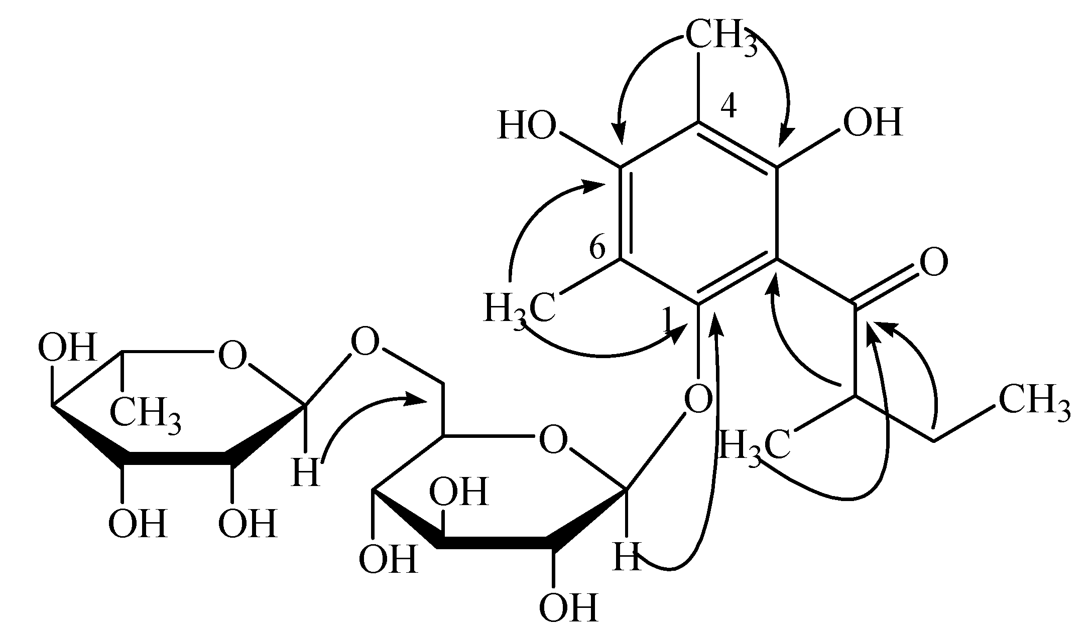
24.5°(c. 0.5, MeOH), m.p. 220~223°C. Its molecular formula was deduced as C25H38O13 from the quasi-molecular ion peak at 569.2200 ([M + Na]+, calcd. 569.2260) in HR-ESI-MS spectrum. The IR spectrum exhibited the absorptions at 3350 cm-1 (OH), 1630 cm-1 (carbonyl), 1598 cm-1 and 1586 cm-1 (phenyl). Its 1H-NMR spectrum (Table 1) showed one hydroxyl proton at δ 12.48 (1H, br s). It also showed five methyl groups at δ 2.10 (3H, s), 1.97 (3H, s), 1.07 (3H, d, J = 6.1 Hz), 0.91 (3H, d, J = 7.2 Hz) and 0.87 (3H, t, J =7.5 Hz), and two anomeric protons at δ 4.42 (1H, br s) and 4.29 (1H, d, J = 7.7 Hz). The 13C-NMR spectrum showed six aromatic signals (δ 160.0, 158.3, 152.7, 110.1, 109.9, 107.5) and one carbonyl signal (δ 210.0). Combining with DEPT and 1H-NMR data, we deduced that the benzene ring was completely substituted. Signals at δ 3.0~3.5 in the 1H-NMR spectrum and signals at δ 60~80 in the 13C-NMR spectrum suggested two sugar units.
Figure 1. Key HMBC correlations of compound 1 (1H →13C).
Molecules 13 02796 g001
Compound 1 was hydrolyzed with 2 M HCl/CH3OH solution to give 4,6-dimethylmultifidol, 4,6-dimethylmultifidol glucoside, one disaccharide and two monosaccharides. Multifidol and multifidol glucoside had been previously identified in the latex of Jatropha multifida in 1989 [2].. Two phloroglucinol glycosides: 2,6-dihydroxy-3,5-dimethyl-1-isobutyrylbenzene-4-O-β-D-glucoside and 2,6-dihydroxy-3,5-dimethyl-1-(2-methylbutyryl)benzene-4-O-β-D-glucoside had been previously identified in Hypericum japonicum [3]. These were very similar to “multifidol glucoside” in chemical structure, except for the glucose linkage with multifidol. The two monosaccharides were identified as rhamnose and glucose by GC-MS analysis using authentic monosaccharide samples as references. Combing the J value between the anomeric proton and the 2-H proton in each monosaccharide with the optical rotation values (the Molecules 13 02796 i001 of rhamnose is -4.4°, the Molecules 13 02796 i001 of glucose is +52.5°), the configuration of the rhamnose was identified as α-L, while that of the glucose was β-D. Thus, compound 1 was identified as 4,6-dimethyl-1-O-[α-L-rhamnopyranosyl-(1→6)-β-D-glucopyranosyl] multifidol. The complete assignments of 1H- and 13C-NMR data were based on the analyses of HSQC, 1H-1H COSY, and HMBC spectra (Figure 1). The key 1H-13C long-range correlations could be observed from H-1 of the rhamnose unit at δ 4.42 to C-6 of the glucopyranose unit at δ 67.4, from H-1 of the glucopyranose unit at δ 4.29 to C-1 of the phenyl unit at ä 152.7, and from H-2 of the methylbutyryl chain at δ 3.88 to C-2 of the phenyl unit at δ 110.1 in the HMBC spectrum.
Table 1. 1H-NMR and 13C-NMR (DMSO-d6 ) data of compound 1.
PositionδHδCPositionδHδC
Methylbutyryl chainGlucopyranose unit
1-210.014.29 (1H, d, 7.7)104.0
23.88 (1H, m)44.923.28 (1H, m)74.1
2-CH30.91 (3H, d, 7.2)17.933.19 (1H, m)76.2
31.31 (1H, m) 1.80 (1H, m)24.643.30 (1H, m)70.7
40.87 (3H, t, 7.5)11.953.10 (1H, m)75.0
Phenyl unit63.69 (2H, d, 10.5)67.4
1-152.7Rhamnose unit
2-110.114.42 (1H, br s)101.0
3-158.323.50 (1H, m)70.1
4-107.533.29 (1H, m)70.2
4-CH31.97 (3H, s)8.643.13 (1H, m)72.0
5-160.053.27 (1H, m)68.3
6-109.961.07 (3H, d, 6.1)17.8
6-CH32.10 (3H, s)9.5
The other eight known compounds were identified as quercetin (2), quercitrin (quercetin 3-O-L-rhamnoside) (3), quercetin 7-O-L-rhamnoside (4), isoquercitrin (quercetin 3-O-β-D-glucoside) (5), dihydrokaempferol (6), dihydroquercetin (7), 3,5,7,3’,5'-pentahydroxydihydroflavonol (8) and chlorogenic acid (9) by direct comparisons of their NMR data with literature data. The structures of compounds 2-9 are shown in Figure 2.
Figure 2. The structures of compounds 2-9 isolated from Hypericum japonicum Thunb.
Molecules 13 02796 g002

Antihypoxic activity of Hypericum japonicum Thunb extracts

Male mice (body weight: 18±2g) were randomly divided into a test group and a control group.. Before the mice were placed under hypoxic conditions for 2 h, the test substances (CHCl3 extract, EtOAc extract, n-BuOH extract and water extract, 20 mg/0.5 mL, equivalent to 1g/kg body weight) and the control solution (DMSO diluted solution, 0.5 mL) were administrated to mice by gastric perfusion. Then each mouse was placed in a 150 mL flask and the flasks were sealed with rubber plugs. Under the sealed and hypoxic situation, the tolerance time in both groups were recorded. The results were as follows:
Table 2. The tolerance time of mice administrated different extracts of Hypericum japonicum Thunb.
SubstancesDoseMice number (n)Tolerance time (min)
DMSO diluted solution0.5 mL2033.30 ± 7.23
60% EtOH extract20 mg/0.5 mL1038.47 ± 9.22
CHCl3 extract20 mg/0.5 mL1040.15 ± 9.58
EtOAc extract20 mg/0.5 mL1044.37 ± 10.25
n-BuOH extract20 mg/0.5 mL1038.37± 8.18
Water extract20 mg/0.5 mL1033.62 ± 11.30

Conclusions

From the study, the EtOAC extract of Hypericum japonicum Thunb. was proven to have antihypoxic activity. A new phloroglucinol diglycoside, 4,6-dimethyl-1-O-[α-L-rhamnopyranosyl-(1°6)-β-D-gluco-pyranosyl] multifidol, along with eight known compounds, were separated from the active fraction.

Experimental

General

Optical rotations were determined on a JASCO P-1020 polarimeter in MeOH and HR-ESI-MS spectra were obtained on a Micromass Q-TOF mass spectrometer. NMR spectra were recorded on a Bruker AVANCE 400 NMR spectrometer (400 MHz for 1H, 100 MHz for 13C). The NMR data were measured in DMSO-d6 with tetramethylsilane (TMS) as internal standard. IR spectra were recorded on a FTIR 8400 spectrophotometer (Shimadzu, Japan) using KBr discs as stated. UV spectra were recorded on a UV2401PC spectrophotometer (Shimadzu, Japan). Column chromatography was carried out on silica gel H60 (Qingdao Haiyang Chemical Group Corporation, Qingdao, P.R. China), Sephadex LH-20 (Amersham Biosciences AB) and ODS (60~80 μm, Merck) as packing materials.

Plant material

In our experiments we used the whole plant. Hypericum japonicum Thunb. herbs were collected from GuangXi province in P.R. China, in July 2003 and identified by Prof. Weichun Wu (Department of Medical Plants, Shenyang Pharmaceutical University, P.R. China). A voucher specimen was deposited at the Department of Natural Products Chemistry of Shenyang Pharmaceutical University.

Extraction

The 60% EtOH extract of Hypericum japonicum Thunb. (4.3 kg) was dissolved and suspended in water and partitioned between chloroform, ethyl acetate and n-BuOH, respectively (three times each). Four fractions, that is, a chloroform fraction (83 g), an ethyl acetate fraction (114 g), an n-butanol fraction (153 g) and an aqueous fraction (228 g) were obtained after evaporation of the corresponding solvents in vacuo. The EtOAc fraction was subjected to silica gel column chromatography (SiO2, 800 g, eluted with 100:0→50:50 CHCl3/MeOH) to obtain fractions E1-E14.. Fraction E2 fraction (2.6 g) was applied to a Sephadex LH-20 column and eluted with CHCl3/MeOH (1:1) to give a subfraction which was subjected to ODS column chromatography with eluted with a H2O/MeOH gradient with increasing MeOH percent, to yield 1 (5.5 mg), 2 (15.3 mg), 3 (20.6 mg) , 4 (19.4 mg) and 5 (10.7 mg). Fraction E14 (2.3 g) was chromatographed over Sephadex LH-20 column (CHCl3-MeOH=1:1) and purified using preparative HPLC (ODS, 35%~60% MeOH) to yield 6 (6.4 mg), 7 (8.1 mg) and 8 (5.9 mg). Fraction E12 (3.4 g) was chromatographed over Sephadex LH-20 column (CHCl3-MeOH=1:1) to yield 9 (1.3 g). Their structures are all shown in Figure 2..
Quercetin (2) [4]: C15H10O7; yellow powder (MeOH); HCl-Mg reaction (+); ESI-MS (positive) m/z 301 [M-H]+; UV (MeOH) λmax nm: 369, 258, 208; IR νmax (KBr): 3450 (br, OH), 2950, l670, l630, l550, l510, 1310 cm-1; 1H-NMR δ: 12.49 (1H, s, OH-5), 10.78 (1H, s, OH-7), 9.59 (1H, s, OH-4’), 9.36 (1H, s, OH-3), 9.30 (1H, s, OH-3’), 7.67 (1H, d, J = 2.2 Hz, H-2’), 7.53 (1H, dd, J = 8.5, 2.2 Hz, H-6’), 6.88 (1H, d, J = 8.5 Hz, H-5’), 6.41 (1H, d, J = 2.0 Hz, H-8), 6.18 (1H, d, J = 2.0 Hz, H-6); 13C-NMR δ: 175.8 (C-4), 163.9 (C-7), 160.7 (C-9), 156.1 (C-5), 147.7 (C-4’), 146.8 (C-2), 145.0 (C-3’), 135.7 (C-3), 121.9 (C-1’), 119.9 (C-6’), 115.6 (C-5’), 115.0 (C-2’), 103.0 (C-10), 98.2 (C-6), 93.3 (C-8).
Quercitrin (quercetin 3-O-L-rhamnoside) (3) [5]: C21H20O11; yellow powder (MeOH); HCl-Mg reaction (+); Molish reaction (+); ESI-MS (positive) m/z 471 [M+Na]+, 447[M-H]+; UV (MeOH) λmax nm: 355, 265, 257; 1H-NMR δ: 12.66 (1H, s, OH-5), 11.00 (1H. s, OH-7), 9.83 (1H. s, OH-4’), 9.42 (1H. s, OH-3’), 7.31 (1H, d, J = 2.0 Hz, H-2’)7.27 (1H, dd, J = 8.3, 2.0 Hz, H-6’), 6.88 (1H, d, J = 8.3 Hz, H-5’), 6.41 (1H, d, J = 2.0 Hz, H-8), 6.22 (1H, d, J = 2.0 Hz, H-6), 5.27 (1H, br s, rha-H-1), 3.99 (1H, m, rha-H-2), 3.55 (1H, m, rha-H-3), 3.18 (1H, m, rha-H-5), 3.10 (1H, m, rha-H-4), 0.82 (3H, d, J = 5.9 Hz, rha-H-5-CH3); 13C-NMR δ: 177.9 (C-4), 164.3 (C-7), 161.4 (C-5), 157.5 (C-9), 156.6 (C-2), 148.6 (C-4’), 145.3 (C-3’), 134.4 (C-3), 121.3 (C-6’), 120.9 (C-1’), 115.8 (C-5’), 115.6 (C-2’), 104.2 (C-10), 101.9 (rha-C-1), 98.8 (C-6), 93.8 (C-8), 71.3 (rha-C-4), 70.7 (rha-C-3), 70.5 (rha-C-2), 70.2 (rha-C-5), 17.6 (rha-C-5-CH3).
Quercetin 7-O-L-rhamnoside (4) [6]: C21H20O11; yellow powder (MeOH); HCl-Mg reaction (+); Molish reaction (+); ESI-MS (positive) m/z 471 [M+Na]+, 447 [M-H]+; 1H-NMR δ: 12.49 (1H, s, OH-5), 9.48 (1H, s, OH-4’), 7.73 (1H, d, J = 2.2 Hz, H-2’), 7.59 (1H, dd, J = 8.5, 2.2 Hz, H-6’), 6.90 (1H, d, J = 8.5 Hz, H-5’), 6.79 (1H, d, J = 2.1 Hz, H-8), 6.42 (1H, d, J = 2.1 Hz, H-6), 5.55 (1H, d, J = 1.4 Hz, rha-H-1), 3.86 (1H, m, rha-H-2), 3.66 (1H, m, rha-H-3), 3.46 (1H, m, rha-H-5), 3.32 (1H, m, rha-H-4), 1.14 (3H, d, J = 6.1 Hz, rha-H-5-CH3); 13C-NMR δ: 175.9 (C-4), 161.4 (C-7), 160.3 (C-5), 155.7 (C-9), 147.9 (C-2), 147.5 (C-4’), 145.1 (C-3’), 136.1 (C-3), 121.8 (C-1’), 120.1 (C-6’), 115.6 (C-2’), 115.2 (C-5’), 104.6 (C-10), 98.8 (rha-C-1), 98.4 (C-6), 94.2 (C-8), 71.6 (rha-C-4), 70.3 (rha-C-3), 70.1 (rha-C-2), 69.8 (rha-C-5), 17.9 (rha-C-5-CH3).
Isoquercitrin(quercetin 3-O-β-D-glucoside) (5) [7]: C21H20O12; yellow powder (MeOH); HCl-Mg reaction (+); Molish reaction (+); ESI-MS (positive) m/z 487 [M+Na]+, 463 [M-H]+; IR νmax (KBr): 3375, 1660, 1606, 1494, 1362, 1304, 1200, 1061, 801, 595 cm-1; 1H-NMR δ: 12.64 (1H, s, OH-5), 10.78 (1H, s, OH-7), 9.60 (1H, s, OH-4’), 9.16 (1H, s, OH-3’), 7.58 (1H, dd, J = 9.0, 2.2 Hz, H-6’), 7.56 (1H, d, J = 2.2 Hz, H-2’), 6.84 (1H, d, J = 9.0 Hz, H-5’), 6.40 (1H, d, J = 2.1 Hz, H-8), 6.20 (1H, d, J = 2.0 Hz, H-6), 5.46 (1H,d, J = 7.5 Hz, glu-H-1), 4.00~3.20 (4H, m, glu-H-2~5), 3.57 (2H, d, J = 11.4 Hz, glu-H-6); 13C-NMR δ: 177.4 (C-4), 164.1 (C-7), 161.2 (C-5), 156.3 (C-2), 156.2 (C-9), 144.8 (C-3’), 148.4 (C-4’), 133.3 (C-3), 121.6 (C-6’), 121.1 (C-1’), 116.2 (C-5’), 115.2 (C-2’), 104.0 (C-10), 100.9 (glu-C-1), 98.6 (C-6), 93.5 (C-8), 77.5 (glu-C-3), 76.5 (glu-C-5), 74.1 (glu-C-2), 69.9 (glu-C-4), 61.0 (glu-C-6).
Dihydrokaempferol (6) [8]: C15H12O6; colorless needle (MeOH); HCl-Mg reaction (+); Molish reaction (-); ESI-MS (positive) m/z 311 [M+Na]+, 287 [M-H]+; 1H-NMR δ: 11.90 (1H, s, OH-5), 10.84 (1H, s, OH-7), 9.55 (1H, s, OH-4’), 7.30 (2H, d, J = 8.4 Hz, H-2’, 6’), 6.78 (2H, d, J = 8.4 Hz, H-3’, 5’), 5.91 (1H, d, J = 2.0 Hz, H-8), 5.86 (1H, d, J = 2.0 Hz, H-6), 5.75 (1H, br s, OH-3), 5.05 (1H, d, J = 11.4 Hz, H-2), 4.58 (1H, d, J = 11.4 Hz, H-3); 13C-NMR δ: 197.8 (C-4), 166.8 (C-7), 163.3 (C-5), 162.5 (C-9), 157.7 (C-4’), 129.4 (C-1’), 127.5 (C-2’, 6’), 114.9 (C-3’, 5’), 100.4 (C-10), 96.0 (C-6), 95.0 (C-8), 82.9 (C-2), 71.4 (C-3).
Dihydroquercetin (7) [9]: C15H12O7; white needle (MeOH); HCl-Mg reaction (+); Molish reaction (-); ESI-MS (positive) m/z 303 [M-H]+; UV (MeOH) λmax nm: 327, 289; IR νmax (KBr): 3410 (OH); 1645 (C=O); 1610, l505 (phenyl C=C); 1120, 975, 770; 1H-NMR δ: 11.89 (1H, s, OH-5), 10.81 (1H, s, OH-7), 9.01 (1H, s, OH-4’), 8.96 (1H, s, OH-3’), 6.87 (1H, s, H-6’), 6.74 (2H, s, H-2’, 5’), 5.90 (1H, d, J = 2.1 Hz, H-8), 5.85 (1H, d, J = 2.1 Hz, H-6), 4.98 (1H, d, J = 11.1 Hz, H-2), 4.50 (1H, d, J = 11.1 Hz, H-3); 13C-NMR δ: 197.7 (C-4), 166.7 (C-7), 163.3 (C-5), 162.5 (C-9), 145.7 (C-4’), 144.9 (C-3’), 128.0 (C-1’), 119.3 (C-6’), 115.3 (C-2’), 115.1 (C-5’), 100.5 (C-10), 95.9 (C-6), 94.9 (C-8), 83.0 (C-2), 71.5 (C-3).
3,5,7,3’,5'-Pentahydroxydihydroflavonol (8) [10]: C15H12O7; yellow powder (MeOH); HCl-Mg reaction (+); Molish reaction (-); ESI-MS (positive) m/z 303 [M-H]+; 1H-NMR δ: 11.89 (1H, s, OH-5), 10.86 (1H, s, OH-7), 8.93 (1H, s, OH-5’), 8.92 (1H, s, OH-3’), 6.93 (1H, d, J = 1.7 Hz, H-4’), 6.72 (2H, d, J = 1.7 Hz, H-2’, 6’), 6.18 (1H, d, J = 6.2 Hz, H-2), 5.92 (1H, d, J = 2.1 Hz, H-8), 5.91 (1H, d, J = 2.1 Hz, H-6), 5.33 (1H, d, J = 2.4 Hz, OH-3), 4.02 (1H, dd, J = 6.2, 2.4 Hz, H-3). 13C-NMR δ: 195.5 (C-4), 166.7 (C-7), 163.9 (C-5), 162.7 (C-9), 144.7 (C-5’), 145.2 (C-3’), 127.0 (C-1’), 118.6 (C-6’), 115.3 (C-2’), 114.9 (C-4’), 100.2 (C-10), 95.8 (C-6), 94.9 (C-8), 81.0 (C-2), 70.9 (C-3).
Chlorogenic acid (9) [11]: C16H18O9; light yellow powder (MeOH); HCl-Mg reaction (-); FeCl3 reaction (+);ESI-MS (positive) m/z 377 [M+Na]+, 353[M-H]+; UV (MeOH) λmax nm: 204.5 sh, 218, 246, 296..5 sh, 329.5; IR νmax (KBr): 3412 (br), 2928, l689, l628, l604, 15l8, 1374, l275, l181, 1160,1120, 1082,1040, 977, 853, 813; 1H-NMR δ: 9.55 (1H, s, caf-OH-7), 9.18 (1H, s, caf-OH-6), 7.46 (1H, d, J = 15.9 Hz, caf-H-3), 7.02 (1H, d, J = 2.0 Hz, caf-H-5), 6.95 (1H, dd, J = 8.2, 2.0 Hz, caf-H-9), 6.76 (1H, d, J = 8.2 Hz, caf-H-8), 6.19 (1H, d, J = 15.9 Hz, caf-H-2), 5.16 (1H, m, qui-H-3), 3.86 (1H, m, qui-H-5), 3.63 (1H, m, qui-H-4), 1.88 (2H, m, qui-H-2), 1.99 (2H, m, qui-H-6). 13C-NMR δ: 176.0 (qui-C-1-COOH), 166.1 (caf-C-1), 148.1 (caf-C-7), 145.6 (caf-C-6), 144.4 (caf-C-3), 125.7 (caf-C-4), 121.1 (caf-C-9), 115.8 (caf-C-5), 115.0 (caf-C-2), 114.6 (caf-C-8), 73.0 (qui-C-1), 71.3 (qui-C-4), 70.4 (qui-C-3), 67.7 (qui-C-5), 35.1 (qui-C-2), 34.9 (qui-C-6).

Acknowledgements

We thank the Key Laboratory for Research and Development of New Drugs, Shenzhen and the Shenzhen Institute for Drug Control for the NMR and MS spectra measurements.

References

  1. Grand Dictionary of Chinese Traditional Medicine; (Book One); Shanghai Science & Technology Press: Shanghai, P.R. China, 1977; pp. 813–814.
  2. Kosasi, S.; van der Sluis, W. G.; Labadie, R. P. Multifidol and multifidol glucoside from the latex of Jatropha multifida. Photochemistry 1989, 28, 2439–2441. [Google Scholar] [CrossRef]
  3. Wu, Q. L.; Wang, S. P.; Wang, L. W.; Yang, J. S.; Xiao, P. G. New phloroglucinol glycosides from Hypericum japonicum. Chin. Chem. Lett. 1998, 9, 469–470. [Google Scholar]
  4. Gao, W.; Shen, Y.; Zhang, H. J.; Tang, H.; Lin, H. W.; Qiu, F. The chemical constituents of Potentilla chinensis. Pharm. Care Res. 2007, 7, 262–264. [Google Scholar]
  5. Ma, X. F.; Tian, W. X.; Wu, L. H.; Cao, X. L.; Ito, Y. C. Isolation of quercetin-3-O-L-rhamnoside from Acer truncatum Bunge by high-speed counter-current chromatography. J. Chromatogr. A 2005, 1070, 211–214. [Google Scholar] [CrossRef]
  6. Zhang, L.; Jin, Y. Y.; Tian, J. K. Studies on Chemical Constituents of Hypericum japonicum. Chin. Pharm. J. 2007, 42, 341–344. [Google Scholar]
  7. Agnihotri, V. K.; ElSohly, H. N.; Khan, S. I.; Smillie, T. J.; Khan, I. A.; Walker, L. A. Antioxidant constituents of Nymphaea caerulea flowers. Phytochemistry 2008, 69, 2061–2066. [Google Scholar] [CrossRef]
  8. Ding, L. S.; Liang, Q. L.; Teng, Y. F. Study on flavonoids in seeds of Hovenia dulcis. Acta Pharm. Sin. 1997, 32, 600–602. [Google Scholar]
  9. Shilova, I. V.; Pisareva, S. I.; Krasnov1, E. A.; Bruzhes1, M. A.; Pyak, A. I. Antioxidant properties of Bergenia crassifolia extract. Pharm. Chem. J. 2006, 40, 620–623. [Google Scholar] [CrossRef]
  10. Fu, F.; Li, T. Z.; Liu, R. H.; Zhang, W.; Zhang, C.; Zhang, W. D.; Chen, H. S. Studies on the Flavonoids of Hypericum japonicum Thunb. ex Murray. Chin. J. Nat. Med. 2004, 2, 283–284. [Google Scholar]
  11. Teng, R. W.; Zhou, Z. H.; Wang, D. Z.; Yang, C. T. Four Caffeoylquinic Acids from Morina Nepalensis Var. Alba Hand.-Mazz. Chin. J. Magn. Reson. 2002, 9, 167–174. [Google Scholar]
  • Sample Availability: Contact the authors.

