Article

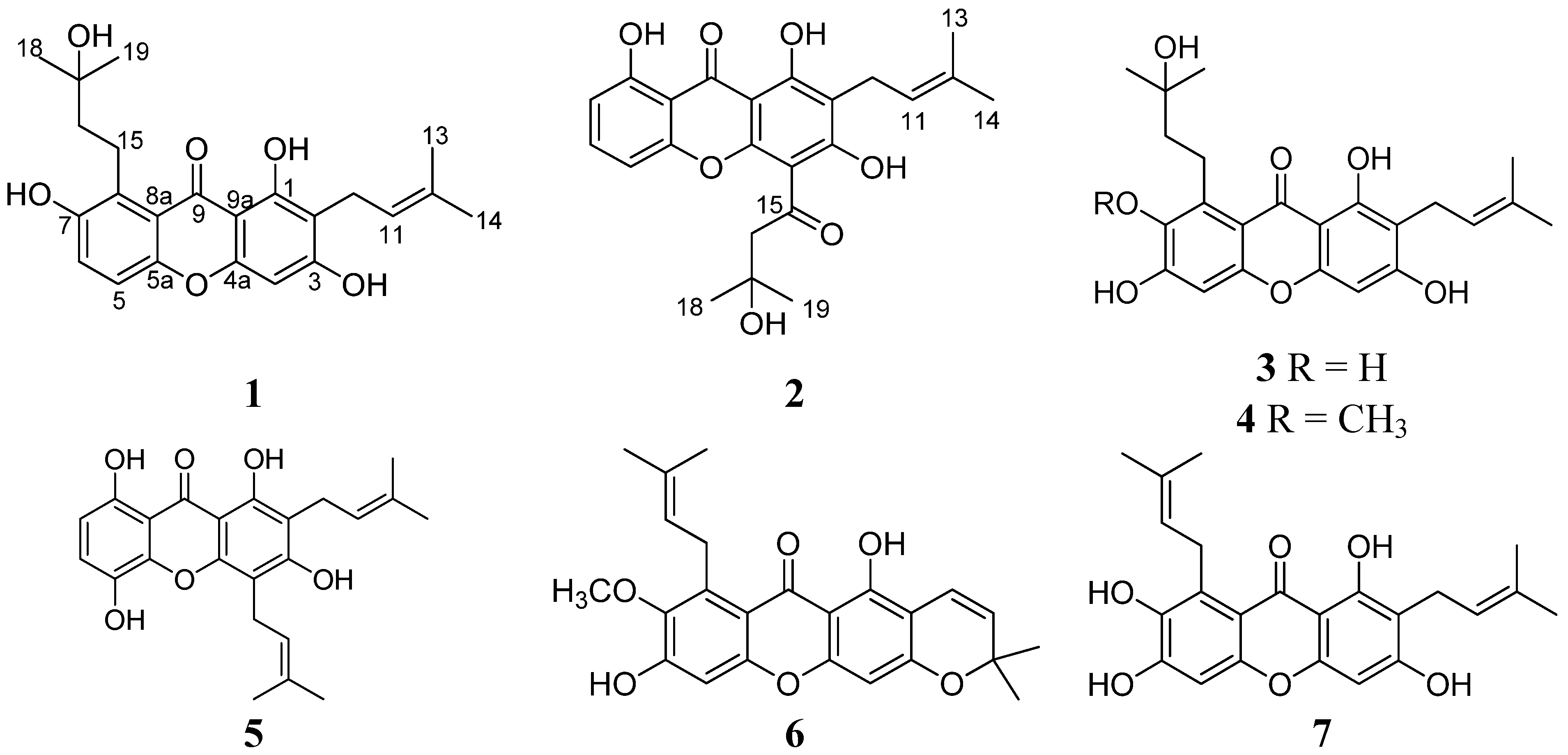
Cytotoxic Prenylated Xanthones from the Pericarps of Garcinia mangostana

1
School of Chemistry and Chemical Engineering, Sun Yat-Sen University, Guangzhou 510275, China
2
Instrumental Analysis and Research Center, Sun Yat-Sen University, Guangzhou 510275, China
3
State Key Laboratory of Oncology in South China, Cancer Center, Sun Yat-Sen University, Guangzhou 510060, China
4
School of Pharmaceutical Sciences, Sun Yat-Sen University, Guangzhou 510006, China
5
Guangdong Technology Research Center for Advanced Chinese Medicine, Guangzhou 510006, China
*
Authors to whom correspondence should be addressed.
Molecules 2014, 19(2), 1820-1827; https://doi.org/10.3390/molecules19021820
Submission received: 30 December 2013 / Revised: 21 January 2014 / Accepted: 27 January 2014 / Published: 6 February 2014
(This article belongs to the Section Natural Products Chemistry)

Abstract

:
Bioassay-guided fractionation of an ethanol extract of the pericarps of Garcinia mangostana led to the isolation of two new prenylated xanthones, named 1,3,7-trihydroxy-2-(3-methyl-2-butenyl)-8-(3-hydroxy-3-methylbutyl)-xanthone (1) and 1,3,8-trihydroxy-2-(3-methyl-2-butenyl)-4-(3-hydroxy-3-methylbutanoyl)-xanthone (2), together with the five known compounds garcinones C (3) and D (4), gartanin (5), xanthone I (6), and γ-mangostin (7). Their structures were elucidated primarily based on MS and NMR data. Compounds 17 showed significant cytotoxic activities against various human cancer cell lines.

1. Introduction

Garcinia mangostana, commonly called mangosteen, is widely distributed in Southeast Asia. This tropical plant is famous for its nutritious and flavorful fruits. The pericarps have been used as folk medicine to treat the diarrhea, bladder infections, gonorrhea, and skin rashes for more than one hundred years. Previous chemical investigation of the whole fruits, trunks, branches, leaves, and pericarps afforded some oxygenated xanthones that have attracted much attention for their fascinating structures and a wide range of biological activities, such as anticancer, antifungal, antibacterial, antiviral, antioxidant, and antiplasmodial properties [1,2,3,4,5,6,7]. Recently, we carried out an HPLC-MS analysis of the ethanol extract of the pericarps, and found that the number of the HPLC peaks was much greater than the number of compounds isolated. Thus, the structures of some of the minor peaks are believed to still be unidentified. The semi-pure fractions were screened for cytotoxicity against the cancer cell lines CNE1 and CNE2, and showed the significant inhibitory activities, with IC50 values of less than 20 μg/mL. These findings aroused our great interest in further investigating these chemical constituents and their pharmacological activities. The ethanol extract of the pericarps was subjected to silica gel column chromatography followed by repeated HPLC to afford two new prenylated xanthone derivatives, 1,3,7-trihydroxy-2-(3-methyl-2-butenyl)-8-(3-hydroxy-3-methylbutyl)-xanthone (1) and 1,3,8-trihydroxy-2-(3-methyl-2-butenyl)-4-(3-hydroxy-3-methylbutanoyl)-xanthone (2), together with the five known compounds garcinones C (3) and D (4), gartanin (5), xanthone I (6), and γ-mangostin (7) (Figure 1). Compounds 17 showed significant cytotoxic activities against various human cancer cell lines. Herein, we describe the isolation, structure elucidation, and biological evaluation of these compounds.
Figure 1. Chemical structures of compounds 17.
Molecules 19 01820 g001

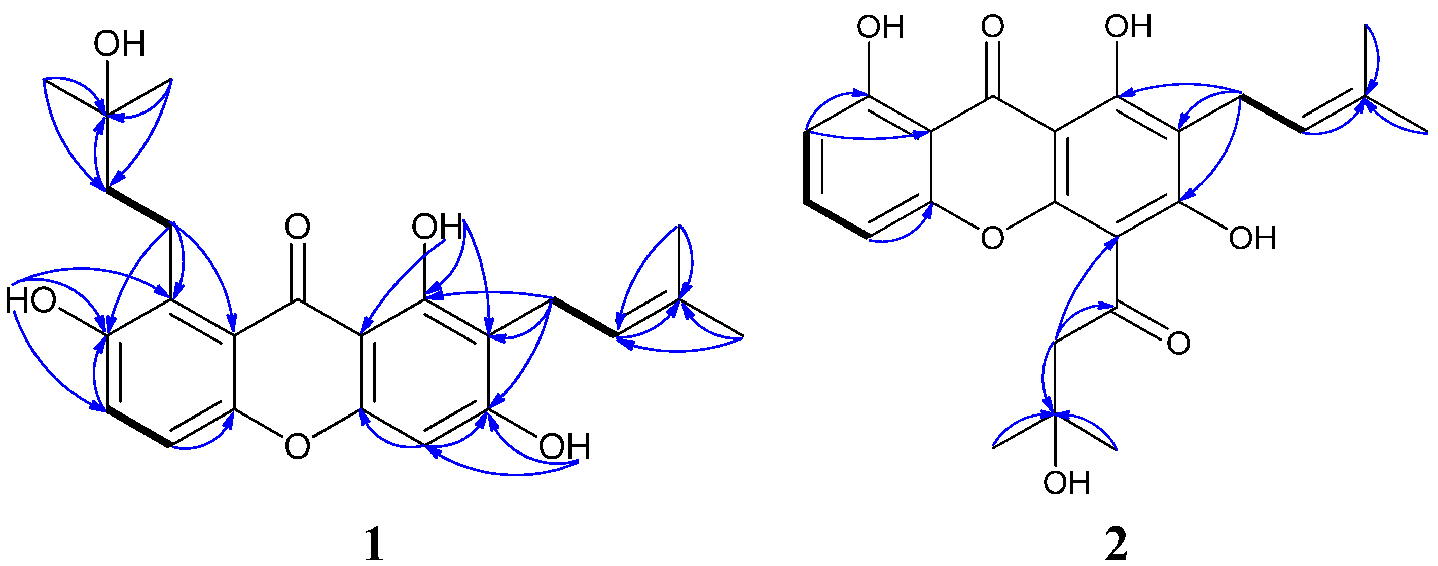
2. Results and Discussion

Compound 1 was obtained as a yellow powder with the molecular formula C23H26O6 and eleven degrees of unsaturation as established by its HREIMS peak at m/z 398.1725 [M]+.. The UV spectrum showed the characteristic absorption bands of a xanthone skeleton at λmax 350, 287, and 247 nm. The 13C-NMR data and DEPT experiments displayed four methyls, three methylenes, four methines, and twelve quarternary carbons (Table 1). One carbonyl group at δC 184.3 and twelve aromatic carbons at δC 163.7, 161.9, 156.3, 152.5, 152.4, 131.1, 124.5, 119.6, 116.8, 111.1, 104.3 and 93.3 constituted the xanthone skeleton. In the 1H-1H COSY spectrum, the allyl proton at H-11 (δH 5.29, t, J = 7.2 Hz, 1H) displaying cross peaks with H-10 (δH 3.37, d, J = 7.2 Hz, 2H), H-13 (δH 1.79, s), and H-14 (δH 1.65, s), combined with the HMBC correlations between H-11, H-13, H-14 and C-12 (δC 131.6 (C)) (Figure 2) revealed that a prenylated 3-methyl-2-butenyl side chain in this molecule. 1H-1H COSY correlations of H-5 (δH 7.23, d, J = 9.2 Hz)/H-6 (δH 7.33, d, J = 9.2 Hz), and H-15 (δH 3.49, t, J = 7.6 Hz, 2H)/H-16 (δH 1.84, t, J = 7.6 Hz, 2H), revealed two partial structures –CHCH– and –CH2CH2–. The quarternary carbon at δC 71.1 (C-17) connected to two methyl groups (δC 29.8, C-18 and C-19), the hydroxyl group (δH 3.81, OH-18), and the above partial structure –CH2CH2–, and formed the other prenylated 3-hydroxy-3-methylbutyl side chain. Three phenolic hydroxyl groups at δH 13.69 (s, OH-1), 9.68 (s, OH-3) and 8.72 (s, OH-7), were placed at C-1, C-3, and C-7, respectively, based on the HMBC correlations from OH-1 to C-1, C-2 and C-9a, from OH-3 to C-2, C-3 and C-4, from OH-7 to C-6, C-7 and C-8. The hydroxyl group at δH 13.69 (s, OH-1) formed an intramolecular hydrogen bond with the carbonyl group at δC 184.3 (C-9). The HMBC correlations between H-10 and C-1, C-2 and C-3 indicated that the prenylated 3-methyl-2-butenyl side chain was connected to C-2, moreover, the correlations between H-15 and C-7, C-8 and C-8a confirmed the other 3-hydroxy-3-methylbutyl side chain was connected to C-8. Therefore, compound 1 was elucidated as 1,3,7-trihydroxy-2-(3-methyl-2-butenyl)-8-(3-hydroxy-3-methylbutyl)-xanthone.
Compound 2, was obtained as a yellow amorphous solid. The molecular formula was determined to be C23H24O7 by analysis of the HREIMS peak at m/z 412.1517 [M]+ and its 13C-NMR data (Table 1). The 13C-NMR data and DEPT experiment displayed four methyls, two methylenes, four methines, and thirteen quarternary carbons. The cross-peaks of H-5 (δH 7.70, d, J = 8.0 Hz)/H-6 (δH 7.37, dd, J = 8.0, 7.6 Hz), H-6/H-7 (δH 7.44, d, J = 7.6 Hz) in 1H-1H COSY showed that three aromatic hydrogens formed a –CHCHCH– partial structure in this molecule (Figure 2). The HMBC correlations of H-16 (δH 3.67, s, 2H)/C-15 (δC 206.1), H-16/C-17 (δC 72.0), H-18 (δH 1.45, s)/C-17, and H-19 (δH 1.45, s)/C-17 indicated a 3-hydroxy-3-methylbutanoyl partial structure. The other 3-methyl-2-butenyl side chain was identical to that of compound 1. The location of the side chains on the xanthone core were assigned at C-2 and C-4 by interpretation of the HMBC correlations of H-10/C-1, H-10/C-2, H-10/C-3, and H-16/C-4. Three phenolic hydroxyl groups at δH 14.33, 14.40 and 15.11 were located at C-1, C-3, and C-8, and they formed intramolecular hydrogen bonds with the carbonyl groups. Based on the analysis above, the structure of compound 2 was assigned as 1,3,8-trihydroxy-2-(3-methyl-2-butenyl)-4-(3-hydroxy-3-methylbutanoyl)-xanthone.
The known compounds were identified as garcinones C [8] and D [9] (3 and 4), gartanin [10,11] (5), xanthone I [12] (6), and γ-mangostin [13] (7), respectively, by comparison of their physical and spectroscopic data with those reported previously.
Table 1. 1H- and 13C- NMR Data of compounds 1 and 2 at 400 and 100 MHz, resp., in acetone-d6. δ in ppm, J in Hz.
Position1 2
δHδCδHδC
1 161.9 165.9
2 111.1 111.8
3 163.7 170.3
46.42 s93.3 105.0
4a 156.3 158.3
57.23 d (9.2)116.8 7.70 d (8.0)115.9
5a 152.4 145.7
67.33 d (9.2)124.5 7.37 dd (8.0, 7.6)126.2
7 152.5 7.44 d (7.6)121.9
8 131.1 147.4
8a 119.6 122.1
9 184.3 181.9
9a 104.3 103.0
103.37 d (7.2)22.1 3.36 d (7.2)21.6
115.29 t (7.2)123.6 5.26 t (7.2)122.3
12 131.6 132.5
131.79 s18.1 1.80 s18.0
141.65 s26.0 1.66 s25.9
153.49 t (7.6)22.6 206.1
161.84 t (7.6)44.5 3.67 s55.3
17 71.1 72.0
181.30 s29.8 1.45 s30.4
191.30 s29.8 1.45 s30.4
OH-113.69 s 14.33 s a
OH-39.68 s 14.40 s a
OH-78.72 s
OH-8 15.12 s a
OH-183.81 s 2.81 s
a These data may be interchanged.
Figure 2. 1H-1H COSY (bold line) and main HMBC (arrow) correlations of compounds 1 and 2.
Molecules 19 01820 g002
The human nasopharyngeal carcinoma cell lines CNE1, CNE2, SUNE1 and HONE1, human lung cancer cell lines A549 and GLC82, human breast cancer cell line MCF-7, and human hepatic cancer cell line Bel-7402 were used to examine the cytotoxic activities of compounds 17 in vitro. Hirsutanol A was used as positive control. As can be seen in Table 2, compounds 17 showed significant cytotoxic activities against these human cancer cell lines. By comparison of their potencies on the tested cancer cell lines, compounds 1 and 2 are similar to compounds 3 and 7, and greater than 46 and hirsutanol A.
Table 2. Cytotoxic activities of 17, and hirsutanol A in vitro. IC50 values in µM a.
Cancer cell Lines1234567Hirsutanol A
Human nasopharyngeal carcinoma cell line CNE11.432.090.7616.286.4615.581.8510.06
Human nasopharyngeal carcinoma cell line CNE20.731.210.549.044.519.771.8112.70
Human nasopharyngeal carcinoma cell line SUNE12.232.762.3024.068.7811.364.413.53
Human nasopharyngeal carcinoma cell line HONE11.1816.727.4011.172.7817.48
Human lung cancer cell line A5491.9720.1715.923.7912.05
Human lung cancer cell line GLC8215.385.5216.973.4610.11
Human breast cancer cell line MCF-72.8519.308.2610.075.2710.35
Human hepatic cancer cell line Bel–74023.3216.737.7811.383.6624.80
a – = not screened.

3. Experimental

3.1. General Procedures

Optical rotations were measured on a Schmidt and Haensch polartronic HNQW5 optical rotation spectrometer (SCHMIDT + HAENSCH GmbH & Co., Berlin, Germany). IR spectra were obtained using a PerkinElmer Frontier FT-IR spectrophotometer (PerkinElmer Inc., Waltham, MA, USA) UV spectra were recorded on a Shimadzu UV-Vis-NIR spectrophotometer (Shimadzu Corporation, Nakagyo-ku, Kyoto, Japan). 1D and 2D NMR spectra were recorded on a Bruker Avance II 400 spectrometer (Bruker BioSpin AG, Industriestrasse 26, Fällanden, Switzerland). The chemical shifts are relative to the residual solvent signals (acetone-d6: δH 2.05 and δC 29.92; DMSO-d6: δH 2.50 and δC 39.51). Mass spectra were obtained on a Thermo DSQ EI low-resolution mass spectrometer (Thermo Fisher Scientific, Waltham, MA, USA) and a Thermo MAT95XP EI high-resolution mass spectrometer (Thermo Fisher Scientific). Preparative HPLC was performed using a Shimadzu LC-20AT HPLC pump equipped with an SPD-20A dual λ absorbance detector and a Shim-pack PRC-ODS HPLC column (250 × 20 mm) (Shimadzu Corporation, Nakagyo-ku, Kyoto, Japan).

3.2. Plant Material

The pericarps of Garcinia mangostana were collected from Guangzhou City, Guangdong Province, China, in June 2013. Voucher specimens are deposited in School of Chemistry and Chemical Engineering, Sun Yat-sen University.

3.3. Extraction and Isolation

The wet pericarps of Garcinia mangostana Linn (1 Kg) were extracted three times with 95% ethanol (2 L) at r.t. for 12 h. The extract was concentrated by low-temperature rotary evaporation. The ethanol extract (20 g) was chromatographed on a silica gel column with petroleum ether–EtOAc (100:0–0:100) and then EtOAc–MeOH (100:0–0:100) as the eluent to afford 15 fractions. Fr. 6 (1.08 g) was further purified by repeated preparative HPLC eluted with H2O–MeCN (60:40, v/v) to yield compounds 1 (6 mg), 3 (146 mg), 4 (168 mg) and 6 (45 mg). Fr. 7 (1.43 g) was further purified by HPLC with a gradient of H2O–MeCN (40:60 up to 0:100, v/v) to afford compounds 2 (12 mg), 5 (78 mg), and 7 (463 mg).

3.4. Spectral Data

1,3,7-Trihydroxy-2-(3-methyl-2-butenyl)-8-(3-hydroxy-3-methylbutyl)xanthone (1). Yellow solid. Molecules 19 01820 i001: + 15.8 (c 0.03, MeOH); UV (MeOH) λmax (ε) 207 nm (9,868), 247 nm (15,547), 287 nm (12,367), 350 nm (2,172); IR vmax 3229, 2964, 2927, 2855, 1642, 1609, 1582, 1485, 1460, 1373, 1319, 1306, 1281, 1231, 1209, 1182, 1161, 1125, 1100, 1083, 1054, 937, 904, 818, 797, 783 cm−1; 1H- and 13C-NMR data, see Table 1; LREIMS m/z 398, 380, 363, 337, 325, 309, 295, 281, 269, 257, 241, 213, 185, 167, 149, 130, 105, 91, 77, 69, 57; HREIMS m/z 398.1725 [M]+ (calcd for C23H26O6, 398.1724).
1,3,8-Trihydroxy-2-(3-methyl-2-butenyl)-4-(3-hydroxy-3-methylbutanoyl)-xanthone (2). Yellow amorphous solid. Molecules 19 01820 i001: + 19.7 (c 0.014, MeOH); UV (MeOH) λmax (ε) 210 nm (34,165), 242 nm (46,002), 264 nm (44,690), 312 nm (25,976), 370 nm (8,733); IR vmax 3466, 3176, 2956, 2926, 2854, 1638, 1600, 1578, 1500, 1464, 1428, 1374, 1356, 1339, 1292, 1238, 1224, 1197, 1167, 1129, 1104, 1170, 964, 847, 789, 779, 748, 723, 661 cm−1; 1H- and 13C-NMR data, see Table 1; LREIMS m/z 412, 394, 379, 354, 339, 323, 311, 299, 283, 271; HREIMS m/z 412.1517 [M]+ (calcd for C23H24O7, 412.1517).
Garcinone C (3). Yellow solid. 13C-NMR (DMSO-d6, 100 MHz) δC : 181.2 (C), 161.6 (C), 159.6 (C), 153.9 (C), 152.1 (C), 151.7 (C), 140.4 (C), 129.9 (C), 129.7 (C), 122.3 (CH), 109.9 (C), 109.1 (C), 101.8 (C), 99.8 (CH), 91.8 (CH), 69.2 (C), 43.6 (CH2), 29.0 (2 × CH3), 25.4 (CH3), 21.8 (CH2), 20.9 (CH2), 17.6 (CH3). 1H-NMR (DMSO-d6, 400 MHz) δH : 13.95 (s, 1H), 10.66 (s, 2H), 8.68 (s, 1H), 6.72 (s, 1H), 6.31 (s, 1H), 5.17 (t, J = 6.9 Hz, 1H), 4.23 (s, 1H), 3.31 (m, 2H), 3.20 (d, J = 6.9 Hz, 2H), 1.73 (s, 3H), 1.62 (s, 3H), 1.57 (m, 2H), 1.20 (s, 6H).
Garcinone D (4). Yellow solid. 13C-NMR (DMSO-d6, 100 MHz) δC : 180.9 (C), 161.9 (C), 159.7 (C), 156.5 (C), 154.3 (C), 153.9 (C), 143.0 (C), 138.3 (C), 130.4 (C), 122.3 (CH), 109.9 (C), 109.4 (C), 101.7 (C), 101.4 (CH), 92.1 (CH), 69.1 (C), 60.4 (CH3), 44.8 (CH2), 29.0 (2 × CH3), 25.5 (CH3), 22.3 (CH2), 21.0 (CH2), 17.7 (CH3) . 1H-NMR (DMSO-d6, 400 MHz) δH: 13.80 (s, 1H), 10.80 (s, 2H), 6.75 (s, 1H), 6.31 (s, 1H), 5.16 (t, J = 6.9 Hz, 1H), 4.17 (s, 1H), 3.74 (s, 3H), 3.27 (m, 2H), 3.19 (d, J = 6.9 Hz, 2H), 1.72 (s, 3H), 1.61 (s, 3H), 1.55 (m, 2H), 1.22 (s, 6H).
Gartanin (5). Yellow solid. 13C-NMR (DMSO-d6, 100 MHz) δC: 183.9 (C), 161.4 (C), 156.8 (C), 152.0 (C), 151.6 (C), 143.5 (C), 137.2 (C), 130.8 (C), 130.7 (C), 123.2 (CH), 122.1 (CH), 122.0 (CH), 110.7 (C), 108.7 (CH), 107.0 (C), 106.8 (C), 101.1 (C), 25.6 (CH3), 25.5 (CH3), 21.4 (CH2), 21.2 (CH2), 17.8 (2 × CH3), 17.8 (CH3). 1H-NMR (DMSO-d6, 400 MHz) δH : 12.15 (s, 1H), 11.16 (s, 1H), 9.56 (s, 2H), 7.21 (d, J = 8.7 Hz, 1H), 6.57 (d, J = 8.7 Hz, 1H), 5.20 (t, J = 6.9 Hz, 1H), 5.13 (t, J = 6.9 Hz, 1H), 3.53 (d, J = 6.9 Hz, 2H), 3.31 (d, J = 6.9 Hz, 2H), 1.81 (s, 3H), 1.74 (s, 3H), 1.63 (s, 6H).
Xanthone I (6). Yellow solid. 13C-NMR (DMSO-d6, 100 MHz) δC : 181.8 (C), 159.8 (C), 157.8 (C), 156.2 (C), 155.6 (C), 154.6 (C), 142.6 (C), 137.0 (C), 132.2 (C), 127.2 (CH), 123.2 (CH), 115.7 (CH), 112.1 (C), 104.0(C), 103.7 (C), 101.8 (CH), 94.2 (CH), 78.0 (C), 62.2 (CH3), 28.5 (2 × CH3), 28.5 (CH3), 26.7 (CH3), 26.1 (CH3), 18.5 (CH3). 1H-NMR (DMSO-d6, 400 MHz) δH: 13.66 (s, 1H), 6.78 (s, 1H), 6.70 (d, J = 10 Hz, 1H), 6.21 (s, 1H), 5.55 (d, J = 10 Hz), 5.25 (t, J = 5.8 Hz, 1H), 4.06 (d, J = 5.8 Hz, 2H), 3.80 (s, 3H), 1.83 (s, 3H), 1.70 (s, 3H), 1.47 (s, 6H).
γ-Mangostin (7). Yellow solid. 13C-NMR (DMSO-d6, 100 MHz) δC : 181.3 (C), 161.8 (C), 159.6 (C), 154.0 (C), 152.3 (C), 151.7 (C), 140.7 (C), 130.1 (C), 129.9 (C), 127.5 (C), 123.6 (CH), 122.5 (CH), 109.9 (C), 109.2 (C), 101.9 (C), 100.0 (CH), 92.0 (CH), 25.7 (CH3), 25.5 (CH3), 25.4 (CH2), 21.0 (CH2), 18.1 (CH3), 17.7 (CH3). 1H-NMR (DMSO-d6, 400 MHz) δH: 13.85 (s, 1H), 10.55 (s, 2H), 8.85 (s, 1H), 6.73 (s, 1H), 6.31 (s, 1H), 5.19 (t, J = 6.3 Hz, 2H), 5.17 (t, J = 6.3 Hz, 2H), 4.03 (d, J = 6.3 Hz, 2H), 3.21 (d, J = 6.9 Hz, 2H), 1.77 (s, 3H), 1.73 (s, 3H), 1.62 (s, 6H).

3.5. Cytotoxicity Assay

The in vitro cytotoxicities of 17 was determined by means of the colorimetric 3-(4,5-dimethylthiazol-2-yl)-2,5-diphenyl-2H-tetrazolium bromide (MTT) assay. The tested human cancer cell lines were seeded in 96-well plates at a density of 3 × 10⁷ cells/L, and the compounds were added at various concentrations (0.125–50 mg/L). After 48 h, MTT was added to the culture medium at a final concentration of 0.5 mg/mL, and the plates were incubated for 4 h at 37 °C. The supernatant was removed. The formazan crystals were dissolved in DMSO (150 µL) with gentle shaking at r.t. The absorbance at 570 nm was recorded with a microplate reader (Bio-Rad, Hercules, CA, USA), and the data were analyzed with the SPSS 13.0 software package. Hirsutanol A, a potent anticancer agent isolated from marine fungal metabolites, was used as a positive control and its cytotoxic activities against the tested cancer cell lines were showed in Table 2 [14].

4. Conclusions

The pericarps of Garcinia mangostana contain numerous xanthones with chemical diversity. The hydroxyl and prenyl groups are the common function groups in the xanthones, however, the variations in the number and position, as well as the diverse prenyl side chain derivatives, including hydroxyl, double bond, and cyclic compounds, make up a large xanthone library. Hopefully, a systematic exploration of the constituents of the pericarps may afford additional xanthones. Compound 2 has a ketone group on the side chain, which is unusual in naturally-occurring xanthones. Compounds 17 showed potent cytotoxic activities. These results provide additional support for the medicinal applications of Garcinia mangostana.

Supplementary Materials

Supplementary materials can be accessed at: https://www.mdpi.com/1420-3049/19/2/1820/s1.

Acknowledgments

This work was supported by the National Natural Science Foundation of China (No. J1103305), the College Students’ Innovative Training Programs of Guangdong Province (No. 1055812020), and the SYSU-IARC Funding (No. 2013-06).

Conflicts of Interest

The authors declare no conflict of interest.

References

  1. Gutierrez-Orozco, F.; Failla, M.L. Biological activities and bioavailability of mangosteen xanthones: A critical review of the current evidence. Nutrients 2013, 5, 3163–3183. [Google Scholar] [CrossRef]
  2. Pedraza-Chaverri, J.; Cardenas-Rodriguez, N.; Orozco-Ibarra, M.; Perez-Rojas, J.M. Medicinal properties of mangosteen (Garcinia mangostana). Food Chem. Toxicol. 2008, 46, 3227–3239. [Google Scholar]
  3. Chin, Y.W.; Kinghorn, A.D. Structural characterization, biological effects, and synthetic studies on xanthones from mangosteen (Garcinia mangostana), a popular botanical dietary supplement. Mini Rev. Org. Chem. 2008, 5, 355–364. [Google Scholar] [CrossRef]
  4. Obolskiy, D.; Pischel, I.; Siriwatanametanon, N.; Heinrich, M. Garcinia mangostana L.: A phytochemical and pharmacological review. Phytother. Res. 2009, 23, 1047–1065. [Google Scholar] [CrossRef]
  5. Suksamrarn, S.; Komutiban, O.; Ratananukul, P.; Chimnoi, N.; Lartpornmatulee, N.; Suksamrarn, A. Cytotoxic prenylated xanthones from the young fruit of Garcinia mangostana. Chem. Pharm. Bull. 2006, 54, 301–305. [Google Scholar] [CrossRef]
  6. Vieira, L.M.M.; Kijjoa, A. Naturally-occurring xanthones: Recent developments. Curr. Med. Chem. 2005, 12, 2413–2446. [Google Scholar] [CrossRef]
  7. Akao, Y.; Nakagawa, Y.; Iinuma, M.; Nozawa, Y. Anti-cancer effects of xanthones from pericarps of mangosteen. Int. J. Mol. Sci. 2008, 9, 355–370. [Google Scholar]
  8. Sen, A.K.; Sarkar, K.K.; Mazumder, P.C.; Banerji, N.; Uusuvori, R.; Hase, T.A. The structures of garcinones A, B, and C: Three new xanthones from Garcinia mangostana. Phytochemistry 1982, 21, 1747–1750. [Google Scholar] [CrossRef]
  9. Sen, A.K.; Sarkar, K.K.; Majumder, P.C.; Banerji, N.; Garcinone, D. A new xanthone from Garcinia mangostana Linn. Indian J. Chem. B 1986, 25, 1157–1158. [Google Scholar]
  10. Parveen, M.; Ud-din Khan, N. Two xanthones from Garcinia mangostana. Phytochemistry 1988, 27, 3694–3696. [Google Scholar] [CrossRef]
  11. Govindachari, T.R.; Kalyanaraman, P.S.; Muthukumaraswamy, N.; Pai, B.R. Xanthones of Garcinia mangostana Linn. Tetrahedron 1971, 27, 3919–3926. [Google Scholar] [CrossRef]
  12. Sen, A.K.; Sarkar, K.K.; Mazumder, P.C.; Banerji, N.; Uusvuori, R.; Hase, T.A. A xanthone from Garcinia mangostana. Phytochemistry 1980, 19, 2223–2225. [Google Scholar] [CrossRef]
  13. Jefferson, A.; Quillinan, A.J.; Scheinmann, F.; Sim, K.Y. Xanthone series. XVIII. Isolation of γ-mangostin from Garcinia mangostana, and preparation of the natural mangostins by selective demethylation. Aust. J. Chem. 1970, 23, 2539–2543. [Google Scholar] [CrossRef]
  14. Li, H.J.; Lan, W.J.; Lam, C.K.; Yang, F.; Zhu, X.F. Hirsutane sesquiterpenoids from the marine-derived fungus Chondrostereum sp. Chem. Biodivers. 2011, 8, 317–324. [Google Scholar] [CrossRef]
  • Sample Availability: Not available.

