Article

Chemical Composition, Biological and Cytotoxic Activities of Plant Extracts and Compounds Isolated from Ferula lutea

1
Laboratoire des IMRCP UMR CNRS 5623, Faculté de pharmacie de Toulouse, Université de Toulouse, Université Paul-Sabatier, 118 route de Narbonne, Toulouse F-31062, France
2
Laboratoire de Chimie Hétérocyclique, Produits Naturels et Réactivité (CHPNR), Equipe Chimie Médicinale et Produits Naturels, Département de Chimie, Faculté des Sciences de Monastir, Université de Monastir, Avenue de l'Environnement, Monastir 5019, Tunisia
*
Authors to whom correspondence should be addressed.
Molecules 2014, 19(3), 2733-2747; https://doi.org/10.3390/molecules19032733
Submission received: 24 December 2013 / Revised: 14 February 2014 / Accepted: 18 February 2014 / Published: 26 February 2014
(This article belongs to the Section Medicinal Chemistry)

Abstract

:
The present work describes the phytochemical study on Ferula lutea flowers. Total phenolics and flavonoids of the n-butanol and ethyl acetate extracts were quantified (phenolics [40.68–52.29 mg gallic acid equivalent/g of dry weight], flavonoids [12.38–14.72 mg quercitin/g dry weight]). Two diastereoisomers were isolated and identified using spectroscopic techniques (1D, 2D NMR and GC-MS). The extracts and diastereoisomers were tested for antioxidant, antiacetylcholinesterase, antimicrobial, antidiabectic, cytotoxic (leukemia cell line) activities and allelopathic potentialities. The strongest antioxidant activity was obtained for the ethyl acetate extract (IC50 = 12.8 ± 1.29 µg/mL). The two extracts exhibited high antidiabetic activity (54.1 and 52.1% at 40 µg/mL).

1. Introduction

Plants still serve as a far reaching source of innovative and original compositions on the structural level. These new compounds serve as a clue for the discovery of new medicines, herbal and practical foods. There is an escalating interest in naturally occurring antioxidants to replace synthetic counterparts used for food conservation, flavoring, and cosmetics, as well as in health care.
The genus Ferula (family Apiaceae) comprises 170 species [1], mostly growing in arid regions of temperate Eurasia, in the Canary Islands and in North Africa. The center of diversity of the genus is situated in W. and C. Asia [2]. In Tunisia, only four species of the Ferula genus have been identified: F. communis, F. lutea, F. tunetana and F. tingitana [3]. Several species of this genus have been used in folk medicine in several countries. The gum resins of the roots from several Ferula species are reported to be used for stomach disorders, rheumatism, headache, arthritis, and dizziness [4,5]. Some species are used in traditional foods as well as in traditional medicine as treatments for skin infections [6] and diabetes, and prevent convulsion and hysteria [7]. It is known that the genus Ferula contains a variety of coumarins [8] and sesquiterpenes [9]. Chemical investigations on F. communis also led to the isolation of coumarins [10,11] and sesquiterpenes [11]. Various biological activities have been reported for the essential oils of some species of Ferula, such as antioxidant [12], cytotoxicity [13] and antibacterial properties [14]. Natural products bearing coumarin moieties have shown a variety of biological activities [15,16]. Coumarins such as (‒)-deltoin are hepatoprotective and show TNF-α inhibitory activity [16]. The roots of F. lutea were the subject of a previous phytochemical investigation, undertaken in our laboratory, leading to the isolation of new dihydrofuranocoumarins as two inseparable isomers, (‒)-5-hydroxyprantschimgin and (‒)-5-hydroxydeltoin, together with eight known compounds, (‒)-prantschimgin, (‒)-deltoin, psoralen, xanthotoxin, umbelliferone, caffeic acid, β-sitosterol and stigmasterol [17]. The essential oil obtained from F. lutea flowers has recently been shown to contain various terpenoidal compounds [18].
In this work, we report the, antioxidant, antiacetylcholinesterase, antimicrobial, antidiabetic, allelopathic and cytotoxic activities of the ethyl acetate and the n-butanol extract of F. lutea flowers and the isolation, and identification of two diastereoisomers in the mixture. This is the first time that extracts and compounds 1a and 1b were evaluated for biological activities.

2. Results and Discussion

2.1. Extraction Yields

Extraction yields of F. lutea flowers are presented in Table 1. The n-butanol extract had the highest yield (3.79%), while the ethyl acetate one gave 0.96%. No data on extraction yields of this plant have been found in the literature.
Table 1. Yields and chemical composition of F. lutea flowers extracts.
ExtractsYield (%)Phenolics (GAE) aFlavonoids (QE) a
Ethyl acetate0.9640.7 ± 0.212.4 ± 0.1
n-Butanol3.7952.3 ± 0.414.7 ± 0.1
GAE: gallic acid equivalents. QE: quercetin equivalent. a: mg/g dry weight.

2.2. Isolation and Structure Elucidation of Compounds 1a + 1b

The mixture of compounds 1a and 1b was isolated as a yellow oil and it showed on TLC a spot featuring a characteristic blue fluorescence under UV light. EI GC-MS analysis of the mixture gave for both a molecular ion peak [M]+ at m/z 328 indicating a common molecular formula of C19H20O4. The structure of 1a and 1b (Figure 1) were elucidated on the basis of the 1H- and 13C-NMR spectral data (Table 2).
Figure 1. Structure of (+)-Z-deltoin (1a, major compound) and (−)-E-deltoin (1b, minor compound).
Molecules 19 02733 g001
Table 2. 1H- and 13C-NMR spectroscopic data of the two diastereoisomers (1a and 1b).
Position1a1b
δHMultiplicity J (Hz)δCδHMultiplicity J (Hz)δC
2--160.99--161.51
36.17d (9.6)111.646.20d (9.6)112.26
47.57d (9.6)143.287.56d (9.6)143.78
57.19s122.767.14s123.20
6--124.04--124.50
7--162,89--163.44
86.69s97.266.76s97.84
4-a--112.11--112.21
8-a--155.18--155.75
2'5.02t (9)88.675.12m88.91
3'-a3.10–3.30m 3.10–3.30m
3'-b3.10–3.30m29.063.10–3.30m29.61
4'--81.49--82.04
5'1.58s20.941.37s20.58
1.59s21.731.39s21.47
1''--166.57--168.67
2''--128.11--127.33
3''5.95qq (7.2; 1.5)137.286.09qq (7.2; 1.2)137.74
4''1.63m19.561.63m20.09
5''1.87m15.131.85m15.64
The 1H-NMR spectrum showed the duplication of all the proton signals of deltoin (Figure 2) [19]. This spectrum showed two signals at δH 6.17 (1H, d, J = 9.6 Hz) and δH 7.57 (1H, d, J = 9.6 Hz) as AB-type signals, attributable to H-3 and H-4 in 1a, respectively. A second AB-type spin system was also observed at δH 6.20 (1H, d, J = 9.6 Hz) and δH 7.56 (1H, d, J = 9.6 Hz), corresponding to H-3 and H-4 in 1b, respectively.
Figure 2. Duplication of signals in 1H-NMR of the mixture of compounds 1a and 1b.
Molecules 19 02733 g002
The same spectrum showed two pair of singlets at δH 6.69 (1H, s, 1a) and δH 7.19 (1H, s, 1a) and at δH 6.76 (1H, s, 1b) and δH 7.14 (1H, s, 1b) corresponding to the aromatic protons H-8 and H-5, respectively. The observation in the same spectrum of a triplet at δH 5.02 (1H, t, J = 9 Hz, H-2', 1a) and a multiplet at δH 5.12 (1H, m, H-2', 1a) a multiplet at δH 3.10–3.30 (4H, m, H-3'a,b, 1a and 1b) suggested the presence of the dihydrofuranic moiety in 1a and 1b [17,19,20,21,22,23,24]. These data and the observed duplication of all the signals are in good concordance with the dihydrofuranocoumarin structure of 1a and 1b. The two methyl groups attached at C-4' in both 1a and 1b were identified according to the two pair of singlets at δH 1.58 (3H, s, H-5') and 1.59 (3H, s, H-6') and at δH 1.37 (3H, s, H-5') and 1.39 (3H, s, H-6'), respectively. The other signals at δH 5.95 (1H, qq, J = 7.2; 1.5 Hz, H-3'', 1a), at δH 1.63 (3H, m, H-4'', 1a), at δH 1.87 (3H, m, H-5'', 1a), at δH 6.09 (1H, qq, J = 7.2; 1.2 Hz, H-3'', 1b), at δH 1.63 (3H, m, H- 4'', 1b), at δH 1.85 (3H, m, H-5'', 1b) coincide with the spectral data of an angeloyl moiety in 1a and 1b with two different configurations (Figure 1).
The 13C-NMR spectrum showed the duplication of most signals including those attributed to the common dihydrofuranocoumarin system and those of the angeloyl moiety confirming that the analyzed sample is a mixture of two diastereoisomeric forms 1a and 1b (Table 2).
The stereochemical proposal for carbon C-2' in 1a has been attributed based on comparison of the chemical shift of H-2' (δH 5.09) to the literature data [20]. On the other hand, the (Z) configuration of the double bond in the angeloyl fragment in 1a was evidenced from the NOESY spectrum showing the dipolar interaction of the ethylenic proton H-3'' (δH 5.95, qq, J1 = 7.2 Hz, J2 = 1.5 Hz) with both methyl groups (CH3)-4'' (δH 1.63, m) and (CH3)-5'' (δH 1.87, m), whereas, the (E) configuration of the same double bond in the angeloyl moiety in 1b was proved by the unique nOe between H-3" (δH 6.09, qq, J = 7.2; 1.2 Hz), and H-5" (δH 1.85, m) revealed from the same NOESY spectrum.
The various plants and organs containing compounds 1a and 1b are grouped in Table 3. Compound 1a was never previously isolated from any Ferula species, but it was previously isolated from Seseli peucedanoides (roots) and Heracleum candolleanum (seeds or roots), while compound 1b was isolated from Peucedanum japonicum (roots), Seseli resinosum (roots) and Ferula lutea (roots). Compounds 1a and 1b were identified for the first time in the F. lutea flowers.
Table 3. Various plants and organs containing compounds 1a and 1b.
CompoundPlantReference
1aSeseli peucedanoides (roots)[21]
Heracleum candolleanum (seeds or roots)[22]
1bFerula lutea (root)[17]
Peucedanum japonicum (root)[23]
Seseli resinosum (root)[24]

2.3. Phenolics and Flavonoids Content

The amount of total phenolics and flavonoids content in the extracts of F. lutea flowers are shown in Table 1. The n-butanol extract had the highest quantity (52.3 ± 0.4 mg GAE/g of dry weight) followed by the ethyl acetate extract (40.7 ± 0.2 mg GAE/g of dry weight). The flavonoids content of the n-butanol extract was the highest (14.7 mg QE/g dry weight) while the ethyl acetate extract had 12.4 ± 0.1 mg QE/g dry weight. The phenolics and flavonoids content of F. lutea has not been reported in the literature. The study of other species and organs (F. assafoetida, aerial parts) showed that the total phenolics were 94.8 ± 5.9 mg GAE/g of dry weight and total flavonoids were 90.9 ± 6.9 mg QE/g dry weight [25].

2.4. Antioxidant Activity

The antioxidant activity of the extracts assayed using two methods (DPPH and ABTS) is presented in Table 4. By the DPPH test, the best result was obtained with the ethyl acetate extract (IC50 = 12.8 ± 1.3 µg/mL) followed by the n-butanol extract (IC50 = 26.0 ± 1.8 µg/mL). The two extracts have good antioxidant activity compared to the positive control, L-ascorbic acid (Vit C, IC50 = 4.4 ± 0.1 µg/mL). The ABTS assay confirmed the DPPH assay results, with the ethyl acetate showing an IC50 = 184.0 ± 7.0 µg/mL and therefore better antioxidant activity than the n-butanol extract (IC50 = 300.0 ± 5.0 µg/mL). The antioxidant activity (DPPH and ABTS) of F. lutea extracts has not been cited in the literature before. For this genus, only F. gummosa Boiss roots [26] was evaluated for antioxidant activity with the DPPH assay giving an IC50 = 579.6 ± 19.4 µg/mL.
Table 4. Antioxidant, antiacetylcholinesterase, antidiabetic and cytotoxic activities of F. lutea extracts and the mixture (1a + 1b).
SamplesDPPH Assay aABTS+ Assay aAntiacetylcholinesterase Activity aAntidiabetic Activity bCytotoxic Activity a
Ethyl acetate extract12.8 ± 1.3184.0 ± 7.0641.054.1>100
n-Butanol extract26.0 ± 1.8300.0 ± 5.0639.552.140
Mixture ( 1a + 1b)nd cnd cna dna d>100
Vitamin C4.4 ± 0.14.1 ± 0.1
Eserine 0.018
Acarbose 30
Doxorubicin 0.1
a: IC50 (µg/mL); b: percent inhibition at 20 µg/mL; c: nd: not determined; d: not active at 1,000 µg/mL.

2.5. Antiacetylcholinesterase Activity

The antiacetylcholinesterase activity of the F. lutea flower extracts was tested (Table 4). The ethyl acetate and n-butanol extracts exhibited weak anti-AChE activity, with IC50 values of 641.0 µg/mL and 639.5 µg/mL, respectively. To our knowledge, no study of the anti-acetylcholinesterase activity of F. lutea or other Ferula species exists in the literature. Compounds 1a and 1b were also evaluated and were considered not active at 1000 µg/mL.

2.6. Antimicrobial Activity

The antimicrobial activities of F. lutea flower extracts against microorganisms was examined in the present study and their potency were qualitatively and quantitatively assessed by the corresponding MIC and MBC values. We did not have enough material to evaluate the antimicrobial activities of the mixture 1a and 1b. The results are given in Table 5 and Table 6. The ethyl acetate and n-butanol extracts had substantial antimicrobial activity against four bacteria and four Candida yeasts. The n-butanol extract (MIC and MBC: 1.25–2.5 mg/mL) exhibited more interesting antimicrobial activities than the ethyl acetate extract (MIC and MBC: 10.00 mg/mL), being especially active against Staphylococcus aureus with a MBC and MIC value of 1.25 mg/mL.
Table 5. Antibacterial activity of F. lutea extracts.
MicroorganismsEthyl Acetaten-ButanolThymolGentamecin
MICMBCMICMBCMICMBC
Staphylococcus aureus ATCC 2592310.0010.001.250.020.200.15
Enterococcus faecalis ATCC 2921210.00>10.001.250.010.600.01
Pseudomonas aeruginosa ATCC 2785310.0010.001.250.501.000.50
Escherichia coli ATCC 2592210.00>10.002.500.010.250.01
MIC: Minimum inhibitory concentration (mg/mL); MFC: Minimum bactericidal concentration (mg/mL); Positive control Gentamecin and thymol (µg/mL).
Table 6. Anticandida activity of F. lutea extracts.
MicroorganismsEthyl acetaten-ButanolThymolAmphotericin B
MICMFCMICMFCMICMFC
Candida glabrata ATCC 900300.631.2510.0010.000.160.50
Candida parapsilosis ATCC 220190.300.6310.0010.000.320.50
Candida albicans ATCC 900280.300.6310.0010.000.130.50
Candida kreseui ATCC 62580.300.6310.0010.000.320.50
MIC: Minimum inhibitory concentration (mg/mL); MFC: Minimum fungicidal concentration (mg/mL). Positive control amphotericin B and thymol (µg/mL).
Concerning Candida yeasts, the ethyl acetate extract (MIC and MBC: 0.30–1.25 mg/mL) exhibited more interesting activities than the n-butanol extract (MIC and MBC: 10.00 mg/mL). The ethyl acetate extract was especially active against C. parapsilosis, C. albicans and C. kreseui, with a MFC value of 0.625 mg/mL and MIC value of 0.3 mg/mL; C. glabrata was found to be the most resistant species, with MIC of 0.625 mg/mL and MFC of 1.25 mg/mL, compared to the positive control, thymol (MIC value of 0.16 µg/mL). No data on the antimicrobial activity of F. lutea or other Ferula spp. extracts is available.

2.7. Antidiabetic Activity

The inhibitory activity of F. lutea extracts against α-amylase is shown in Table 4. The ethyl acetate and n-butanol extracts showed very good α-amylase inhibitory activity. At 20 μg/mL of extracts, the inhibitory activity was 54.1% and 52.1% for the ethyl acetate and n-butanol extracts, respectively. The mixture of compounds 1a and 1b was poorly active (IC50 > 100 µg/mL). Acarbose is less active than these two extracts. To our knowledge, no previously study on the antidiabetic activity of any Ferula species has been reported.

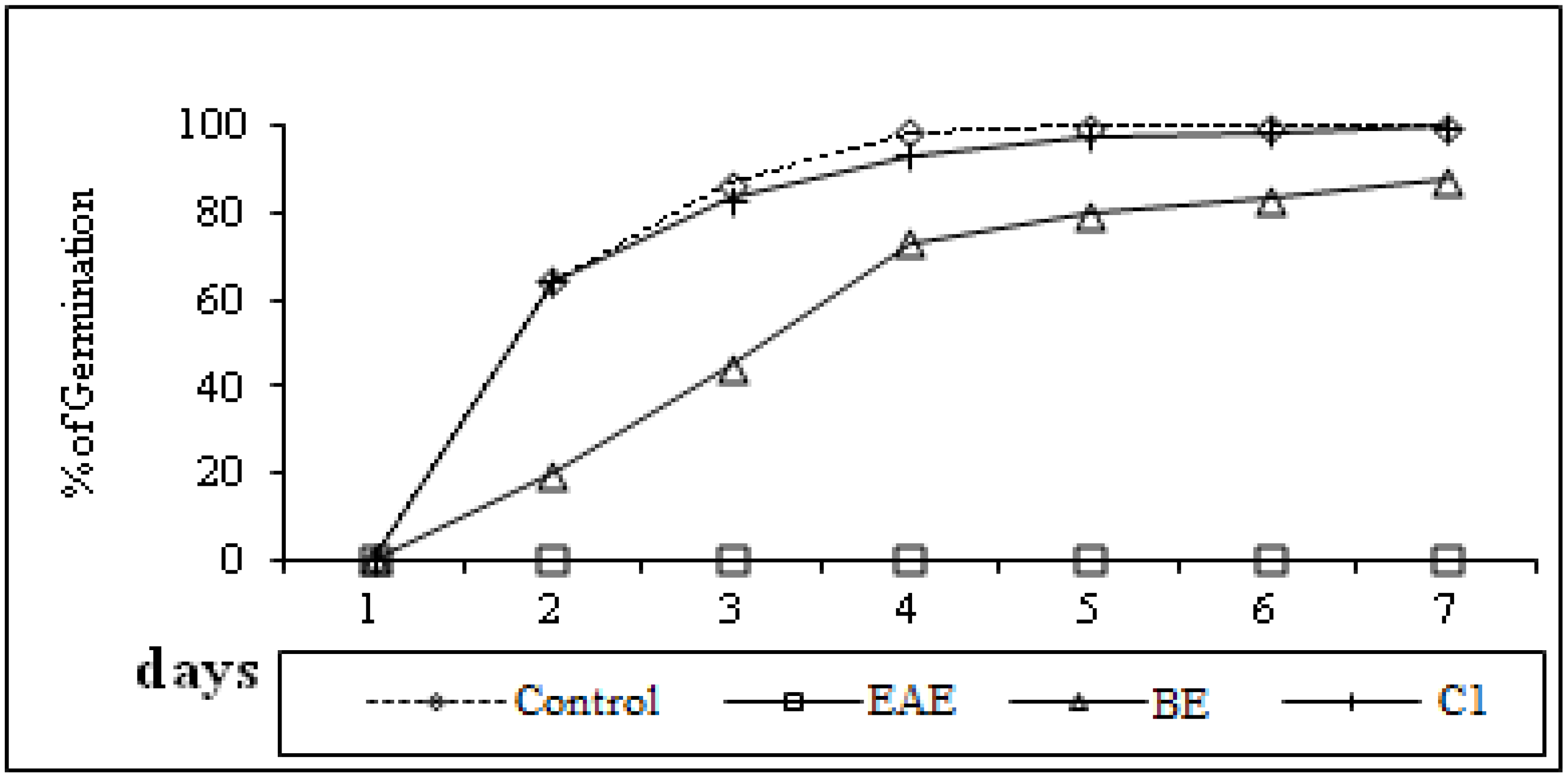
2.8. Allelopathic Potential

Methanol, in which the residues were dissolved, had no effect on germination hence any observed effects could be attributed to allelochemicals present in the organic extracts and compounds. Bioassays in the presence of extracts showed that the ethyl acetate extracts were the most effective. At the seventh day, the maximum toxicity of these extracts was registered, whereby the germination percentage was 0% (Figure 3). In the others cases, the maximum germination percentage values were 100% for the control and the n-butanol extract, but 87% for the mixture of compounds 1a and 1b(Figure 4).
For seedling lettuce growth, we recorded a strong activity for the ethyl acetate extract followed by the butanolic extract (BE), then compounds 1a and 1b (Figure 4). Indeed, we registered a total inhibition of root and shoot growth in presence of the ethyl acetate extract of Ferula plants (Figure 4). Relative toxicity of organic extracts is reported in the literature [27] where a higher phytotoxicity of methanol solutions of ethyl acetate extract is observed on lettuce compared to the n-butanol and water fractions. The result suggests that more phytotoxic substances were present in the ethyl acetate fraction than in the butanol one, resulting in more inhibitory effects on the test plant. Allelopathic potential of Ferula species is not cited in the literature.
Figure 3. Roots (R) and shoots length (PA), expressed in percent of control, of lettuce in presence (1 mg/mL) of (EAE), n-butanol extract (BE) and two diastereoisomers (1a and 1b) (C1) of F. lutea; 7 days after germination.
Molecules 19 02733 g003
Figure 4. Germination of lettuce seeds in the presence (1 mg/mL) of distilled water (control), ethyl acetate (EAE), n-butanolic extract (BE) and two diastereoisomers (1a and 1b) (C1) of F. lutea.
Molecules 19 02733 g004

2.9. Cytotoxicity Evaluation

The results of the anticancer test on the isolated compounds, ethyl acetate and n-butanol extracts using the cancer cell Line, K562 (leukemia cell line) are given in Table 4. The cytotoxicity of the n-butanol extract was superior to that of all samples tested with an IC50 = 40 µg/mL, while the ethyl acetate extract and compounds 1a and 1b were poorly active (IC50 > 100 µg/mL). The value of the cytotoxic activity of the n-butanol extract and is good, but not very high compared with the standard substance (doxorubicin).This is the first work citing the cytotoxic activity of F. lutea or other Ferula spp.

3. Experimental

3.1. General Experimental Procedures

1H- (300 MHz), 13C- (75 MHz) and 2D NMR spectra of compounds 1a and 1b were recorded in CDCl3 with a Bruker NMR-300 spectrometer. The residual solvent resonances were used as internal references. Chemical shifts are expressed in ppm and coupling constants are given in Hertz.

3.2. Collection of Plant Material

F. lutea flowers were collected in the region of Béja (Tunisia) on April 2010 and identified by Professor Féthia Harzallah Skhiri, at the Laboratory of Genetic Biodiversity and Valorisation of Bioresources of the Higher Institute of Biotechnology of Monastir, Tunisia. A voucher specimen (F.L.F-10) was deposited in the same laboratory.

3.3. Extraction and Isolation

The fresh flowers of F. lutea (5.8 kg) were macerated at room temperature with MeOH–H2O (7:3, 25 L) at room temperature for 7 days and then concentrated by rotary evaporation leaving an oil layer (5.8 g) and an aqueous residue (only the organic solvent was evaporated at the low temperature used ‒30 °C) which was partitioned successively with ethyl acetate and n-butanol extracts, yielding, after evaporation of the solvents, the corresponding ethyl acetate (56 g) and n-butanol (220 g) extracts. From the all extracts, only the oil layer (5 g) was subjected to silica gel column chromatography eluting successively with pentane–ethyl acetate (95:5 to 100% ethyl acetate) to afford 14 fractions. The ninth subfraction (2.4 g) was chromatographed on silica gel eluting with pentane–ethyl acetate (7:3) to afford a mixture of compounds 1a and 1b (2 g) that was difficult to separate. The 1H- and 13C-NMR spectral data of two diastereoisomers compounds (1a, 1b) are listed in Table 3.

3.4. Determination of Total Polyphenol Contents

The amount of total phenolic compounds was determined according to the method of Velioglu et al., [28] which uses the Folin-Ciocalteu reagent. Tested samples were prepared at a concentration of 1 mg/mL. The sample (100 µL) was transferred into a test tube and Folin-Ciocalteu reagent (750 µL, previously diluted 10-times with deionised water) were added and mixed. The mixture was allowed to stand at a temperature of 25 °C for 5 min, then saturated sodium carbonate (Na2CO3) solution (750 µL) was added to the mixture and then gently mixed. After standing at 25 °C for 90 min, the absorbance was read at 725 nm using an UV–Visible spectrophotometer. A standard curve of gallic acid was used. Total phenolic content of plant parts was expressed as mg gallic acid equivalents per gram of dry weight (mg GAE/g DW) through the calibration curve with gallic acid. The calibration curve range was 0–250 μg/mL (R2 = 0.99).

3.5. Estimation of Total Flavonoid Contents

The AlCl3 method [29] was used to determine the total flavonoid content of the sample extracts. Each extract (1.5 mL) was added to equal volumes of a solution of 2% AlCl3·6H2O (2 g in 100 mL methanol). The mixture was vigorously shaken, and absorbance at 367 nm was read after 10 min of incubation. Total flavonoid content was expressed as mg quercetin/g dry weight (mg QE/g DW), through the calibration curve of quercetin. The calibration curve range was 0–50 μg/mL (R2 = 0.99).

3.6. Antioxidant Activity by DPPH Assay

Diluted methanol solutions of the samples at different concentrations (1 mL each) were mixed with 1 mL of a freshly prepared (80 µg/mL) methanol solution of DPPH. The resulted solution was incubated at 37 °C for 30 min after what the absorbance of the solution was measured at 517 nm [30]. Lower absorbance of the reaction mixture indicates higher free radical scavenging activity. Tests were carried out in triplicate. Decrease in absorption induced by the tested sample was compared to that of the positive control ascorbic acid. The capability to scavenge the DPPH.radical was calculated using the following equation:
Inhibition ratio (DPPH. scavenging effect) (%) = [(Acontrol − Asample) / Acontrol] × 100
The IC50 was calculated using the linear relation between the compound concentration and the probability of the percentage of DPPH inhibition.

3.7. Antioxidant Activity by ABTS Assay

ABTS was dissolved in water to a 7 mM concentration and the ABTS radical cation was produced by adding potassium persulfate to a final concentration of 2.45 mM [31]. The radical generation was completed in the dark at room temperature for 12 h. This solution was then diluted with methanol to adjust its absorbance at 734 nm to 0.706 ± 0.009. To determine the scavenging activity, 1 mL of diluted ABTS+. solution was added to 1 mL of methanolic solutions of extract at different concentrations and the absorbance at 734 nm was measured 10 min after the initial mixing, using methanol as the blank. The percentage of inhibition was calculated by the equation:
[(Acontrol − Asample)/Acontrol] × 100
where Acontrol is the absorbance of the control reaction containing all reagents except the tested sample, and Asample is the absorbance of the test compound. Tests were carried out in triplicate. Ascorbic acid was used as the positive control. The IC50 was calculated using linear relation between the compound concentration and probability of the percentage of ABTS inhibition.

3.8. Antiacetylcholinesterase Activity

Briefly, 50 mM Tris–HCl buffer (100 µL, pH 8), the extract (50 µL) and acetylcholinesterase solution (10 µL) containing 0.26 U/mL were mixed in a microwell plate and left to incubate for 15 min. Subsequently, a solution of AChI (0.023 mg/mL, 50 µL) and 3 mM DTNB (140 µL) were added. The absorbance was read at 405 nm when the reaction reached equilibrium [32]. Eserine was used as a positive control and water served as a negative control and it was considered 100% activity. The percentage inhibition ((%) IP) is given as follows:
(%) IP = 100 − (Asample/Acontrol) × 100
where Acontrol is the absorbance of the control reaction containing all reagents except the tested sample, and Asample is the absorbance of the test compounds. Tests were carried out in triplicate and a blank with Tris-HCl buffer instead of enzyme solution was used.

3.9. Cytotoxic Activity

Cytotoxic activity of extracts against the human chronic myelogenous leukaemia cell line K562 was estimated by the 3-(4,5-dimethylthiazol-2-yl)-2,5-diphenyltetrazolium bromide (MTT) assay of Khlifi et al. [33]. The cells were grown in RPMI- 1640 medium supplemented with 10% faetal calf serum (Gibco, Gaithersburg, MD, USA), air and 5% CO2. The resulting blue formazan product can be measured spectrophotometrically. The MTT colorimetric assay was performed in 96-well plates. Adherent human chronic myelogenous leukaemia K562 cells were seeded in a 96-well plate at a concentration of 104 cells/well and incubated at 37 °C overnight in a 5% CO2 enriched atmosphere. Cells in exponential growth phase were incubated at 37 °C for 72 h with each tested compound at concentrations ranging from 3.12 to 400 μg/mL. After that, the medium was removed and cells were treated with MTT solution (100 μL, 0.2 mg/mL in PBS) at 37 °C for 2 h. MTT solution was then discarded and DMSO (500 μL) was added to dissolve insoluble formazan crystals. Optical density was measured at 540 nm. Each extract concentration was tested in triplicate. Doxorubicin was used as positive control.

3.10. GC-MS Analysis

The mass spectra of compounds 1a and 1b (diluted to 1% in the hexane) was achieved by coupled GC/MS analysis, on a system composed of a gas (HP-5890 II, Hewlett Packard Corporation, Palo Alto, California, CA, USA) coupled to a quadrupolar mass spectrometer (HP-MSD 5972 TO) operated in electron impact mode. The column used was an HP polar INNOWAX column of 30 m, 0.25 mm, and 0.25 μm film thickness. Its stationary phase was constituted of polyethylene glycol.

3.11. Antidiabetic Activity

Porcine pancreatic α-amylase inhibitory activity was determined using a literature method [34]. Starch azure (2 mg) was used as substrate and was suspended in 0.5 M Tris-HCl (pH 6.9, 0.2 mL) containing 0.01 M CaCl2. The tubes containing substrate solution were boiled for 5 min. After 5 min of preincubation at 37 °C, extract dissolved in DMSO (0.2 mL) was added to the tube containing the substrate solution. A volume of porcine pancreatic amylase in Tris-HCl buffer (0.1 mL) was added and then incubated at 37 °C for 10 min and the reaction was stopped by adding 50% acetic acid (0.5 mL). The reaction mixture was centrifuged at 3,000 rpm for 5 min at 4 °C and the absorbance was measured at 595 nm.

3.12. Antimicrobial Activity: Determination of Minimum Inhibitory (MIC), Minimum Bactericidal (MBC), and Minimum Fungicidal Concentrations (MFC)

The test microorganisms included the following Gram-positive bacteria: Staphylococcus aureus ATCC 25923, Enterococcus faecalis ATCC 29212; Gram-negative bacteria: Escherichia coli ATCC 25922 and Pseudomonas aeruginosa ATCC 27853 and four Candida yeasts: Candida albicans ATCC 90028, C. glabrata ATCC 90030, C. kreusei ATCC 6258 and C. parapsilosis ATCC 22019).
The MIC values for the antibacterial and anticandidal screening were determined with the broth dilution method (microdilution using 96-well microplates) following the procedure described by Cintia et al. [35]. Briefly, all samples were prepared at a concentration of 10 mg/mL by dissolution of the extracts in 10% DMSO. The final concentrations of the plant samples tested ranged from 40 to 2 × 10⁴ µg/mL. The MIC of each sample was defined as the lowest concentration of oil that inhibited either the bacterial or candidal growth, after incubation at 37 °C for 18 to 24 h. The MBC and the MFC were determined by subculture on blood agar at 37 °C for 18 to 24 h. Thymol and gentamicin or thymol and amphotericin B were used as positive controls against the bacterial or candidal strains, respectively. The highest activities for each sample are presented in Table 4 and Table 5 with the most noticeable antimicrobial activities against different strains shown in bold face. All tests were performed in triplicate.

3.13. Allelopathic Potential

The two samples concentrated from the ethyl acetate (EAE) and n-butanol (BE) extracts and the mixture (1a + 1b) (C1) were again dissolved in MeOH to compare their phytotoxic effects. Five mL of each organic fraction at 6,000 ppm and mixture (1a + 1b) at 1,000 ppm were placed in a Petri dish lined with a sheet of Whatman No. 1 filter paper and evaporated to dryness for 24 h at 24 °C. After evaporation, distilled water (5 mL) was pipetted onto the filter paper. Methanol and distilled water were both were used as negative control. Seeds of lettuce were surface sterilized by immersing in 0.525 mg/mL sodium hypochlorite for 15 min, and then were rinsed four times with deionised water, imbibed in deionised water at 22 °C for 4 h, and carefully blotted using a folded paper towel. Lettuce has been used as tested plant because it is very sensitive to chemicals at low concentration, although it sometimes overestimates the actual allelopathy [36]. Thirty swollen seeds were evenly placed on filter paper wetted with sample in each Petri dish. The germination was recorded at 24-h intervals till 144 h. A seed was considered germinated when the radical protruded ≥2 mm [37]. Data were transformed to percent of control for further analysis. At 7th day after sowing, germination, shoot and root length of target species seedlings were measured. Data were transformed to percent of control for analysis.

4. Conclusions

F. lutea flowers were investigated for their chemical composition. Total phenols and flavonoids contents of extracts were determined and two diastereoisomers were isolated and identified. To the best of our knowledge, we report the first study on the antiacetylcholinesterase, antidiabectic, antimicrobial, allelopathic and cytotoxic activities of F. lutea flower constituents. We can conclude that the ethyl acetate extract has the highest antioxidant activity, the n-butanol one possesses an interesting cytotoxicity and finally both extracts have good antidiabetic activity. Nevertheless, it is interesting to continue the fractionation work to isolate the molecules responsible for the antioxidant, antidiabetic and cytotoxic activities. Compounds 1a and 1b were isolated as a mixture and their activities were evaluated for the first time.

Conflictts of Interest

The authors declare no conflict of interest.

References

  1. Downie, S.R.; Watson, M.F.; Spalik, K.; Katz-Downie, D.S. Molecular systematics of old world 2. Apioideae (Apiaceae): Relationships among some members of tribe Peucedaneaesensulato, the placement of several islandendemic species, and resolution within the apioidsuperclade. Can. J. Bot. 2000, 78, 506–528. [Google Scholar]
  2. Drude, O. Die Natürlichen Pflanzenfamilien; (In German). Engler, A., Ed.; Wilhelm Engelmann: Leipzig, Germany, 1898; Volume 3, pp. 228–232. [Google Scholar]
  3. Pottier-Alapetite, G. Flore de la Tunisie: Angiospermes-Dicotyledones, Apétales-Dialypétales; (In Spanish). Official printing: Tunis, Tunisia, 1979; pp. 607–609. [Google Scholar]
  4. Tamemoto, K.Y.; Takaishi, B.; Chen, K.; Kawazoe, H.; Shibata, T.; Higuti, G.; Honda, M.; Ito, Y.; Takeda, O.K.; Kodzhimatov, O. Ashurmetov sesquiterpenoids from the fruits of Ferula kuhistanica and antibacterial activity of the constituents of F. kuhistanica. Phytochemistry 2001, 58, 763–767. [Google Scholar] [CrossRef]
  5. Gonzalez, A.G.; Barrera, J.B. Chemistry and sources of mono- and bicyclic sesquiterpenes from Ferula species. Prog. Chem. Org. Nat. Prod. 1995, 64, 1–92. [Google Scholar]
  6. Appendino, G.; Spagliardi, P.; Cravotto, G.; Pocock, V.; Milligan, S. Daucane phytoestrogens: A structure—activity study. J. Nat. Prod. 2002, 65, 1612–1615. [Google Scholar] [CrossRef]
  7. Boulos, L. Algonac, Medicinal Plants of North Africa; ML. Reference Publications Inc.: Alyonae, MI, USA, 1983. [Google Scholar]
  8. El-Razek, A.M.H.; Ohta, S.; Hirata, T. Terpenoid coumarins from the genus Ferula. Heterocycles 2003, 60, 689–716. [Google Scholar] [CrossRef]
  9. Suzuki, K.; Okasaka, M.; Kashiwada, Y.; Takaishi, Y.; Honda, G.; Ito, M.; Takeda, Y.; Kodzhimatov, O.K.; Ashurmetov, O.; Sekiya, M.; et al. Sesquiterpene lactones from the roots of Ferula varia and their cytotoxic activity. J. Nat. Prod. 2007, 70, 1915–1918. [Google Scholar] [CrossRef]
  10. Lamnaouer, D. Anticoagulant activity of coumarins from Ferula communis L. Therapie 1999, 54, 747–751. [Google Scholar]
  11. Valle, M.G.; Appending, G.; Nano, G.M.; Picci, V. Prenylatedcoumarins and sesquiterpenoids from Ferula communis. Phytochemistry 1986, 23, 253–256. [Google Scholar]
  12. Kartal, N.; Sokmen, M.; Tepe, B.; Daferera, D.; Polissiou, M.; Sokmen, A. Investigation of the antioxidant properties of Ferula orientalis L. using a suitable extraction procedure. Food Chem. 2006, 100, 584–589. [Google Scholar]
  13. Alkhatib, R.; Hennebelle, T.; Joha, S.; Idziorek, T.; Preudhomme, C.; Quesnel, B.; Sahpaz, S.; Bailleul, F. Activity of elaeochytrin A from Ferula elaeochytris on leukemia cell lines. Phytochemistry 2008, 69, 2979–2983. [Google Scholar] [CrossRef]
  14. Abedi, D.; Jalali, M.; Asghari, G.; Sadeghi, N. Composition and antimicrobial activity of oleogumresin of Ferula gumosa Bioss. essential oil using Alamar Blue. Res. Pharm. Sci. 2008, 3, 41–45. [Google Scholar]
  15. Fylaktakidou, K.C.; Hadjipavlou-Litina, D.J.; Litinas, K.E.; Nicolaides, D.N. Natural and synthetic coumarin derivatives with anti-inflammatory/antioxidant activities. Curr. Pharm. Des. 2004, 10, 3813–3833. [Google Scholar] [CrossRef]
  16. Özlem, B.; Gülçin, S.Ç.; Hanefi, Ö.; Stefano, D.; Karel, Š.J.H. Hepatoprotective and TNF-α inhibitory activity of Zosima absinthifolia extracts and coumarins. Fitoterapia 2011, 82, 454–459. [Google Scholar] [CrossRef]
  17. Salem, B.S.; Jabrane, A.; Harzallah-Skhiri, F.; Jannet, B.H. New bioactive dihydrofuranocoumarins from the roots of the Tunisian Ferula lutea (Poir.) Maire. Bioorg. Med. Chem. Lett. 2013, 23, 4248–4252. [Google Scholar] [CrossRef]
  18. Znati, M.; Jabrane, A.; Hajlaoui, H.; Bouajila, J.; Casanova, J.; Jannet, B.H. Chemical composition and in vitro evaluation of antimicrobial and anti-acetylcholinesterase of the flower oil of Ferula lutea L. Nat. Prod. Commun. 2012, 7, 947–950. [Google Scholar]
  19. Razavi, S.M.; Nazemiyeh, H.; Hajiboland, R.; Kumarasamy, Y.; Delazar, A.; Nahar, L.; Sarker, S.D. Coumarins from the aerial parts of Prangos uloptera (Apiaceae). Rev. Bras. Farmacogn. 2008, 18, 1–5. [Google Scholar]
  20. Baba, K.; Yoneda, Y.; Kozawa, M.; Fujita, E.; Wang, N.H.; Yuan, C.Q. Studies on Chinese traditional l medicine ‘Fang-Feng’ (II). Comparison of several Fang-Feng by coumarins, chromones and polyacetylenes. Jpn. J. Pharmacogn. 1989, 43, 216–221. [Google Scholar]
  21. Abyshev, A.Z.; Abyshev, A.D. Coumarins of Seseli peucedanoides. Chem. Nat. Compd. 1984, 20, 230–231. [Google Scholar] [CrossRef]
  22. Chacko, S.; Singh, O.V.; Sethuraman, M.G.; George, V. Coumarins from Heracleum. candolleanum. Indian Drugs. 2001, 38, 594–596. [Google Scholar]
  23. Chen, I.S.; Chang, C.T.; Sheen, W.S.; Teng, C.M.; Tsai, I.L.; Duh, C.Y.; Ko, F.N. Coumarins and antiplatelet aggregation constituents from Formosan Peucedanum. japonicum. Phytochemistry 1996, 41, 525–530. [Google Scholar] [CrossRef]
  24. Tosun, A.; Baba, M.; Bahadir, O.; Okuyama, T. Coumarins isolated from the roots of Seseli resinosum in Turkey. Pharma. Biol. 2006, 44, 528–533. [Google Scholar] [CrossRef]
  25. Dehpour, A.A.; Ebrahimzadeh, M.A.; Nabavi, S.F.; Mohammad, N.S. Antioxidant activity of the methanol extract of Ferula assafoetida and its essential oil composition. Grasas Aceites 2009, 60, 405–412. [Google Scholar] [CrossRef]
  26. Ebrahimzadeh, M.A.; Nabavi, S.M.; Nabavi, S.F.; Dehpour, A.A. Antioxidant activity of hydroalcholic extract of Ferula gummosa Boiss roots. Eur. Rev. Med. Pharmacol. Sci. 2011, 15, 658–664. [Google Scholar]
  27. Chon, S.S.; Jang, H.G.; Kim, D.K.; Kim, Y.M.; Boo, H.O.; Kim, Y.J. Allelopathic potential in lettuce (Lactuca. sativa L.). Plants. Sci. Hortic. 2005, 106, 309–317. [Google Scholar] [CrossRef]
  28. Velioglu, Y.S.; Mazza, G.; Gao, L.; Oomah, B.D. Antioxidant activity and total phenolics in selected fruits, vegetables, and grain products. J. Agric. Food Chem. 1998, 46, 4113–4117. [Google Scholar] [CrossRef]
  29. Lamaison, J.L.C.; Carnet, A. Teneurs en principaux flavonoids des fleurs de Crataegeus. monogyna Jacq et de Crataegeus. laevigata (Poiret D. C) en fonction de la vegetation. Pharm. Acta. Helv. 1990, 65, 315–320. (In French) [Google Scholar]
  30. Ebrahimabadi, A.H.; Ebrahimabadi, E.H.; Djafari-Bidgoli, Z.; Kashi, F.J.; Mazoochi, A.; Batooli, H.J. Composition and antioxidant and antimicrobial activity of the essential oil and extracts of Stachys. inflate Benth from Iran. Food Chem. 2010, 119, 452–458. [Google Scholar] [CrossRef]
  31. Re, R.; Pellegrini, N.; Proteggente, A.; Pannala, A.; Yang, M.; Rice-Evans, C.J. Antioxidant activity applying an improved ABTS radical cation decolorization assay. Free Radic. Biol. Med. 1999, 26, 1231–1237. [Google Scholar] [CrossRef]
  32. Falé, P.L.; Borges, C.; Madeira, P.J.A.; Ascensão, L.; Araujo, M.E.M.; Florêncio, M.H.J. Rosmarinic acid, scutellarein 4'-methyl ether 7-O-glucuronide and (16S)-coleon E are the main compounds responsible for the antiacetylcholinesterase and antioxidant activity in herbal tea of Plectranthus. barbatus (“Falso Boldo”). Food Chem. 2009, 114, 798–805. [Google Scholar] [CrossRef]
  33. Khlifi, D.; Sghaier, R.M.; Amouri, S.; Laouini, D.; Hamdi, M.; Bouajila, J. Composition and anti-oxidant, anti-cancer and anti-inflammatory activities of Artemisia herba-alba, Ruta. chalpensis L. and Peganum harmala L. Food Chem. Toxicol. 2013, 55, 202–208. [Google Scholar] [CrossRef]
  34. Hansawasdi, C.; Kawabata, J.; Kasai, T. Amylase inhibitors from Roselle (Hibiscus sabdariffia Linn) Tea. Biosci. Biotechnol. Biochem. 2000, 64, 1041–1043. [Google Scholar] [CrossRef]
  35. Cintia, S.G.K.; Smania, A., Jr.; Pedrosa, R.C.; Ferreira, S.R.S.J. Antioxidant and antimicrobial activities of shiitake (Lentinulaedodes) extracts obtained by organic solvents and supercritical fluids. Food Eng. 2007, 80, 631–638. [Google Scholar] [CrossRef]
  36. Olofsdotter, M. Rice-astep toward use of allelopathy. Agron. J. 2001, 93, 3–8. [Google Scholar] [CrossRef]
  37. Hou, J.Q.; Romo, J.T. Effects of chemicals stimulators on germination of Ceratoids lanata. Seed Sci. Technol. 1998, 26, 9–163. [Google Scholar]
  • Sample Availability: Samples of the compounds are available from the authors.

