Article

Sesquiterpenes from the Brazilian Red Alga Laurencia dendroidea J. Agardh

by
Fernanda Lacerda Da Silva Machado
1,2,
Thatiana Lopes Biá Ventura
3,
Lísia Mônica de Souza Gestinari
2,
Valéria Cassano
4,
Jackson Antônio Lamounier Camargos Resende
5,
Carlos Roland Kaiser
1,
Elena B. Lasunskaia
3,
Michelle Frazão Muzitano
6 and
Angélica Ribeiro Soares
1,2,*
1
Instituto de Química, Universidade Federal do Rio de Janeiro, Avenida Athos da Silveira Ramos, 149, 21941-909, Rio de Janeiro, RJ, Brazil
2
Grupo de Produtos Naturais de Organismos Aquáticos (GPNOA), Núcleo de Estudos em Ecologia e Desenvolvimento Sócioambiental de Macaé, Universidade Federal do Rio de Janeiro – Campus Macaé, Av. São José do Barreto, 764, 27965-045, Macaé, RJ, Brazil
3
Laboratório de Biologia do Reconhecer, Centro de Biociências e Biotecnologia, Universidade Estadual do Norte Fluminense Darcy Ribeiro, Avenida Alberto Lamego, 2000, 28013-602, Campos dos Goytacazes, RJ, Brazil
4
Laboratório de Algas Marinhas Professor Édison José de Paula, Departamento de Botânica, Universidade de São Paulo, Rua do Matão, 277, 05508-090, São Paulo, SP, Brazil
5
Laboratório Regional de Difração de Raios X (LDRX), Universidade Federal Fluminense, Avenida Litorânea, s/n, 24020-150, Niterói, RJ, Brazil
6
Laboratório de Produtos Naturais, Curso de Farmácia, Universidade Federal do Rio de Janeiro, Campus Macaé, Pólo Novo Cavaleiro – IMMT, Rua Alcides da Conceição, 159, 27933-378, Macaé, RJ, Brazil
*
Author to whom correspondence should be addressed.
Molecules 2014, 19(3), 3181-3192; https://doi.org/10.3390/molecules19033181
Submission received: 15 January 2014 / Revised: 3 March 2014 / Accepted: 5 March 2014 / Published: 17 March 2014
(This article belongs to the Section Natural Products Chemistry)

Abstract

:
Two new chamigrane sesquiterpenes 12 and three known compounds 35 were isolated from a lipophilic extract of the red alga Laurencia dendroidea collected from the Southeastern Brazilian coast. Dendroidone (1) and dendroidiol (2) were isolated from samples collected at Biscaia Inlet, Angra dos Reis, Rio de Janeiro and at Manguinhos Beach, Serra, Espírito Santo, respectively. Debromoelatol (3), obtusane (4) and (1S*,2S*,3S*,5S*,8S*,9S*)-2,3,5,9-tetramethyltricyclo[6.3.0.01.5]undecan-2-ol (5) were obtained from specimens collected at Vermelha Beach, Parati, Rio de Janeiro. The structures of new compounds were elucidated by extensive NMR (1H-, 13C-, COSY, HSQC, HMBC and NOESY) and high resolution mass spectrometry analysis. Additionally, the absolute configuration of compound 2 was assigned by X-ray analysis. Full spectroscopic data is described for the first time for compound 3. Anti-inflammatory and antimycobacterial activities of compounds 25 were evaluated. Compounds 35 inhibited the release of inflammatory mediator NO while TNF-α levels were only affected by 3. All compounds tested displayed moderate antimycobacterial action.

1. Introduction

It is estimated that more than 700 compounds with unique structural features have already been isolated from red algae of genus Laurencia, family Rhodomelaceae, order Ceramiales [1,2], which occurs on temperate to tropical shores of the world inhabiting intertidal and subtidal areas [3]. This remarkable chemical diversity comprises sesquiterpenes, diterpenes, triterpenes and acetogenins, mainly halogenated [4,5,6,7,8,9]. Several studies suggest that in the marine environment these compounds have a role as chemical defenses against herbivores, fouling organisms and pathogens [10,11]. Despite the high number of isolated compounds, recent reports confirm the potential of Laurencia to produce unknown structures [12]. Chamigrane-type compounds are the main class of sesquiterpenes isolated [2], for which some interesting pharmacological actions are described, including antibacterial [13], cytotoxic [14], and antileishmanial [15]. In addition, L. undulata and L. snackeyi extracts exhibited anti-inflammatory action [16,17], and the tricyclic brominated diterpene neorogioltriol, isolated from L. glandulifera, displayed both in vivo and in vitro anti-inflammatory activity [18].
The pathogenesis of several diseases such as tuberculosis, caused mainly by Mycobacterium tuberculosis, is highly influenced by the inflammatory response. Some anti-inflammatory drugs are employed as an adjunctive therapy for tuberculosis [19]. Therefore, combined anti-inflammatory and antimycobacterial properties in a single compound or class of compounds could be relevant for the treatment of tuberculosis. Furthermore, the long duration of current therapy as well as the associated side effects often compromises its effectiveness and it is intimately linked to the emergence of drug resistance [20]. Thus, there is an urgent need for short and simple regimens, which are both effective and safe.
In our ongoing study on structurally diverse and biologically active compounds from Laurencia species, specimens of the Brazilian red alga L. dendroidea J. Agardh were collected from three different places along the southeastern coast, and extracted with dichloromethane. Herein we report the extraction, isolation and structure elucidation of compounds 15 (Figure 1) along with anti-inflammatory and antimycobacterial activities of compounds 25.
Figure 1. Structures of compounds 1–5.
Molecules 19 03181 g001

2. Results and Discussion

The organic extracts were subjected to chromatographic separations yielding dendroidone (1) from Biscaia Inlet, Angra dos Reis, Rio de Janeiro (population A), dendroidiol (2) from Manguinhos Beach, Serra, Espirito Santo (population B) and debromoelatol (3), obtusane (4) and (1S*,2S*,3S*,5S*,8S*,9S*)-2,3,5,9-tetramethyltricyclo[6.3.0.01.5]undecan-2-ol (5) from Vermelha Beach, Parati, Rio de Janeiro (population C). Compounds 25 were tested for anti-inflammatory and antimycobacterial activities.
Compound 1 was isolated as an optically active colorless oil, Molecules 19 03181 i001 = +6.0° (c 0.06, CHCl3). The molecular formula was established as C15H20BrClO2 on the basis of HR-APCI-MS data (m/z 349.0381 [M+H]+, calcd. for C15H21BrClO2, 349.0393), implying five degrees of unsaturation. The infrared (IR) spectrum exhibited absorptions of hydroxyl (3,457 cm−1) and carbonyl (1,675 cm−1) groups. The 1H-NMR spectrum displayed signals corresponding to two methyl groups at δH 1.09 (H3-12) and 1.11 (H3-13), two methines geminal to heteroatoms at δH 4.67 (H-10) and δH 4.21 (H-9) and three olefinic hydrogens at δH 5.16 (H-14a), δH 4.77 (H-14b) and one highly deshielded at δH 7.24 (H-15), suggesting it was part of a conjugated system. 13C and HSQC experiments revealed the presence of two methyls, four aliphatic methylenes, two deshielded methines, two quaternary sp3 carbons and five sp2 carbon resonances that were assigned to a ketone [δC 197.6 (C-4)] and two olefins [δC 117.5 (C-14), 131.3 (C-15), 136.1 (C-3), 142.6 (C-7)]. The positions of the bromine and the hydroxyl groups were deduced from the carbon chemical shifts of carbons at δC 69.6 (C-10) and 72.0 (C-9), respectively [21]. The presence of an exomethylene group involving C-7 and C-14 was suggested by HMBC correlation between broadened olefinic singlets at δH 5.16 (H-14a) and δH 4.77 (H-14b) with carbons at δC 48.7 (C-6) and 38.6 (C-8). From the 1H-1H-COSY NMR spectrum the coupling between methylene protons at δH 2.58 (H2-8) and the proton on carbinolic group at δH 4.21 (H-9) was observed. The latter also couples with the proton on carbon bearing a bromine at δH 4.67 (H-10). Ring A was established by HMBC correlations from methyls at δH 1.09 (H3-12) and 1.11 (H3-13) to the respective carbons [δC 22.7 (C-13); δC 20.8 (C-12)] and to δC 48.7 (C-6), 69.6 (C-10) and 43.0 (C-11).
Ring B was proposed based on correlations between methylenes protons at δH 2.81 (H-2a) and δH 1.76 (H-1b) on the COSY spectrum along with HMBC correlations from δH 2.46 (H-5b) to δC 197.6 (C-4) and from δH 1.76 (H-1b) to δC 48.7 (C-6). Based on the 13C-NMR data, a chlorine atom was assigned to C-15 [22]. The Z configuration of C-3 double bond was proposed from the observation of correlation between H2-2/H-15 on NOESY spectrum. The relative configuration was determined by NOESY correlations and 1H-1H coupling constants. NOE correlations of H-14b to H-5a demonstrated that exomethylene and methylene CH2-5 groups were positioned on the same face. Moreover, halomethine proton H-10 displayed correlations to H-1a and H3-13 indicating that H-10 occupied an axial position. Based on the axial orientation of H-10 and the small coupling constant of the carbinolic H-9 (3 Hz) it was suggested that it was equatorial, therefore bromine and hydroxyl were in a cis configuration. Hence, the combined data established the structure of compound 1 as (Z)-10-bromo-15-chloro-11,11-dimethyl-7-methylidenespiro[5.5]undec-3(15)-ene-4-one which represents a new chemical entity, which was trivially named dendroidone.
Table 1. 1H (500 MHz) and 13C (125 MHz) NMR data for compounds 13 (CDCl3, δ in ppm).
No.123
δCδH, mult. (J in Hz)δCδH, mult. (J in Hz)δCδH, mult. (J in Hz)
1a
1b
25.62.16 dm (13.6)
1.76 ddd (14.0, 13.6, 4.8)
22.62.00 brd (13.0)
1.60 brd (13.0)
25.81.89 m
1.50 m
2a
2b
24.22.81 m
2.08 m
33.81.70 dt (13.0, 4.0, 3.2)
1.20 m
29.51.86 m
1.25 m
3136.1-70.3-124.6-
4197.6-67.44.23 dd (11.0, 5.0)128.2-
5a
5b
45.52.85 dd (17.5, 3.5)
2.46 d (17.5)
35.12.10 m37.52.48 m
2.15 m
648.7-50.5-46.6-
7142.6-141.9-145.2-
8a
8b
38.62.58 d (3.0)38.52.32 dd (14.0, 2.7)
2.51 dd (14.0, 2.7)
41.72.45 m
2.16 m
972.04.21 q (3.0)72.04.07 m67.93.76 ddd (16.0, 10.5, 5.0)
1069.64.67 d (3.0)70.74.52 d (3.0)45.91.66 m
1.53 m
1143.0-44.2-37.5-
1220.81.09 s20.41.03 s23.80.84 s
1322.71.11 s24.11.06 s24.80.94 s
14a
14b
117.55.16 s
4.77 s
116.75.25 s
4.97 s
113.55.01 s
4.60 s
15131.37.24 m28.51.21 s19.51.70 s
Compound 2 was isolated as colorless crystals, Molecules 19 03181 i001 
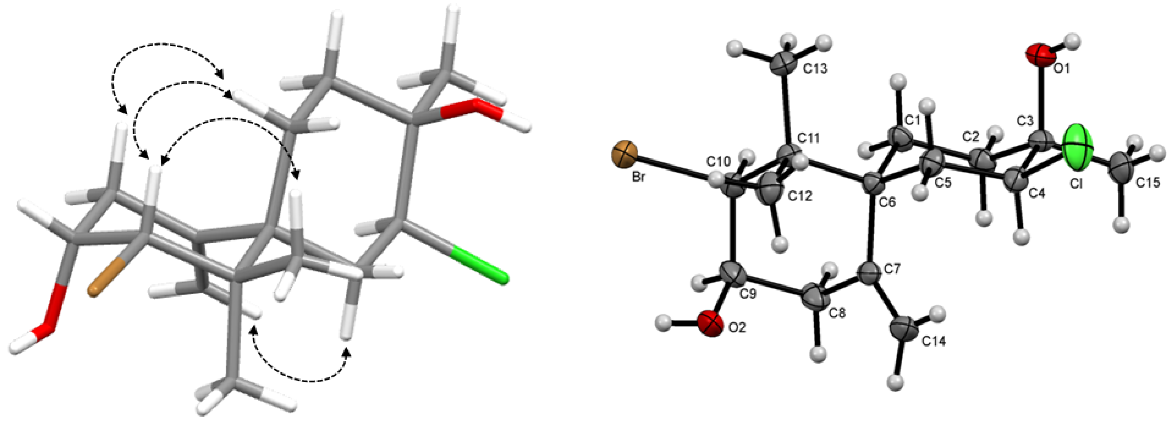
= −19.2° (c 0.08, CHCl3), with the molecular formula C15H24BrClO2 deduced by HR-ESI-MS from the pseudomolecular ion peak [M+Na]+ at m/z 375.0417 (calcd. for C15H24BrClO2Na, 375.0525), requiring three degrees of unsaturation. The IR spectrum exhibited absorptions of a hydroxyl group at 3,463 cm−1. The 1H-NMR spectrum (Table 1) also displayed a pair of broad singlets characteristic of an exocyclic methylene group at δH 5.25 (H-14a) and 4.97 (H-14b), three tertiary methyl groups at δH 1.03 (H3-13), 1.06 (H3-12) and 1.21 (H3-15) and three hydrogens of heterosubstituted carbons at δH 4.07 (H-9), 4.23 (H-4) and 4.52 (H-10). The 13C-NMR spectrum and DEPT-135 experiment revealed the presence of fifteen carbon atoms corresponding to three methyls, five methylenes, three methines and four quaternary carbons, including two olefinic carbons and four carbons attached to heteroatoms. 1H-1H COSY and HMBC correlations indicated that the first ring was similar to compound 1 while the second was proposed based on 1H-1H COSY correlations between δH 4.23 (H-4) and 2.10 (2H, m, H2-5) and correlations from δH 1.21 (H3-15) to δC 33.8 (C-2), 70.3 (C-3) and 67.4 (C-4) in HMBC spectrum. Chemical shifts of C-3 and H-4/C-4 indicated the presence of a tertiary alcohol and chlorine substitution, respectively [23].
The relative configuration was determined on the basis of measured coupling constants and NOESY spectrum (Figure 2). Taken together, the data suggested that the compound 2 represented a new chamigrane sesquiterpene with chlorohydrin function 4,10-dibromo-4-chloro-3,11,11-trimethyl-7-methylidenespiro[5.5]undec-3,9-diol, for which the trivial name dendroidiol was proposed. Further X-ray crystallographic data of 2 confirmed the suggested structure and defined the absolute configuration as 3R, 4S, 6S, 9R, 10S as depicted (Figure 2). The rings A (C1—C2—C3—C4—C5—C6) and B (C7—C8—C9—C10—C11—C12) adopted chair configuration, with ring-puckering parameters q2 = 0.060(7)Å; ϕ2 = 352(7)° and q2 = 0.042(5) Å; ϕ2 = 233(7)°, respectively [24].
Figure 2. Key NOESY correlations and ORTEP drawing of 2.
Molecules 19 03181 g002
Compound 3 was isolated as colorless oil. The 13C-NMR spectra along with HSQC experiment revealed the presence of fifteen carbons distributed as five quaternary carbons, one methine, six methylenes and three methyls, including four olefinic carbons (Table 1). The molecular formula C15H23OCl was deduced by NMR and EI-MS data. Like compounds 1 and 2, the 1H-NMR spectrum displayed singlets of an exocyclic methylene group (δH 5.01, 4.60). Additionally, it also displayed one carbinolic proton at δH 3.76 and three quaternary methyls (δH 0.84, 0.94, 1.70). Comparison of NMR spectra of compound 3 to 1 and 2 revealed that ring A differed only on bromine substitution at C-10. The correlations in the HMBC spectrum from methyl H3-15 to C-2, C-3 and C-4 indicated the second ring was similar to the described for elatol [15]. Thus, the present data suggested that compound 3 was debromoelatol previously isolated from L. obtusa [25]. Full spectroscopic data for compound 3 is described for the first time.
Furthermore, two additional known sesquiterpenes obtusane (4) and (1S*,2S*,3S*,5S*,8S*,9S*)-2,3,5,9-tetramethyltricyclo[6.3.0.01.5]undecan-2-ol (5) were isolated and identified by comparison of their spectroscopic and physical data to those reported in the literature [15].
Compounds 25 were submitted to tests evaluating immunomodulatory and antimycobacterial actions (Table 2). These two pharmacological approaches were supported by previous studies reporting immunomodulatory [26] and antimycobacterial activities [27] of Laurencia species and also due to the expertise of the group in these two areas.
Table 2. IC50 values for the compounds isolated from L. dendroidea in the production of NO and TNF-α by LPS-stimulated macrophages, against M. bovis BCG and in LDH cytotoxicity assay. Values in the same column with different superscript letters (a–d) are significantly different (p < 0.05 or p < 0.001; results of the Tukey test).
SampleIC50 (μM)
NOTNF-αM. bovis BCGCytotoxicity
2>284.3 a>284.3 a80.2 ± 4.3 a>284.3 a
369.1 ± 4.7 b133.8 ± 7.4 b82.4 ± 4.7 a>392.5 b
444.9 ± 3.0 c>250.9 c44.7 ± 4.0 b197.2 ± 1.0 c
574.6 ± 5.8 b393.4 ± 0.4 d204.6 ± 7.6 c416.4 ± 4.9 d
l-NMMA71.3 ± 4.4 b---
Rifampicin--0.004 ± 1.3 d-
The in vitro anti-inflammatory potential of isolated compounds was evaluated in a preliminary study of immunomodulatory properties, which was assessed by their inhibitory effects on NO and TNF-α productions from LPS-activated RAW 264.7 macrophages. Compounds 35 inhibited NO release by stimulated macrophages, with IC50 values ranging from 44.9 ± 3.0 to 74.6 ± 5.8 μM (Table 2). Compound 4 was significantly more active (p < 0.05) while compounds 3 and 5 displayed similar activity to the positive control L-NMMA (L-N-monomethyl-arginine), a selective iNOS synthase inhibitor (p > 0.05). TNF-α production was moderately inhibited by compound 3 (IC50 133.8 ± 7.4 µM), however, the remaining compounds did not show promising effects.
In the second part of the preliminary pharmacological study, antimycobacterial activity of isolated sesquiterpenes was evaluated using rapidly-growing strain Mycobacterium bovis BCG (Table 2). In this test, the sesquiterpene 4 (IC50 44.7 ± 4.0 μM) was the most active compound, but it was less effective than the positive control Rifampicin.
In order to determine whether there was any selectivity, cell viability was assessed by lactate dehydrogenase (LDH) release (Table 2). Compound 4 was considered only moderately toxic while compounds 2, 3 and 5 showed no toxicity whatsoever.

3. Experimental

3.1. General Procedures

Optical rotations were measured on a Perkin Elmer model 341LC polarimeter using a Na lamp at 20 °C. IR spectra were obtained with a Perkin Elmer spectrum one FT-IR. 1H-NMR, 13C-NMR, DEPT-135, COSY, HSQC, HMBC and NOESY spectra were measured employing a Bruker Avance III instrument operating at 500 MHz for 1H-NMR and at 125 MHz for 13C NMR in CDCl3. EI-MS spectra were obtained with a Shimadzu GCMSQP-2010 Plus. HR-APCI-MS spectra were recorded on a MicrOTOF (Bruker Daltonics, Billerica, MA, USA) mass spectrometer. HR-ESI-MS spectra were recorded on an UltrOTOF (Bruker Daltonics) mass spectrometer. Column chromatography was performed with Silicycle SiliaFlash F60 (230–400 mesh) silica and Sephadex LH-20 (Fluka, Steinheim, Germany). Thin layer chromatography was carried out with silica gel GF254 plates. The spray reagent was a solution of 2% of Ce(SO4)2 in H2SO4. HPLC separations were performed with a Shimadzu instrument equipped with an LC-6AD pump, CBM-20A and SPD-20AV detector using a Shim-pack Prep-ODS, 250 × 20 mm, 5 μm column.

3.2. Plant Material

The red seaweed Laurencia dendroidea J. Agardh (Rhodomelaceae, Ceramiales) was collected from three distinct areas of the Southeastern Brazilian coast: Biscaia inlet - Angra dos Reis - Rio de Janeiro state (23° 01' S, 44° 14' W), in April, 2011 (Population A); Manguinhos Beach – Serra – Espírito Santo state (20°11' S, 40°11' W), in March, 2010 (Population B) and Vermelha Beach – Parati – Rio de Janeiro state (23°11' S, 44°38' W), in April, 2011 (Population C). Botanical identification was made by L. M. Gestinari and V. Cassano and voucher specimens (Biscaia Inlet: RFA 36068, Manguinhos Beach: RFA 35887 and Vermelha Beach: RFA 36045) were deposited at the Herbarium of the Rio de Janeiro Federal University, Brazil (RFA).

3.3. Extraction and Isolation

The air-dried algae of each collection, 476 g (Biscaia Inlet), 422 g (Manguinhos Beach) and 111 g (Vermelha Beach), were extracted three times with CH2Cl2 (8.0 L, 3.9 L and 2.2 L, respectively) with the assistance of ultrasonication. The solvent was removed under reduced pressure, yielding 12.8 (2.7%), 15.0 (3.5%) and 2.5g (2.3%) of dark green oils, respectively.
Biscaia Inlet crude extract (3.47 g) was separated by silica gel column chromatography (50.5 g) eluted in n-hexane–CH2Cl2 (100:0, 75:25, 50:50; 48:52; 45:55; 40:60; 25:75; 0:100), CH2Cl2–EtOAc (50:50, 0:100) and MeOH (200 mL of each mixture), resulting in 27 sub-fractions (1–27). Fraction 23 (443 mg), eluted with CH2Cl2–EtOAc (50:50) was submitted to gel filtration column on Sephadex LH-20 with a mixture of n-hexane–CH2Cl2–MeOH (1:1:1, 250 mL). This process resulted in the separation of six sub-fractions (23.1–23.6). Fraction 23.4 (28 mg) was purified with preparative HPLC (column Shim-pack Prep-ODS, 250 × 20 mm, 5 μm.) using a linear gradient of 50% of CH3CN in H2O at a flow rate of 20 mL/min and monitoring wavelength of 210 nm, resulting in the isolation of compound 1 (4 mg).
Manguinhos Beach crude extract (4.0 g) was separated by silica gel column chromatography (62.0 g) eluted in n-hexane–CH2Cl2 (100:0, 98:2, 95:5; 90:10, 75:25, 50:50; 0:100), CH2Cl2–EtOAc (50:50, 0:100) and MeOH (200 mL of each mixture), resulting in 16 sub-fractions (1–16). Fraction 13 (114 mg), eluted with CH2Cl2, was purified with preparative HPLC (column Shim-pack Prep-ODS, 250 × 20 mm, 5 μm) using a linear gradient of 60% of CH3CN in H2O at a flow rate of 20 ml/min and monitoring wavelength of 210 nm, resulting in the separation of three sub-fractions (13.1–13.3). Compound 2 (41.0 mg) was identified from fraction 13.1.
Vermelha Beach crude extract (1.9 g) was purified by silica gel column chromatography (52.0 g) using a mixture of n-hexane–CH2Cl2 (100:0, 95:5, 90:10, 75:25, 50:50, 0:100), CH2Cl2–EtOAc (50:50, 0:100) and MeOH (200 mL of each mixture). As a result, 24 sub-fractions (1–24) were obtained. Compounds 4 (4.0 mg) and 5 (12.0 mg) were identified in fractions 7 [n-hexane–CH2Cl2 (90:10)] and 13 [n-hexane–CH2Cl2 50:50], respectively. Fraction 22 (336.0 mg) was submitted to gel filtration column on Sephadex LH-20 with a mixture of n-hexane–CH2Cl2–MeOH (1:1:1, 300 mL). This process resulted on the separation of five sub-fractions (22.1–22.5). Fraction 22.3 (111.0 mg) was further chromatographed in silica gel column chromatography (7.0 g) using a gradient of CH2Cl2–EtOAc (100:0, 98:2, 95:5, 90:10, 80:20) resulting in five sub-fractions (22.3.1–22.3.5). Compound 3 (23.0 mg) was identified from fraction 22.3.1, eluted in CH2Cl2.

3.4. Spectral Data

Dendroidone (1). Colorless oil; Molecules 19 03181 i001 = +6,0° (c 0.06, CHCl3); IR (film) νmax 3457, 2927, 1675 cm−1; 1H-NMR and 13C-NMR data, see Table 1; EI-MS (rel. int.) m/z 320 (2), 318 (2), 313 (3), 311 (2), 308 (3), 306 (15), 304 (12), 269 (3), 267 (9), 249 (26), 221 (19), 207 (75), 183 (33), 175 (22), 171 (24), 157 (23), 147 (38), 143 (59), 133 (27), 131 (28), 129 (35), 119 (66), 117 (32), 115 (34), 107 (50), 105 (63), 93 (32), 91 (96), 85 (95), 83 (41), 79 (58), 77 (61), 41 (100); HR-APCI-MS [M+H]+ m/z 349.0381 (calcd for C15H20BrClO2, 349.0393).
Dendroidiol (2). Colorless crystal; m.p. 116 °C; Molecules 19 03181 i001 = −19.2° (c 0.08, CHCl3); IR (film) νmax 3463, 2971, 1918, 1640, 1453, 757, 622 cm−1; 1H-NMR and 13C-NMR data, see Table 1; EI-MS (rel. int.) m/z 334 (2), 316 (5), 314 (6), 299 (7), 297 (5), 253 (4), 237 (15), 235 (46), 217 (10), 199 (25), 173 (5), 157 (16), 133 (22), 119 (42), 107(73), 105 (74), 85 (91), 69 (47), 55 (53), 43 (100). HR-ESI-MS [M+Na]+ m/z 375.0417 (calcd for C15H24BrClO2Na, 375.0525).
Debromoelatol (3). Colorless oil; Molecules 19 03181 i001 = +9.0° (c 0.2, CHCl3); IR (film) νmax 3391, 2918, 2849, 1702, 1464, 1296, 939, 758, 720 cm−1; 1H-NMR and 13C-NMR data, see Table 1; EI-MS (rel. int.) m/z 254 [M+] (1), 239 (15), 237 (6), 236 (34), 223 (10), 221 (29), 219 (6), 210 (8), 209 (4), 208 (25), 201,15 (27), 195 (11), 173 (31), 119 (88), 105 (74), 91 (91), 85 (100).

3.5. X-ray Crystallography

X-ray diffraction data was carried out in Nonius Kappa CCD diffractometer at room temperature with radiation MoKα. The collect was performed utilizing Collect software [28] and the data was reduced with EvallCCD [24]. The structure was solved by direct methods and refined by full-matrix least squares on F2 with SHELX-97 package [29]. The positions of hydrogen atoms were generated geometrically and refined according to a riding model. All non-hydrogen atoms were refined anisotropically. The supplementary crystallographic data for 2 reported in this paper have been deposited at the Cambridge Crystallographic Data Center, under the reference number CCDC 950138. Copies of the data can be obtained, free of charge, on application to the Director, CCDC, 12 Union Road, Cambridge CB2 1EZ, UK, fax: +44 1223 336033 or [email protected].
Compound 2 was crystallized from n-hexane to give colorless crystals. Crystal data: C15H24O2ClBr, M = 351.7, colorless block, size 0.30 × 0.26 × 0.16 mm3, T = 293(2) K, Orthorhombic, space group P212121, a = 9.2197(8) Å, b = 11.2080(8) Å, c = 15.6121(10) Å, V = 1613.3(2) Å3, Z = 4, Dc = 1.448 g/cm3, µ = 2.71 mm−1, F(000) = 728, 14001 reflections measured in the range 3.14° ≤ θ ≤ 25.65°, completeness θmax = 99.7%, 3044 independent, with Rint = 0.045; 178 parameters; Final agreement factors: R1 = 0.037 [F2 > 2σ(F2)], wR2 = 0.079 and GOOF = 1.09; largest difference peak and hole = 0.73, −0.62 eÅ−3. Flack parameter value x = −0.024(13) [30].

3.6. Antimycobacterial Activity

Samples were evaluated using a tetrazole salt assay to measure mycobacterial growth in liquid medium [31]. Initially, a suspension of Mycobacterium bovis BCG strain Moreau was grown in Middlebrook 7H9 medium supplemented with 0.05% Tween 80 and ADC. At a middle logarithmic growth phase, the bacterial suspension was diluted to obtain a concentration of 2 × 10⁷ CFU/mL, and 50 µL of the resulting suspension was plated in a 96-well plate (1 × 10⁶ CFU/well) and supplemented with 50 µL of each sample in three concentrations. The sealed plate was incubated at 37 °C and 5% CO2 for 7 days. After this period, 10 µL of tetrazolium salt (MTT: 3-[4,5-dimethylthiazol-2-yl]-2,5-diphenyltetrazole, Sigma-Aldrich, St. Louis, MO, USA - 5 mg/mL in sterile PBS) was added. After 3 h of incubation, the cells were lysed through the treatment with 100 µL of lyses buffer (20% w/v SDS/50% DMF - dimethylformamide in distilled water - pH 4.7). The plate was incubated overnight and measured using a spectrophotometer at 570 nm. As a positive control, a bacterial suspension treated with the standard antimycobacterial drug rifampicin (Sigma-Aldrich-95% purity) at concentrations of 0.0011, 0.0033, 0.01 and 0.03 µg/mL, was used. As a negative control, an untreated bacterial suspension was employed. The test was performed in triplicate and the mean value and standard deviation were calculated.

3.7. Determination of Nitric Oxide and TNF-α Production by the RAW 264.7 Macrophage

The murine peritoneal macrophage cell line RAW 264.7 was obtained from the American Type Culture Collection (ATCC, Rockville, MD, USA) and grown at 37 °C and 5% CO2 in DMEM F-12 that was supplemented with 10% FCS and gentamicin (50 µg/mL). RAW 264.7 cells (1 × 10⁵ cells/well) were seeded in flat bottom 96-well tissue culture plates (Corning Inc., Corning, NY, USA) in the presence or absence of various concentrations of the samples (100, 20 and 4 µg/mL) and/or LPS (Escherichia coli 055:B5; Sigma-Aldrich). After a 24 h incubation period, culture supernatants were collected for NO and TNF-α assays. Nitrite, a stable NO metabolite, was determined by using the Griess test [32]. As a positive control of inhibitory activity, intact, untreated macrophages were used. As a negative control, macrophages stimulated with 1 µg/mL LPS were used. A nitric oxide inhibitor, L-NMMA (Sigma-Aldrich - 98% purity), was also used as a positive control at 20 µg/mL, inhibiting 59.22% ± 2.96% of the NO production. TNF-α was measured by an L929 fibroblast bioassay. This assay system uses murine L929 cells sensitive to TNF-α. For this, murine fibroblast cell line L929 cells (ATCC) (2 × 10⁵ cells/well) were seeded in flat bottom 96-well tissue culture plates (Corning Inc.) 24 h before of being inoculated with macrophage culture supernatant and actinomycin D (2 µg/mL) added. After 24 h of incubation with macrophage culture supernatant, L929 viability was assayed by MTT [3-(4,5-dimethylthiazol-2-yl)-2,5diphenyltetrazolium bromide] method [33]. The cytokine levels were calculated by using a purified recombinant mouse cytokine to obtain a standard curve that correlates cellular viability and TNF-α concentration.

3.8. Lactate Dehydrogenase Cytotoxicity Assay

LDH assay was used to evaluate the toxicity of the studied samples towards macrophage cultures. The release of LDH (cytoplasmic enzyme lactate dehydrogenase) from RAW 264.7 cells treated with samples was determined using 50 µL of cell culture supernatant collected 24 h after the treatment, as described in the previous section [34]. The LDH release, which represents an indirect indication of cytotoxicity, was determined colorimetrically using a commercial kit (Doles Reagentes e Equipamentos para Laboratorios Ltda., Goiânia, Brazil). The specific release was calculated as a percentage of the controls: non-treated macrophages as the negative control (O.D. 0.249, cytotoxicity 1.99% ± 0.62%) and 1% Triton X-100 (Vetec Chem., Duque de Caxias, Brazil) detergent treated macrophages as the positive control (O.D. 1.278, cytotoxicity 99.95% ± 2.26%). Final concentrations of DMSO, used as the carrier solvent for the samples, were tested in parallel as a control. Cytotoxicity was shown as percentage of controls. Tests were performed in triplicate and the mean value and standard deviation were calculated.

4. Conclusions

In conclusion, the present study resulted in the isolation of five sesquiterpenes from Brazilian specimens of L. dendroidea. Compound 1 represents a new compound with a chloroenone group while compound 2 displayed a chlorohydrin function. Full spectroscopic data is described for the first time for compound 3. Moreover, compound 4 significantly suppressed NO production in LPS-stimulated RAW 264.7 macrophages and also displayed antimycobacterial action against M. bovis BCG. Thus, present data showed that L. dendroidea is a promising source of immunomodulatory and antimycobacterial drugs.

Supplementary Materials

Supplementary materials can be accessed at: https://www.mdpi.com/1420-3049/19/3/3181/s1.

Acknowledgments

The authors thank FINEP (3175/06) and Fundação de Amparo à Pesquisa do Rio de Janeiro (FAPERJ) for financial support. We are also grateful to Conselho Nacional de Desenvolvimento Científico e Tecnológico (CNPq) for the productivity fellowships to A.R. Soares and the fellowships to F.L.S. Machado. T.L.B. Ventura thanks Coordenação de Aperfeiçoamento de Pessoal de Nível Superior (CAPES) for the fellowship. We thank Ricardo Moreira Borges and Ari Miranda da Silva from Central Analitica do IPPN, UFRJ for HR-APCI-MS analysis. We would like to thank Ricardo Moreira Borges for the valuable comments.

Author Contributions

Fernanda Lacerda da Silva Machado performed the experiments, analyzed the data and wrote the paper.Thatiana Lopes Biá Ventura, Elena B. Lasounskaia and Michelle Frazão Muzitano carried out biological studies and critical reading of the manuscript. Lísia Mônica de Souza Gestinari and Valéria Cassano were responsible for sample identification. Jackson Antônio Lamounier Camargos Resende conducted X-ray crystallographic experiments and the analysis of resulting data. Carlos Roland Kaiser performed NMR analysis and Angélica Ribeiro Soares collected the plant material, supervised the entire work, analyzed the data and made the critical reading of the manuscript.

Conflictts of Interest

The authors declare no conflict of interest.

References

  1. Kamada, T.; Vairappan, S. A new bromoallene-producing chemical type of the red alga Laurencia nangii Masuda. Molecules 2012, 177, 2119–2125. [Google Scholar] [CrossRef]
  2. Wang, B.G.; Gloer, J.B.; Ji, N.Y.; Zhao, J.C. Halogenated organic molecules of Rhodomelaeae origin: Chemistry and biology. Chem. Rev. 2013, 113, 3632–3685. [Google Scholar] [CrossRef]
  3. Cassano, V.; Metti, Y.; Millar, A.J.K.; Gil-Rodríguez, M.C.; Sentíes, A.; Díaz-Larrea, J.; Oliveira, M.C.; Fujii, M.T. Redefining the taxonomic status of Laurencia dendroidea (Ceramiales, Rhodophyta) from Brazil and the Canary Island. Eur. J. Phycol. 2012, 47, 67–81. [Google Scholar] [CrossRef]
  4. Blunt, J.W.; Copp, B.R.; Hu, W.P.; Munro, M.H.G.; Northcote, P.T.; Prinsep, M.R. Marine natural products. Nat. Prod. Rep. 2007, 24, 31–86. [Google Scholar] [CrossRef]
  5. Blunt, J.W.; Copp, B.R.; Keyzers, R.A.; Munro, M.H.G.; Prinsep, M.R. Marine natural products. Nat. Prod. Rep. 2013, 30, 237–323. [Google Scholar] [CrossRef]
  6. Cabrita, M.T.; Vale, C.; Rauter, A.P. Halogenated compounds from marine algae. Mar. Drugs 2010, 8, 2301–2317. [Google Scholar] [CrossRef]
  7. Cen-Pacheco, F.; Nordström, L.; Souto, M.L.; Martín, M.N.; Fernández, J.J.; Daranas, A.H. Studies on polyethers produced by red algae. Mar. Drugs 2010, 8, 1178–1188. [Google Scholar] [CrossRef]
  8. Dembitsky, V.M.; Tolstikov, G.A. Natural halogenated sesquiterpenes from marine organisms. Chem. Sustain. Dev. 2004, 12, 1–12. [Google Scholar] [CrossRef]
  9. Díaz-Marrero, A.R.; Brito, I.; de la Rosa, J.M.; D’Croz, L.; Fabelo, O.; Ruiz-Perez, C.; Darias, J.; Cueto, M. Novel lactone chamigrene-derived metabolites from Laurencia majuscula. Eur. J. Org. Chem. 2009, 9, 1407–1411. [Google Scholar]
  10. Konig, G.M.; Wright, A.D. Laurencia rigida: Chemical investigations of its antifouling dichloromethane extract. J. Nat. Prod. 1997, 60, 967–970. [Google Scholar] [CrossRef]
  11. Pereira, R.C.; da Gama, B.A.; Teixeira, V.L.; Yoneshigue-Valentin, Y. Ecological roles of natural products of the Brazilian red seaweed Laurencia obtusa. Braz. J. Biol. 2003, 63, 665–672. [Google Scholar]
  12. Li, X.D.; Ding, W.; Miao, F.P.; Ji, N.Y. Halogenated chamigrane sesquiterpenes from Laurencia okamurae. Magn. Reson. Chem. 2012, 50, 174–177. [Google Scholar] [CrossRef]
  13. Vairappan, C.S.; Kawamoto, T.; Miwa, H.; Suzuki, M. Potent antibacterial activity of halogenated compounds against antibiotic-resistant bacteria. Planta Med. 2004, 70, 1087–1090. [Google Scholar] [CrossRef]
  14. Dias, T.; Brito, I.; Moujir, L.; Paiz, N.; Darias, J.; Cueto, M.J. Cytotoxic sesquiterpenes from Aplysia dactylomela. Nat. Prod. 2005, 68, 1677–1679. [Google Scholar] [CrossRef]
  15. Machado, F.L.S.; Pacienza-Lima, W.; Rossi-Bergmann, B.; Gestinari, L.M.S.; Fujii, M.T.; de Paula, J.C.; Costa, S.S.; Lopes, N.P.; Kaiser, C.R.; Soares, A.R. Antileishmanial sesquiterpenes from the Brazilian red alga Laurencia dendroidea. Planta Med. 2011, 77, 733–735. [Google Scholar] [CrossRef]
  16. Jung, W.K.; Choi, I.; Oh, S.; Park, S.G.; Seo, S.K.; Lee, S.W.; Lee, D.S.; Heo, S.J.; Jeon, Y.J.; Je, J.Y.; et al. Anti-asthmatic effect of marine red alga (Laurencia undulata) polyphenolic extracts in a murine model of asthma. Food Chem. Toxicol. 2009, 47, 293–297. [Google Scholar] [CrossRef]
  17. Vairappan, C.S.; Kamada, T.; Lee, W.W.; Jeon, Y.J. Anti-inflammatory activity of halogenated secondary metabolites of Laurencia snackeyi (Weber-van Bosse) Masuda in LPS-stimulated RAW 264.7 macrophages. J. Appl. Phycol. 2013, 25, 1805–1813. [Google Scholar] [CrossRef]
  18. Chatter, R.; Othman, R.B.; Rabhi, S.; Kladi, M.; Tarhouni, S.; Vagias, C.; Roussis, V.; Guizani-Tabbane, L.; Kharrat, R. In vivo and in vitro anti-inflammatory activity of neorogioltriol, a new diterpene extracted from the red alga Laurencia glandulifera. Mar. Drugs 2011, 9, 1293–1306. [Google Scholar] [CrossRef]
  19. McGee, S.; Hirschmann, J. Use of corticosteroids in treating infectious diseases. Arch. Intern. Med. 2008, 168, 1034–1046. [Google Scholar] [CrossRef]
  20. Lawn, S.D.; Zumla, A. Tuberculosis. Lancet 2011, 378, 58–72. [Google Scholar]
  21. Wessels, M.; König, G.M.; Wright, A.D. New natural product isolation and comparison of the secondary metabolite content of three distinct samples of the sea hare Aplysia dactylomela from Tenerife. J. Nat. Prod. 2000, 63, 920–928. [Google Scholar] [CrossRef]
  22. Li, X.D.; Miao, F.P.; Liang, X.R.; Wang, B.G.; Ji, N.Y. Two halosesquiterpenes from Laurencia composita. RSC Adv. 2013, 3, 1953–1956. [Google Scholar] [CrossRef]
  23. Brennan, M.R.; Erickson, K.L.; Minott, D.A.; Pascoe, K.O. Chamigrane metabolites from a Jamaican variety of Laurencia obtusa. Phytochemistry 1987, 26, 1053–1057. [Google Scholar] [CrossRef]
  24. Cremer, D.; Pople, J.A. A general definition of ring puckering coordinates. J. Am. Chem. Soc. 1975, 97, 1354–1358. [Google Scholar] [CrossRef]
  25. González, A.G.; Darias, J.; Diaz, A.; Fourneron, J.D.; Martin, J.D.; Perez, C. Evidence for the biogenesis of halogenated chamigrenes from the red alga Laurencia obtusa. Tetrahedron Lett. 1976, 17, 3051–3054. [Google Scholar] [CrossRef]
  26. Yang, E.J.; Moon, J.Y.; Kim, M.J.; Kim, D.S.; Kim, C.S.; Lee, W.J.; Lee, N.H.; Hyun, C.G. Inhibitory effect of Jeju endemic seaweeds on the production of pro-inflammatory mediators in mouse macrophage cell line RAW 264.7. J. Zhejiang Univ. Sci. B 2010, 11, 315–322. [Google Scholar]
  27. Konig, G.M.; Wright, A.D.; Franzblau, S.G. Assessment of antimycobacterial activity of a series of mainly marine derived natural products. Planta Med. 2000, 66, 337–342. [Google Scholar] [CrossRef]
  28. Hooft, R.W.W. Collect Software; Nonius BV: Delft, The Netherlands, 1998. [Google Scholar]
  29. Sheldrick, G.M. A short history of SHELX. Acta Crystallogr. 2008, A64, 112–122. [Google Scholar] [CrossRef]
  30. Flack, H.D. On enantiomorph-polarity estimation. Acta Crystallogr. 1983, A39, 876–881. [Google Scholar] [CrossRef]
  31. Gomez-Flores, R.; Gupta, S.; Tamez-Guerra, R.; Mehta, R.T.J. Determination of MICs for Mycobacterium avium-M. intracellulare complex in liquid medium by a colorimetric method. Clin. Microbiol. 1995, 33, 1842–1846. [Google Scholar]
  32. Da Silva, S.A.G.; Costa, S.S.; Rossi-Bergmann, B. The anti-leishmanial effect of Kalanchoe is mediated by nitric oxide intermediates. Parasitology 1999, 118, 575–582. [Google Scholar] [CrossRef]
  33. Mosmann, T. Rapid colorimetric assay for cellular growth and survival: Application to proliferation and citotoxicity assays. J. Immunol. Methods 1983, 65, 55–63. [Google Scholar] [CrossRef]
  34. Muzitano, M.F.; Cruz, E.A.; Almeida, A.P.; Silva, S.A.G.; Kaiser, C.R.; Guette, C.; Rossi-Bergmann, B.; Costa, S.S. Quercitrin: na antileishmanial flavonoid glycoside from Kalanchoe pinnata. Planta Med. 2006, 72, 81–83. [Google Scholar] [CrossRef]
  • Sample Availability: Samples of compounds 15 are not available.

