Article

Synthesis, Crystal Structure, Spectra and Quantum Chemical Study on 1-Phenyl-3-(4-nitrophenyl)-5-(2-thienyl)-2-pyrazoline

1
Microscale Science Institute, Weifang College, Weifang 261061, China
2
Jiangsu Key Laboratory for Chemistry of Low-Dimensional Materials, Huaiyin Normal University, Huaian 223300, China
*
Author to whom correspondence should be addressed.
Molecules 2014, 19(4), 5313-5324; https://doi.org/10.3390/molecules19045313
Submission received: 24 March 2014 / Revised: 14 April 2014 / Accepted: 17 April 2014 / Published: 23 April 2014

Abstract

:
1-Phenyl-3-(4-nitrophenyl)-5-(2-thienyl)-2-pyrazoline was synthesized and characterized by elemental analysis, IR and X-ray single crystal diffraction. UV-Vis spectra and fluorescence spectra were measured. Density functional theory calculations on the structure of the title compound were performed at the B3LYP/6-311G** level of theory. NPA atomic charge distributions indicate that, although the S atom in the thienyl ring loses coordination capacity, the title compound still may be used as a potential multi-dentate ligand to coordinate with metallic ions. The calculation of the second order optical nonlinearity was carried out. Natural bond orbital analyses indicate that the electronic absorption bands are mainly derived from the contribution of nπ* and ππ* transitions. Fluorescence spectra determination shows that the title compound is a potential orange-light emitting material.

1. Introduction

Pyrazoline derivatives are five-membered, nitrogen-containing heterocyclic compounds with high hole-transport efficiency, excellent blue emission and high quantum yield [1,2,3], which have made them useful as fluorescent brightening agents, fluorescence chemosensors, hole-transport materials in electrophotography, OLED and as fluorescent materials [4,5,6,7,8]. Since 2001, different groups have prepared pyrazoline nanoparticles ranging from tens to hundreds of nanometers in size by using the re-precipitation method and explored their size-tunable optical properties for application in optoelectronic devices [9,10,11]. Our group has also reported the synthesis, structure and spectra properties of a series of 2-pyrazolines by experimental and theoretical methods [12,13,14,15,16,17,18].
Among numerous pyrazoline derivatives, one type of pyrazolines containing thienyl groups has been proved very important. For example, they can be introduced at the C-4 position of a 1,8-naphthalimide derivative and lead to stronger fluorescence emission [19]. Various synthetic methods for this type of pyrazolines have been reported, such as a microwave method [20], a catalytic method in the present of base [21] a refluxing method [22], and so on.
On the other hand, with the development of theoretical chemistry, density functional theory (DFT) has become an increasingly useful tool to compare and verify experimental studies. The success of DFT is mainly due to the fact that it describes small molecules more reliably than Hartree-Fock theory. It is also computationally more economic than wave function based methods with inclusion of electron correlation [23,24].
However, to our knowledge, no structural data obtained either by experimental or theoretical methods have been reported so far for the title compound, 1-phenyl-3-(4-nitrophenyl)-5-(2-thienyl)-2-pyrazoline, which contains both a pyrazolinyl and a substituted thienyl group. Thus, in order to characterize the correlation between molecular structure and macroscopic properties in the studied compound, it seemed essential to undertake a detailed comparative study of the isolated molecule and the solid state unit. In this paper, after the title compound was synthesized by a refluxing method (see Scheme 1), a concerted approach by X-ray crystallography and DFT calculations was used, which takes advantage of both the high interpretative power of the theoretical studies and the precision and reliability of the experimental method. We hope that the research presented herein will be helpful for the design of pyrazoline-based electroluminescent devices and materials.
Scheme 1. Synthetic route to the title compound.
Molecules 19 05313 g006

2. Results and Discussion

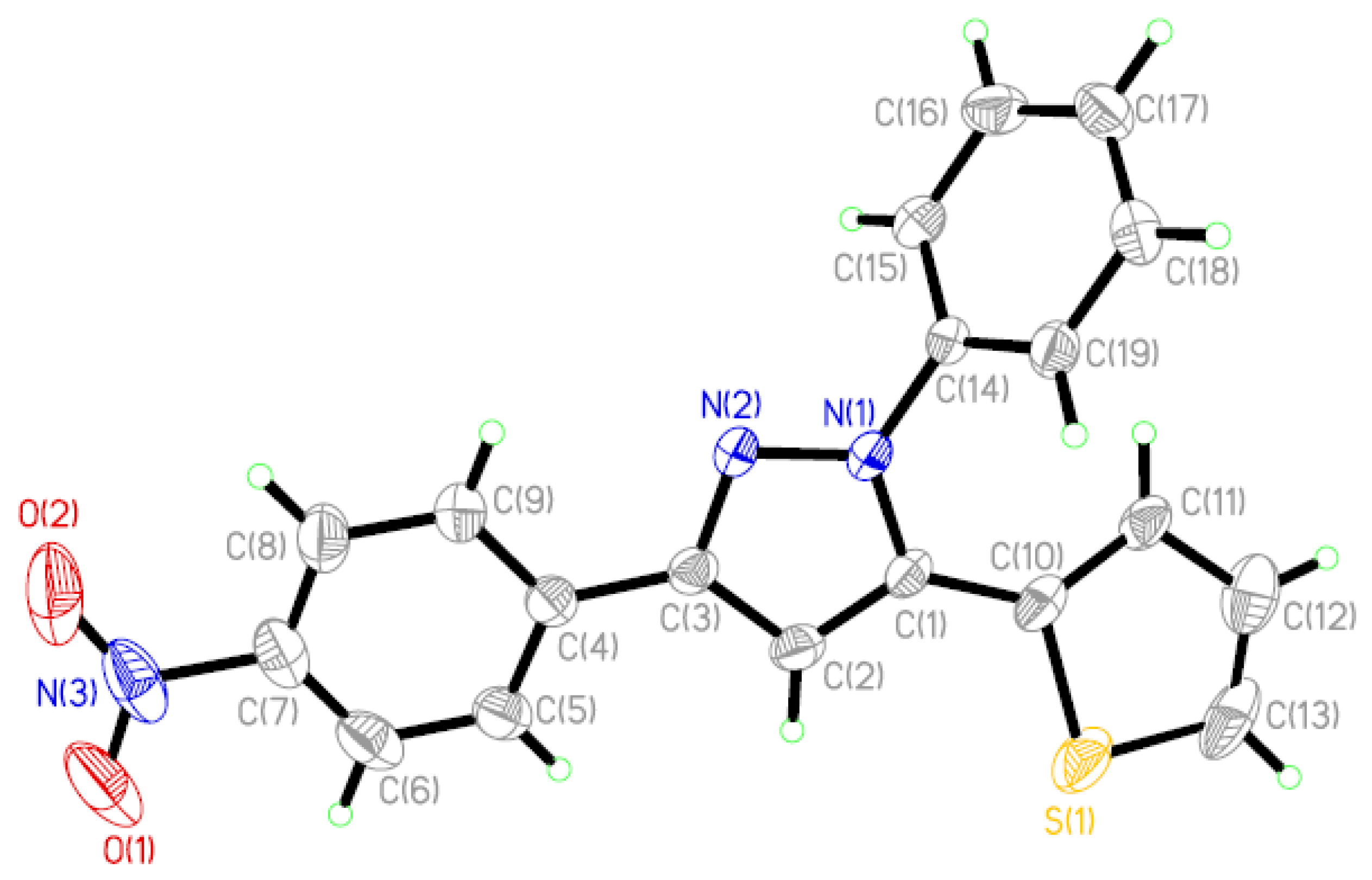
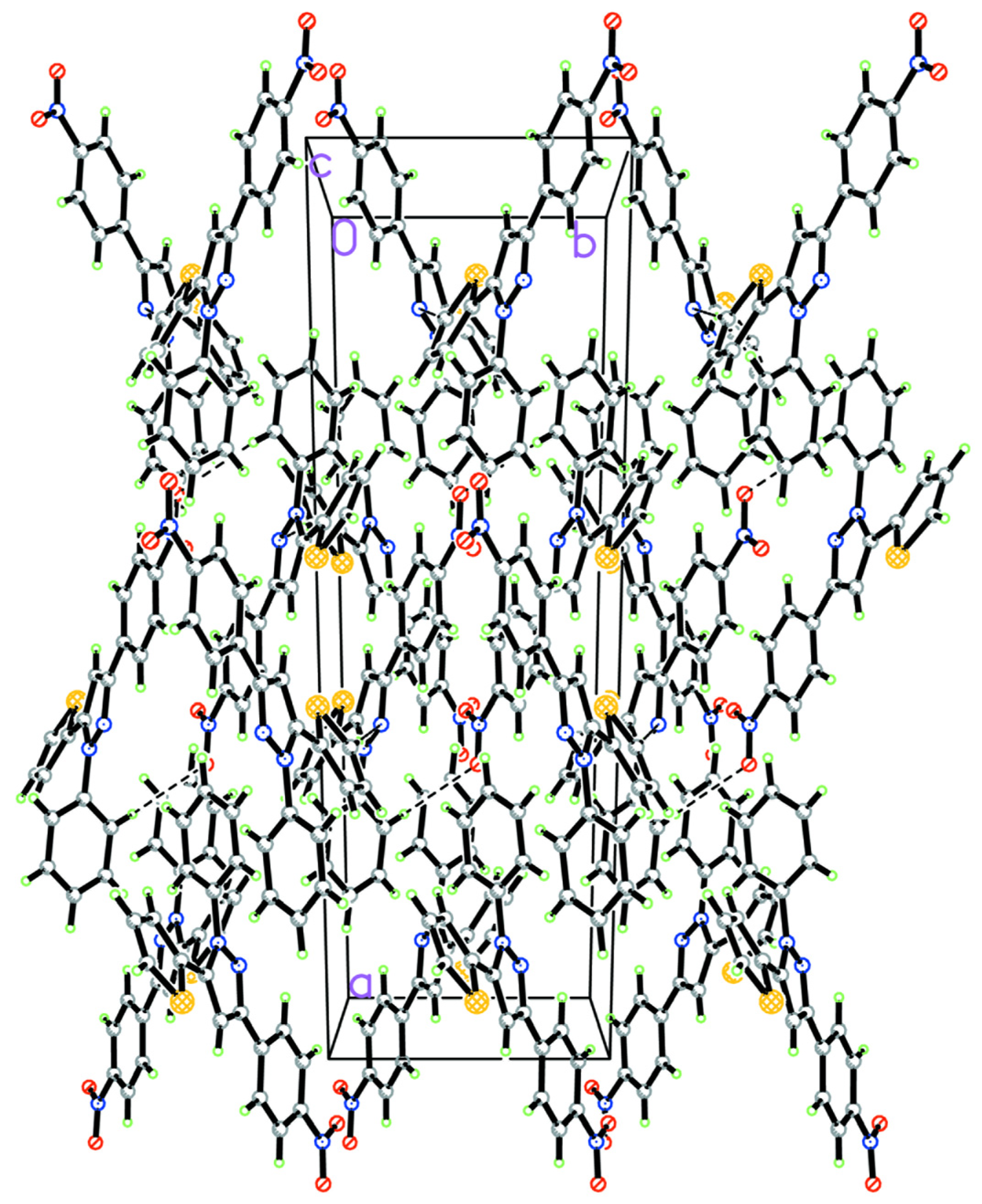
2.1. Description of the Crystal Structure

The displacement ellipsoid plot with the numbering scheme for the title compound is shown in Figure 1. Figure 2 shows a perspective view of the crystal packing in the unit cell. Selected bond lengths and bond angles by X-ray diffractions are listed in Table 1 along with the calculated bond parameters.
Figure 1. Molecular structure with the atomic numbering scheme for the title compound.
Molecules 19 05313 g001
Figure 2. A view of the crystal packing down the c axis for the title compound.
Molecules 19 05313 g002
The molecular structure of the title compound consists of discrete [PhC3HN2PhNO2C4H3S] entities. All of the bond lengths and bond angles in the phenyl rings are in the normal range. In the pyrazolinyl ring of the molecule, the C=N bond length [1.332(4) Å] is longer than that in the similar structure [C=N 1.297(2) Å] [25]. The bond lengths of C–N [1.365(4) Å] and N–N [1.356(3) Å] are shorter than those found in the above-cited structure [C–N 1.474(2) Å, N–N 1.380(2) Å] [25]. The pyrazolinyl ring makes dihedral angles of 5.39(3), 60.10(3) and 19.27(11), respectively, with the p-nitrophenyl, phenyl and 2-thienyl rings.
Table 1. Selected structural parameters by X-ray and theoretical calculations.
Bond Lengths (Å)ExperimentB3LYP/6-311G**Bond Lengths (Å)ExperimentB3LYP/6-311G**
S(1)–C(13)1.675(5)1.730C(1)–C(10)1.458(4)1.457
S(1)–C(10)1.704(3)1.750C(2)–C(3)1.402(4)1.415
O(1)–N(3)1.201(6)1.225C(3)–C(4)1.466(4)1.468
O(2)–N(3)1.225(6)1.225C(4)–C(5)1.389(5)1.403
N(1)–N(2)1.356(3)1.348C(4)–C(9)1.393(5)1.405
N(1)–C(1)1.365(4)1.380C(5)–C(6) 1.381(5)1.387
N(1)–C(14)1.430(4)1.429C(10)–C(11)1.402(5)1.373
N(2)–C(3)1.332(4)1.334C(14)–C(19)1.373(4)1.394
N(3)–C(7)1.480(5)1.474C(15)–C(16)1.381(5)1.391
C(1)–C(2)1.374(4)1.383C(17)–C(18)1.362(5)1.394
Bond Angles (°) Bond Angles (°)
C(13)–S(1)–C(10)92.7(2)91.6 C(8)–C(7)–C(6)121.6(4)121.6
N(2)–N(1)–C(1)112.0(2)111.9C(11)–C(10)–S(1)109.6(2)110.5
C(3)–N(2)–N(1)105.4(2)106.0 C(10)–C(11)–C(12)111.4(4)112.9
O(1)–N(3)–O(2)125.1(5)124.6 C(13)–C(12)–C(11)113.4(4)113.3
N(1)–C(1)–C(2)105.8(2)105.8C(12)–C(13)–S(1)112.9(3)111.7
C(1)–C(2)–C(3)106.2(3)105.7 C(19)–C(14)–C(15)121.0(3)120.6
N(2)–C(3)–C(2)110.6(3)110.6C(17)–C(16)–C(15)120.5(3)120.3
C(5)–C(4)–C(9)117.9(3)118.7 C(17)–C(18)–C(19)119.9(3)120.4
In the crystal lattice, there are two intermolecular interactions (C–H···Y, Y=N) [26,27] and some C–H···π supramolecular interactions (see Table 2) [28]. In the solid state, all above supramolecular interactions stabilize the crystal structures.
Table 2. Hydrogen bonds and C–H···π supramolecular interactions a.
D–H···ASymmetryH···A (Å)D···A (Å)∠D–H···A (°)
C(13)–H(13)···N(2)x, 1 − y, −1/2 + z2.510(3)3.439(3) 176.98
C(19)–H(19)···O(1)−x, −y, −z2.552(2)3.279(1) 135.30
C(11)–H(11)···Cg(4)x, y, z3.037(1)3.743(1)134
C(16)–H(16)···Cg(4)1/2 − x, 1/2 + y, 1/2 − z3.209(2)3.879(3)131
C(18)–H(18)···Cg(1)1/2 − x, 1/2 − y, −z2.809(2)3.596(2)143
a Cg(1) and Cg(4) denote thienyl ring and phenyl ring C(14)–C(19), respectively.

2.2. Optimized Geometry

DFT calculations were performed on the title compound at B3LYP/6–311G** level of theory. Some optimized geometric parameters are also listed in Table 1. In view of the bond lengths in Table 1, most predicted values are longer than experimental ones and the biggest difference between the theoretical and experimental values occurs at S(1)–C(13) bond, with the different values being 0.0554 Å. As for the bond angles, most predicted values correspond with the experimental values and the biggest difference is seen in the bond angle of C(12)–C(13)–S(1), with a difference between values of 1.22°. The reasons for the above discrepances maybe as follows: (1) the theoretical values correspond to the isolated molecule in the gas-phase and the experimental values are from the molecule in the solid state. The geometry of the solid-state structures is subject to intermolecular forces, such as van der Waals interactions and crystal packing forces, which make most of the experimental bond lengths shorter than the theoretical ones; (2) in the solid state, there exists a C–H···π supramolecular interaction corresponding with the thienyl ring (see Table 2), while in theoretical calculations, supramolecular interactions are neglected, which, to some extent, may lead to bigger bond length and bond angle differences between the experiments and calculations corresponding to the thienyl ring. Despite of some differences, the DFT method used here can reproduce the molecular geometry on the whole and it is the basis for our following discussion.

2.3. Atomic Charge Distributions

Based on B3LYP/6-311G** optimized geometry, NPA atomic charge distributions of the title compound were calculated. All of the atomic charges of non-hydrogen atoms are listed in Table 3.
Table 3. NPA atomic charge distributions obtained at B3LYP/6-311G** level.
AtomCharges (e)AtomCharges (e)AtomCharges (e)
N(1)−0.16920C(4)−0.03329C(11)−0.22313
N(2)−0.28191C(5)−0.18105C(12)−0.24353
C(1)0.13219C(6)−0.17616C(13)−0.37017
C(2)−0.27626C(7)0.05712C(14)0.14793
C(3)0.13913C(8)−0.17632C(15)−0.19406
N(3)0.51394C(9)−0.16957C(16)−0.18771
O(1)−0.38790S(1)0.41951C(17)−0.19584
O(2)−0.38977C(10)−0.22371C(18)−0.18649
C(19)−0.20411
As seen from Table 3, all non-hydrogen atomic charge distributions are not only influenced by the atomic electronegativity, but also by the conjugation mode around the atom. For example, for the nineteen C atoms, there are three atomic charge distribution phenomena: (1) in the two phenyl rings and one thienyl ring, because C atoms have higher electronegativity than H atoms, thirteen C atoms conjugated with H atoms have negative atomic charges; (2) since the electronegativity of C is smaller than that of N, four C atoms joined with N atoms have positive atomic charges; (3) for the C(4) and C(10) atoms, they are indirectly conjugated with the N atoms, which makes them have negative atomic charges. In addition, for three N atoms, N(1) and N(2) atoms are connected with C atoms, so they both carry negative atomic charges. However, for the N(3) atom, there are two O atoms and one C atom bond with it, which leads the N(3) atom to have a positive charge. It is remarkable that, although the S atom has higher electronegativity than the C atoms, in the thienyl ring, the atomic charge values of S are positive, indicating that this S atom loses the capacity to coordinate with metallic ions, which is very different from that in a free thiophene molecule. On the whole, considering the atomic charge distributions and the steric effect, the title compound has three sites, namely N(2), O(1) and O(3), which have negative charge values and allow it to act as a multi-dentate ligand to coordinate with metallic ions.

2.4. Calculations of Nonlinear Optical Property

Since 2-pyrazoline derivatives have non-linear optical (NLO) properties [29], so we also made a prediction on the title compound. The predicted results were compared with the traditional NLO materials of urea and para-nitroaniline (PNA). On the basis of the MNDO Hamiltonian and PM3 parametrization with the MOPAC program package, the molecular hyperpolarizability value, βμ, the vector components along the dipole moment direction, of the title compound were calculated to be 4.997 × 10⁻³⁰ esu, which is greater than the value of urea (0.14 × 10⁻³⁰ esu calculated using the same method) [30] and smaller than that of PNA (6.801 × 10⁻³⁰ esu calculated using the same method). The comparisons indicate that the title compound is a potential NLO material.

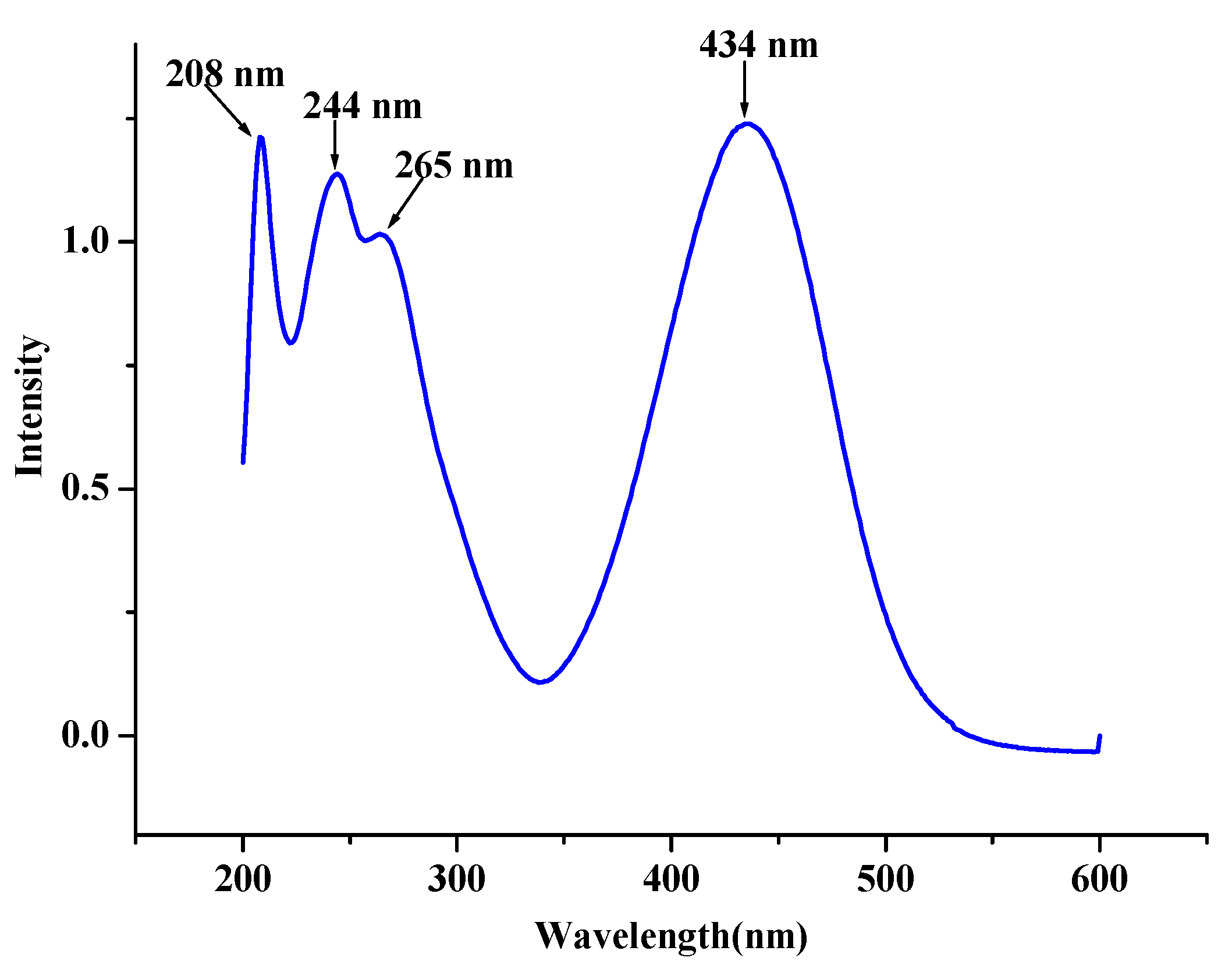
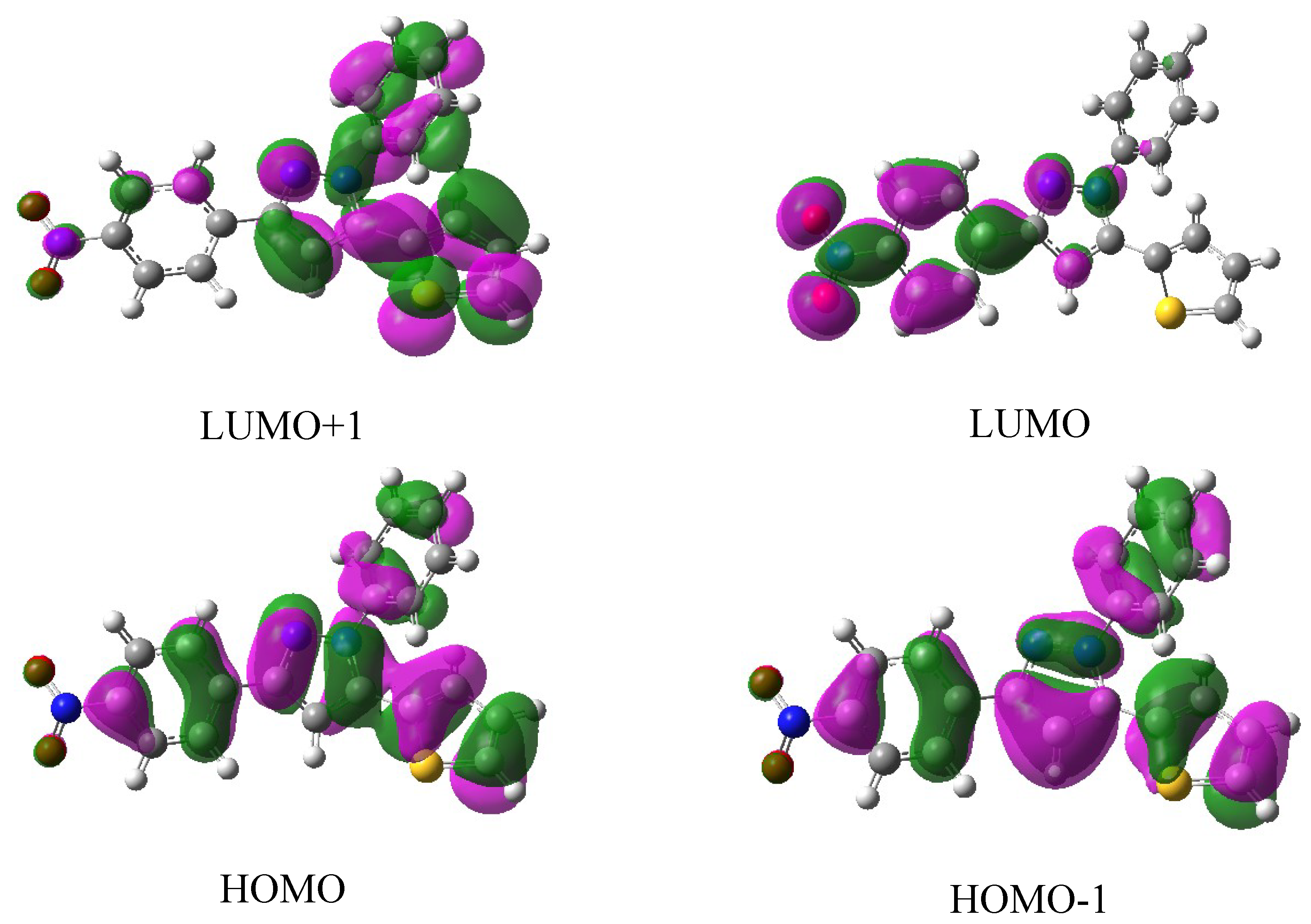
2.5. Electronic Absorption Spectra

The electronic absorption spectrum of the title compound have been measured in EtOH solution and is shown in Figure 3. From Figure 3, one can find that there are four electronic transition peaks and three of them locate at about 208, 244 and 265 nm, respectively, which are in the ultraviolet region range. The fourth peak is at 434 nm, indicating the title compound solution has absorbed the blue-light and led to the solution being yellow.
Figure 3. Electronic absorption spectra of the title compound measured in EtOH solution.
Molecules 19 05313 g003
Natural population analyses based on the B3LYP/6-311G** optimized geometry show that the frontier molecular orbitals of the title compound are mainly composed of p atomic orbitals, so electronic transitions corresponding to above electronic spectra are mainly assigned to nπ* and ππ* electronic transitions. Figure 4 shows the surfaces of the HOMO-1, HOMO, LUMO and LUMO+1 for the title compound. As seen in Figure 4, when electron transitions take place, electrons are mainly transferred among the phenyl ring, nitrophenyl ring and thienyl ring, they correspond to the nπ* and ππ* electronic transitions.
Figure 4. Some frontier molecular orbital stereographs for the title compound.
Molecules 19 05313 g004

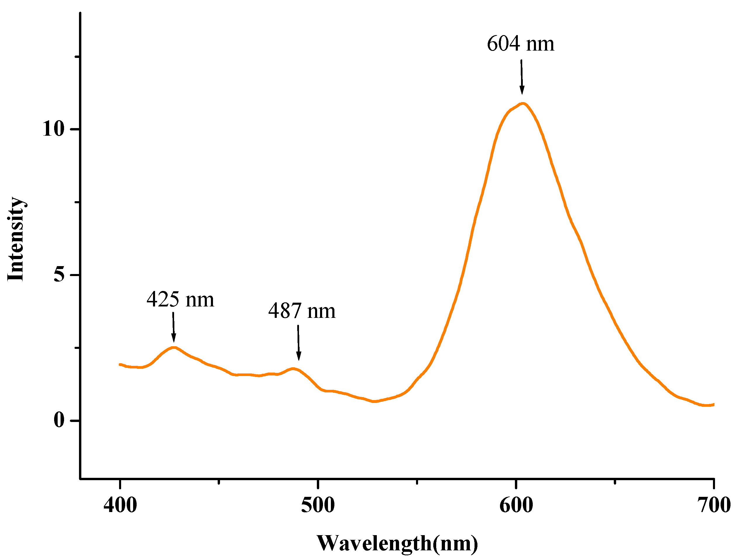
2.6. Fluorescence Spectra

The solid-state fluorescence spectrum of the title compound is shown in Figure 5. The spectrum exhibits two weak emissions at 425 and 487 nm, respectively, which are in the violet-light region. A maximum emission band is at 604 nm, which is in the orange-light region. This result suggests the title compound is a potential orange-light emitting material.
Figure 5. Solid-state fluorescence spectra of the title compound.
Molecules 19 05313 g005

3. Experimental and Theoretical Methods

3.1. Physical Measurements
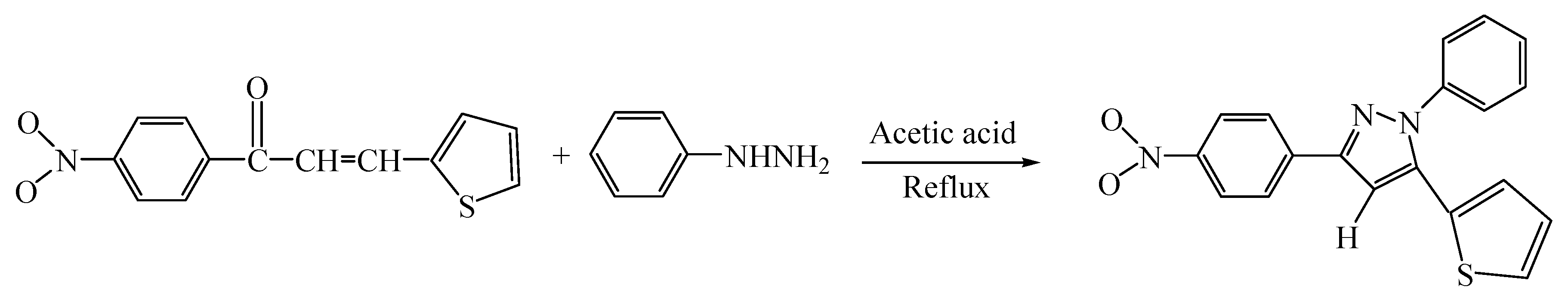
Elemental analyses for carbon, hydrogen and nitrogen were performed by a Perkin-Elmer 240C elemental instrument. IR spectra (4000–400 cm−1) were recorded on a Nicolet FT-IR spectrophotometer as KBr pellets. Electronic absorption spectra were measured on a Shimadzu UV3100 instrument in EtOH solution and solid-state fluorescence spectra were measured on a F96-fluorospectrophotometer.

3.2. Synthesis

All chemicals were obtained from a commercial source and used without further purification. 1-(4-Nitrophenyl)-3-(2-thienyl)-2-propenyl-1-ketone (0.01 mol) and phenylhydrazine (0.015 mol) were mixed in acetic acid (40 mL) and stirred during reflux for 6 h. Then, the mixture was poured into ice-water to afford light-yellow solids. The solids were filtered and washed with water until the pH of the solution was about 7. Finally, the light-yellow solid crystals of the title compound were dried at room temperature. Yield 78.6%. Mp. 168.1–169.2. IR: v 3433(m), 3055(m), 1588(vs), 1550(s), 1492(vs), 1390(s), 1338(vs), 1267(m), 1127(s), 1107(s), 1062(m), 1034(w), 992(m), 839(s), 742(vs), 710(m), 685(s), 512(w) cm−1. Found: C, 65.51; H, 3.89; N, 12.23%. Calc. for C19H13N3O2S: C, 65.69, H, 3.77, N, 12.10%.

3.3. Crystallographic Study

The selected crystal of the title compound was mounted on an Rigaku raxis Rapid IP Area Detector diffractometer. Reflection data were measured at 293(2) K using graphite monochromated Mo-Kα (λ = 0.71073 Å) radiation and a ωscan mode. The correction for Lp factors and empirical absorption were applied to the data. The structures were solved by direct methods and refined by full-matrix least-squares method on Fobs2 using the SHELXTL software package [31]. All non-H atoms were anisotropically refined. The hydrogen atom positions were fixed geometrically at calculated distances and allowed to ride on the parent C atoms. The final least-square cycle gave R = 0.0707, wR2 = 0.1922. Atomic scattering factors and anomalous dispersion corrections were taken from International Table for X-ray Crystallography [32]. A summary of the key crystallographic information is given in Table 4.
CCDC-990511 contains the supplementary crystallographic data for this paper. These data can be obtained free of charge at www.ccdc.cam.ac.uk/conts/retrieving.html (or from the Cambridge Crystallographic Data Centre (CCDC), 12 Union Road, Cambridge CB2 1EZ, UK; Fax: +44(0)1222-336033; E-Mail: [email protected]).

3.4. Computational Methods

Initial molecular geometry of the title compound was taken from its crystal structure. Then, DFT calculations at B3LYP/6-311G** levelof theory by the Berny method [33] were performed with the Gaussian 03 software package [34]. Vibrational frequencies calculated ascertain the structure was stable (no imaginary frequencies). Natural Bond Orbital (NBO) analyses were also performed on the optimized structure. On the basis of the MNDO Hamiltonian [35] and PM3 parametrization [36] with the MOPAC [37] program package, the molecular hyperpolarizability value was also calculated. All calculations were performed on a DELL PE 2850 server and a Pentium IV computer using the default convergence criteria.
Table 4. Summary of crystallographic results for the title compound.
Empirical FormulaC19H13N3O2S
Formula weight347.38
Temperature293(2) K
Wavelength0.71073 Å
Crystal system, space groupMonoclinic, C2/c
Unit cell dimensionsa = 28.023(7) Å
b = 7.8005(15) Å β = 122.69(2)°
c = 18.008(7) Å
Volume3312.9(17) Å3
Z, Calculated density8, 1.393 Mg/m3
Absorption coefficient0.213 mm−1
F(000)1440
θ range for data collection3.26 to 25.00°
Limiting indices−33 ≤ h ≤ 33, −9 ≤ k ≤ 8, −21 ≤ l ≤ 21
Reflections collected/unique10,658/2887 [Rint = 0.0476 ]
Refinement method Full-matrix least-squares on F2
Data/restraints/parameters2887/0/226
Goodness-of-fit on F21.120
Final R indices [I > 2σ (I)]R1 = 0.0707, wR2 = 0.1922
R indices (all data)R1 = 0.0911, wR2 = 0.2071
Largest diff. peak and hole0.402 and −0.404 e. Å−3

4. Conclusions

1-Phenyl-3-(4-nitrophenyl)-5-(2-thienyl)-2-pyrazoline has been synthesized and characterized, by a variety of methods including IR, X-ray single crystal diffraction, UV-Vis and fluorescence spectroscopy. For the title compound, DFT calculations at B3LYP/6-311G** of the structure and NPA atomic charge distributions have been carried out. Experimental and predicted results indicate that the optimized geometry at B3LYP/6-311G** level can represent the molecular structure. NPA atomic charge distributions show the title compound may be acted as multi-dentate ligand to coordinate with metallic ions. The electronic absorption spectra measurement of the title compound exhibits four absorption bands and DFT calculations indicate the above electronic spectra are mainly assigned to nπ* and ππ* electronic transitions. The solid-state fluorescence spectrum reveals the title compound is a potential orange-light emitting material.

Acknowledgments

This work was supported by Fund of Jiangsu Key Laboratory for Chemistry of Low-Dimensional Materials (JSKC12107 and JSKC12106) and Huaian Science & Technology Bureau, Jiangsu Province, China (HAG2012005, HAG2013009 and HC201307).

Author Contributions

Huan-Mei Guo for synthesis and writing the manuscript; Pu-Su Zhao for calculations and analysis; Qian Wu for characterizations of the title compound; Yu-Feng Li for single crystal structural refinement.

Conflicts of Interest

The authors declare no conflict of interest.

References

  1. Xiao, D.; Xi, L.; Yang, W.; Fu, H.; Shuai, Z.; Fang, Y.; Yao, J. Size-Tunable Emission from 1,3-Diphenyl-5-(2-anthryl)-2-pyrazoline Nanoparticles. J. Am. Chem. Soc. 2003, 125, 6740–6745. [Google Scholar] [CrossRef]
  2. Ji, S.J.; Shi, H.B. Synthesis and fluorescent property of some novel benzothiazoyl pyrazoline derivatives containing aromatic heterocycle. Dyes Pigm. 2006, 70, 246–250. [Google Scholar] [CrossRef]
  3. Bian, B.; Ji, S.J.; Shi, H.B. Synthesis and fluorescent property of some novel bischromophore compounds containing pyrazoline and naphthalimide groups. Dyes Pigm. 2008, 76, 348–352. [Google Scholar] [CrossRef]
  4. Zhang, X.H.; Lai, W.Y.; Gao, Z.Q.; Wong, T.C.; Lee, C.S.; Kwong, H.L. Photoluminescence and electroluminescence of pyrazoline monomers and dimers. Chem. Phys. Lett. 2000, 320, 77–80. [Google Scholar] [CrossRef]
  5. Wei, X.Q.; Yang, G.; Cheng, J.B.; Lu, Z.Y.; Xie, M.G. Synthesis of novel light-emitting calix[4]arene derivatives and their luminescent properties. Opt. Mater. 2007, 29, 936–940. [Google Scholar] [CrossRef]
  6. Pramanik, S.; Banerjee, P.; Sarkar, A.; Mukherjee, A.; Mahalanabis, K.K.; Bhattacharya, S.C. Spectroscopic investigation of 3-pyrazolyl 2-pyrazoline derivative in homogeneous solvents. Spectrochim. Acta A 2008, 71, 1327–1332. [Google Scholar] [CrossRef]
  7. Pokladko, M.; Gondek, E.; Sanetra, J.; Nizioł, J.; Danel, A.; Kityk, I.V.; Reshak Ali, H. Spectral emission properties of 4-aryloxy-3-methyl-1-phenyl-1H-pyrazolo[3,4-b]quinolines. Spectrochim. Acta A 2009, 73, 281–285. [Google Scholar] [CrossRef]
  8. Sun, Y.F.; Cui, Y.P. The synthesis, structure and spectroscopic properties of novel oxazolone-, pyrazolone- and pyrazoline-containing heterocycle chromophores. Dyes Pigm. 2009, 81, 27–34. [Google Scholar] [CrossRef]
  9. Fu, H.B.; Yao, J.N. Size Effects on the Optical Properties of Organic Nanoparticles. J. Am. Chem. Soc. 2001, 123, 1434–1439. [Google Scholar] [CrossRef]
  10. Oh, S.W.; Kang, Y.S. The size-dependent optical properties of 1-phenyl-3-naphthyl-5-((dimethyl amino) phenyl)-2-pyrazoline nanoparticles. Colloids Surf. A: Physicochem. Eng. Aspects 2005, 257, 415–418. [Google Scholar]
  11. Fu, H.B.; Loo, B.H.; Xiao, D.B.; Xie, R.M.; Ji, X.H.; Yao, J.N.; Zhang, B.W.; Zhang, L.Q. Multiple Emissions from 1,3-Diphenyl-5-pyrenyl-2-pyrazoline Nanoparticles: Evolution from Molecular to Nanoscale to Bulk Materials. Angew. Chem. Int. Ed. 2002, 41, 962–965. [Google Scholar] [CrossRef]
  12. Zhao, P.S.; Li, Y.F.; Guo, H.M.; Jian, F.F.; Wang, X. Synthesis, Crystal Structure and Density Functional Calculations on 1-Phenyl-3-p-fluorophenyl-5-p-chlorophenyl-2-pyrazoline. Bull. Korean Chem. Soc. 2007, 28, 1539–1544. [Google Scholar] [CrossRef]
  13. Zhao, P.S.; Li, Y.F.; Guo, H.M.; Wang, X.; Jian, F.F. Synthesis, Characterization and ab initio Calculations on 1-Phenyl-3-p-fluorophenyl-5-p-methoxyphenyl-2-pyrazoline. Pol. J. Chem. 2007, 81, 1735–1742. [Google Scholar]
  14. Jian, F.F.; Zhao, P.S.; Guo, H.M.; Li, Y.F. Synthesis, characterization, crystal structure and DFT studies on 1-acetyl-3-(2,4-dichloro-5-fluoro-phenyl)-5-phenyl-pyrazoline. Spectrochim. Acta A 2008, 69, 647–653. [Google Scholar] [CrossRef]
  15. Zhao, P.S.; Wang, H.Y.; Li, R.Q.; Guo, H.M. Synthesis, crystal structure, electronic spectra and density functional studies on 1N-phenyl-3-(3,4-dichlorophenyl)-5-phenyl-2-pyrazoline. Indian J. Chem. A 2008, 47, 986–991. [Google Scholar]
  16. Zhao, P.S.; Li, R.Q.; Sun, X.J.; Guo, H.M.; Jian, F.F. Comparative study on two 2-pyrazoline derivatives with experimental and theoretical methods. Struct. Chem. 2009, 20, 443–451. [Google Scholar] [CrossRef]
  17. Zhao, P.S.; Li, R.Q.; Wang, H.Y.; Jian, F.F.; Guo, H.M. Experimental and theoretical comparative studies on two 2-pyrazoline derivatives. Spectrochim. Acta A 2009, 74, 87–93. [Google Scholar] [CrossRef]
  18. Zhao, P.S.; Zhou, S.S.; Guo, Z.Y.; Zhu, Y. Crystal structure, spectra properties and comparative studies on a 2-pyrazoline derivative. Spectrochim. Acta A 2012, 94, 65–71. [Google Scholar] [CrossRef]
  19. Jin, Z.N.; Wu, J.S.; Wang, C.F.; Dai, G.L.; Liu, S.Y.; Lu, J.M.; Jiang, H.J. Novel fluorescent 1,8-naphthalimide derivatives containing thiophene and pyrazole moieties: Synthesis by direct C–H arylation and evaluation of photophysical and electrochemical properties. Spectrochim. Acta A: Mol. Biomol. Spectr 2014, 117, 527–534. [Google Scholar] [CrossRef]
  20. Willy, B.; Müller, T.J.J. Regioselective Three-Component Synthesis of Highly Fluorescent 1,3,5-Trisubstituted Pyrazoles. Eur. J. Org. Chem. 2008, 24, 4157–4168. [Google Scholar] [CrossRef]
  21. Yang, Y.W.; Kuang, C.X.; Jin, H.; Yang, Q.; Zhang, Z.K. Efficient synthesis of 1,3-diaryl-4-halo-1H-pyrazoles from 3-arylsydnones and 2-aryl-1,1-dihalo-1-alkenes. Beilstein. J. Org. Chem. 2011, 7, 1656–1662. [Google Scholar] [CrossRef]
  22. Raghava, B.; Prasad, T.N.M.; Lakshminarayana, B.N.; Sridhar, M.A.; Prasad, J.S.; Rangappa, K.S. Synthesis and crystal structure of 3-(2,5-dimethylphenyl)-1-(4-methoxy phenyl)-5-(thiophen-2-yl)-1H-pyrazole. X-ray Struct. Anal. Online 2012, 28, 51–52. [Google Scholar] [CrossRef]
  23. Koch, W.; Holthausen, M.C. A Chemistry Guide to Density Functional Theory; Wiley-VCH: Weinheim, Germany, 2000. [Google Scholar]
  24. Parr, R.R.; Yang, R.G. Density Functional Theory of Atoms and Molecules; Oxford University Press: New York, NY, USA, 1989; and references therein. [Google Scholar]
  25. Guo, H.M.; Jian, F.F.; Zhao, P.S.; Zhang, Y.C.; Li, Y.F. 5-(2-Chlorophenyl)-3-(4-chlorophenyl)-1-phenyl-2-pyrazoline. Acta. Cryst. 2007, E63, o215–o216. [Google Scholar]
  26. Steiner, T. C–H–O Hydrogen Bonding in Crystals. Cryst. Rev. 1996, 6, 1–51. [Google Scholar] [CrossRef]
  27. Jeffrey, G.A.; Maluszynska, H.; Mitra, J. Hydrogen bonding in nucleosides and nucleotides. Int. J. Biol. Macromol. 1985, 7, 336–348. [Google Scholar] [CrossRef]
  28. Hunter, R.H.; Haueisen, R.H.; Irving, A. The First Water-Dependent Liquid Clathrate: X-Ray Evidence in the Solid for a C–H···π (Heteroarene) π···H–C Interaction. Angew. Chem. Int. Ed. Engl. 1994, 33, 566–568. [Google Scholar] [CrossRef]
  29. Joaquín, B.; Koen, C.; Raquel, G.; Stephan, H.; André, P.; José, L.S. Versatile optical materials: Fluorescence, non-linear optical and mesogenic properties of selected 2-pyrazoline derivatives. J. Mater. Chem. 1998, 8, 1725–1730. [Google Scholar] [CrossRef]
  30. You, X.Z. Molecular-based Materials -Opto-electronic Functional Compounds; Science and Technology Publishing Company: Shanghai, China, 2001; pp. 1–164. [Google Scholar]
  31. Sheldrick, G.M. SHELXTL; v5 Reference Manual; Siemens Analytical X-Ray Systems: Madison, WI, USA, 1997. [Google Scholar]
  32. Wilson, A.J. International Table for X-Ray Crystallography; Kluwer Academic: Dordrecht, The Netherlands, 1992; Volume C, Tables 4.2.6.8 and 6.1.1.4; pp. 219–222, 500–502, respectively. [Google Scholar]
  33. Peng, C.; Ayala, P.Y.; Schlegel, H.B.; Frisch, M.J. Using redundant internal coordinates to optimize equilibrium geometries and transition states. J. Comput. Chem. 1996, 17, 49–56. [Google Scholar] [CrossRef]
  34. Frisch, M.J.; Trucks, G.W.; Schlegel, H.B.; Scuseria, G.E.; Robb, M.A.; Cheeseman, J.R.; Montgomery, J.A.; Vreven, T., Jr.; Kudin, K.N.; Burant, J.C.; et al. Gaussian 03W; Gaussian, Inc.: Wallingford, CT, USA, 2004. [Google Scholar]
  35. Dewar, M.J.S.; Thiel, W. Ground states of molecules. 38. The MNDO method. Approximations and parameters. J. Am. Chem. Soc. 1977, 99, 4899–4907. [Google Scholar] [CrossRef]
  36. Stewart, J.J.P. Optimization of parameters for semiempirical methods I. Method. J. Comput. Chem. 1989, 10, 209–220. [Google Scholar] [CrossRef]
  37. Stewart, J.J.P. QCPE Program 455 (1983); Version 6.0; Indiana University: Bloomington, IN, USA, 1990. [Google Scholar]
  • Sample Availability: Sample of the title compound is available from authors.

