Article

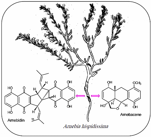
Two Polycyclic Geranylhydroquinone-Derived Metabolites from Roots of Arnebia hispidissima (Lehm.) DC.

by
Atallah F. Ahmed
1,2,*,
Hassan-Elrady A. Saad
2 and
Eman M. Abd El-Karim
2
1
Department of Pharmacognosy, College of Pharmacy, King Saud University (KSU), Riyadh 11451, Kingdom of Saudi Arabia
2
Department of Pharmacognosy, Faculty of Pharmacy, Mansoura University (MU), Mansoura 35516, Egypt
*
Author to whom correspondence should be addressed.
Molecules 2014, 19(5), 5940-5951; https://doi.org/10.3390/molecules19055940
Submission received: 20 March 2014 / Revised: 23 April 2014 / Accepted: 4 May 2014 / Published: 9 May 2014
(This article belongs to the Section Natural Products Chemistry)

Abstract

:
A phytochemical investigation of the least polar organic extracts of Arnebia hispidissima (Lehm.) DC. roots has led to the isolation of two unique polycyclic geranylhydroquinone-derived metabolites, arnebacene (1) and arnebidin (2), along with some known phenolic metabolites viz., arnebin-7 (3) and vanillic acid (4). The chemical identification of the new isolated compounds, including their relative stereochemistry, was achieved via spectroscopic analyses, including 2D NMR, and spectral comparison with related compounds. A biosynthetic pathway is proposed for the new compounds on the basis of their structure-relationship with previously isolated metabolites.

Graphical Abstract

1. Introduction

Arnebia hispidissima (Lehm.) DC. (Arabian primrose), which belongs to the family Boraginaceae, is widely distributed in the northern Africa area, through Egypt, to northern India [1]. To date, the roots of eight Arnebia species were studied phytochemically, and several naphthoquinone metabolites possessing various biological activities, have been isolated [2,3,4,5,6,7]. The root of A. hispidissima has been used as a food colorant whereas the flowering shoot has been employed in diseases of the tongue and throat as well as fevers and cardiac disorders [4,8]. Moreover, the organic extracts of A. hispidissima exhibited antibacterial [3,9] and antitumor activity [10] which were attributable to the presence of naphthoquinones, triterpenoids, and pyrrolizidine alkaloids. Our phytochemical investigation on the root of Arnebia hispidissima growing in Sinai Peninsula, has led to the isolation of new furanohexahydroanthracene-based metabolite arnebacene (1) and the arnebin-7-related dimer arnebidin (2), along with two known phenolic compounds 3 and 4. The structures of the new metabolites, including their stereochemistry, were elucidated on the basis of extensive spectroscopic analyses (including 1D and 2D NMR) and by comparison of their spectral data with those of related compounds. Compound 2 is reported herein as a novel heptacyclic arnebin-7 dimer with an unprecedented tricyclo[3.3.0.01,3]octane core.

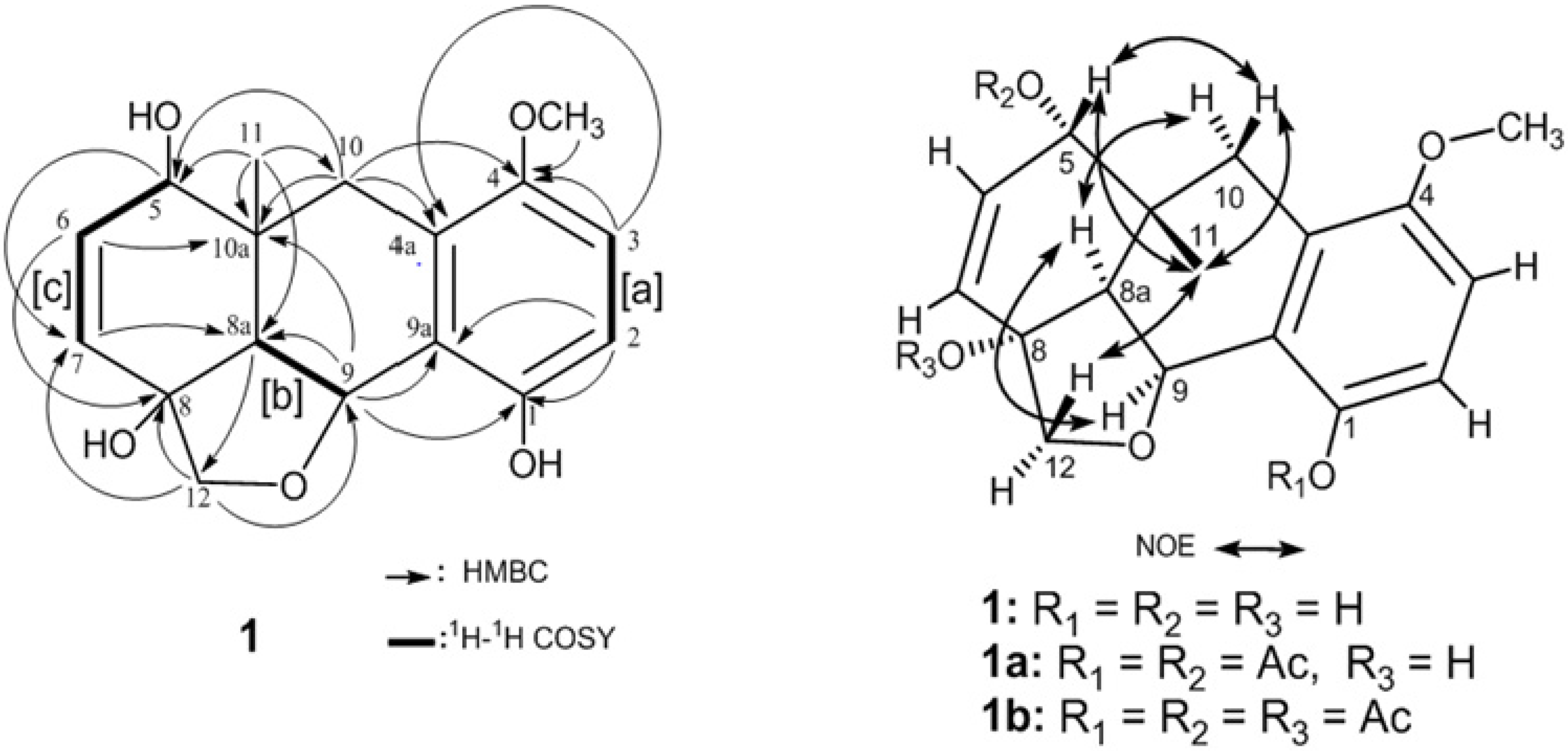
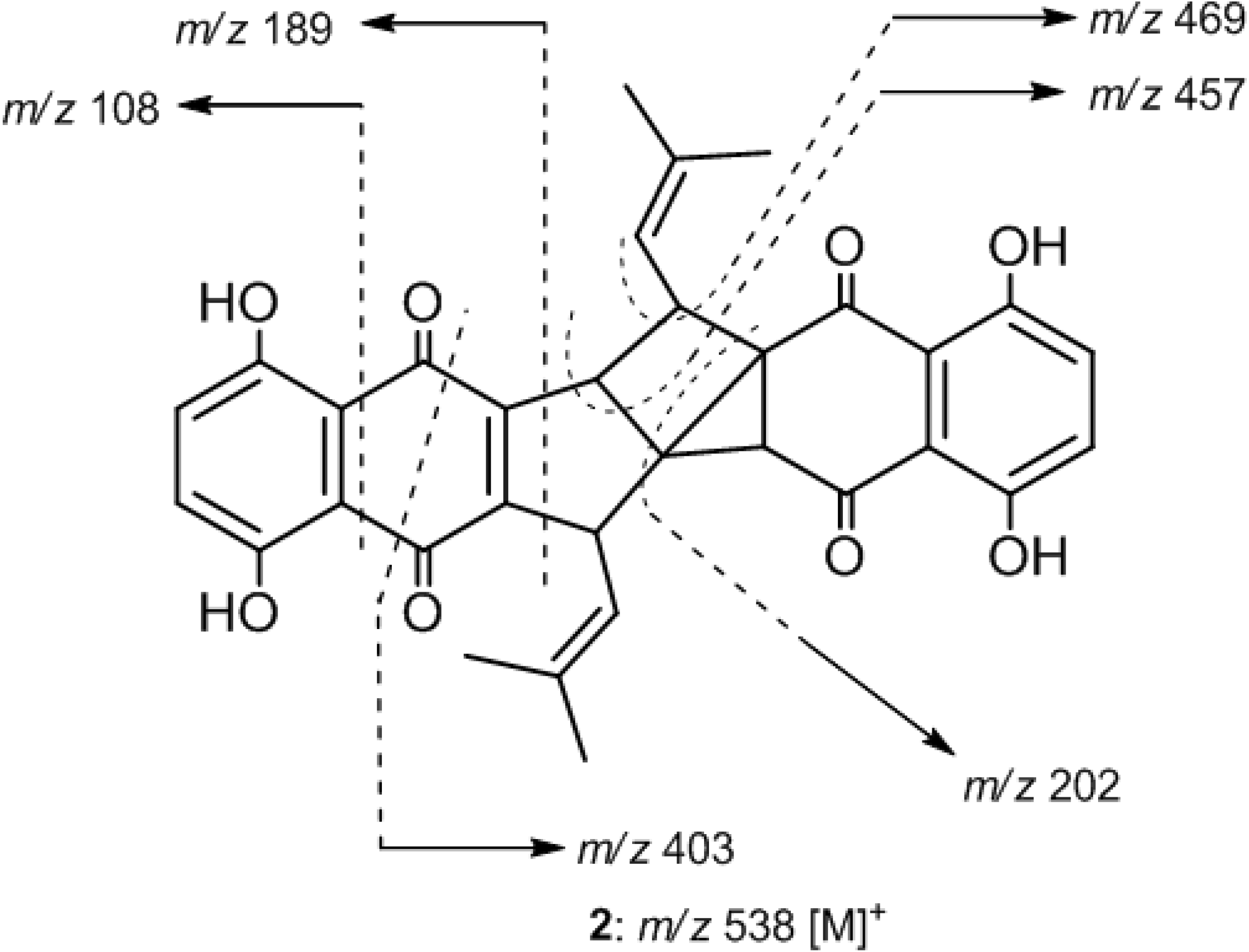
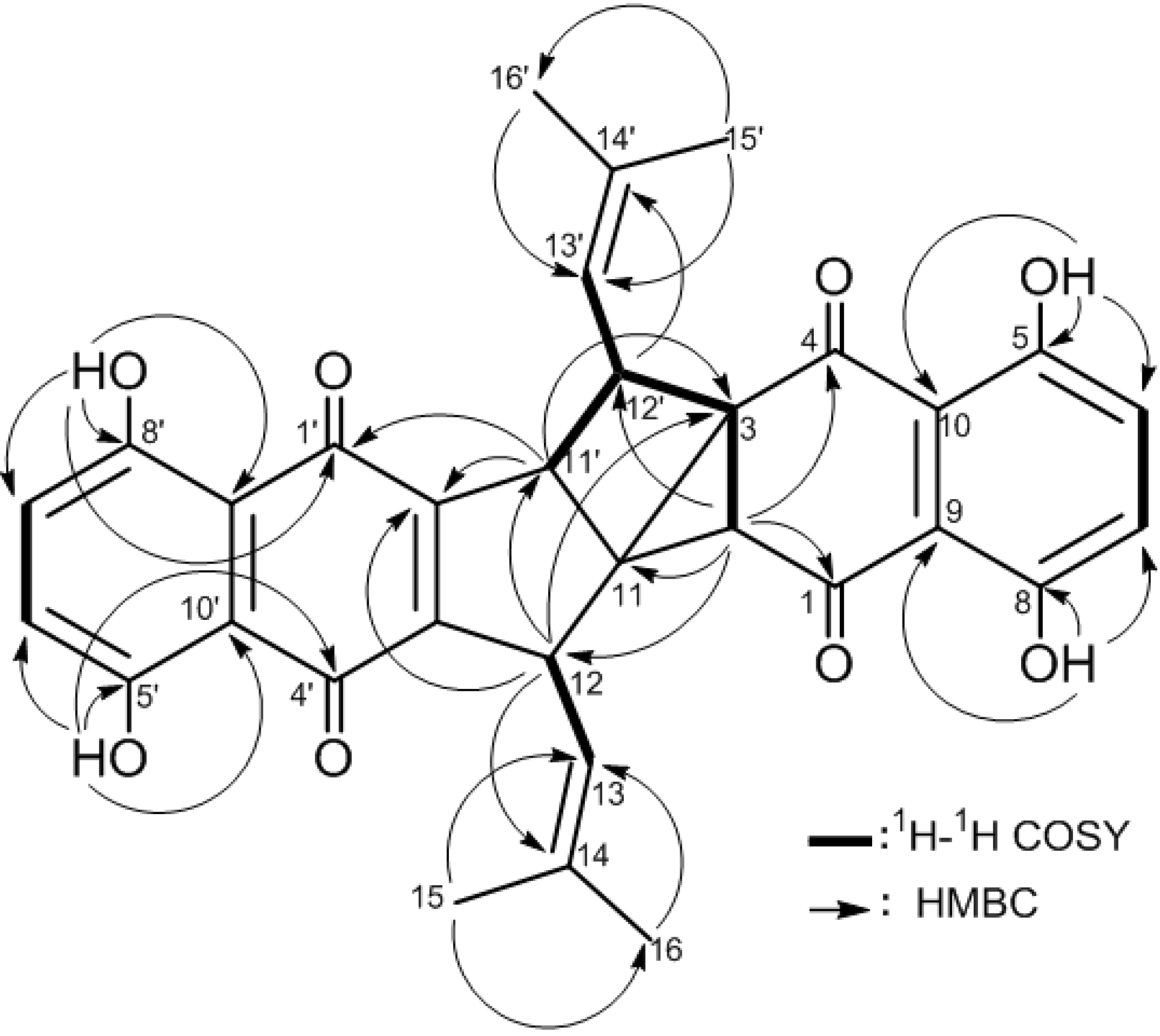
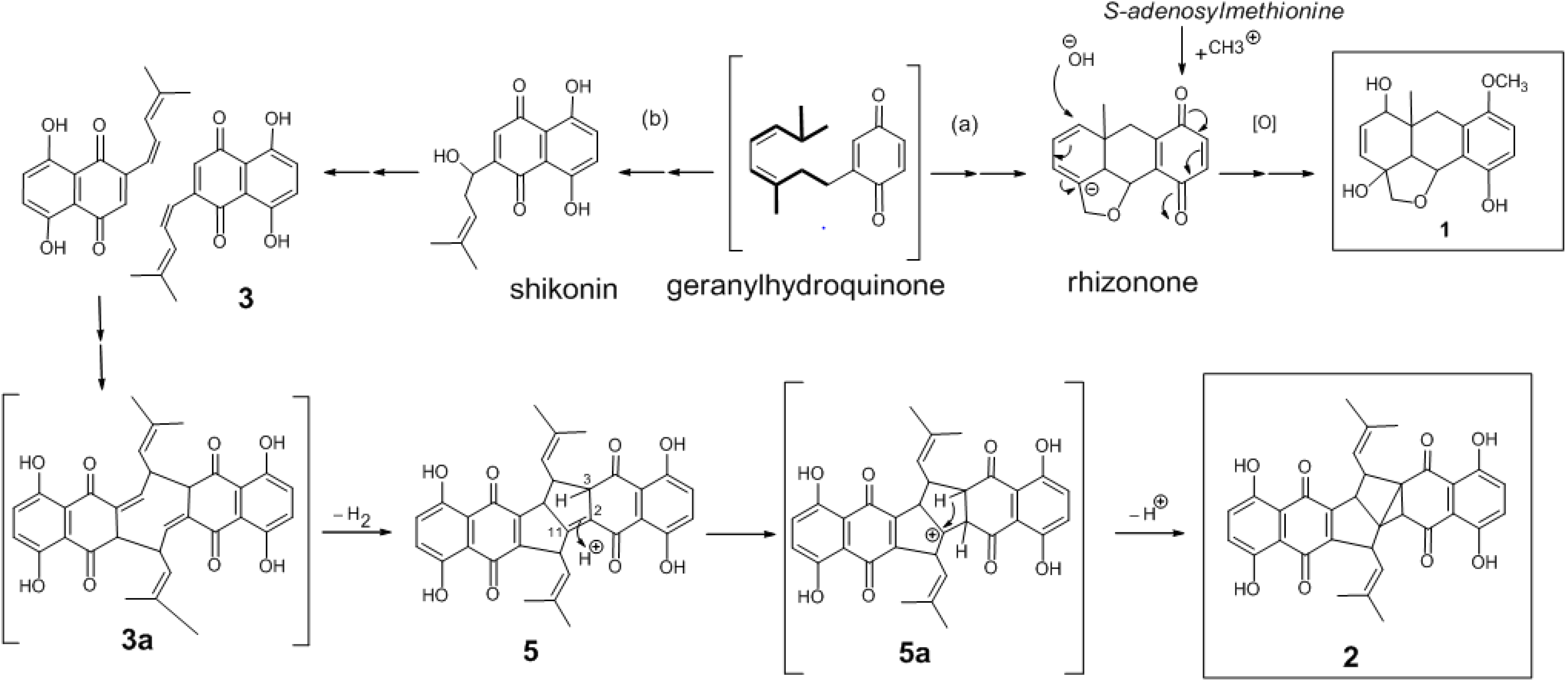
2. Results and Discussion
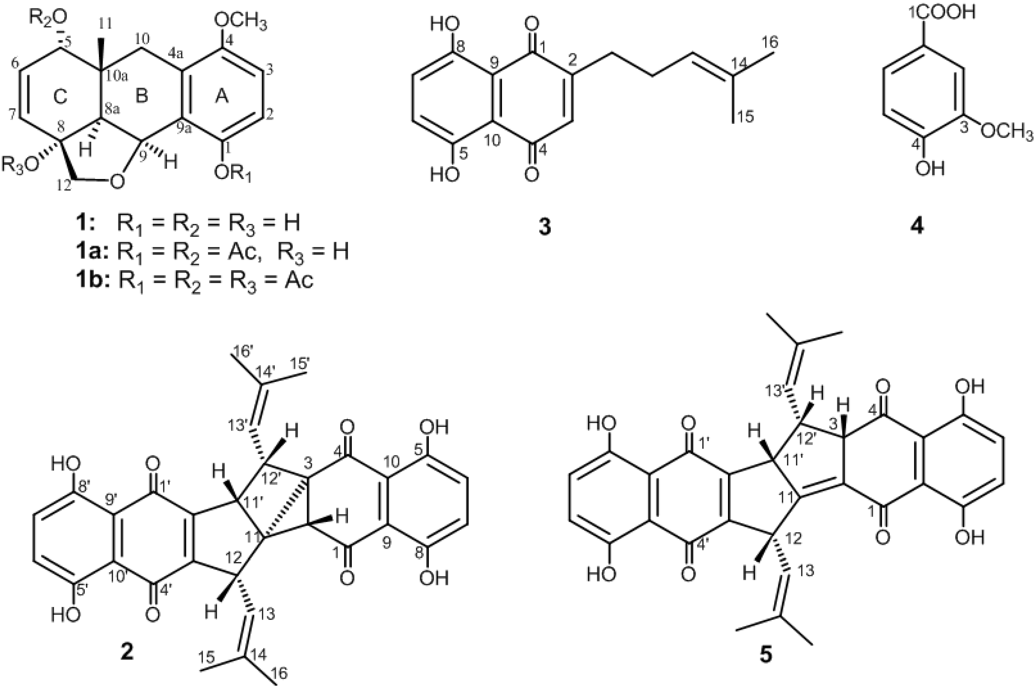
The dried root of A. hispidissima was powdered, sequentially extracted with light petroleum and then MeOH. The MeOH extract was further partitioned with H2O/n-hexane, H2O/CH2Cl2, and then with H2O/EtOAc to yield MHF, MCF, and MEF fractions, respectively. The light petroleum extract and MeOH-derived fractions were separately subjected to chromatographic fractionation and purification, utilizing a series of normal-phased chromatographic systems, to afford compounds 2 and 3 from the light petroleum extract, 1 from MCF, and 4 from MEF (Figure 1).
Figure 1. Phenolic constituents (14) isolated from A. hispidissima roots and shikometabolin D (5).
Molecules 19 05940 g001
Compounds 3 and 4 were identified by comparison of their physical and spectroscopic (MS and NMR) data with those of the previously isolated compounds as arnebin-7 [11] (syn. deoxyalkannin [12] or deoxyshikonin [7]) and vanillic acid [13], respectively. Compound 4 is reported herein from genus Arnebia for the first time, although it was isolated once from the genus Onosma [14] of the same family (Boragenaceae).
Compound 1 was isolated as colorless feathery crystals with a molecular formula of C17H20O5, implying eight degrees of unsaturation, as established by the HRFAB-MS m/z 304.1307 (M+). Its absorption bands in the UV (281 nm) and IR (3356 and 1601 cm−1) spectra revealed the presence of a phenolic moiety. The peaks appearing in the in EI-MS at m/z 286 [M-H2O]+ and 239 [M-H2O-Me-MeOH]+ and in FAB-MS at m/z 269 [M-H2O+H]+ indicated the presence in the structure of 1 of at least two hydroxyl groups, a methyl and a methoxy group. The 13C-NMR spectrum displayed 17 carbon signals (Table 1) which were assigned, by the assistance of DEPT spectra, into two methyls, two methylenes, seven methines (including two sp3 oxymethines), and six quaternary carbons (including one sp3 and two sp2 oxygenated carbons). The eight sp2 carbons resonating at δC 111.4–151.3 indicated the presence of four double bonds. Thus, the remaining four degrees of unsaturation indicated a tetracyclic skeleton for compound 1. The HMQC spectrum showed δHC correlations at 2.67 (2H, s)/33.2; 3.92 and 4.08 (each 1H, d, J = 10.0 Hz)/77.9; 0.85 (3H, s)/19.5; and 3.79 (3H, s)/56.1 attributable to the presence of an isolated methylene (10-CH2), an isolated oxymethylene (12-CH2), a ring-junction tertiary methyl (10a-CH3) and an aromatic methoxy group (4-OCH3), respectively. The 1H-1H COSY experiment established three partial proton spin systems structure (Figure 2) of a 1,2,3,4-tetrasubstituted benzene [a] (δH 6.77 and 6.80, each 1H, d, J = 8.8 Hz), a 1,2-disubstituted oxyethylene unit [b] (δH 2.59 and 5.39, each 1H, d, J = 6.0 Hz), and a 1-oxyallyl moiety [c] (δH 6.01, 1H, d, J = 10.0Hz; 6.11, 1H, dd, J = 10.0, 6.0 Hz; and 3.84, 1H, d, J = 6.0 Hz). The linking of the above mentioned three partial structures [a–c], the isolated two methylenes, the tertiary methyl, the methoxy group with the remaining six quaternary carbons in the molecule (Figure 2) was established by the HMBC correlations as follows: the aromatic protons (H-2 and H-3) and the olefinic protons (H-6 and H-7) exhibited 3J correlations to C-9a (qC), C-4a (qC), C-10a (qC) and C-8a (CH), respectively. Since the isolated methylene protons H2-10 and the oxymethine proton H-9 were found correlated to C-4a and C-10a and to C-9a and C-8a, respectively, therefore ring B should be fused with rings A and C through C-4a/C-9a and C-10a/C-8a, respectively (Figure 1 and Figure 2). The HMBC correlations observed from upfield shifted protons at δH 0.85 (3H, s) to C-5, C-8a, C-10, and C-10a positioned the single methyl group in the molecule at the ring juncture carbon C-10a. Furthermore, the HMBC correlations exhibited from H2-12 to C-8 (δC 78.8, qC) and C-9 (δC 73.9, CH) and from H-8a (δH 2.59, 1H, d, J = 6.0 Hz), to C-12 indicated a trisubstituted furan ring to be located at C-8, C-8a, and C-9. Acetylation of compound 1 afforded diacetate and triacetate derivatives 1a and 1b. The latter compound 1b displayed ester carbonyl absorptions at 1770 and 1734 cm−1 and no hydroxyl absorptions in the IR spectrum, whereas it exhibited successive ions peaks at m/z 370 [M-AcOH]+, 310 [M-2AcOH]+, and 250 [M-3AcOH]+ in the EI-MS due to elimination of three acetoxyl groups. Therefore, the three hydroxyl groups in 1 were determined. The C-1, C-5, and C-8 positions of these hydroxyl groups were suggested by detailed analysis of HMBC correlations of compound 1 (Figure 2). Moreover, the NMR data of 1b showed an upfield shift at C-1 (ΔδC-5.9) and downfield shifts at 5-CH (ΔδH + 1.06 and ΔδC + 0.8) and C-8 (ΔδC + 8.3), relative to those of compound 1, which further supported the locations of hydroxyl groups to be at C-1, C-5, and C-8, respectively. It is noteworthy to mention that the 2H singlet of the methylene protons at C-10 (δH 2.67) was transformed into two 1H doublets in the diacetate 1aH 2.49, H-10α and 2.73, H-10β, each 1H, d, J = 15.6 Hz) and in the triacetate 1bH 2.54, H-10α and 2.73, H-10β, each 1H, d, J = 16.4 Hz) derivatives as a result of acetylation of 5-OH in compound 1. On the basis of the above findings, the gross structure of compound 1 was thus deduced as illustrated in Figure 2.
Table 1. 1H and 13C-NMR chemical shifts for 1.
Atom1 a
δCδH
1149.9 (qC)
2113.3 (CH) b6.80, d (8.8) c
3111.4 (CH)6.77, d (8.8)
4151.3 (qC)
4a124.7 (qC)
570.3 (CH)3.84, d (6.0)
6129.7 (CH)6.11, dd (10.0, 6.0)
7131.2 (CH)6.01, d (10.0)
878.8 (qC)
8a49.8 (CH)2.59, d (6.0)
973.9 (CH)5.39, d (6.0)
9a122.0 (qC)
1033.2 (CH2)2.67, 2 H, s
10a35.9 (qC)
1119.5 (CH3)0.85, 3H, s
1277.9 (CH2)4.08, d (10.0), α
3.92, d (10.0), β
4-OMe56.1 (CH3)3.79, 3H, s
a Spectra recorded at 500 MHz in CDCl3 for 1H and 125 MHz for 13C; b Attached protons were determined by DEPT and HMQC experiments; c The J values are in Hz in parentheses. The chemical shift values (δH/ δC) are in ppm downfield from TMS.
Figure 2. 1H-1H COSY and HMBC correlations in 1 and key NOESY correlations in 1, 1a and 2b.
Molecules 19 05940 g002
The relative stereochemistry of compound 1 was interpreted from the NOE interactions (Figure 2) observed in the NOESY spectra of 1, 1a and 1b. Supposing the β-orientation of the ring-juncture methyl at C-10a, the significant NOESY correlations observed between Me-11 and H-5 revealed the α-orientation of the 5-OH group in 1. Although the methine protons H-8a in 1, 1a, and 1b displayed a strong NOE interaction with H-9, both protons lacked NOE cross-peaks with Me-11. The α-orientation for H-8a and H-9 in 1 was thus suggested. One of the methylene protons at C-10 in 1aH 2.73) or 1bH 2.73) displayed NOE interactions with Me-11 and H-5 and designated as Hβ-10 whereas the other one (δH 2.49 in 1a or δH 2.54 in 1b) was found to be NOE correlated with H-8a. The α-orientation for H-8a and consequently for H-9 was thus confirmed. Moreover, one oxymethylene proton at C-12 in 1aH 3.92) or 1bH 4.05) exhibited significant NOE response with Me-11 of 1a or 1b, respectively, disclosing the β-configuration of the furan ring and thus 8-OH should be α-oriented. From the above findings, the structure and relative configuration of 1 was unambiguously established as (5S*,8R*,8aS*,9S*,10aR*)-1,5,8-trihydroxy-4-methoxy-10a-methyl-8,9-oxymethylene-5,10a,8,8a,9,10-hexahydroanthracene and named arnebacene.
Compound 2 was isolated as an orange-red amorphous powder. The molecular ion peak appearing in the EI-MS (m/z 538 [M]+, 100% abundance) and the NMR spectral data (Table 2) established a molecular formula of C32H26O8. Therefore, twenty degrees of unsaturation were determined for compound 2. The EI-MS exhibited ion peaks at m/z 189 [C10H5O4]+, and 108 [C6H4O2]+ (Figure 3) diagnostic for naphthoquinone-derived monomers such as arnebin-7 (3) [11,15]. The 1H-NMR spectrum showed 26 proton signals (Table 2) attributed to four vinylic methyls (δH 1.54, 1.61, 1.72 and 1.83, 3H each, d, J = 1.5 Hz), two vinylic methines (δH 4.39 and 4.63, 1H each, ddd, J = 8.5, 1.5, 1.5 Hz) and four sp3 methines (δH 2.68, 1H, d, J = 2.5 Hz; 3.35, 1H, ddd, J = 8.5, 4.5, 2.5 Hz; 3.82, 1H, dd, J = 8.5, 4.5 Hz and 3.89, 1H, dd, J = 4.5, 1.5 Hz). The remaining eight proton signals indicated the presence of two 1,2,3,4-tetrasubstituted phenyl moieties (δH 7.22, 7.25, 7.29 and 7.30, 1H each, d, J = 8.5 Hz), each possesses 1,4-dihydroxy groups (δH 12.07, 12.30, 12.40 and 12.50, 1H each, s). Moreover, the 13C-NMR data (Table 2) displayed 32 carbon signals, indicating the presence of four keto-carbonyls (δC 182.2–195.5, 4C, each qC) and eighteen sp2 carbons (δC 111.4–159.1, 12 C, each qC and 116.9–130.0, 6C, each CH) of nine carbon-carbon double bonds, four methyls (δC 18.6–26.0, 4C, each CH3), four sp3 methine (δC 37.6–45.3, 4C, each CH), and two sp3 quaternary carbons (δC 49.2 and 38.3, each qC). Comparison of NMR data of 2 with those of arnebin-7 (3) (Table 2) suggested compound 2 as a dimer of 3 in which six protons were eliminated. Therefore, compound 2 should possess an heptacyclic structure. Moreover, comparison of 13C-NMR data (Table 2) with those of shikometabolin D (5), another arnebin-7 dimer produced by anaerobic incubation of shikonin with Bacteroides fragilis [15,16], revealed high similarity. The only difference is the disappearance of a tetrasubstituted olefinic carbon-carbon double bond at δC 117.4 (C-11, qC) and 137.5 (C-2, qC) in 5 and the appearance of two quaternary sp3 carbons at δ 38.3 and 49.2 in 2 instead. Thus, compound 2 differs in the presence of an additional ring, including C-2 and C-11 as ring-juncture carbons. The 1H/1H correlations observed in the COSY spectrum of 2 (Figure 4) revealed the presence of two consecutive spin-spin system extending from H-12 to H-13 and from H-11' to H-13' through H-12'. On the other hand a long-range 1H/1H correlation was observed from H-12' (δH 3.35, 1H, ddd, J = 8.5, 4.5, 2.5 Hz) to H-2 (δH 2.68, 1H, d, J = 2.5 Hz). Thus, the only sp3 quaternary carbons in 2 should be C-11 and C-3. This result was further evidenced from the 1H/13C long-range correlations observed in the HMBC spectrum (Figure 4 from H-2 to C-11 (δC 38.3, qC) and from H-11' (δH 3.89, 1H, dd, J = 4.5, 1.5 Hz) to C-2 (δC 45.3, CH) and C-3 (δ 49.2, qC). Therefore, an additional tricyclic ring is formed in the structure of 2connecting C-2 with both C-3 and C-11 to form a strained bicyclo[2.1.0]pentanyl moiety. Furthermore, other detailed COSY and HMBC correlations (Figure 4) along with EI-MS fragmentation pattern (Figure 3) established the planar structure of compound 2.
Table 2. 1H and 13C-NMR chemical shifts for 2 and 3.
Atom2 a3 a
δCδHδCδH
1193.1 (qC)
245.3 (CH) b2.68, d (2.5) c
349.2 (qC)
4195.5 (qC)
5156.3 (qC)
6129.1 (CH)7.29, d (8.5)
7128.9 (CH)7.30, d (8.5)
8157.4 (qC)
9111.7 (qC)
10112.7 (qC)
1138.3 (qC)
1240.7 (CH)3.82, dd (8.5, 4.5)
13116.9 (CH)4.39, ddd (8.5, 1.5, 1.5)
14138.6 (qC)
1'182.2 (qC) 183.0 (qC)
2'144.3 (qC) 151.5 (qC)
3'140.0 (qC) 134.5 (CH)6.84, s
4'182.6 (qC) 183.0 (qC)
5'159.1 (qC) 162.3 (qC)
6'129.3 (CH)7.25, d (8.5)131.2 (CH)7.19, s
7'129.9 (CH)7.22, d (8.5)130.9 (CH)7.19, s
8'158.6 (qC) 163.0 (qC)
9'112.5 (qC) 111.7 (qC)
10'111.4 (qC) 112.0 (qC)
11'38.1 (CH)3.89, dd (4.5, 1.5)26.6 (CH2)2.64, 2H, ddd (7.5, 1)
12'37.6 (CH)3.35, ddd (8.5, 4.5, 2.5)29.7 (CH2)2.30, 2H, q, (13.5, 7.5)
13'117.4 (CH)4.63, ddd (8.5, 1.5, 1.5)122.4 (CH)5.13, ddd (7.5, 1.5, 1.5)
14'137.8 (qC) 133.6 (qC)
Me-1518.6 (CH3)1.83, 3H, d (1.5)17.8 (CH3)1.69, 3H, d (1.5)
Me-1626.0 (CH3)1.54, 3H, d (1.5)25.7 (CH3)1.60, 3H, d (1.5)
Me-15'18.7 (CH3)1.61, 3H, d (1.5)
Me-16'25.8 (CH3)1.72, 3H, d (1.5)
OH-5 12.29, s 12.46, s
OH-8 12.48, s 12.56, s
OH-5' 12.40, s
OH-8' 12.07, s
a Spectra recorded at 500 MHz in CDCl3 for 1H and 125 MHz for 13C; b The attached protons were determined by DEPT and HMQC experiments; c The J values are in Hz in parentheses. The chemical shift values (δHC) are in ppm downfield from TMS.
Figure 3. EI-MS fragmentation pattern of 2.
Molecules 19 05940 g003
Figure 4. 1H-1H COSY and HMBC correlations for 2.
Molecules 19 05940 g004
From the above findings, compound 2 was identified as a novel heptacyclic arnebin-7 dimer. Comparison of proton chemical shifts and J values of H-12, H-11' and H-12' of 2 with those of 5 (Table 2), suggested the relative stereochemistries at C-12, C-11' and C-12' to be the same. The 4J long-range (J = 2.5 Hz) coupling occurred between H-2 and H-12' disclosed the cis configuration of H-2 [17]. Compound 2 was thus proposed to be 2R*, 3S*,11R*,11'R*, 12S*,12'S*-configured and named arnebidin (Figure 1).
The new metabolite 1 is proposed to be biosynthetically derived from a monoterpenoid benzoquinone (rhizonone) [18], obtained by cyclization of an intermediate precursor (geranylhydroquinone), through hydroxylation of C-5, oxidation of C-8, and monomethylation of the p-quinonoid moiety (pathway a). The production of monomeric metabolites from geranylhydroquinone e.g., shikonin and arnebin-7 (3) (pathway b), followed by condensation of two molecules of 3 from their side chain was suggested to yield the intermediate 3a then the hexacyclic dimeric metabolite shikometabolin D (5) with a bicylic pentalene core. Protonation of metabolite 5 at C-2 affords the carbonium ion 5a. The subsequent deprotonation at C-3 of 5a and the following electrophilic aliphatic substitution at C-11could generate the heptacyclic derivative (2) with a tricyclo[3.3.0.01,3]octane core. The presence of 2,11-double bond is thought to catalyze the intramolecular rearrangement (cyclopropanation) in metabolite 5 to yield compound 2. Although they are few in number, cyclopropyl-containing natural products, in which the double bonds of their precursors involved in the cyclopropanation by a similar mechanism, have been reported. These can be exemplified by the biosynthesis of crythanthemic acid from dimethylallyl diphosphate [19] and sabinene or thujone from terpinen-4-yl cation [20] and substantiated the proposed biosynthetic pathway for arnebidin (2) (Figure 5).
Figure 5. Proposed biosynthetic pathways for 1 and 2.
Molecules 19 05940 g005

3. Experimental

3.1. General

IR and UV spectra were recorded on Hitachi I-2001 infrared and Hitachi U-3210 spectrophotometers, respectively. Mass spectral data were obtained by EI and FAB with a VG Quattro GC/MS spectrometer. HRMS spectra were obtained by ESI on a Bruker APEX II mass spectrometer. NMR spectra were recorded on Brukers Avance DPX-300 and 400 and Varian INOVA-500 NMR spectrometers at 300, 400, and 500 MHz for 1H, and at 75, 100, and 125 MHz for 13C, respectively, in CDCl3 or CD3OD. Silica gel 60 (Merck, 230–400 mesh) were used for column chromatography. Precoated Si gel plates (Merck, Kieselgel 60 F254, 0.25 mm) were used for analytical thin layer chromatography (TLC). Preparative high-performance liquid chromatography (HPLC) was performed on a Hitachi L-7100 apparatus using a Merck Hibar Si-60 column (250 × 21 mm, 7 μm) and Hitachi L-7400 UV detector (detection wavelength = 280 nm).

3.2. Plant Materials

The roots of A. hispidissima were collected from Wady Khashaba, South of Sinai Peninsula, Egypt, during spring, and identified by Ibrahim A. Mashaly, Department of Botany, Faculty of Science, MU. A voucher sample (S-98-1) was deposited at the Department of Pharmacognosy, Faculty of Pharmacy, MU. All freshly prepared organic extracts and fractions of the air-dried roots were kept in freezer at −20 °C until use.

3.3. Extraction and Isolation of Compounds

The air-dried roots of A. hispidissima (1 Kg) were powdered and exhaustively extracted with light petroleum (boiling point 60–80 °C). The light petroleum extract was concentrated under vacuum to afford a reddish brown viscous residue (13.8 g). The marc was exhaustively extracted with MeOH and the solvent-free extract (106.7 g) was then successively partitioned with H2O/n-hexane, H2O/CH2Cl2 and then with H2O/EtOAc. The three organic partitions were separately evaporated under vacuum to give the MeOH-derived n-hexane, CH2Cl2, and EtOAc fractions (MHF: 4.9 g, MCF: 3.6 g, and MEF: 6.5 g, respectively). The light petrol extract was chromatographed on a column of Si gel and eluted with benzene in n-hexane then CH2Cl2 in benzene (0%–100%, gradient) to afford six fractions (P1–P6). The orange red fractions P2 eluted with benzene-n-hexane (1:1 to 7:3, gradient) and P5 eluted with benzene-n-hexane (1:0) were purified separately on preparative Si gel TLC using benzene-n-hexane (2:3, double run) or CH2Cl2-benzene-acetone (50:50:1) to obtain 3 (6 mg) from P2 and 2 (7 mg) from P5. MCF was chromatographed on Si gel column using MeOH in CH2Cl2 (0% to 2%) to give a UV-absorbing fraction. This fraction, eluted with 2% MeOH in CH2Cl2, was then purified on Si gel column using 1.5% MeOH in CH2Cl2 followed by preparative Si gel HPLC using 1.5% MeOH in CH2Cl2 (flow rate 2.5 mL/min) to yield 1 (3.2 mg). MEF was fractionated on Si gel column using EtOAc-MeOH-H2O (95:5:0.5 to 60:40:4, gradient) to obtain a UV-absorbing fraction which was further purified by preparative Si gel TLC using 12% MeOH in CH2Cl2 to afford 4 (2.5 mg).
Compound 1. Colorless feathery crystals, sublimable at 190–191 °C. TLC (Si gel, solvent: MeOH-CH2Cl2 [1:19], Rf 0.17). UV λmax (CHCl3) 281 nm. IR (neat) νmax 3356 (broad), 2960, 2838, 1601, 1510, 1458, 1408, 1272, 1093, 1030 cm−1. 1H and 13C-NMR data (CDCl3), see Table 1. FAB-MS m/z: 327 (12.3, [M+Na]+), 305 (26.8, [M+H]+), 304 (100, [M] +), 287 (17.3, [M-H2O+H]+), 269 (8.9, [M-2 H2O+H]+), 239 (11.9 [M-H2O-MeOH-Me]+), 241 (12.5 [M-H2O-CH2O-Me]+), 189 (26.0, [M-3H2O-MeOH-CH2O+H]+). EI-MS 70 eV m/z: 304 (38.8, [M]+), 286 (3.4, [M-H2O]+), 268 (11.9, [M-2H2O]+), 239 (12.4, [M-H2O-MeOH-Me]+), 188 (50.0, [M-3H2O-MeOH-CH2O+H]+). HRFAB-MS m/z 304.1307 (calcd for C17H20O5, 304.1311).
Acetylation of compound 1: A solution of 1 (2.2 mg, 0.007 mM) in pyridine (0.3 mL) was mixed with Ac2O (0.2 mL), and the mixture was stirred at RT for 24 h. The mixture was diluted to 5 mL with distilled water, neutralized with sodium carbonate powder, extracted with ether (2 × 3 mL). The ether extract was then evaporated to dryness, dissolved in acetone-n-hexane (1:3) and subjected to normal phase HPLC using the same solvent system at a flow rate of 1 mL/min. Two UV-absorbing products: a diacetyl derivative 1a (1.2 mg, 0.0031 mmol, 42.7%) and a triacetyl derivative 1b (0.6 mg, 0.0014 mmol, 19.3%) were separately collected.
1a: A white amorphous powder. TLC (Si gel, solvent: acetone-n-hexane [1:3], Rf 0.11). IR (neat) νmax 3350, 2950, 2830, 1770, 1734, 1481, 1369, 1240, 1201, 1084 cm1. 1H-NMR (300 MHz, CDCl3) δH: 6.95 (1H, d, J = 8.8 Hz, H-2), 6.84 (1H, d, J = 8.8 Hz, H-3), 6.07 (2H, m, H-6 and H-7), 5.23 (1H, d, J = 5.1 Hz, H-9), 4.93 (1H, br s, H-5), 3.99 (1H, d, J = 10.0 Hz, Hα-12), 3.92 (1H, d, J = 10.0 Hz, Hβ-12), 3.82 (3H, s, MeO-4), 2.73 (1H, d, J = 15.6 Hz, Hβ-10), 2.49 (1H, d, J = 15.6 Hz, Hα-10), 2.47 (1H, d, J = 5.1 Hz, H-8a), 2.33 (3H, s, AcO-1), 2.11 (3H, s, AcO-5), 0.98 (3H, s, Me-11), 13C-NMR (75 MHz, CDCl3) δC: 170.5 (1C, qC, 5-OAc), 170.4 (1C, qC, 1-OAc), 155.0 (1C, qC, C-4), 144.0 (1C, qC, C-1), 133.0 (1C, CH, C-7), 127.1 (1C, qC, C-9a), 126.0 (1C, CH, C-6), 125.8 (1C, qC, C-4a), 120.5 (1C, CH, C-2), 110.1 (1C, CH, C-3), 78.8 (1C, qC, C-8), 77.9 (1C, CH2, C-12), 72.7 (1C, CH, C-9), 71.4 (1C, CH, C-5), 55.6 (1C, CH3, 4-OMe), 50.5 (1C, CH, C-8a), 34.0 (1C, qC, C-10a), 33.0 (1C, CH2, C-10), 21.1 (2C, CH3, 1-OAc and 5-OAc), 20.0 (1C, CH3, C-11). EI-MS 70 eV m/z: 388 (1.7, [M]+), 370 (2.3, [M-H2O]+), 328 (1.7, [M-AcOH]+), 295 (32.2 [M-AcOH-H2O-Me]+), 268 (7.7, [M-2AcOH]+), 253 (49.4, [M-2AcOH-Me]+), 239 (12.5).
1b: A white amorphous powder. TLC (Si gel, solvent: acetone-n-hexane [1:3], Rf 0.22). IR (neat) νmax 2950, 2830, 1770, 1734 (strong), 1479, 1371, 1240, 1201, 1024 cm−1. 1H-NMR (300 MHz, CDCl3) δH: 6.95 (1H, d, J = 8.8 Hz, H-2), 6.85 (1H, d, J = 8.8 Hz, H-3), 6.61 (1H, d, J = 10.0 Hz, H-7), 6.10 (1H, dd, J = 10.0, 5.6 Hz, H-6), 5.05 (1H, d, J = 5.0 Hz, H-9), 4.90 (1H, d, J = 5.6 Hz, H-5), 4.40 (1H, d, J = 11.5 Hz, Hα-12), 4.05 (1H, d, J = 11.5 Hz, Hβ-12), 3.83 (3H, s, MeO-4), 2.73 (1H, d, J = 16.4 Hz, Hβ-10), 2.54 (1H, d, J = 16.4 Hz, Hα-10), 2.83 (1H, d, J = 5.0 Hz, H-8a), 2.30 (3H, s, AcO-1), 2.12 (3H, s, AcO-8), 2.10 (3H, s, AcO-5), 0.97 (3H, s, Me-11), 13C-NMR (75 MHz, CDCl3C: 170.7 (2C, qC, 5-OAc and 8-OAc), 170.5 (1C, qC, 1-OAc), 155.0 (1C, qC, C-4), 144.0 (1C, qC, C-1), 130.0 (1C, CH, C-7), 129.0 (1C, CH, C-6), 128.0 (1C, qC, C-4a), 127.1 (1C, qC, C-9a), 120.5 (1C, CH, C-2), 110.2 (1C, CH, C-3), 87.1 (1C, qC, C-8), 76.0 (1C, CH2, C-12), 72.6 (1C, CH, C-9), 71.1 (1C, CH, C-5), 56.3 (1C, CH3, 4-OMe), 47.1 (1C, CH, C-8a), 34.0 (1C, qC, C-10a), 33.0 (1C, CH2, C-10), 22.2 (1C, CH3, 8-OAc), 21.1 (2C, CH3, 1-OAc and 5-OAc), 18.3 (1C, CH3, C-11). EI-MS 70 eV m/z: 370 (2.6, [M-AcOH]+), 310 (1.1, [M-2AcOH]+), 295 (32.2 [M-2AcOH-Me]+), 253 (28.4), 250 (3.9, [M-3AcOH]+), 241 (17.6), 227 (26.7).
Compound 2. Orange red amorphous powder. TLC (Si gel, solvent: benzene-CH2Cl2-acetone [50:50:1], Rf 0.34). 1H and 13C-NMR data (CDCl3), see Table 2. EI-MS 70 eV m/z: 538 (100, [M]+), 523 (7.9, [M-CH3]+), 495 (7.8, [M-isopropyl]+), 469 (12.2, [M-isopentenyl]+), 457 (8.5, [M-isohexenyl]+), 415 (6.0), 403 (10.6, [M-C7H4O3-H]+), 391 (4.0), 202 (4.0, [C11H6O4]+), 391 (5.0), 317 (3.0), 279 (3.5), 239 (3.0), 202 (4.0, [C11H6O4]+), 189 (4.0, [C10H5O4]+), 149 (6.5), 108 (4.0, [C6H4O2] +).
Compound 3: Reddish violet amorphous powder, m.p. 94–95 °C. TLC (Si gel, solvent: benzene-CH2Cl2-acetone [50:50:1], Rf 0.82). 1H and 13C-NMR data (CDCl3), see Table 2. EI-MS 70 eV m/z: 272 (100, [M]+), 254 (15.5, [M-H2O]+), 229 (37.2), 216 (37.1, [M-2CO]+), 204 (98.4, [M-C5H8]+), 189 (3.7, [M-C10H5O4]+), 108 (4.0, [M-C6H4O2]+).
Compound 4: White amorphous powder; m.p. 210–212 °C. TLC (Si gel, solvent: MeOH-CH2Cl2 [6:94], Rf 0.36). 1H-NMR (400 MHz, CD3OD) δH: 7.60 (1H, d, J = 2.0 Hz, H-2), 7.51 (1H, dd, J = 8.0, 2.0 Hz, H-6), 6.90 (1H, d, J = 8.0 Hz, H-2), 3.87 (3H, s, OCH3). 13C -NMR (100 MHz, CDCl3) δC: 174.0 (1C, qC, C-7), 150.4 (1C, qC, C-4), 148.2 (1C, qC, C-3), 129.6 (1C, qC, C-1), 124.4 (1C, CH, C-6), 115.3 (1C, CH, C-5), 114.1 (1C, CH, C-2), 56.7 (1C, CH3, OCH3). EI-MS 70 eV m/z (intensity): 530 (2.9, [M]+), 194 (35.0, [C10H10O4]+), 177 (36.5), 150 (15.4). FAB-MS m/z: 553 (0.7, [M+Na]+), 531 (1.0, [M+H]+), 530 (1.0, [M]+), 195 (9.4, [C11H6O4+H]+), 194 (10.7, [C11H6O4]+), 177 (57.1).

4. Conclusions

The phytochemical study of Arnebia hispidissima (Lehm.) DC. roots resulted in the isolation two new polycyclic gernaylhydroquinone-derived metabolites: arnebacene (1) and arnebidin (2). Compound 1 was defined as (5S*,8R*,8aS*,9S*,10aR*)-1,5,8-trihydroxy-4-methoxy-10a-methyl-8,9-oxymethylene-5,10a,8,8a,9,10-hexahydroanthracene. Compound 2 is reported herein as an heptacyclic arnebin-7 dimer with an unprecedented tricyclo[3.3.0.01,3]octane core formed by the coupling of side chains.

Acknowledgments

The authors would like to extend their sincere appreciation to the Deanship of Scientific Research at King Saud University for its funding of this research through the Research Group Project no RGP-VPP-272. The authors would also like to thank Fathi Halaweish, South Dakota State University, USA, for some NMR measurements. The authors are indebted to the late Hosni A. E-F Abd El-Rahman, Mansoura University, Egypt for plant collection and some academic support.

Author Contributions

Ahmed and Saad observed the proposal and contributed to data analysis and interpretations. Abd El-Karim did the survey data in the database and wrote the experimental part. Saad and Ahmed wrote the chemical part and final version of manuscript.

Conflicts of Interest

The authors declare no conflict of interest.

References

  1. Boulos, L. Flora of Egypt, Geraniaceae-Boraginaceae; Al Hadara Publishing: Cairo, Egypt, 2000; Volume 2, pp. 291–352. [Google Scholar]
  2. Hamdard, M.E.; Badar, Y.; Khan, M.S.; Shamsi, M.A. Revised phytochemical study of Arnebia hispidissima. Pak. J. Pharm. Sci. 1988, 1, 19–20. [Google Scholar]
  3. Jain, S.C.; Singh, B.; Jain, R. Arnebins and antimicrobial activities of Arnebia hispidissima DC. cell cultures. Phytomedicine 2000, 6, 474–476. [Google Scholar] [CrossRef]
  4. Shukla, Y.N.; Srivastava, A.; Singh, S.C.; Kumar, S. New naphthoquinones from Arnebia hispidissima roots. Planta Med. 2001, 67, 575–577. [Google Scholar] [CrossRef]
  5. Singh, B.; Sahu, P.M.; Jain, S.C.; Singh, S. Estimation of naphthaquinones from Arnebia hispidissima (Lehm.) DC. In vivo and in vitro. I. Anti-inflammatory screening. Phytother. Res 2004, 18, 154–159. [Google Scholar] [CrossRef]
  6. Singh, B.; Sharma, M.K.; Meghwal, P.R.; Sahu, P.M.; Singh, S. Anti-inflammatory activity of shikonin derivatives from Arnebia hispidissima. Phytomedicine 2003, 10, 375–380. [Google Scholar]
  7. Papageorgiou, V.P.; Assimopoulou, A.N.; Couladouros, E.A.; Hepworth, D.; Nicolaou, K.C. Chemistry and biology of alkannins, shikonins and related naphthazarin natural products. Angew. Chem. Int. Ed. 1999, 38, 270–300. [Google Scholar] [CrossRef]
  8. Ghazanfar, S.A. Handbook of Arabian Medicinal Plants; CRC Press: London,UK, 1994; pp. 52–58. [Google Scholar]
  9. Jain, S.C.; Jain, R.; Singh, B. Antimicrobial principles from Arnebia hispidissima. Pharmaceut. Biol. 2003, 41, 231–233. [Google Scholar] [CrossRef]
  10. Wassel, G.; el-Menshawi, B.; Saeed, A.; Mahran, G.; el-Merzabani, M. Screening of selected plants for pyrrolizidine alkaloids and antitumor activity. Pharmazie 1987, 42, 709. [Google Scholar]
  11. Shukla, Y.N.; Tandon, J.S.; Dhar, M.M. Arnebin-7 A new Naphthaquinone from Arnebia nobilis. Indian J. Chem. 1973, 11, 528–529. [Google Scholar]
  12. Damianakos, H.; Kretschmer, N.; Syklowska-Baranek, K.; Pietrosiuk, A.; Bauer, R.; Chinou, I. Antimicrobial and cytotoxic isohexenylnaphthazarins from Arnebia euchroma (Royle) Jonst. (Boraginaceae) callus and cell suspension culture. Molecules 2012, 17, 14310–14322. [Google Scholar] [CrossRef]
  13. Chang, S.W.; Kim, K.H.; Lee, I.K.; Choi, S.U.; Ryu, S.Y.; Lee, K.R. Phytochemical constituents of Bistorta manshuriensis. Nat. Prod. Sci. 2009, 15, 234–240. [Google Scholar]
  14. Naz, S.; Ahmad, S.; Ajaz, R.S.; Asad, S.S.; Siddiqi, R. Antibacterial activity directed isolation of compounds from Onosma hispidum. Microbiol. Res. 2006, 161, 43–48. [Google Scholar] [CrossRef]
  15. Meselhy, M.R.; Kadota, S.; Tsubono, K.; Hattori, M.; Namba, T. Biotransformation of shikonin by human intestinal bacteria. Tetrahedron Lett. 1994, 50, 3081–3098. [Google Scholar] [CrossRef]
  16. Meselhy, M.R.; Kadota, S.; Tsubono, K.; Kusai, A.; Hattori, M.; Namba, T. Shikometabolins A, B, C and D, novel dimeric naphthoquinone metabolites obtained from shikonin by human intestinal bacteria. Tetrahedron Lett. 1994, 3, 583–586. [Google Scholar]
  17. Silverstein, R.M.; Webster, F.X. Spectrometric Identification of Organic Compounds, 6th ed.; John Wiley & Sons, Inc.: New York, NY, USA, 2002; pp. 185–187. [Google Scholar]
  18. Fukui, H.; Feroj, H.A.F.M.; Kyo, M. Formation and secretion of a uinque quinone by hairy root cultures of Lithospermum erythrorhizon. Phytochemistry 1999, 51, 511–515. [Google Scholar] [CrossRef]
  19. Rivera, S.B.; Swedlund, B.D.; King, G.J.; Bell, R.N.; Hussey, C.E., Jr.; Shattuck-Eidens, D.M.; Wrobel, W.M.; Peiser, G.D.; Poulter, C.D. Chrysanthemyl diphosphate synthase: Isolation of the gene and characterization of the recombinant non-head-to-tail monoterpene synthase from Chrysanthemum cinerariaefolium. Proc. Natl. Acad. Sci. USA 2001, 98, 4373–4378. [Google Scholar] [CrossRef]
  20. Dewick, P.M. Medicinal Natural Products. In A Biosynthetic Approach, 3th ed.; JohnWiley & Sons, Inc.: New York, NY, USA, 2009; pp. 193–198. [Google Scholar]
  • Sample Availability: Not available.

