Article

Antileishmanial and Cytotoxic Compounds from Valeriana wallichii and Identification of a Novel Nepetolactone Derivative †

1
Institute of Pharmacy and Food Chemistry, University of Wuerzburg, Am Hubland, 97074 Wuerzburg, Germany
2
Institute for Molecular Infection Biology, University of Wuerzburg, Josef-Schneider-Str. 2, 97080 Wuerzburg, Germany
3
Department of Pharmaceutical Technology, Jadavpur University, Kolkata-700032, India
*
Author to whom correspondence should be addressed.
Dedicated to the memory of Dr. R.D. Singh, Chief Scientist, Council of Scientific & Industrial Research (CSIR), Institute of Himalayan Bioresource Technology (IHBT), Palampur, India.
Molecules 2015, 20(4), 5740-5753; https://doi.org/10.3390/molecules20045740
Submission received: 24 February 2015 / Revised: 26 March 2015 / Accepted: 27 March 2015 / Published: 1 April 2015

Abstract

:
The chloroform extract of Valeriana wallichii (V. wallichii) rhizomes was investigated to elucidate the structures responsible for reported antileishmanial activity. Besides bornyl caffeate (1, already been reported by us previously), bioassay-guided fractionation resulted in two additional cinnamic acid derivatives 23 with moderate leishmanicidal activity. The structure of a novel nepetolactone derivative 4 having a cinnamic acid moiety was elucidated by means of spectral analysis. To the best of our knowledge villoside aglycone (5) was isolated from this plant for the first time. The bioassay-guided fractionation yielded two new (compounds 67) and two known valtrates (compounds 89) with leishmanicidal potential against Leishmania major (L. major) promastigotes. In addition, β-bisabolol (10), α-kessyl alcohol (11), valeranone (12), bornyl isovalerate (13) and linarin-2-O-methylbutyrate (14) were identified. This is the first report on the isolation of 4'-demethylpodophyllotoxin (15), podophyllotoxin (16) and pinoresinol (17) in V. wallichii. In total thirteen known and four new compounds were identified from the extract and their cytotoxic and antileishmanial properties were evaluated.

1. Introduction

Valeriana wallichii DC (syn. V. jatamansi Jones) is endemic to the Himalayan regions of India, Nepal and China and is used in folk and ayurvedic medicine, for instance, against sleep disorders, skin disorders or pain [1]. Antimicrobial [2], anti-inflammatory [3], insecticidal [4], antiviral [5] and antioxidant [6,7] properties of the extract are known. Additionally, the antileishmanial activity of the chloroform extract of the rhizomes was reported previously [8] and studies to isolate and identify the responsible compounds by bioassay-guided fractionation were undertaken and produced bornyl caffeate (1) [9]. In continuation of our search for antileishmanial compounds in V. wallichii we herein reveal the antileishmanial activity of valtrates and we isolated additional cytotoxic compounds which are to date unheard in Valeriana. We successfully isolated and characterized two more cinnamic acid derivatives 2, 3 showing leishmanicidal properties. Additionally, we discovered a nepetolactone derivative 4 and villoside aglycone 5, both being novel natural products. Two new (compounds 67) and two known valtrates (compounds 89) were found. For all valtrates we could show strong antileishmanial activity. Furthermore, β-bisabolol (10), α-kessyl alcohol (11), valeranone (12), bornyval (13), linarin-O-2-methylbutyrate (14), 4'-demethylpodophyllotoxin (15), podophyllotoxin (16) and pinoresinol (17) were isolated from the extract (cf. Figure 1).

2. Results and Discussion

2.1. Bioassay-Guided Fractionation of the Extract

The dried chloroform extract of V. wallichii rhizomes was slurried in MeOH, filtered and fractionated into 12 fractions F1–F12 by preparative HPLC. After screening for antileishmanial activity on L. major promastigotes in vitro the most promising fractions F4–F8 were further investigated by subjecting them to preparative HPLC and multiple column chromatographic separations (cf. Figure 2) which led to the isolation and identification of highly active valtrates 69 and cinnamic acid derivatives 13 with moderate antileishmanial activity. β-Bisabolol (10) and valeranone (12) were found in a subfraction of F7 and bornyl isovalerate (13) and α-kessyl alcohol (11) were isolated from fraction F8. The non-active fractions F1–F3 gave the highest amount of substances. To avoid missing small amounts of potentially active compounds we have carefully investigated these fractions. This resulted in the discovery of a novel cinnamic acid derivative 4 with a nepetolactone skeleton and villoside aglycone 5. From these fractions 4'-demethylpodophyllotoxin (15), podophyllotoxin (16), pinoresinol (17) and linarin-O-2-methylbutyrate (14) were isolated as well (cf. Figure 1).
Figure 1. Isolated and identified compounds from V. wallichii extract. Abbr.: Iv = isovaleryl; Miv = β-methylisovaleryl; IvaIva = α-isovaleroyloxyisovaleryl; Ac = acetyl.
Molecules 20 05740 g001
Figure 2. Overview of the bioassay-guided fractionation of V. wallichii extract. Method A: preparative HPLC (250 mm × 10 mm, 5 µm, Nucleosil 100-5); H2O (a): MeOH (b), gradient: 70% b (0 min), 75% b (7 min), 100% b (25 min), 70% b (30 min); flow rate: 3.3 mL/min. Method B: preparative HPLC (125 mm × 10 mm, 5 µm, Nucleodur Sphinx RP); H2O (a): CH3CN (b), gradient: 10% b (0–2 min), 30% b (2–15 min), 65% b (15–22 min), 10% b (22–24 min); flow rate: 4.4 mL/min. Method C: flash chromatography (silica gel; MeOH/CHCl3, 4.8:0.2, v/v). Method D: column chromatography (silica gel; MeOH/CHCl3, 4.8:02, v/v).
Molecules 20 05740 g002

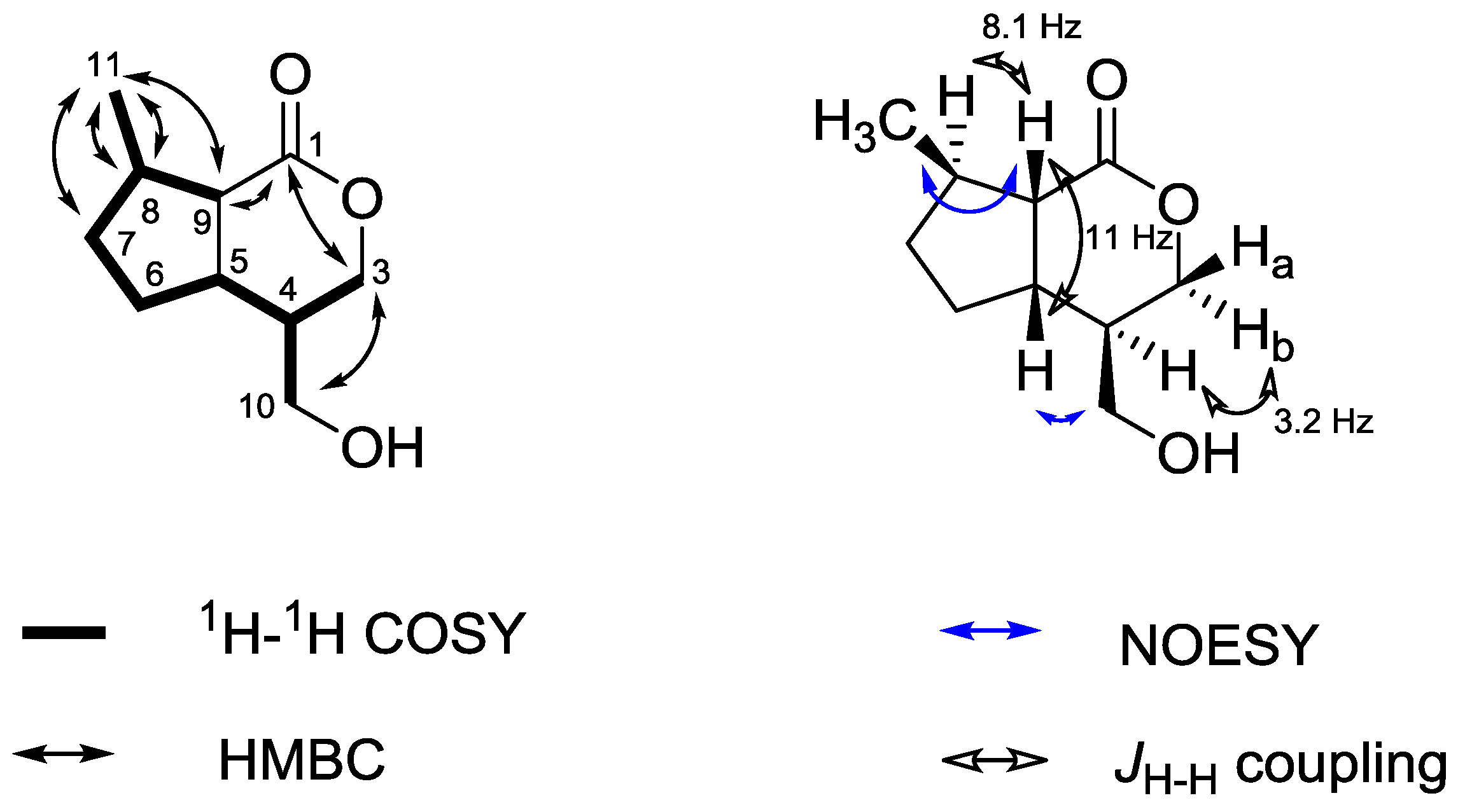
2.2. Structure Elucidation of Novel Nepetolactone Derivative 4

Compound 4 was isolated as a yellowish syrup after multiple fractionation of F1. The IR spectrum showed absorption bands for hydroxyl groups (3382 cm−1), carbonyl functions (1749 and 1697 cm−1) and a double bond (1603 cm−1). The ESI-MS investigation yielded mass signals at m/z 506.9 [M−H], 621.4 [M−H+TFA] and 326.8 [M−Glc] in the negative mode, respectively, supporting the proposed structure. In the positive mode multiple adducts (526.2 [M+H3O]+, 531.1 [M+Na]+) were detected besides the molecular ion signal at m/z 509.0 [M+H]+. Fragmentation of m/z 526.2 and 531.1 gave a fragment of m/z 347.1 [M−Glc+Na]+. In the NMR spectra the characteristic 1H and 13C signals for a trans-cinnamic acid with a para-substituted phenyl ring were present. 13C signals in the range of 70–80 ppm represented a sugar moiety. The assignment of the sugar as β-d-glucose was supported by comparison of the coupling constants of the protons to data from the literature [10] while the connectivity of the sugar and the cinnamic acid derivative to the nepetolactone skeleton [11] was established by HMBC cross peaks. Interpretation of coupling constants and NOESY experiments revealed the stereochemistry (cf. Figure 3): NOESY correlations of H-8 with other protons were the most prominent ones. H-8 gave NOESY cross peaks with H-1' of the sugar moiety, H-10, H-2" and H-3" of the cinnamic acid rest and the H-11 protons of the methyl group which led to the conclusion that all these groups are in close spatial proximity. The cinnamic acid seems to be located in the space above H-8. Therefore the proposed configuration is consistent with the configuration of the villoside aglycone 5 and other similar compounds from Valeriana [12]. Additional evidence came from a weak NOESY correlation between the protons of the C-9 methyl group and H-5b. Thus, compound 4 was assigned to be {[(3S,4S,7R,8S)-1-oxo-3,7-dimethyl-4-(O-β glucopyranosyl)oxy]hexahydrocyclopenta-[c]furan-3-yl}-methyl-trans-4-hydroxycinnamate.
Figure 3. Key 1H-1H-COSY, HMBC and NOESY correlations of 4.
Molecules 20 05740 g003

2.3. Structure Elucidation of Villoside Aglycone 5

Villoside is an iridoid glycoside first isolated by Taguchi et al. [13] from Patrinia villosa Juss., a Valerianaceae. However, the villoside aglycone was only obtained semi-synthetically [13]. This is the first report on the isolation of the aglycone 5 directly from a plant, after chromatographic separation of F1 (cf. Figure 2). Similar compounds (jatamanines) have been isolated from V. jatamansi before [12]. Compound 5 was obtained as a yellow syrup. The IR spectrum is characterized by absorption bands at 3387 cm−1 indicating a hydroxyl group and at 1,705 cm−1 caused by a carbonyl function. The ESI-MS showed signals at m/z 185.1 [M+H]+ and 207.0 [M+Na]+ which was consistent with the calculated mass of m/z 184.1 for the proposed structure. The 1H spectrum did not show any aromatic proton signals. The 13C spectrum exhibited ten signals including a typical signal for a lactone functionality at 175.7 ppm. A 13C signal at 19.8 ppm with a corresponding 1H signal at 1.13 ppm for three protons suggested the presence of a methyl group (C-11). The methylene groups next to an oxygen gave characteristic 13C signals at 61.7 and 68.7 ppm (C-3 and C-10, respectively). The remaining signals were found in the aliphatic region. The 1H-1H COSY data showed the presence of a spin system involving the hydrogens H-5 to H-9 and, therefore, suggested the presence of a 5-membered ring. All signals have been fully assigned by means of 1H-1H COSY, HMBC and HMQC experiments. The configuration was determined by using coupling constants and NOESY data (cf. Figure 4). NOESY correlations between H-11 and H-9, H-5 and H-10, respectively, suggested the methyl group (C-11) and the proton at C-9 as well as the methylene group at C-10 and the proton at C-5 to be located in the same plane of the molecule. The small coupling constant of H-4 and H-3b (3.2 Hz) supports this hypothesis of a quasi-equatorial position of H-4. Additionally, the coupling constant between H-9 and H-8 (Jaa = 8.1 Hz) was backing a trans-configuration. Furthermore, the coupling constant between H-5 and H-9 (11.0 Hz) was consistent with the reported values of the structurally similar jatamanines having the same configuration [12]. Taking biogenetic reasons into account, compound 5 can be assigned to (4R,5S,8R,9S)-4-(hydroxymethyl)-8-methylhexahydro-cyclopenta[c]pyranone (villoside aglycone).
Figure 4. Key 1H-1H-COSY, HMBC and NOESY correlations of villoside aglycone 5.
Molecules 20 05740 g004

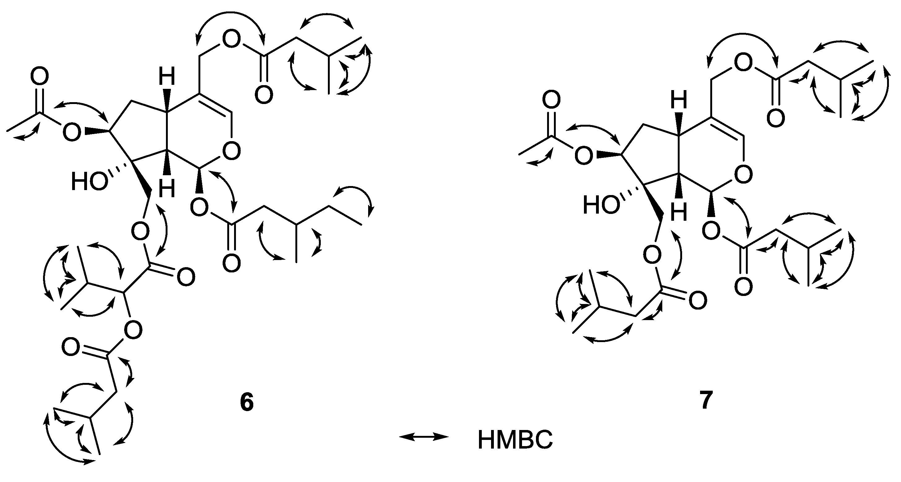
2.4. Valtrates with Antileishmanial Activity

Typical and well known constituents of Valeriana species are iridoid esters e.g., of the valepotriate and valtrate type, which have been previously investigated in detail because of their in vitro antifungal [14] and cytotoxic activity [15,16,17] and their potential use as antitumor agents [18]. In the active fractions F4–F7 two known valtrates were found, one (compound 8) of the diene- and one (compound 9) of the hydrine-type [16], and two iridoid esters 6, 7 with an as yet unreported substitution pattern. The known valtrates were identified by comparing their spectral data to the literature data (cf. Table 1). Compound 8 has been reported before [14,19,20] and valechlorine (9) too [17,21,22]. The new compounds consist of a typical valtrate skeleton of the hydrine-type and are esterified with various acids e.g. isovaleric acid, acetic acid. The connections of the esters were assigned by HMBC experiments (cf. Figure 5). By comparing coupling constants and taking biogenetic considerations into account the configuration was determined to be the same as reported in the literature.
Most of the terpenes have been found in subfractions of F7 and F8 (cf. Figure 2). β-Bisabolol (10) [23], α-kessyl alcohol (11) [24,25], valeranone (12) [26] and bornyl isovalerate (13) are well known in the Valeriana species [27,28,29,30] since a plethora of terpenes has been identified from the essential oil of Valeriana, especially by GC/MS analysis [31,32] in the past. Linarin-O-2-methylbutyrate (14) has been reported from V. wallichii before [33]. Additionally the following known natural products were isolated from V. wallichii for the first time: the phenylpropanoids caffeic (1), isoferulic (2) and p-hydroxycinnamic acid bornyl ester (3), the lignans 4´-demethylpodophyllotoxin (15), podophyllotoxin (16) and pinoresinol (17).
Table 1. 1H- and 13C-NMR data of new and known valtrates 69 in CDCl3.
Position13C of 61H of 613C of 71H of 713C of 8 [20]1H of 813C of 9 [17]1H of 9 [17]
189.26.22 (d; 4.7; 1H)89.26.27 (d; 4.2; 1H)92.56.25 (d; 10.0; 1H)89.46.19 (d; 5.1; 1H)
3141.06.44 (br s; 1H)140.96.44 (br s; 1H)148.06.68 (br s; 1H)141.06.45 (br s; 1H)
4112.8 112.9 108.7 113.0
531.82.90 (dd; 9.2; 11.0; 1H)31.42.90 (dd; 7.8; 8.0; 1H)139.1 32.62.91 (m; 1H)
634.92.1 (m; 2H)34.72.12 (m; 2H)117.65.77 (t; 2.8; 1H)35.02.04–2.13 (m; 2H)
780.14.99 (t; 4.5; 1H)80.45.03 (t; 4.7; 1H)83.15.47 (d; 2.8; 1H)80.05.01 (t; 3.9; 1H)
880.9 80.9 80.2 81.8
944.52.42 (dd; 6.1; 15.0; 1H)44.72.44 (dd; 4.1; 9.6; 1H)48.42.94 (dd; 2.5; 10.0; 1H)45.42.45 (dd; 5.9; 8.1; 1H)
1066.84.29 (d; 11.5; 1H)
4.35 (d; 11.5; 1H)
66.64.22 (s; 2H)65.54.32 (d; 11.6; 1H)
4.39 (d; 11.6; 1H)
49.13.71 (d; 11.4; 1H)
3.82 (d; 11.4; 1H)
1163.44.42 (d; 12.3; 1H)
4.62 (d; 12.3; 1H)
63.34.42 (d; 12.3; 1H)
4.62 (d; 12.3; 1H)
60.94.64 (d; 12.5; 1H)
4.71 (d; 12.5; 1H)
63.34.42 (d; 12.3; 1H)
4.60 (d; 12.3; 1H)
R11171.3 171.1 170.8 d 171.1
241.32.40 (m; 2H)43.3 b2.23 (m; 4H)43.22.32 (d; 7.0; 2H)43.4 g2.09–2.13 (m; 4H)
331.81.90 (m; 1H)25.6 c2.07–2.15 (m; 3H)25.72.05–2.09 (m; 2H)25.7 h2.00–2.06 (m; 2H)
419.50.97 (m; 3H)22.3 a0.96 (m; 18H)22.3 f0.93 (d; 6.6; 6H) g22.4 i0.97 (d; 6.7; 6H) g
529.31.40 (m; 2H)22.3 a0.96 (m; 18H)22.3 f0.93 (d; 6.6; 6H) g22.4 i0.97 (d; 6.7; 6H) g
611.20.90 (m; 3H)
R21169.7 173.0 170.8 d 172.9
243.02.30–2.35 (m; 4H)43.52.19 (m; 2H)20.92.04 (s; 3H)43.3 g2.09–2.13 (m; 4H)
325.72.11 (m; 2H)25.7 c2.07–2.15 (m; 3H) 25.6 h2.00–2.06 (m; 2H)
4 and 522.40.98 (d; 6.9; 12H)22.4 a0.96 (m; 18H) 22.4 i0.95 (d; 6.6; 6H) g
R31170.2 170.3 171.8 169.8
221.02.07 (s; 3H)21.02.05 (s; 3H)43.42.14 (m; 2H)21.02.08 (s; 3H)
3 25.6 e2.05–2.09 (m; 2H)
4 and 5 22.3 f0.95 (d; 6.6; 6H) g
R41173.2 172.9 173.8
276.94.74 (d; 4.8; 1H)43.1 b2.23 (m; 4H)43.02.20 (m; 2H)
330.02.23 (m; 1H)25.7 c2.07–2.15 (m; 3H)25.6 e2.17–2.25 (m; 1H)
4 18.81.02 (m; 3H)22.3 a0.96 (m; 18H)22.3 f0.99 (d; 6.6; 6H) g
517.31.00 (m; 3H)22.3 a0.96 (m; 18H)22.3 f0.99 (d; 6.6; 6H) g
6172.9
743.02.30–2.35 (m; 4H)
825.72.11 (m; 2H)
9 and 1022.40.98 (d; 6.9; 12H)
a–i Assignments may be interchanged.
Figure 5. Key HMBC correlations of valtrates 6 and 7.
Molecules 20 05740 g005

2.5. Antileishmanial Activity and Cytotoxicity

All compounds isolated were subjected to the evaluation of their antileishmanial activity against L. major promastigotes in vitro and cytotoxicity against macrophages (J774.1 murine cell line) using the corresponding AlamarBlue© assays [34] (cf. Table 2).
Table 2. Antileishmanial activity and cytotoxicity of fractions containing isolated compounds.
CompoundIC50 (L. major) [µg/mL]IC50 (J774.1) [µg/mL]
Bornyl caffeate (1)48.8 *8.3 *
216.734.7
312.28.6
4>100>100
5>10017.5
61.9 a<0.8 a
70.8<0.8
81.7 *1.0 *
92.31.9
β-Bisabolol (10)52.237.1
α-Kessyl alcohol (11)5.8 b1.7 b
Valeranone (12)60.842.6
Bornyl isovalerate (13)>100 *>100 *
Linarin-O-2-methylbutyrate (14)>10010.8
15>100<0.8
16>100<0.8
Pinoresinol (17)>100<0.8
Miltefosine36.2 *56.5 *
* These data were gained from pure or synthesized compounds and are therefore given in µM. a There was a small amount of another valtrate present. b The fraction of kessyl alcohol was contaminated with an non-identified valtrate (~10% by NMR integration).
The valtrates 69 showed the highest antileishmanial activity (IC50 0.8–2.3 µg/mL). Their cytotoxicity is in the same range. It is unknown whether the cytotoxicity is directly responsible for the leishmanicidal potential. Nevertheless, the valtrates represent the main active principle of the extract. Fractions with moderate antileishmanial activity in the range of the standard drug miltefosine were phenylpropanoids bornyl caffeate (1), bornyl isoferulate (2) and bornyl 3-hydroxycinnamate (3). This has led to the synthesis of a compound library and QSAR studies recently reported by us [2].
Furthermore, valeranone (12) and β-bisabolol (10) exhibited moderate antileishmanial and cytotoxic activity. The kessyl alcohol fraction showed rather good antileishmanial activity. However, it was contaminated with an active but unidentified valtrate, thus it remains unclear whether kessyl alcohol (8) is really active. Bornyl isovalerate (13), linarin-O-2-methylbutyrate (14), podophyllotoxin (11), 4'-demethylpodophyllotoxin (15) and pinoresinol (16) showed no antileishmanial activity. However, podophyllotoxin and 4'-demethylpodophyllotoxin were highly cytotoxic (IC50 < 0.8 µg/mL). This is not surprising as podophyllotoxin and derivatives are widely used as anticancer agents. However, cytotoxicity and antileishmanial activity are not linked to each other in this case. Fractions with novel compounds 4 and 5 exhibited no antileishmanial activity (IC50 > 100 µg/mL). Nevertheless, villoside aglycone (5) showed moderate cytotoxicity (IC50 17.5 µg/mL). Taken together the antileishmanial activity was found in valtrate derivatives, mainly, and cinnamic acid derivatives with exception of novel compound 4. However the valtrates were found to be highly cytotoxic in contrast to the cinnamic acid derivatives having moderate cytotoxicity.

3. Experimental Section

3.1. General

1H (400.132 MHz), 13C (100.613 MHz) and 2D NMR spectra were recorded on a Bruker Avance 400 Ultra Shield™ (Bruker Biospin, Ettlingen, Germany) spectrometer. As internal standard the signals of the deuterated solvents were used (CDCl3: 1H 7.26 ppm, 13C 77.0 ppm). IR spectra were recorded on a Jasco FT/IR-6100 spectrometer (Groß-Umstadt, Germany) with an ATR unit at room temperature. LC/MS was conducted on an Agilent 1100 analytical HPLC with DAD detection and an Agilent LC/MSD Trap (Agilent Technologies, Böblingen, Germany). For flash chromatography a Reveleris Flash system (Grace, Columbia, MD, USA) was used. Preparative HPLC was conducted using an Agilent 1100 preparative HPLC with fraction collector and multiple wavelength detector (MWD). TLC plates (60 F254) were purchased from Macherey-Nagel. Column chromatography was carried out on silica gel (Kieselgel 60, 0.063–0.2 mm, 70–320 mesh, Merck, Darmstadt, Germany), filtration on LiChroprep© RP-18 (40–63 µm, Merck).

3.2. Plant Material

Dried rhizomes of V. wallichii were acquired from a local herb shop in Kolkata, India. The identity as V. jatamansi Jones syn. V. wallichii DC. was confirmed by comparison with an authentic sample cultivated at the Institute of Himalayan Bioresource Technology in Palampur, Himachal Pradesh, India by R.D. Singh. A voucher specimen of the herbarium was preserved in the laboratory of Banasri Hazra (IHBT Ref. No. 11666).

3.3. Extraction and Isolation

The pulverized rhizome (10 g) was refluxed with chloroform (100 mL) for 2 h and filtered. The solvent was removed from the filtrate in a rotary evaporator, followed by complete drying in vacuo. Thus, the chloroform extract was obtained (yield = 1.1%; w/w) and preserved at 4 °C. The process was repeated to get higher amounts of the extract as per experimental requirements. Dry extract (11 g) was slurried in MeOH and filtrated using a short column filled with LiChroprep RP-18 silica gel which yielded 9.5 g of extract after evaporation. This material was fractionated into 12 fractions (F1–F12) by preparative HPLC method A (250 mm × 10 mm, 5 µm, Macherey-Nagel Nucleosil 100-5; A) H2O, B) MeOH, gradient: 70% B (0 min), 75% B (7 min), 100% B (25 min), 70% B (30 min); flow rate: 3.3 mL/min).
A portion of 600 mg of fraction F1 was further fractionated by preparative HPLC method B (125 mm × 10 mm, 5 µm, Macherey-Nagel Nucleodur Sphinx RP; A) H2O, B) CH3CN, gradient: 10% B (0–2 min), 30% B (2–15 min), 65% B (15–22 min), 10% B (22–24 min); flow rate: 4.4 mL/min) resulting in 9 fractions (F1-1 to F1-9). F1-1 (1.5 mg) yielded villosid aglycone 5, F1-4 (9 mg) the novel nepetolactone derivative 4. From F1-5 (3.7 mg) and F1-6 (9.1 mg) podophyllotoxin (16) and 4'-demethylpodophyllotoxin (15) were identified and F1-7 (2.9 mg) contained pinoresinol (17).
A 150 mg portion of fraction F2 was partitioned in two fractions (F2-I and F2-II) using the above HPLC method B. In F2-I linarin-O-2-methylbutyrate (14) (6.0 mg) was found and F2-II (12.5 mg) yielded podophyllotoxin (16)
Fractionation of F3 into three fractions (F3-I to F3-III) yielded again the linarin derivative 14 in F3-II. Fraction F4 was subjected to flash chromatography (silica gel, MeOH/CHCl3, 4.8:0.2 v/v) and gave 6 fractions (4α–4ζ) from which valechlorine (9) was identified in fraction 4γ (32.6 mg).
Fraction F5 was separated by column chromatography (CC) on silica gel (MeOH/CHCl3, 4.8:0.2 v/v) and yielded 5 fractions (5α, 5β1, 5β2, 5β3, 5γ). Repeating the chromatography with fraction 5β3 (258 mg) gave three fractions (5β3a–5β3c) of which the last fraction 5β3c yielded 135 mg of bornyl caffeate (1). Fractionation of 5β2 (168 mg) by CC gave four fractions (5β2a–5β2c) of which fraction 5β2a gave 14.2 mg of valtrate 7. Fractions 5β2b and 5β2c were combined and subjected to preparative HPLC method B). This process resulted in six fractions (5β2bc1–5β2bc6) from which fraction 5β2bc yielded 6.1 mg of valtrate 9.
Fraction F6 was again fractionated like F5 by CC which gave seven fractions 6α–6η. 6γ (24.2 mg) and 6δ (25.7 mg) were combined and subjected to preparative HPLC method B which gave fractions 6dega1-6dega5. 6dega2 again yielded valtrate 7 (4.9 mg) and 6dega3 valtrate 8 (4.0 mg).
F7 was fractionated by column chromatography as above and gave four fractions (7α–7δ). 7α (57.6 mg) was further partitioned by CC into four fractions (7α1–7α4) of which 7α2 yielded 1.5 mg of a mixture of valeranone (12) and β-bisabolol (10). Further fractionation of 7α3 (19.7 mg) by the same CC method gave another 3 fractions (7α3a–7α3c) of which 7α3a consisted of 5.0 mg pure β-bisabolol (10). 7α3c yielded 2.1 mg of caffeic acid derivative 2. Fractions 7β and 7γ were combined and fractionated by preparative HPLC method B which resulted in 10 fractions (7bega1–7bega10) of which fraction 7bega2 gave 7.6 mg cinnamic acid derivative 3 and fraction 7bega9 2.5 mg of valtrate 6.
F8 was fractionated into 6 fractions (8α–8ζ) by CC. Fraction 8β yielded 1.7 mg of bornyl isovalerate (13). 8ε was fractionated further by HPLC method B and gave six fractions (8ε1–8ε6) with fraction 8ε4 yielding 6.8 mg kessyl alcohol (11).

3.4. Characterization of Novel Nepetolactone Derivative {[(3S,4S,7R,8S)-1-Oxo-3,7-dimethyl-4-(O-β-d-glucopyranosyl)oxy]hexahydrocyclopenta[c]furan-3-yl}-methyl-trans-4-hydroxycinnamate (4)

Yellowish syrup; IR (ATR): 3382 (OH); 1749, 1697 (C=O), 1603 (C=C) cm-1; 1H-NMR (CDCl3): δ = 1.19 (3H, d, J = 6.9 Hz, H-11), 1.73 (3H, s, H-9), 1.41 (1H, m, H-6b), 1.68 (1H, m, H-5b), 1.97 (1H, m, H-6a), 2.20 (1H, m, H-5a), 2.35 (1H, m, H-7), 2.83 (1H, m, H-8), 3.22 (1H, m, H-2'), 3.26 (1H, m, H-5'), 3.41 (1H, t, J = 9.1 Hz, H-3'), 3.50 (1H, t, J = 9.6 Hz, H-4'), 3.79 (2H, dd, J = 3.2, 7.6 Hz, H-6'), 4.37 (1H, d, J = 7.5 Hz, H-1'), 4.52 (2H, m, H-10), 6.21 (1H, d, J = 15.9 Hz, H-2"), 6.79 (2H, J = 8.6 Hz, H-6", H-8"), 7.36 (2H, d, J = 8.6 Hz, H-5", H-9"), 7.60 (1H, d, J = 15.9 Hz, H-3") ppm; 13C-NMR (CDCl3): δ = 17.1 (C-9), 21.5 (C-11), 30.6 (C-5), 33.0 (C-6), 37.6 (C-7), 59.8 (C-8), 61.3 (C-6'), 67.3 (C-10), 69.4 (C-4'), 73.2 (C-2'), 76.0 (C-5'), 76.3 (C-3'), 86.0 (C-4), 93.4 (C-3), 96.8 (C-1'), 113.5 (C-2"), 115.8 (C-6", C-8"), 125.7 (C-4"), 130.2 (C-5", C-9"), 146.4 (C-3"), 159.5 (C-7"), 167.5 (C-1"), 177.2 (C-1) ppm; ESI-MS: m/z 621.4 [M−H+TFA], 506.9 [M−H], 326.8 [M−Glc]; HRMS (ESI-TOF+): m/z [M+Na]+ calculated for C25H32O11Na: 531.1842; found: 531.1836.

3.5. Characterization of Villoside Aglycone (4R,5S,8R,9S)-4-(Hydroxymethyl)-8-methylhexahydro-cyclopenta[c]pyranone (5)

Yellowish syrup; IR (ATR): 3387 (OH); 1705 (C=O) cm−1; 1H-NMR (CDCl3): δ = 1.11–1.18 (1H, m, H-7a), 1.13 (3H, d, J = 6.4 Hz, H-11), 1.21–1.31 (1H, m, H-6a), 1.63–1.72 (1H, m; H-4), 1.82–1.88 (1H, m, H-7b), 1.97–2.05 (1H, m, H-6b), 2.12–2.27 (2H, m, H-8, H-5), 2.32 (1H, dd, J = 8.4, 11.0 Hz, H-9), 3.44 (1H, dd, J = 8.00, 11.1 Hz, H-10a), 3.64 (1H, dd, J = 4.70, 11.1 Hz, H-10b), 4.10 (1H, m, H-3a), 4.35 (1H, dd, J = 3.20, 11.1 Hz, H-3b) ppm; 13C-NMR (CDCl3): δ = 19.8 (C-11), 32.3 (C-6), 34.6 (C-7), 38.4 (C-8), 38.9 (C-5), 42.4 (C-4), 48.9 (C-9), 61.7 (C-10), 68.7 (C-3), 175.7 (C-1) ppm; ESI-MS: m/z 185.1 [M+H]+, 207.0 [M+Na]+; HRMS (ESI-TOF+): m/z [M+Na]+ calculated for C10H16O3Na: 207.0997; found: 207.0993.

3.6. Antileishmanial and Cytotoxicity Assays

Antileishmanial activity against L. major promastigotes and cytotoxicity against a murine macrophage cell line (J774.1) was determined using an AlamarBlue© assay. Methods for both procedures have been reported previously [34]. IC50 values are presented as mean values of two experiments.

4. Conclusions

The valtrates 69 represent the main active antileishmanial principles of the V. wallichii extract, but they are toxic. Additionally, the cinnamic acid derivatives 13 showed moderate activity, interestingly except for novel compound 4. This could be due to the larger size of the molecule or the higher polarity caused by the sugar moiety. However, the previously reported cinnamic acid derivatives exhibited higher activity, even without the double bond [2].

Acknowledgments

Thanks are due to the SFB 630 (DFG) for financial support and to MS and Svetlana Sologub for conducting the antileishmanial and cytotoxicity assays, respectively. BH wishes to dedicate this work to the memory of R.D. Singh, Chief Scientist, CSIR, at IHBT, Palampur, and acknowledge his immense contribution towards identification of the V. wallichii plant sample used in this work. We regret his sad demise in a tragic road accident in October 2014. This publication was funded by the German Research Foundation (DFG) and the University of Wuerzburg in the funding programme Open Access Publishing.

Author Contributions

JG fractionated the extract, isolated the compounds, elucidated structures and prepared the manuscript. MS performed the antileishmanial assays. BH provided the plant material and prepared the extract. UH and BH helped preparing the manuscript and provided scientific input and discussion. HM read the manuscript.

Conflicts of Interest

The authors declare no conflict of interest.

References

  1. Sah, S.P.; Mathela, C.; Chopra, K. Valeriana wallichii: A phyto-pharmacological review. J. Pharm. Res. 2010, 3, 2337–2339. [Google Scholar]
  2. Khuda, F.; Iqbal, Z.; Khan, A.; Nasir, F. Antimicrobial and anti-inflammatory activities of leaf extract of Valeriana wallichii DC. Pak. J. Pharm. Sci. 2012, 25, 715–719. [Google Scholar] [PubMed]
  3. Agnihotri, S.; Wakode, S.; Ali, M. Chemical composition, antimicrobial and topical anti-inflammatory activity of Valeriana jatamansi Jones. essential oil. J. Essent. Oil Bear. Pl. 2011, 14, 417–422. [Google Scholar] [CrossRef]
  4. Dua, V.; Alam, M.; Pandey, A.; Rai, S.; Chopra, A.; Kaul, V.; Dash, A. Insecticidal activity of Valeriana jatamansi (Valerianaceae) against mosquitoes. J. Am. Mosq. Control Assoc. 2008, 24, 315–318. [Google Scholar] [CrossRef] [PubMed]
  5. Ming, D.S.; Yu, D.Q.; Yang, Y.Y.; He, C.H. The structures of three novel sesquiterpenoids from Valeriana jatamansi Jones. Tetrahedron Lett. 1997, 38, 5205–5208. [Google Scholar] [CrossRef]
  6. Sudhanshu, N.R.; Sandhya, M.; Ekta, M. Evaluation of antioxidant properties of Valeriana wallichi to scavenge free radicals. Asian J. Pharm. Clin. Res. 2012, 5, 238–240. [Google Scholar]
  7. Thusoo, S.; Gupta, S.; Sudan, R.; Kour, J.; Bhagat, S.; Hussain, R.; Bhagat, M. Antioxidant activity of essential oil and extracts of Valeriana jatamansi roots. BioMed. Res. Int. 2014. [Google Scholar] [CrossRef]
  8. Ghosh, S.; Debnath, S.; Hazra, S.; Hartung, A.; Thomale, K.; Schultheis, M.; Kapkova, P.; Schurigt, U.; Moll, H.; Holzgrabe, U. Valeriana wallichii root extracts and fractions with activity against Leishmania spp. Parasitol. Res. 2011, 108, 861–871. [Google Scholar] [CrossRef] [PubMed]
  9. Glaser, J.; Schultheis, M.; Hazra, S.; Hazra, B.; Moll, H.; Schurigt, U.; Holzgrabe, U. Antileishmanial lead structures from nature: Analysis of structure-activity relationships of a compound library derived from caffeic acid bornyl ester. Molecules 2014, 19, 1394–1410. [Google Scholar] [CrossRef] [PubMed]
  10. Sobolev, A.P.; Brosio, E.; Gianferri, R.; Segre, A.L. Metabolic profile of lettuce leaves by high-field NMR spectra. Magn. Reson. Chem. 2005, 43, 625–638. [Google Scholar] [CrossRef] [PubMed]
  11. Eisenbraun, E.J.; Browne, C.E.; Irvin-Willis, R.L.; McGurk, D.J.; Eliel, E.L.; Harris, D.L. Structure and stereochemistry of 4aβ,7α,7aβ-nepetalactone from Nepeta mussini and its relationship to the 4aα,7α,7aα -and 4aα,7α,7aβ-nepetalactones from N. cataria. J. Org. Chem. 1980, 45, 3811–3814. [Google Scholar] [CrossRef]
  12. Lin, S.; Chen, T.; Liu, X.-H.; Shen, Y.-H.; Li, H.-L.; Shan, L.; Liu, R.-H.; Xu, X.-K.; Zhang, W.-D.; Wang, H. Iridoids and lignans from Valeriana jatamansi. J. Nat. Prod. 2010, 73, 632–638. [Google Scholar] [CrossRef] [PubMed]
  13. Taguchi, H.; Yokokawa, Y.; Endo, T. Studies on the constituents of Patrinia villosa Juss. Yakugaku Zasshi 1973, 93, 607–611. [Google Scholar] [PubMed]
  14. Fuzzati, N.; Wolfender, J.; Hostettmann, K.; Msonthi, J.; Mavi, S.; Molleyres, L. Isolation of antifungal valepotriates from Valeriana capense and the search for valepotriates in crude Valerianaceae extracts. Phytochem. Anal 1996, 7, 76–85. [Google Scholar] [CrossRef]
  15. Bos, R.; Hendriks, H.; Scheffer, J.J.C.; Woerdenbag, H.J. Cytotoxic potential of valerian constituents and valerian tinctures. Phytomedicine 1998, 5, 219–225. [Google Scholar] [CrossRef] [PubMed]
  16. Lin, S.; Shen, Y.-H.; Li, H.-L.; Yang, X.-W.; Chen, T.; Lu, L.-H.; Huang, Z.-S.; Liu, R.-H.; Xu, X.-K.; Zhang, W.-D.; et al. Acylated iridoids with cytotoxicity from Valeriana jatamansi. J. Nat. Prod. 2009, 72, 650–655. [Google Scholar]
  17. Lin, S.; Zhang, Z.-X.; Chen, T.; Ye, J.; Dai, W.-X.; Shan, L.; Su, J.; Shen, Y.-H.; Li, H.-L.; Liu, R.-H.; et al. Characterization of chlorinated valepotriates from Valeriana jatamansi. Phytochemistry 2013, 85, 185–193. [Google Scholar]
  18. Bounthanh, C.; Bergmann, C.; Beck, J.P.; Haag-Berrurier, M.; Anton, R. Valepotriates, a new class of cytotoxic and antitumor agents. Planta Med. 1981, 41, 21–28. [Google Scholar] [CrossRef] [PubMed]
  19. Finner, E.; David, S.; Thies, P. Über die Wirkstoffe des Baldrians. Planta Med. 1984, 50, 4–6. [Google Scholar] [CrossRef] [PubMed]
  20. Koch, U.; Hölzl, J. The compounds of Valeriana alliariifolia. Planta Med. 1985, 51, 172–173. [Google Scholar] [CrossRef]
  21. Xu, J.; Li, Y.; Guo, Y.; Guo, P.; Yamakuni, T.; Ohizumi, Y. Isolation, structural elucidation, and neuroprotective effects of iridoids from Valeriana jatamansi. Biosci. Biotechnol. Biochem. 2012, 76, 1401–1403. [Google Scholar] [CrossRef] [PubMed]
  22. Yang, X.P.; Li, E.W.; Zhang, Q.; Yuan, C.S.; Jia, Z.J. Five new iridoids from Patrinia rupestris. Chem. Biodivers. 2006, 3, 762–770. [Google Scholar] [CrossRef] [PubMed]
  23. Minyard, J.P.; Thompson, A.C.; Hedin, P.A. Constituents of the cotton bud. VIII. β-Bisabolol, a new sesquiterpene alcohol. J. Org. Chem. 1968, 33, 909–911. [Google Scholar]
  24. Itô, S.; Kodama, M.; Nozoe, T.; Hikino, H.; Hikino, Y.; Takeshita, Y.; Takemoto, T. Structure and absolute configuration of α-kessyl alcohol and kessyl glycol. Tetrahedron 1967, 23, 553–563. [Google Scholar] [CrossRef]
  25. Nishiya, K.; Kimura, T.; Takeya, K.; Itokawa, H. Sesquiterpenoids and iridoid glycosides from Valeriana fauriei. Phytochemistry 1992, 31, 3511–3514. [Google Scholar] [CrossRef] [PubMed]
  26. Vite, G.D.; Spencer, T.A. Synthesis of (+)-7-epivaleranone and (+)-valeranone. J. Org. Chem. 1988, 53, 2560–2565. [Google Scholar] [CrossRef]
  27. Gränicher, F.; Christen, P.; Kapetanidis, I. Essential oils from normal and hairy roots of Valeriana officinalis var. sambucifolia. Phytochemistry 1995, 40, 1421–1424. [Google Scholar] [CrossRef]
  28. Mathela, C.S.; Chanotiya, C.S.; Sammal, S.S.; Pant, A.K.; Pandey, S. Compositional diversity of terpenoids in the himalayan Valeriana genera. Chem. Biodivers. 2005, 2, 1174–1182. [Google Scholar] [CrossRef] [PubMed]
  29. Wang, J.; Zhao, J.; Liu, H.; Zhou, L.; Liu, Z.; Wang, J.; Han, J.; Yu, Z.; Yang, F. Chemical analysis and biological activity of the essential oils of two valerianaceous species from China: Nardostachys chinensis and Valeriana officinalis. Molecules 2010, 15, 6411–6422. [Google Scholar] [CrossRef] [PubMed]
  30. Houghton, P.J. The biological activity of valerian and related plants. J. Ethnopharmacol. 1988, 22, 121–142. [Google Scholar] [CrossRef] [PubMed]
  31. Bos, R.; Woerdenbag, H.J.; Hendriks, H.; Smit, H.F.; Wikström, H.V.; Scheffer, J.J. Composition of the essential oil from roots and rhizomes of Valeriana wallichii DC. Flavour Fragr. J. 1997, 12, 123–131. [Google Scholar] [CrossRef]
  32. Sati, S.; Mathela, C. Essential oil composition of Valeriana hardwickii var. arnottiana from the Himalayas. Flavour Fragr. J. 2005, 20, 299–301. [Google Scholar] [CrossRef]
  33. Chari, V.M.; Jordan, M.; Wagner, H.; Thies, P.W. A 13C-NMR study of the structure of an acyl-linarin from Valeriana wallichii. Phytochemistry 1977, 16, 1110–1112. [Google Scholar] [CrossRef]
  34. Bringmann, G.; Thomale, K.; Bischof, S.; Schneider, C.; Schultheis, M.; Schwarz, T.; Moll, H.; Schurigt, U. A novel Leishmania major amastigote assay in 96-well format for rapid drug screening and its use for discovery and evaluation of a new class of leishmanicidal quinolinium salts. Antimicrob. Agents Chemother. 2013, 57, 3003–3011. [Google Scholar] [CrossRef] [PubMed]
  • Sample Availability: Samples of novel compounds 4 and 5 are available from the authors.

