
High Fluorescent Porphyrin-PAMAM-Fluorene Dendrimers

Abstract
:1. Introduction
2. Results and Discussion


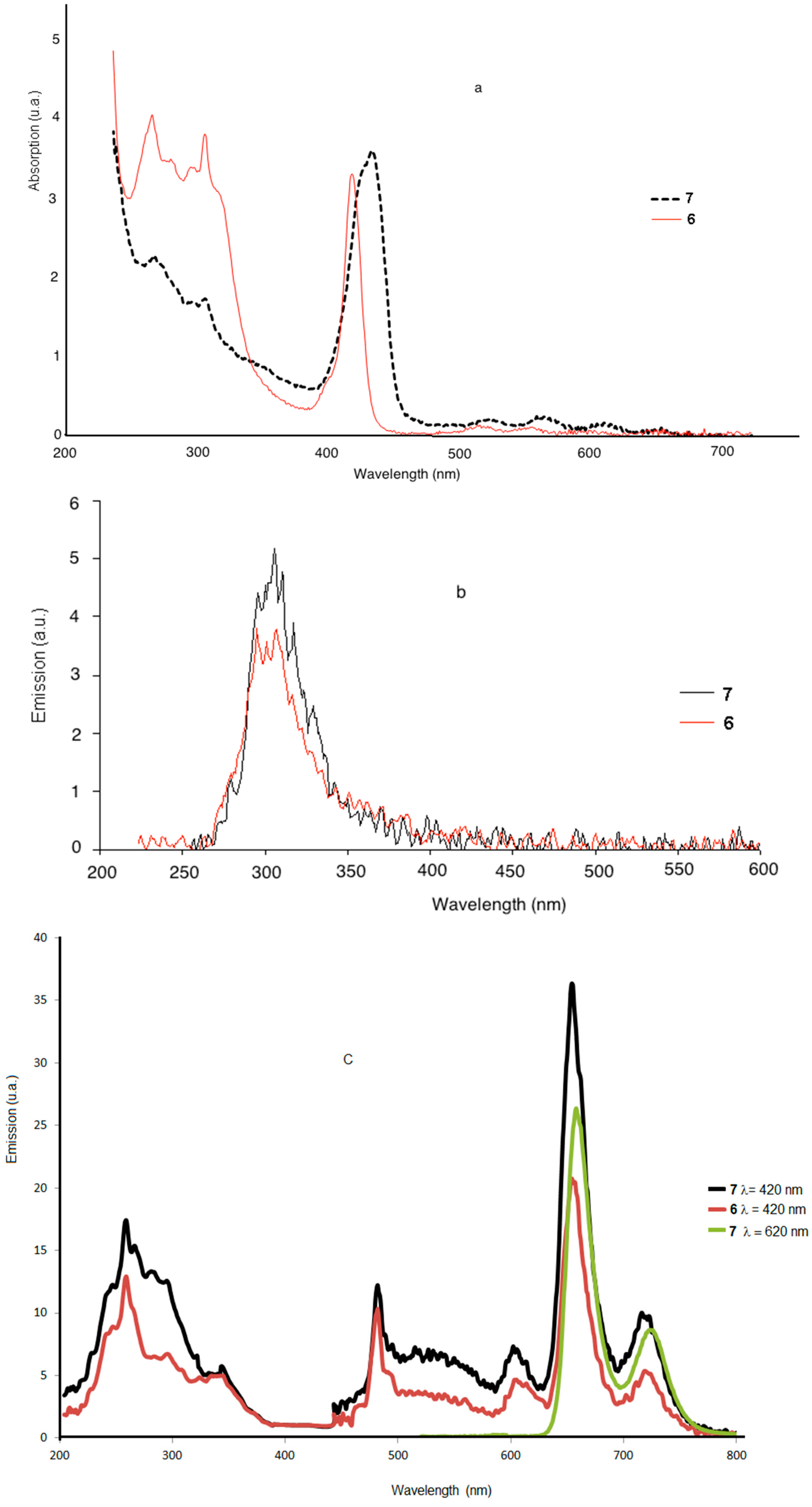
2.1. Optical Properties in Solution


| Sample | Fluorenemax (nm) | Soret Bandmax (nm) | Qmax (nm) | ɛ (×10−5 M−1 cm−1) |
|---|---|---|---|---|
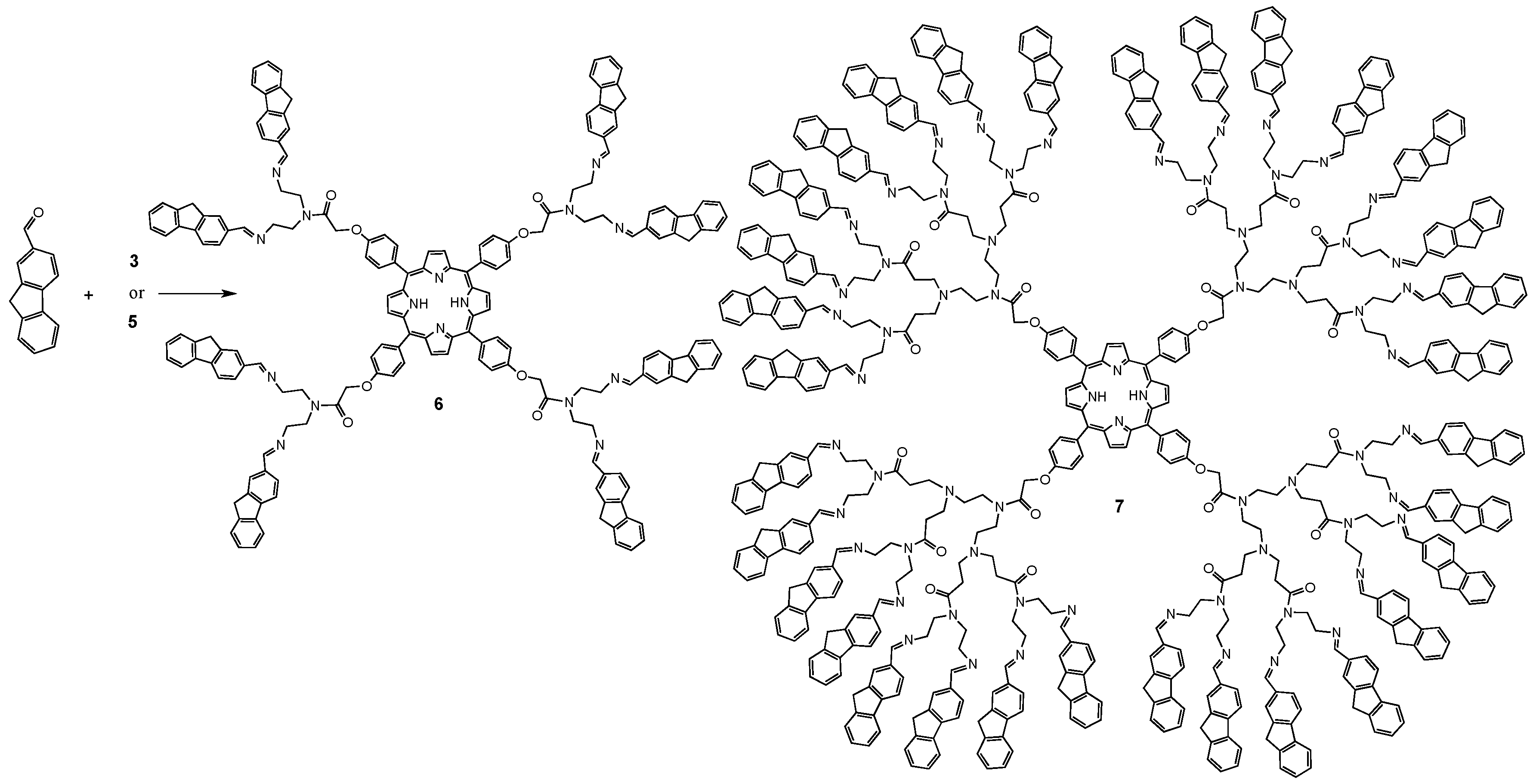
| 6 | 272, 309 | 419 | 557 | 0.235 |
| 7 | 266, 307 | 436 | 564 | 0.247 |
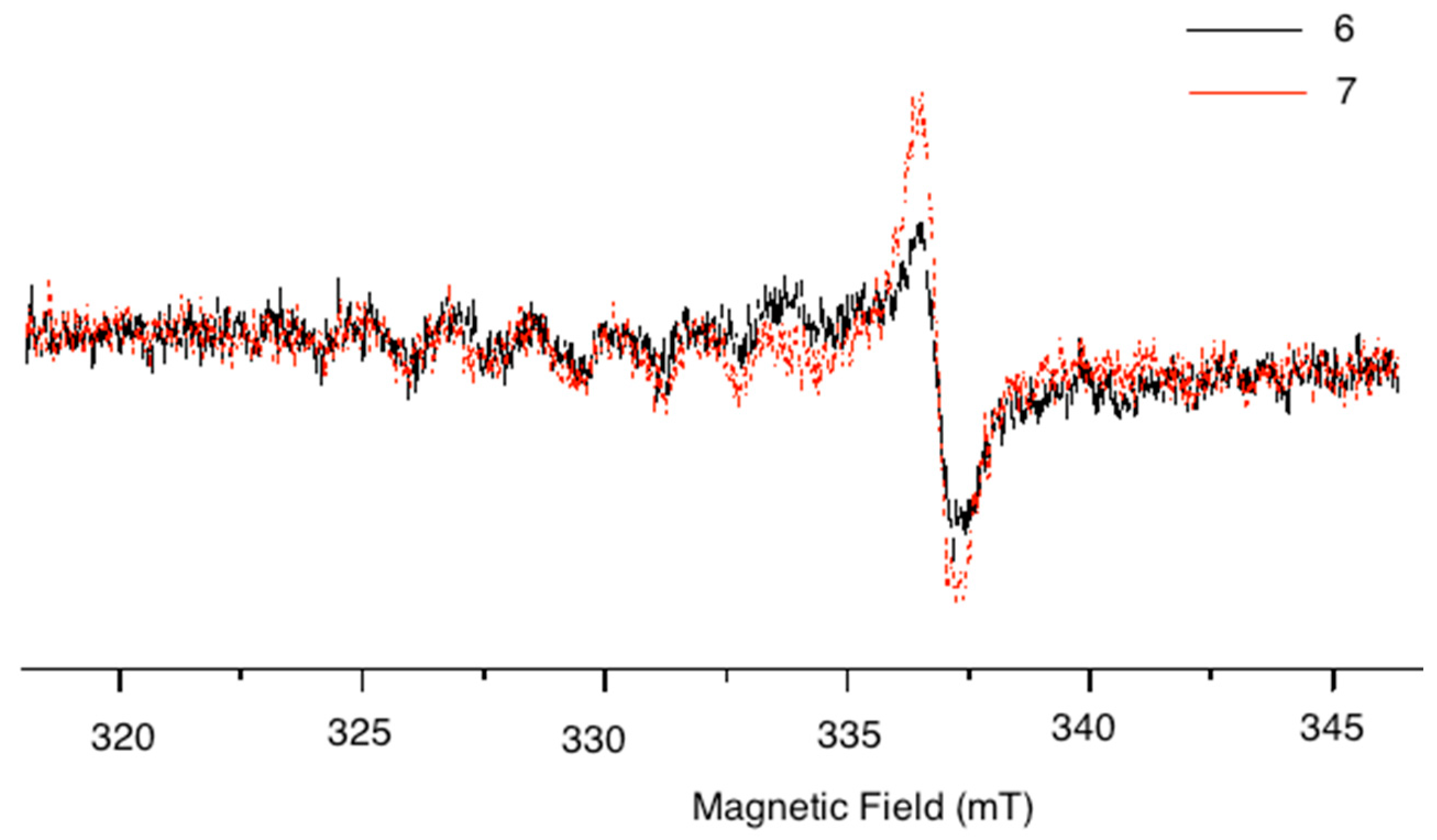
2.2. EPR Studies

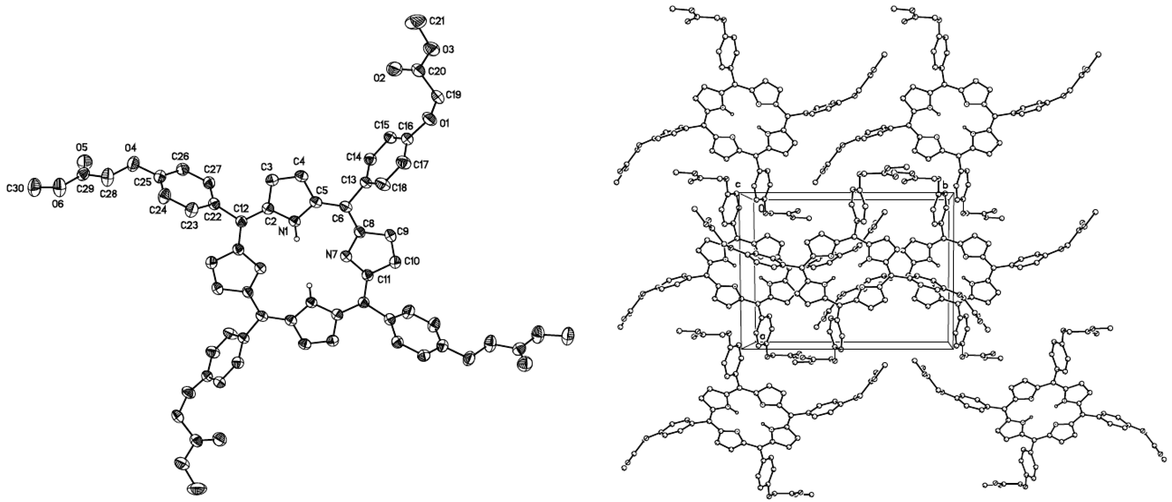
2.3. Crystal Structure Determination

| Empirical formula | C56 H46 N4O12 |
| Formula weight | 966.97 |
| Temperature | 298(2) K |
| Wavelength | 1.54178 Å |
| Crystal system | Monoclinic |
| Space group | P 21/c |
| Unit cell dimensions | a = 12.3084(3) Å α = 90° |
| b = 16.4548(4) Å β = 98.2840(10)° | |
| c = 11.8932(2) Å γ = 90° | |
| Volume | 2383.62(9) Å3 |
| Z | 2 |
| Density (calculated) | 1.347 Mg/m3 |
| Absorption coefficient | 0.789 mm−1 |
| F(000) | 1012 |
| Crystal size | 0.364 × 0.105 × 0.017 mm3 |
| Theta range for data collection | 3.629 to 68.264° |
| Index ranges | −14 ≤ h ≤ 14, −19 ≤ k ≤ 19, −14 ≤ l ≤ 12 |
| Reflections collected | 27,160 |
| Independent reflections | 4359 [R(int) = 0.1102] |
| Completeness to theta = 67.679° | 99.8% |
| Refinement method | Full-matrix least-squares on F2 |
| Data/restraints/parameters | 4359/1/330 |
| Goodness-of-fit on F2 | 1.048 |
| Final R indices [I > 2sigma(I)] | R1 = 0.0668, wR2 = 0.1691 |
| R indices (all data) | R1 = 0.0988, wR2 = 0.1936 |
| Largest diff. peak and hole | 0.747 and −0.269 e·Å−3 |
3. Experimental Section
3.1. General Information
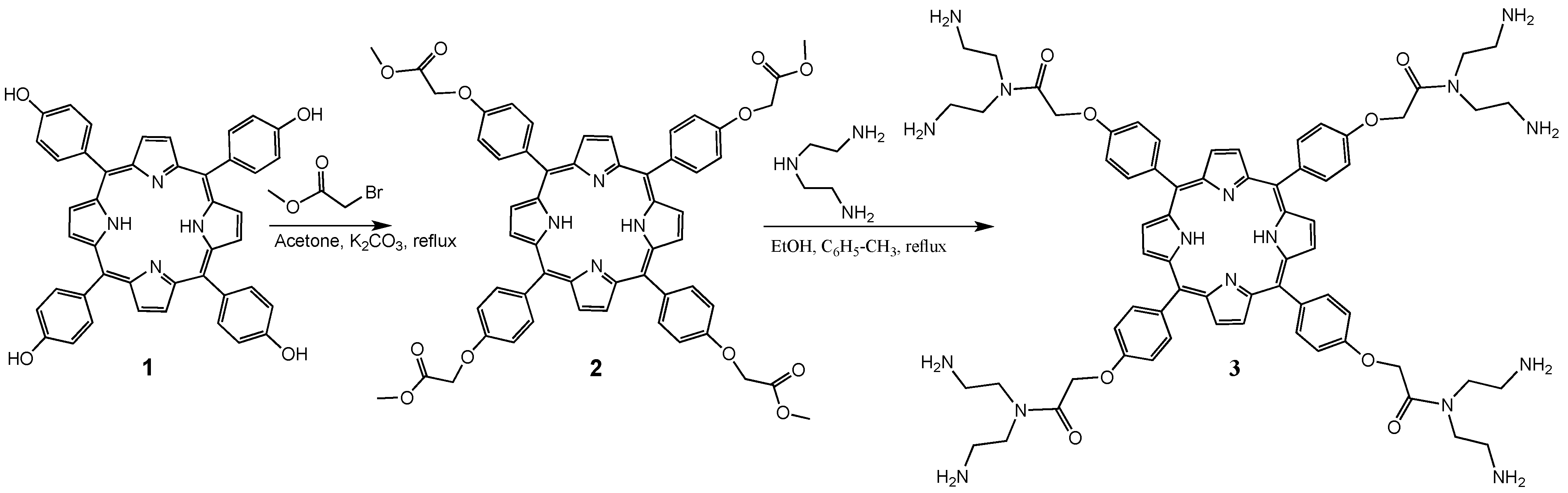
3.2. Synthesis of Generation 0.5 Dendrimer 2
3.3. Synthesis of Generation 1.0 Dendrimer 3
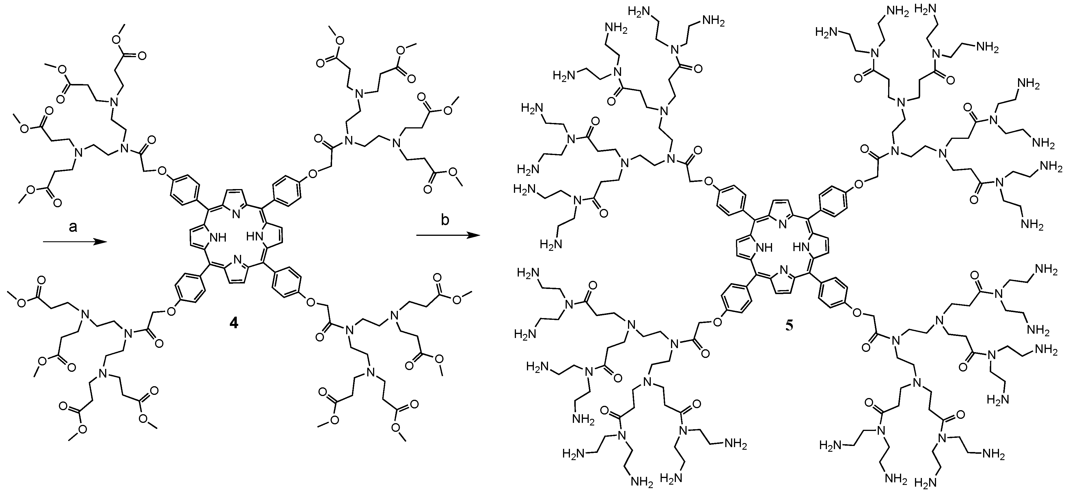
3.4. Synthesis of Generation 1.5 Dendrimer 4
3.5. Synthesis of Generation 2.0 Dendrimer 5
3.6. Synthesis of Dendrimers 6 and 7 with Fluorene in the Periphery
4. Conclusions
Acknowledgments
Author Contributions
Conflicts of Interest
References
- Tomalia, D.A.; Fréchet, J.M.J. Dendrimers and other Dendritic Polymers; Wiley: New York, NY, USA, 2001. [Google Scholar]
- Hecht, S.; Fréchet, J.M.J. Dendritic Encapsulation of Function: Applying Nature’s Site Isolation Principle from Biomimetics to Materials Science. Angew. Chem. Int. Ed. 2001, 40, 74–91. [Google Scholar] [CrossRef]
- Piotti, M.E.; Rivera, F., Jr.; Bond, R.; Hawker, C.J.; Frechet, J.M.J. Synthesis and Catalytic Activity of Unimolecular Dendritic Reverse Micelles with ‘Internal’ Functional Groups. J. Am. Chem. Soc. 1999, 121, 9471–9472. [Google Scholar] [CrossRef]
- Gunes, S.; Neugebauer, H.; Sariciftci, N.S. Conjugated polymer-based organic solar cells. Chem. Rev. 2007, 107, 1324–1338. [Google Scholar] [CrossRef] [PubMed]
- Kira, A.; Umeyama, T.; Matano, Y.; Yoshida, K.; Isoda, S.; Park, J.K.; Kim, D.; Imahori, H. Supramolecular Donor−Acceptor Heterojunctions by Vectorial Stepwise Assembly of Porphyrins and Coordination-Bonded Fullerene Arrays for Photocurrent Generation. J. Am. Chem. Soc. 2009, 131, 3198–3200. [Google Scholar] [CrossRef] [PubMed]
- Nishiyama, N.; Stapert, H.R.; Zhang, G.-D.; Takasu, D.; Jiang, D.-L.; Nagano, T.; Aida, T.; Kataoka, K. Light-Harvesting Ionic Dendrimer Porphyrins as New Photosensitizers for Photodynamic Therapy. Bioconjugate Chem. 2003, 14, 58–66. [Google Scholar] [CrossRef]
- Finikova, O.; Galkin, A.; Rozhkov, V.; Cordero, M.; Hägerhäll, C.; Vinogradov, S. Porphyrin and Tetrabenzoporphyrin Dendrimers: Tunable Membrane-Impermeable Fluorescent pH Nanosensors. J. Am. Chem. Soc. 2003, 125, 4882–4893. [Google Scholar] [CrossRef] [PubMed]
- Kiran, P.P.; Reddy, D.R.; Maiya, B.G.; Dharmadhikari, A.K.; Kumar, G.R.; Desai, N.R. Enhanced optical limiting and nonlinear absorption properties of azoarene-appended phosphorus (V) tetratolylporphyrins. Appl. Opt. 2002, 41, 7631–7636. [Google Scholar] [CrossRef] [PubMed]
- Minghao, S.; Zhishan, B. Tuning the optical properties of hyperbranched polymers through the modification of the end groups (pages 111–124). J. Polym. Sci. Part A: Polym. Chem. 2007, 45, 111–124. [Google Scholar] [CrossRef]
- Poriel, C.; Ferrand, Y.; Le-Maux, P.; Paul-Roth, C.; Simonneaux, G.; Rault-Berthelot, J. Anodic oxidation and physicochemical properties of various porphyrin-fluorenes or -spirobifluorenes: Synthesis of new polymers for heterogeneous catalytic reactions. J. Electroanal. Chem. 2005, 583, 92–103. [Google Scholar] [CrossRef]
- Paul-Roth, C.; Rault-Berthelot, J.; Simonneaux, G. New polymers for catalytic carbene transfer: Electropolymerization of tetrafluorenylporphyrinruthenium carbon monoxide. Tetrahedron 2004, 60, 12169–12175. [Google Scholar] [CrossRef]
- Kazuya, O.; Naoyuki, M.; Yoshiaki, K. Synthesis and Self-Organization of Fluorene-Conjugated Bisimidazolylporphyrin and Its Optical Properties. Int. J. Mol. Sci. 2013, 14, 322–331. [Google Scholar]
- Cannistraro, S.; van Vorst, A.; Jorit, G. EPR studied on singlet oxygen production by porphyrins. Photochem. Photobiol. 1978, 28, 257–279. [Google Scholar] [CrossRef]
- Lin, Q.S.; Zhang, T.L.; Yuan, L.B. Study of ESR spectra and photochemical reaction of porphyrin derivatives. Chin. J. Appl. Chem. 1988, 5, 57–61. [Google Scholar]
- Minbo, L.; Hongli, Z.; Huihui, Y.; Chengrui, J.; Shaohua, Z.; Ying, J. Absorption and EPR spectra of some porphyrins and metalloporphyrins. Dyes Pigm. 2007, 74, 357–362. [Google Scholar] [CrossRef]
- Wenqi, Z.; Ning, S.; Lianxiang, Y.; Xingqiao, W. UV-visible, fluorescence and EPR properties of porphyrins and metalloporphyrins. Dyes Pigm. 2008, 77, 153–157. [Google Scholar] [CrossRef]
- Sample Availability: Not available.
© 2015 by the authors. Licensee MDPI, Basel, Switzerland. This article is an open access article distributed under the terms and conditions of the Creative Commons Attribution license ( http://creativecommons.org/licenses/by/4.0/).
Share and Cite
Garfias-Gonzalez, K.I.; Organista-Mateos, U.; Borja-Miranda, A.; Gomez-Vidales, V.; Hernandez-Ortega, S.; Cortez-Maya, S.; Martínez-García, M. High Fluorescent Porphyrin-PAMAM-Fluorene Dendrimers. Molecules 2015, 20, 8548-8559. https://doi.org/10.3390/molecules20058548
Garfias-Gonzalez KI, Organista-Mateos U, Borja-Miranda A, Gomez-Vidales V, Hernandez-Ortega S, Cortez-Maya S, Martínez-García M. High Fluorescent Porphyrin-PAMAM-Fluorene Dendrimers. Molecules. 2015; 20(5):8548-8559. https://doi.org/10.3390/molecules20058548
Chicago/Turabian StyleGarfias-Gonzalez, Karla I., Ulises Organista-Mateos, Andrés Borja-Miranda, Virginia Gomez-Vidales, Simon Hernandez-Ortega, Sandra Cortez-Maya, and Marcos Martínez-García. 2015. "High Fluorescent Porphyrin-PAMAM-Fluorene Dendrimers" Molecules 20, no. 5: 8548-8559. https://doi.org/10.3390/molecules20058548