Article

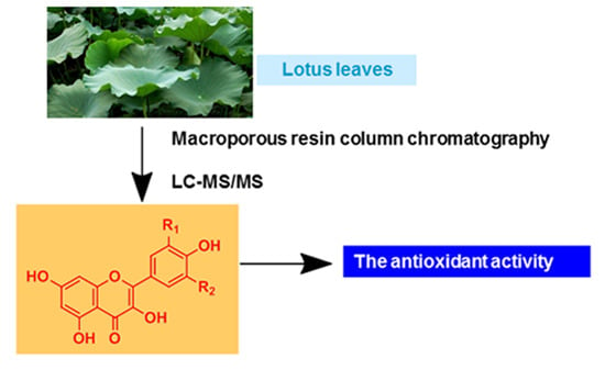
Analysis of Flavonoids in Lotus (Nelumbo nucifera) Leaves and Their Antioxidant Activity Using Macroporous Resin Chromatography Coupled with LC-MS/MS and Antioxidant Biochemical Assays

1
Key Laboratory of Plant Germplasm Enhancement and Specialty Agriculture, Wuhan Botanical Garden of Chinese Academy of Sciences, Wuhan 430074, China
2
Changchun University of Chinese Traditional Medicine, Changchun 130000, China
*
Author to whom correspondence should be addressed.
Molecules 2015, 20(6), 10553-10565; https://doi.org/10.3390/molecules200610553
Submission received: 27 March 2015 / Accepted: 29 May 2015 / Published: 8 June 2015
(This article belongs to the Collection Bioactive Compounds)

Abstract

:
Lotus (Nelumbo nucifera) leaves, a traditional Chinese medicinal herb, are rich in flavonoids. In an effort to thoroughly analyze their flavonoid components, macroporous resin chromatography coupled with HPLC-MS/MS was employed to simultaneously enrich and identify flavonoids from lotus leaves. Flavonoids extracted from lotus leaves were selectively enriched in the macroporous resin column, eluted subsequently as fraction II, and successively subjected to analysis with the HPLC-MS/MS and bioactivity assays. Altogether, fourteen flavonoids were identified, four of which were identified from lotus leaves for the first time, including quercetin 3-O-rhamnopyranosyl-(1→2)-glucopyranoside, quercetin 3-O-arabinoside, diosmetin 7-O-hexose, and isorhamnetin 3-O-arabino- pyranosyl-(1→2)-glucopyranoside. Further bioactivity assays revealed that these flavonoids from lotus leaves possess strong antioxidant activity, and demonstrate very good potential to be explored as food supplements or even pharmaceutical products to improve human health.

Graphical Abstract

1. Introduction

Lotus (Nelumbo nucifera), a common perennial aquatic herb, is extensively cultivated in eastern Asia, particularly in China [1]. All parts of lotus, including the leaves, stamens, flowers, seeds and rhizomes, have been used as traditional Chinese medicines or vegetables for thousands of years [2]. The leaves of lotus are traditionally used for the treatment of haematemesis, haematuria, metrorrhagia, hyperlipidaemia, fever and inflammatory skin conditions [3]. In recent years, the antioxidant [4], antiviral [5], anti-obesity [6] and lipolytic activities [7] of lotus leaves have been reported and attracted more and more interest, while phytochemicals from lotus leaves and their associated potential activities have not yet been fully explored. The annual production of lotus leaves now exceeds 800,000 tons in China, but most of them are discarded as agricultural wastes by farmers [8]. It is thus highly desirable to expedite the research on how to make the best use of lotus leaves as potential products for the food or pharmaceutical industries. In this regard, it is of primary importance to conduct a thorough analysis of the major chemical components from lotus leaves and their associated bioactivities.
It is reported that the lotus leaves are rich in flavonoids [5] and alkaloids [9], and several flavonoids have been isolated. Kashiwada et al. isolated and identified five flavonoid glycosides (quercetin 3-O-β-d-glucuronide, quercetin 3-O-β-d-xylopyranosyl-(1→2)-β-d-galactopyranoside, rutin, isoquecitrin and hyperin) by nuclear magnetic resonance spectroscopy (NMR) [5]. Ohkoshi et al. also identified eight flavonoids (quercetin 3-O-α-arabinopyranosyl-(1→2)-β-d-galactopyranoside, rutin, (+)-catechin, hyperoside, isoquercitrin, quercetin, and astragalin) [7]. Till now, ten flavonoids were isolated from lotus leaves [5,7,10]. Flavonoids, a type of phenolic compounds, have attracted extensive attention because of their strong antioxidant activity and their ability to reduce the formation of free radicals and to scavenge free radicals [11,12,13]. It is considered that oxidative damage is attributable to excess active oxygen species generated in the body [14], and the antioxidant defense system plays an important role in human health [15]. In order to reduce damage to the human body and prolong the storage stability of foods, synthetic antioxidants are often used in these processes [16]. However, the use of synthetic antioxidants, such as butylated hydroxyanisole (BHA) and butylated hydroxytoluene (BHT), has been questioned for the possibility of being carcinogenic and causing liver damage [17]. Compared with synthetic antioxidants, antioxidants from natural sources are characterized by their less toxicity, and better health effects. Thus, the focuses of this study were put on the flavonoids from lotus leaves and their associated antioxidant activity.
So far, several methods for the enrichment and separation of flavonoids in lotus leaves including liquid-liquid extraction [10], SPE [18], high-speed counter-current chromatography (HSCCC) [19] have been developed. However, these methods have several limitations, such as low capacity, low yields, or the need for special instrumentation. Furthermore, these methods share similar difficulties in completely separating flavonoids and alkaloids, which are the major active ingredients of lotus leaves. Comparatively, macroporous resin chromatography, with its properties of high adsorption capacity, good stability, low operational cost, and simple procedure, is one of the most efficient methods to separate bioactive components from crude herbal material extracts [20]. Nowadays macroporous resins chromatography has been successfully applied in industry for separation and preparation of flavonoids, glycosides, saponins, and so on [21]. In this context, we strove to develop an off-line two-dimensional chromatography method combining macroporous resin and reverse phase liquid chromatography to firstly enrich the flavonoids from the crude extracts of lotus leaves in the first dimension, then conduct the subsequent chemical and activity analysis of the flavonoid components.

2. Results and Discussion

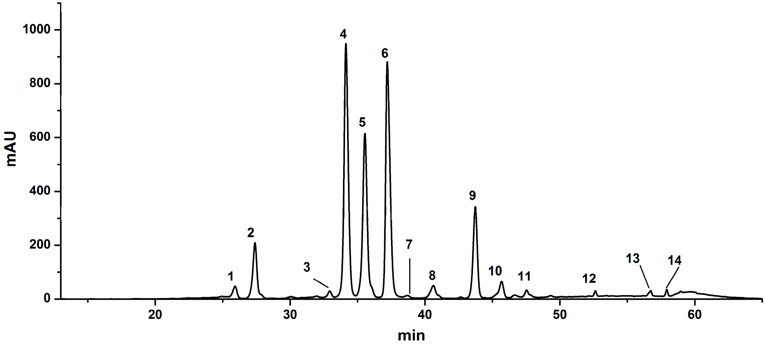
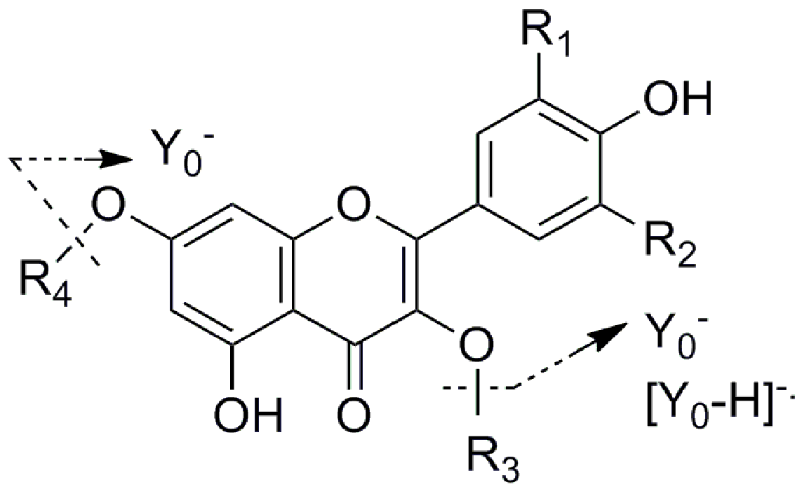
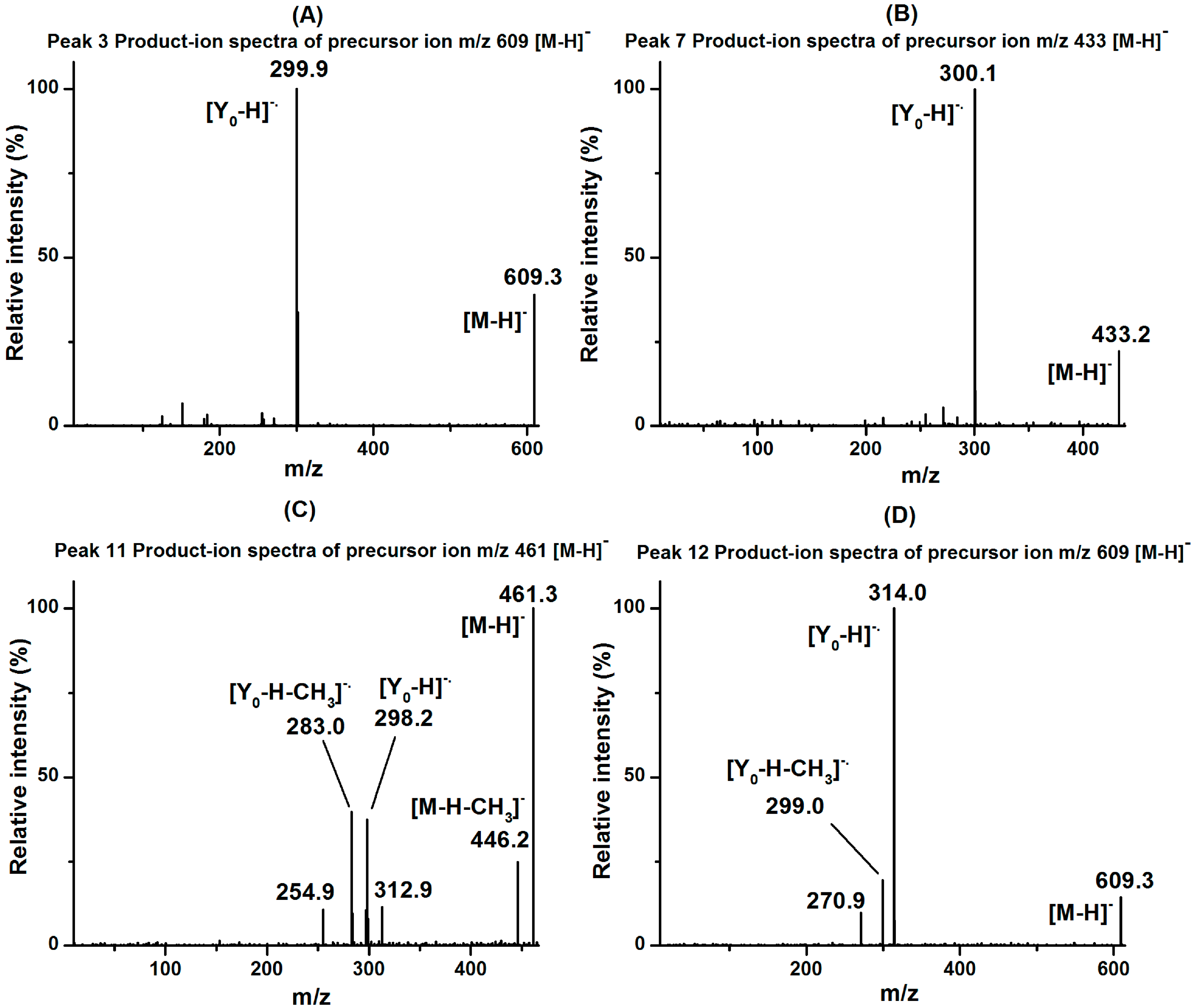
2.1. Analysis of Flavonoids by HPLC-MS/MS

The highest total flavonoid content (TFC) was determined in fraction II (690.5 mg isoquercetin equivalents/g sample, Table 1), while very few peaks were detected in fractions I, III and IV in chromatographic profiles recorded at 350 nm, so fractions I, III and IV were thus deemed to have very few or even no flavonoids. A successful enrichment and separation of total flavonoids from lotus leaves was thus achieved by the D101 macroporous resin chromatography, and the resultant flavonoids were then ready for the subsequent chemical and bioactivity analysis. After the flavonoids from the lotus leaves extracts were successfully enriched in fraction II using the D101 macroporous resin chromatography, HPLC-MS/MS was then employed to identify those flavonoids. Figure 1 shows the chromatographic profile of fraction II at 350 nm, where fourteen peaks were well detected and resolved, representing at least 14 lotus leaves flavonoids. To identify these already resolved flavonoids, LC-MS/MS experiments were conducted. The LC and MS/MS data, including retention times, molecular ions, aglycone ions and some important fragment ions are listed in Table 1. The fragmentation of O-glycosyl flavonoids in the negative ion mode (NI) is characterized by the loss of the sugar moieties, and deprotonated aglycone species (Y0) or radical aglycone ion ([Y0− H]−.) fragments are obtained [22,23]. The MS/MS model of flavonoid O-glycosides is shown in Figure 2. For flavonol mono-O-glycosides, glycosylation took place at the 3-position if the relative abundance of the [Y0 − H]. ion was significantly higher than that of the Y0 ion, and the situation is reversed when glycosylation happens at the 7-position [22]. Till now, five aglycones, kaempferol, quercetin, isorhamnetin, myricetin and diosmetin, were identified from lotus [24].
Figure 1. The HPLC profile of flavonoids in fraction II from lotus leaves recorded at 350 nm.
Molecules 20 10553 g001
Figure 2. The MS/MS model of flavonoid O-glycosides.
Molecules 20 10553 g002
As listed in Table 1, peak 1 showed a [M − H] ion at m/z 479, with its [Y0− H]−. ion at m/z 316 (loss of a hexose moiety), indicating that it was myricetin monohexoside, and the glycosylation took place at the 3-position based on the characteristic [Y0− H]−. fragment ion. Peak 1 was therefore identified as myricetin 3-O-hexose, which has been previously found in lotus leaves and flowers [24]. Similar to the MS/MS model of peak 1, the presence of a [M − H] ion at m/z 477 and the corresponding [Y0 − H]−. ion at m/z 314 for peak 13 indicated that it was an isorhamnetin monohexoside identified as isorhamnetin 3-O-hexose, which has been previously reported [18]. Peak 2 exhibited the [M − H] ion at m/z 595 with its [Y0 − H]−. ion at m/z 300 (loss of a pentose and a hexose moiety), indicating that it was a quercetin diglycoside. For flavonol O-diglycosides, the mass spectrometric behaviors of diglycosides are notably different depending on the linkage between the two monosaccharides. A [Y0 − H]−. ion tends to be generated in the case of a C1→C2 linkage between the two monosaccharides, while the Y0 ion is indicative of a C1→C6 linkage [22]. The higher abundance of [Y0− H]−. ion at m/z 300 for peak 3 indicated that the interglycosidic linkage between the two monosaccharides in this compound was C1→C2, and peak 2 was thus identified as quercetin 3-O-arabinopyranosyl-(1→2)-galactopyranoside, which has been reported in lotus leaves [25].
Table 1. Identification of flavonoids in fraction II of lotus leaves by LC-MS/MS.
Peak No.Rt (min) aNI-MSMS/MSIdentification
125.9479316Myricetin 3-O-hexose
227.4595300Quercetin 3-O-arabinopyranosyl-(1→2)-galactopyranoside
333.0609300Quercetin 3-O-rhamnopyranosyl-(1→2)-glucopyranoside
434.1463300Quercetin 3-O-galactoside (hyperoside)
535.5463300Quercetin 3-O-glucoside (isoquercitrin)
637.2477301Quercetin 3-O-glucuronide
738.7433300Quercetin 3-O-arabinoside
840.6447284Kaempferol 3-O-galactoside
943.7447284Kaempferol 3-O-glucoside (astragalin)
1045.7461285Kaempferol 3-O-glucuronide
1147.5461446; 298; 283Diosmetin 7-O-hexose
1252.6609314; 299Isorhamnetin 3-O-arabinopyranosyl-(1→2)-glucopyranoside
1356.7477314Isorhamnetin 3-O-hexose
1457.9491315Isorhamnetin 3-O-glucuronide
a Rt: retention time on HPLC.
As shown in Figure 3A,D, peaks 3 and 12 exhibited the same [M − H] ions at m/z 609 with [Y0 − H]· ions at m/z 300 and 314, indicating that they were quercetin diglycoside and isorhamnetin diglycoside, respectively. Considering the similar MS/MS spectrometric behaviors with peak 2, peaks 3 and 12 were identified as quercetin 3-O-rhamnopyranosyl-(1→2)-glucopyranoside and isorhamnetin 3-O-arabinopyranosyl-(1→2)-glucopyranoside. These two flavonoids were identified in this study for the first time. In addition, the presence of the fragment ion at m/z 299 ([Y0 −H − CH3]·) of peak 12 indicated that the methoxyflavone readily lost a methyl group [26].
Figure 3. The MS/MS spectra of newly identified flavonoids in fraction II from lotus leaves: (A) peak 3; (B) peak 7; (C) peak 11; (D) peak 12.
Molecules 20 10553 g003
Peaks 4 and 5 exhibited the same [M − H] ions at m/z 463 with [Y0− H]−. ions at m/z 300 (loss of a hexose moiety), indicating that they are quercetin monohexoside isomers with a hexose conjugated at the 3-position. In addition, peak 4 had a shorter retention time than peak 5. Considering that glycosides linked with galactose elute before glucose linkages [27,28], peaks 4 and 5 were identified as quercetin 3-O-galactoside (hyperoside) and quercetin 3-O-glucoside (isoquercitrin) [25], and the results were further validated by comparing their LC-MS with the corresponding standards.
Peak 6 showed a [M − H] ion at m/z 477, with a Y0 ion at m/z 301 (loss of a glucuronic acid). For glucuronic acid glycosides, only the Y0 ion was observed during the MS/MS process, and the glucuronic acid glycoside was conjugated at the 3-position. Thus, peak 6 was identified as quercetin 3-O-glucuronide, which has previously been reported to be the major flavonoid in lotus leaves [25]. Like the MS/MS spectrometric behavior of peak 6, the ions at m/z 461 ([M − H]), m/z 285 (Y0), 491 ([M − H]) and m/z 315 (Y0) for peaks 10 and 14 indicated that they were kaempferol and isorhamnetin glucuronic acid glycosides, respectively. Thus, peaks 10 and 14 were identified as kaempferol 3-O-glucuronide and isorhamnetin 3-O-glucuronide [24,29].
As shown in Figure 3B, peak 7 showed a [M − H] ion at m/z 433, with a [Y0− H]−. ion at m/z 300 (loss of a pentose moiety), indicating that it was quercetin monopentoside, and glycosylation took place at the 3-position. Peak 7 was therefore identified as quercetin 3-O-arabinoside. Its MS/MS spectra agreed with a compound identified in Juglans regia L. leaves [30], while it has been found in lotus leaves in this work for the first time.
For peaks 8 and 9, the same [M − H] ions were observed at m/z 447, with their [Y0− H]−. ions at m/z 284 (loss of a hexose moiety), indicated that both were kaempferol 3-O-hexoses. Furthermore, peak 8 had a shorter retention time than that of peak 9. Thus, peaks 8 and 9 were identified as kaempferol 3-O-galactoside and kaempferol 3-O-glucoside (astragalin), which have previously been reported in lotus petals [29]. In addition, peak 9 was further confirmed by comparing the LC-MS/MS spectra with the corresponding standard.
As shown in Figure 3C, peak 11 showed a [M − H] ion at m/z 461, with a [Y0− H]−. ion at m/z 298 (loss of a hexose moiety), indicating that it was diosmetin monohexoside, and glycosylation took place at the 7-position. The presence of the ions at m/z 446 ([M – CH3]) and 283 ([Y0 – H − CH3]−.) indicated that the aglycone of diosmetin easily lost a methyl group. Peak 11 was identified as diosmetin 7-O-hexose, which has also been found in lotus leaves for the first time.

2.2. Antioxidant Activity of Flavonoids from Lotus Leaves

The antioxidant activity of plant extracts cannot be evaluated by only one single method due to the complex nature of phytochemicals, and antioxidant activity determination is highly reaction-mechanism dependent [12]. Multiple chemical or biological assays have been developed to evaluate the antioxidant activity and explain the antioxidant mechanism of action of plant extracts. Of those, the DPPH assay, ABTS assay and reducing power assay are the most commonly used assays to evaluate the antioxidant activities of plant extracts [31]. In view of this, a series of assays including DPPH scavenging activity, ABTS scavenging activity and FRAP were used for the determination of the antioxidant activity of the fraction of lotus leaves. Results of these evaluations were expressed as Trolox equivalents and IC50 values, which are shown in Table 2. Fraction II showed good free radical scavenging activity in the DPPH assay. The activity of fraction II (4695.3 μmol·TE/g) was higher than that of BHT (3612.3 μmol·TE/g, positive control). In terms of IC50, fraction II (IC50 = 0.101 mg/mL) had a lower value than BHT (IC50 = 0.121 mg/mL), which implied that fraction II possessed a stronger radical scavenging activity in the DPPH assay. For scavenging activity pattern in the ABTS assay, the activity of fraction II (5012.3 μmol·TE/g) was higher than that of BHT (4567.0 μmol·TE/g), while the IC50 value of fraction II (IC50 = 0.138 mg/mL) was lower than that of BHT (IC50 = 0.143 mg/mL). With regard to the ferric reducing capacity of fraction II, the trend was almost the same as that of the DPPH and ABTS assay. The reducing power activity of fraction II (500.5 mmol Fe2+/100 g) was nearly the same with that of Trolox (642.1 mmol Fe2+/100 g).
Table 2. Total flavonoids in fraction II from lotus leaves a and their corresponding antioxidant activity.
SampleTotal Flavonoid (mg IE/g)DPPHABTSFRAP (mmol Fe2+/100 g)
(μmol TE/g)IC50 value b (mg/mL)(μmol TE/g)IC50 value b (mg/mL)
Fraction II690.5 ± 35.84695.3 ± 144.30.101 ± 0.0075012.3 ± 133.80.138 ± 0.007500.5 ± 62.8
BHTnt3612.3 ± 170.90.121 ± 0.0044567.0 ± 155.60.143 ± 0.004nt
Troloxntnt0.112 ± 0.005nt0.119 ± 0.005642.1 ± 55.7
a Each value is presented as the mean ± SD of three replicate determinations; b IC50 value was determined to be the effective concentration at which DPPH and ABTS radicals were inhibited by 50%, respectively. IE, isoquercetin equivalents; TE, Trolox equivalents; nt, not tested.
Taken together, the above results demonstrate that fraction II from lotus leaves possesses good antioxidant potential, and the antioxidant activity of fraction II was nearly the same as that of BHT. This capability may be correlated to the different flavonoid components identified in the lotus leaves, whose activity has been relative to the electron donating ability associated with the degree and position of hydroxylation and methoxylation on the B-ring [32]. The flavonoids possessing a catecholic B-ring, such as hyperoside (4) isoquercitrin (5) and quercetin 3-O-arabinoside (7) (which also possess a double bond at the 2 position of the C ring, conjugated with the 4-oxo group), are probably the derivatives that contribute the most to the total antioxidant activity, because the presence of a catechol moiety confers greater stability to the aroxyl radicals formed upon reaction with radical compounds. Kaempferol 3-O-galactoside (8) and astragalin (9), which only possess a double bond at the 2 position of the C ring (again conjugated with the 4-oxo function group) and a phenolic B-ring, probably contribute to the total antioxidant activity to a lesser extent than those of the compounds mentioned above.

3. Experimental Section

3.1. Chemicals and Materials

Three flavonoid glycoside standards (quercetin 3-O-galactoside (hyperoside), quercetin 3-O-glucoside (isoquercitrin), kaempferol 3-O-glucoside (astragalin)) were purchased from Shanghai Tauto Biotech (Shanghai, China). HPLC-grade solvents (acetonitrile and formic acid), butylated hydroxytoluene (BHT), 1,3,5-tri(2-pyridyl)-2.4.6-triazine (TPTZ), 6-hydroxy-2,5,7,8-tetramethyl-chroman-2-carboxylic acid (Trolox), α,α-diphenyl-β-picrylhydrazyl (DPPH), and 2,2′-azinobis-(3-ethylbenzthiazoline-6-sulfonic acid) (ABTS) were purchased from Sigma-Aldrich Corp. (Shanghai, China). HPLC-grade water was obtained using a Milli-Q System (Millipore, Billerica, MA, USA). Other chemicals of analytical grade were obtained from Shanghai Chemical Reagent Corp. (Shanghai, China). Millipore membranes (0.22 μm) were purchased from Jinteng Experiment Equipment Corp. (Tianjin, China). D101 macroporous resin was purchased from an industrial chemical company affiliated with Nan Kai University (Tianjin, China). Fresh lotus leaves were collected from Guangchang (Jiangxi, China) in 2013 and stored at −40 °C. Lotus leaves were dried at 45 °C, and then stored at 4 °C before use.

3.2. Extraction and Fractionation of Crude Lotus Leaves Extracts

The dried lotus leaves were powdered (100 g) and ultrasonically extracted at room temperature for 40 min with 1000 mL of 70% ethanol. After three extractions, the extracts were combined and filtered, and the supernatants were evaporated under reduced pressure and lyophilized to afford dark-green residues which were dissolved in H2O (50 mL) and were subjected to liquid/liquid partitioning with petroleum ether (B.P. 60–90 °C) in order to remove chlorophyll. Later the lower layer was evaporated, and fractionated by the D101 macroporous resin chromatography. The column was washed with distilled water to remove water soluble impurities (sugar, protein, and other water-soluble molecules) and then eluted successively with 30, 50, 70, and 90% ethanol and named fractions I, II, III and IV, respectively. The weights of crude extracts and fractions (I–IV) were 27.6 g, 3.1 g, 3.7 g, 1.0 g and 1.1 g, respectively. Aliquots of the eluents were then subjected to further analysis.

3.3. Determinations of Total Flavonoids Content (TFC)

Total flavonoids content was determined using a previously described colorimetric method [33]. Briefly, a 30 μL aliquot of appropriately diluted sample solution was mixed with 180 μL of distilled water in a well of a 96-well plate and 10 μL of a 5% NaNO2 solution was added subsequently. After 6 min, 20 μL of 10% AlCl3 solution was added and allowed to stand for 6 min before an addition of 60 μL of 4% NaOH solution. The absorbance of the mixture was determined at 510 nm vs. a water blank using a multifunctional microplate reader (Infinite M200 PRO, Tecan, Männedorf, Switzerland) after 15 min. Isoquercitrin was used as standard compound for the quantification of total flavonoids. All values were expressed as milligrams of isoquercitrin equivalents per gram of sample (mg IE/g sample).

3.4. HPLC Analysis of Flavonoids

The analysis of flavonoids was carried out using a Thermo Accela 1250 U-HPLC system (Thermo Fisher Scientific, San Jose, CA, USA) equipped with a binary solvent pump, column oven, auto-sampler and UV detector. A 10-μL aliquot of each sample solution was injected and analyzed on a Sunfire C18 column (150 mm × 4.6 mm, 3.5 μm, Waters, MA, USA). The separation was conducted at 30 °C (column temperature) using a gradient elution method with 0.5% formic acid in distilled water (solvent A) and 0.1% formic acid in acetonitrile (solvent B). The solvent gradient in volumetric ratios was set as follows: 0–10 min at 88% A; 10–42 min from 88% A to 80% A; 42–55 min from 80% A to 70% A; 55–63 min from 70% A to 40% A; 63–64 min from 40 % A to 88% A; and 64–65 min at 88% A. The flow rate was 0.6 mL/min and the effluents were monitored at 350 nm.

3.5. Identification of Flavonoids

Flavonoids were identified using a Thermo Accela 600 HPLC system with a UV detector coupled to a TSQ Quantum Access MAX triple-stage quadropole mass spectrometer (Thermo Fisher Scientific). Electrospray ionization (ESI) was applied in the negative ion mode (NI) for the MS analysis. The operation conditions of mass analysis were set as follows: capillary temperature, 350 °C; vaporizer temperature, 300 °C; sheath gas (N2) pressure, 40 arbitrary units; auxiliary gas (N2) pressure, 10 arbitrary units; spray voltage, 3 kV. The mass spectra were recorded in the mass range from m/z 150 to 1500. The MS/MS spectra were obtained using the Data-Dependent mode and the collision energy was set as following: collision energy (CE), 10 V; collision energy grad (CE grad), 0.035 V/m.

3.6. Determination of Antioxidant Activity of Flavonoids

3.6.1. DPPH Free Radical Scavenging Activity

DPPH scavenging activity was determined by the method described by Brand-Williams et al. with slight modifications [34]. Ten μL of appropriately diluted sample or Trolox solution (31.25–1000 μM) was added to 190 μL of DPPH solution (final concentration was 0.1 mM in methanol) in a 96-well plate. Then, the sample mixture was shaken gently and kept in the dark at room temperature for 30 min. Thereafter, the absorbance at 517 nm was measured and methanol was used for the baseline correction with a multifunctional microplate reader. The DPPH radical scavenging activity of extracts was calculated from the standard curve of Trolox and expressed as micromoles of Trolox equivalents (TE) per gram of sample (μmol TE/g). In addition, to determine the IC50 of samples on DPPH, six different concentrations were used. Following the same procedure above, the methanol instead of sample was made as blank controls, while BHT and Trolox were used as positive controls. The ability to scavenge the DPPH radical was calculated as a percentage according to the following equation:
DPPH-scavenging effect (%) = [(ADPPH − AS)/ADPPH] × 100
where ADPPH = absorbance of control, AS = absorbance of sample), and the IC50 value was determined to be the effective concentration at which DPPH radicals were inhibited by 50%.

3.6.2. ABTS Free Radical Scavenging Activity

ABTS free radical scavenging activity was determined according to the method adopted by Zou et al. with some minor modifications [33]. The ABTS assay is based on the capacity to quench ABTS radical cationic (ABTS+) formation relative to Trolox. Briefly, a solution of ABTS+ was prepared by mixing equal volumes of potassium persulfate (4.9 mM in H2O) and ABTS (7 mM in H2O), and the solution was incubated in the dark for 12–16 h. The radical was stable in this form for more than two days when stored in the dark at room temperature. The ABTS+ solution was then diluted with 80% ethanol to obtain an absorbance of 0.700 ± 0.005 at 734 nm. Afterwards, ten microliters of appropriately diluted samples was added to 190 μL of ABTS+ solution in a 96-well plate. The mixture was incubated in the dark at room temperature for 30 min and then the absorbance was recorded at 734 nm. Trolox was used as standard, and a standard calibration curve was obtained for Trolox at concentrations ranging from 31.25 μM to 500 μM. The ABTS free radical scavenging activity of samples was calculated from the standard curve of Trolox and expressed as micromoles of Trolox equivalents (TE) per gram of sample (μmol·TE/g). The scavenging activities of different concentrations of samples against ABTS+ were also measured to calculate the IC50, and the procedure was similar to the DPPH scavenging method described before.

3.6.3. Ferric Reducing/Antioxidant Power (FRAP) Assay

FRAP assay was performed as described previously by Benzie and Strain [35]. This method measures the change in absorbance at 593 nm owing to the formation of a blue colored ferrous 2,4,6-tripyridyl-s-triazine complex (Fe2+-TPTZ) from colorless oxidized ferric form (Fe3+-TPTZ) by the action of electron donating antioxidants. The stock solutions included 300 mM acetate buffer, pH 3.6 (3.1 g C2H3NaO2·3H2O and 16 mL C2H4O2), 10 mM TPTZ solution in 40 mM HCl, and 20 mM FeCl3·6H2O solution. The working solution was prepared by mixing 10 volumes of acetate buffer, 1 volume of TPTZ solution, and 1 volume of FeCl3·6H2O solution and then warmed at 37 °C before use. Then, 10 μL of properly diluted samples and 30 μL of distilled water was added to 260 μL of freshly prepared FRAP reagent in a 96-well plate and mixed thoroughly. The mixture was incubated at 37 °C for 10 min, and the absorbance was measured at 593 nm. The FRAP value was calculated and expressed as millimoles of Fe2+ equivalents per 100g of sample (mmol Fe2+equiv/100 g) based on a calibration curve plotted using FeSO4·7H2O as standard at a concentration ranging from 0.125 to 2 mM. All solutions were freshly made and used on the day of preparation.

3.7. Statistical Analysis

Data were expressed as the mean ± standard deviation of triplicate measurements. The data were statistically analyzed using statistical software, OriginPro 8.6.

4. Conclusions

To extend our research on the correlations and underlying mechanisms between chemical components and their bioactivities, a more efficient macroporous resin chromatography with much better resolution and compatibility coupled with HPLC-MS was developed for the simultaneous separation and biochemical analysis of flavonoids from the complex extracts of lotus leaves. Altogether, fourteen flavonoids from lotus leaves were identified in this work, which greatly improved the previous LC-MS/MS method. More importantly, the newly developed method has led to some significant new findings. Among fourteen flavonoids identified, quercetin 3-O-rhamnopyranosyl-(1→2)-gluco- pyranoside, quercetin 3-O-arabinoside, diosmetin 7-O-hexose and isorhamnetin 3-O-arabino- pyranosyl-(1→2)-glucopyranoside were identified in lotus leaves for the first time. In addition, this macroporous resin chromatography method is very compatible with flexible downstream biological activity studies, and greatly facilitated the antioxidant activity screening in this study. It is expected that with some slight modifications this new method will prove useful to explore more important applications in food and pharmaceutical industries.

Acknowledgments

This work was jointly supported by “the Hundred Talents Program” from Chinese Academy of Sciences (Grant No. 29Y429291a0129 to M. Guo), The Knowledge Innovation Project of Chinese Academy of Sciences (Grant No. Y455421Z02), and the Key Laboratory of Plant Germplasm Enhancement and Specialty Agriculture of Wuhan Botanical Garden, Chinese Academy of Sciences.

Author Contributions

Ming-Quan Guo and Ming-Zhi Zhu conceived and designed the experiments; Ming-Zhi Zhu, Wei Wu, Li-Li Jiao and Ping-Fang Yang performed the experiments or helped with the data analysis; Ming-Zhi Zhu and Ming-Quan Guo wrote the paper. All authors read and approved the final manuscript.

Conflicts of Interest

The authors declare no conflict of interest.

References

  1. Mukherjee, P.K.; Mukherjee, D.; Maji, A.K.; Rai, S.; Heinrich, M. The sacred lotus (Nelumbo nucifera)—Phytochemical and therapeutic profile. J. Pharm. Pharmacol. 2009, 61, 407–422. [Google Scholar] [CrossRef] [PubMed]
  2. Kredy, H.M.; Huang, D.H.; Xie, B.J.; He, H.; Yang, E.N.; Tian, B.Q.; Xiao, D. Flavonols of lotus (Nelumbo nucifera, gaertn.) seed epicarp and their antioxidant potential. Eur. Food Res. Technol. 2010, 231, 387–394. [Google Scholar] [CrossRef]
  3. National Commission of Chinese Pharmacopoeia. Pharmacopoeia of the People’s Republic of China; China Medical Science and Technology Press: Beijing, China, 2010; pp. 258–259. [Google Scholar]
  4. Wu, M.J.; Wang, L.; Weng, C.Y.; Yen, J.H. Antioxidant activity of methanol extract of the lotus leaf (Nelumbo nucifera gertn.). Am. J. Chin. Med. 2003, 31, 687–698. [Google Scholar] [CrossRef] [PubMed]
  5. Kashiwada, Y.; Aoshima, A.; Ikeshiro, Y.; Chen, Y.P.; Furukawa, H.; Itoigawa, M.; Fujioka, T.; Mihashi, K.; Cosentino, L.M.; Morris-Natschke, S.L.; et al. Anti-HIV benzylisoquinoline alkaloids and flavonoids from the leaves of Nelumbo nucifera, and structure-activity correlations with related alkaloids. Bioorg. Med. Chem. 2005, 13, 443–448. [Google Scholar] [CrossRef] [PubMed]
  6. Ono, Y.; Hattori, E.; Fukaya, Y.; Imai, S.; Ohizumi, Y. Anti-obesity effect of Nelumbo nucifera leaves extract in mice and rats. J. Ethnopharmacol. 2006, 106, 238–244. [Google Scholar] [CrossRef] [PubMed]
  7. Ohkoshi, E.; Miyazaki, H.; Shindo, K.; Watanabe, H.; Yoshida, A.; Yajima, H. Constituents from the leaves of Nelumbo nucifera stimulate lipolysis in the white adipose tissue of mice. Planta Med. 2007, 73, 1255–1259. [Google Scholar] [CrossRef] [PubMed]
  8. Huang, B.; Ban, X.Q.; He, J.S.; Tong, J.; Tian, J.; Wang, Y.W. Hepatoprotective and antioxidant activity of ethanolic extracts of edible lotus (Nelumbo nucifera gaertn.) leaves. Food Chem. 2010, 120, 873–878. [Google Scholar] [CrossRef]
  9. Luo, X.B.; Chen, B.; Liu, J.J.; Yao, S.Z. Simultaneous analysis of N-nornuciferine, O-nornuciferine, nuciferine, and roemerine in leaves of Nelumbo nucifera gaertn by high-performance liquid chromatography-photodiode array detection-electrospray mass spectrometry. Anal. Chim. Acta 2005, 538, 129–133. [Google Scholar] [CrossRef]
  10. Elegami, A.A.; Bates, C.; Gray, A.I.; Mackay, S.P.; Skellern, G.G.; Waigh, R.D. Two very unusual macrocyclic flavonoids from the water lily Nymphaea lotus. Phytochemistry 2003, 63, 727–731. [Google Scholar] [CrossRef]
  11. Pietta, P.G. Flavonoids as antioxidants. J. Nat. Prod. 2000, 63, 1035–1042. [Google Scholar] [CrossRef] [PubMed]
  12. Singh, M.; Kaur, M.; Silakari, U. Flavones: An important scaffold for medicinal chemistry. Eur. J. Med. Chem. 2014, 84, 206–239. [Google Scholar] [CrossRef] [PubMed]
  13. Guerra-Araiza, C.; Alvarez-Mejia, A.L.; Sanchez-Torres, S.; Farfan-Garcia, E.; Mondragon-Lozano, R.; Pinto-Almazan, R.; Salgado-Ceballos, H. Effect of natural exogenous antioxidants on aging and on neurodegenerative diseases. Free Radic. Res. 2013, 47, 451–462. [Google Scholar] [CrossRef] [PubMed]
  14. Lopez-Alarcon, C.; Denicola, A. Evaluating the antioxidant capacity of natural products: A review on chemical and cellular-based assays. Anal. Chim. Acta. 2013, 763, 1–10. [Google Scholar] [CrossRef] [PubMed]
  15. Flora, S.J.S.; Dikshit, M.; Flora, G. Role of free radicals and antioxidants in health and disease. Cell. Mol. Biol. 2007, 53, 1–3. [Google Scholar] [PubMed]
  16. Wan, P.F.; Sheng, Z.L.; Han, Q.; Zhao, Y.L.; Cheng, G.D.; Li, Y.H. Enrichment and purification of total flavonoids from Flos Populi extracts with macroporous resins and evaluation of antioxidant activities in vitro. J. Chromatogr. B 2014, 945, 68–74. [Google Scholar] [CrossRef] [PubMed]
  17. Zou, Y.P.; Lu, Y.H.; Wei, D.Z. Antioxidant activity of a flavonoid-rich extract of Hypericum perforatum L. in vitro. J. Agric. Food Chem. 2004, 52, 5032–5039. [Google Scholar] [CrossRef] [PubMed]
  18. Chen, S.; Wu, B.H.; Fang, J.B.; Liu, Y.L.; Zhang, H.H.; Fang, L.C.; Guan, L.; Li, S.H. Analysis of flavonoids from lotus (Nelumbo nucifera) leaves using high performance liquid chromatography/photodiode array detector tandem electrospray ionization mass spectrometry and an extraction method optimized by orthogonal design. J. Chromatogr. A 2012, 1227, 145–153. [Google Scholar] [CrossRef] [PubMed]
  19. Deng, S.G.; Deng, Z.Y.; Fan, Y.W.; Peng, Y.; Li, J.; Xiong, D.M.; Liu, R. Isolation and purification of three flavonoid glycosides from the leaves of Nelumbo nucifera (lotus) by high-speed counter-current chromatography. J. Chromatogr. B 2009, 877, 2487–2492. [Google Scholar] [CrossRef] [PubMed]
  20. Du, Z.; Wang, K.; Tao, Y.; Chen, L.; Qiu, F. Purification of baicalin and wogonoside from Scutellaria baicalensis extracts by macroporous resin adsorption chromatography. J. Chromatogr. B 2012, 908, 143–149. [Google Scholar] [CrossRef] [PubMed]
  21. Li, J.; Chase, H.A. Development of adsorptive (non-ionic) macroporous resins and their uses in the purification of pharmacologically-active natural products from plant sources. Nat. Prod. Rep. 2010, 27, 1493–1510. [Google Scholar] [CrossRef] [PubMed]
  22. Ablajan, K.; Abliz, Z.; Shang, X.Y.; He, J.M.; Zhang, R.P.; Shi, J.G. Structural characterization of flavonol 3,7-di-O-glycosides and determination of the glycosylation position by using negative ion electrospray ionization tandem mass spectrometry. J. Mass Spectrom. 2006, 41, 352–360. [Google Scholar] [CrossRef] [PubMed]
  23. Liu, Y.; Yang, J.S.; Cai, Z.W. Chemical investigation on Sijunzi decoction and its two major herbs Panax ginseng and Glycyrrhiza uralensis by LC/MS/MS. J. Pharmaceut. Biomed. 2006, 41, 1642–1647. [Google Scholar] [CrossRef] [PubMed]
  24. Chen, S.; Fang, L.C.; Xi, H.F.; Guan, L.; Fang, J.B.; Liu, Y.L.; Wu, B.H.; Li, S.H. Simultaneous qualitative assessment and quantitative analysis of flavonoids in various tissues of lotus (Nelumbo nucifera) using high performance liquid chromatography coupled with triple quad mass spectrometry. Anal. Chim. Acta 2012, 724, 127–135. [Google Scholar] [CrossRef] [PubMed]
  25. Goo, H.R.; Choi, J.S.; Na, D.H. Simultaneous determination of quercetin and its glycosides from the leaves of Nelumbo nucifera by reversed-phase high-performance liquid chromatography. Arch. Pharm. Res. 2009, 32, 201–206. [Google Scholar] [CrossRef] [PubMed]
  26. Wu, W.; Yan, C.Y.; Li, L.; Liu, Z.Q.; Liu, S.Y. Studies on the flavones using liquid chromatography-electrospray ionization tandem mass spectrometry. J. Chromatogr. A 2004, 1047, 213–220. [Google Scholar] [CrossRef]
  27. Abad-Garcia, B.; Berrueta, L.A.; Garmon-Lobato, S.; Gallo, B.; Vicente, F. A general analytical strategy for the characterization of phenolic compounds in fruit juices by high-performance liquid chromatography with diode array detection coupled to electrospray ionization and triple quadrupole mass spectrometry. J. Chromatogr. A 2009, 1216, 5398–5415. [Google Scholar] [CrossRef] [PubMed]
  28. Zhou, H.; Xing, J.P.; Liu, S.; Song, F.R.; Cai, Z.W.; Pi, Z.F.; Liu, Z.Q.; Liu, S.Y. Screening and determination for potential a-glucosidase inhibitors from leaves of acanthopanax senticosus harms by using UF-LC/MS and ESI-MSn. Phytochem. Anal. 2012, 23, 315–323. [Google Scholar] [CrossRef] [PubMed]
  29. Yang, R.Z.; Wei, X.L.; Gao, F.F.; Wang, L.S.; Zhang, H.J.; Xu, Y.J.; Li, C.H.; Ge, Y.X.; Zhang, J.J.; Zhang, J. Simultaneous analysis of anthocyanins and flavonols in petals of lotus (Nelumbo) cultivars by high-performance liquid chromatography-photodiode array detection/electrospray ionization mass spectrometry. J. Chromatogr. A 2009, 1216, 106–112. [Google Scholar] [CrossRef] [PubMed]
  30. Zhao, M.H.; Jiang, Z.T.; Liu, T.; Li, R. Flavonoids in juglans regia l. Leaves and evaluation of in vitro antioxidant activity via intracellular and chemical methods. Sci. World J. 2014, 2014. [Google Scholar] [CrossRef]
  31. Niki, E. Assessment of antioxidant capacity in vitro and in vivo. Free Radic. Bio. Med. 2010, 49, 503–515. [Google Scholar] [CrossRef] [PubMed]
  32. RiceEvans, C.A.; Miller, N.J.; Paganga, G. Structure-antioxidant activity relationships of flavonoids and phenolic acids. Free Radic. Bio. Med. 1996, 20, 933–956. [Google Scholar] [CrossRef]
  33. Zou, Y.P.; Chang, S.K.C.; Gu, Y.; Qian, S.Y. Antioxidant activity and phenolic compositions of Lentil (Lens culinaris var. Morton) extract and its fractions. J. Agric. Food Chem. 2011, 59, 2268–2276. [Google Scholar] [CrossRef] [PubMed]
  34. Brandwilliams, W.; Cuvelier, M.E.; Berset, C. Use of a free-radical method to evaluate antioxidant activity. Food Sci. Technol. 1995, 28, 25–30. [Google Scholar]
  35. Benzie, I.F.F.; Strain, J.J. The ferric reducing ability of plasma (FRAP) as a measure of “antioxidant power”: The FRAP assay. Anal. Biochem. 1996, 239, 70–76. [Google Scholar] [CrossRef] [PubMed]
  • Sample Availability: Samples of the compounds (quercetin 3-O-galactoside (hyperoside), quercetin 3-O-glucoside (isoquercitrin), kaempferol 3-O-glucoside (astragalin)) are available from the authors.

