Article

Phytochemical Screening of Quaking Aspen (Populus tremuloides) Extracts by UPLC-QTOF-MS and Evaluation of their Antimicrobial Activity

by
Annabelle St-Pierre
1,
Dorian Blondeau
1,
André Lajeunesse
1,
Julien Bley
2,
Nathalie Bourdeau
2 and
Isabel Desgagné-Penix
1,3,*
1
Department of Chemistry, Biochemistry and Physics, Université du Québec à Trois-Rivières, Trois-Rivières, QC G9A 5H7, Canada
2
Innofibre, Trois-Rivières, QC G9A 5H7, Canada
3
Plant Biology Research Group, Trois-Rivières, QC G9A 5H7, Canada
*
Author to whom correspondence should be addressed.
Molecules 2018, 23(7), 1739; https://doi.org/10.3390/molecules23071739
Submission received: 29 June 2018 / Revised: 9 July 2018 / Accepted: 12 July 2018 / Published: 16 July 2018
(This article belongs to the Section Chemical Biology)

Abstract

:
The continual emergence of pathogen resistance is a recurring challenge and pushes for the development of antimicrobial compounds. Here, we investigated compounds from quaking aspen trees (Populus tremuloides) as potential antimicrobial agents. Several extractions using different solvents were realized, and corresponding antimicrobial activity was tested against eight microorganisms. Results revealed that polar extraction solvents including water, ethanol and methanol gave the best extraction yields (>15.07%). Minimal inhibition concentration (MIC) and minimal bactericidal/fungicidal concentration (MBC/MFC) demonstrated that water extracts had the best antimicrobial activity by a weak to moderate inhibition of growth of all eight tested microorganisms in addition to having a bactericidal effect on three of them. The quaking aspen methanol extract also displayed antimicrobial activity but to a lower level than the water extract. Ultra-performance liquid chromatography quadrupole time-of flight mass spectrometry (UPLC-QTOF-MS) analysis led to the identification of 92 compounds, mainly polyphenols in both extracts, with 22 molecules previously known for their antimicrobial properties. According to the relative abundance, 4-hydroxybenzaldehyde (5.44% in methanol extract) and kaempferol (5.03% in water extract) were the most abundant antimicrobial compounds. Among antimicrobial molecules identified, nine were from the flavonoid family. The results of our study demonstrate the interest of using quaking aspen as source of antimicrobial compounds.

Graphical Abstract

1. Introduction

Continual emergence of pathogens resistant to antibiotics, antiseptics and disinfectants is a real challenge and it motivates innovation in the field of antimicrobial agent development [1,2]. This research field is in perpetual expansion in order to discover new molecules with antimicrobial activity. Although synthetic antimicrobial agent development has largely contributed to the field, the current environmental concerns coupled with the need to deal with our natural resource residues has prompted a renewed interest in natural antimicrobial molecules. Thus, it is relevant to look at plant specialized (aka secondary) metabolites known for their biological and therapeutic activities. Indeed, several current studies are using this alternative approach to investigate molecules produced by plants with the purpose of developing antimicrobial agents. In fact, it was estimated that there are around 25% of prescribed drugs on the market that come from plant metabolites [3,4].
Quaking aspen (Populus tremuloides) is a tree largely present in the boreal forests of North America and Eurasia and is poorly studied and exploited for its biological properties. This broadleaf tree exhibits the largest spatial distribution of all deciduous species in Canada. It stretches from Newfoundland and Labrador to British Columbia and Yukon [5]. Quaking aspen was formerly used in traditional medicine by Native American nations of Québec for its various therapeutic functions, either as a vermifuge, to treat venereal disease, as an anti-arthritis agent or as a cure for colds [5,6]. Since some of these applications suggest antimicrobial activities, quaking aspen has potential in the development of antimicrobial agents through the extraction of its specialized metabolites. Previous studies on the chemical composition of quaking aspen have revealed that this species is an important source of extractive compounds, i.e., molecules easily extractive with a solvent. It was reported that quaking aspen trees are rich in polyphenols, but also in sterols, steryls, triterpenes and alkaloids [7,8,9]. Polyphenols and alkaloids are families of compounds highly sought for their antioxidant, antitumor and antimicrobial activities [10,11,12,13]. These classes of compounds are recognized to be synthesized by plants to protect themselves against biotic stress. These molecules, according to their chemical structure, can act as toxins that alter biological processes of predators such as herbivores, fungi or bacteria [14,15]. Triterpenes are also known for their antimicrobial potential and are often involved in plant defense mechanisms against pathogens [16,17,18]. Currently, there are very few studies linking specialized (aka secondary) metabolites of quaking aspen to biological activities of interest. It would be advantageous to characterize specialized metabolites of this species and their respective antimicrobial efficiency.
The residual biomass of quaking aspen still remains sparsely exploited and valued while it is very abundant and easily accessible. Currently in Québec, this tree is mainly used by the forest industry for its transformation into pulp and paper, lumber and building materials [5,6]. During the industrial process of sawing wood, residues such as bark, knots and small branches are removed from the sapwood and discarded because of high content of undesirable compounds (lignins, polyphenols and resins) which contribute to low mechanical properties in the manufacture of lumber [19]. In this way, a significant volume of wood residues remains unexploited while they contain a large proportion of interesting metabolites. Since bark residues contain a lot of lignocellulosic compounds rich in organic matter, they are commonly burned and used as a bioenergy source. In some cases, bark ends up buried in the ground and is not valued [20,21]. Thus, it is necessary to focus on quaking aspen in the context of forest residue management and valorisation through the potential antimicrobial activities that its metabolites can offer.
Thus, the objective of this study was to evaluate the antimicrobial potential of quaking aspen through its active specialized metabolites extracted from bark residues. To achieve this goal, the general composition and extractive yield of different fraction sizes of bark residues were determined, and multiple solvents extractions for optimal yield of bioactive metabolites were performed. Next, the families of compounds present in the extracts were screened qualitatively using thin layer chromatography (TLC) analysis, and specific compounds were identified using ultra-performance liquid chromatography time of flight quadrupole mass spectrometry (UPLC-QTOF-MS) analysis. Antimicrobial activity was then evaluated in vitro by determining the minimal inhibition concentration (MIC) and the minimal bactericidal/fungicidal concentration (MBC/MFC) of extracts. These were evaluated on different bacterial and fungal strains including three Gram-negative bacteria, two Gram-positive bacteria and three fungi (two yeasts and one mold). The comparative analysis of the chemical composition of the extracts with their antimicrobial activity supports the use of quaking aspen as a potential antimicrobial source.

2. Results

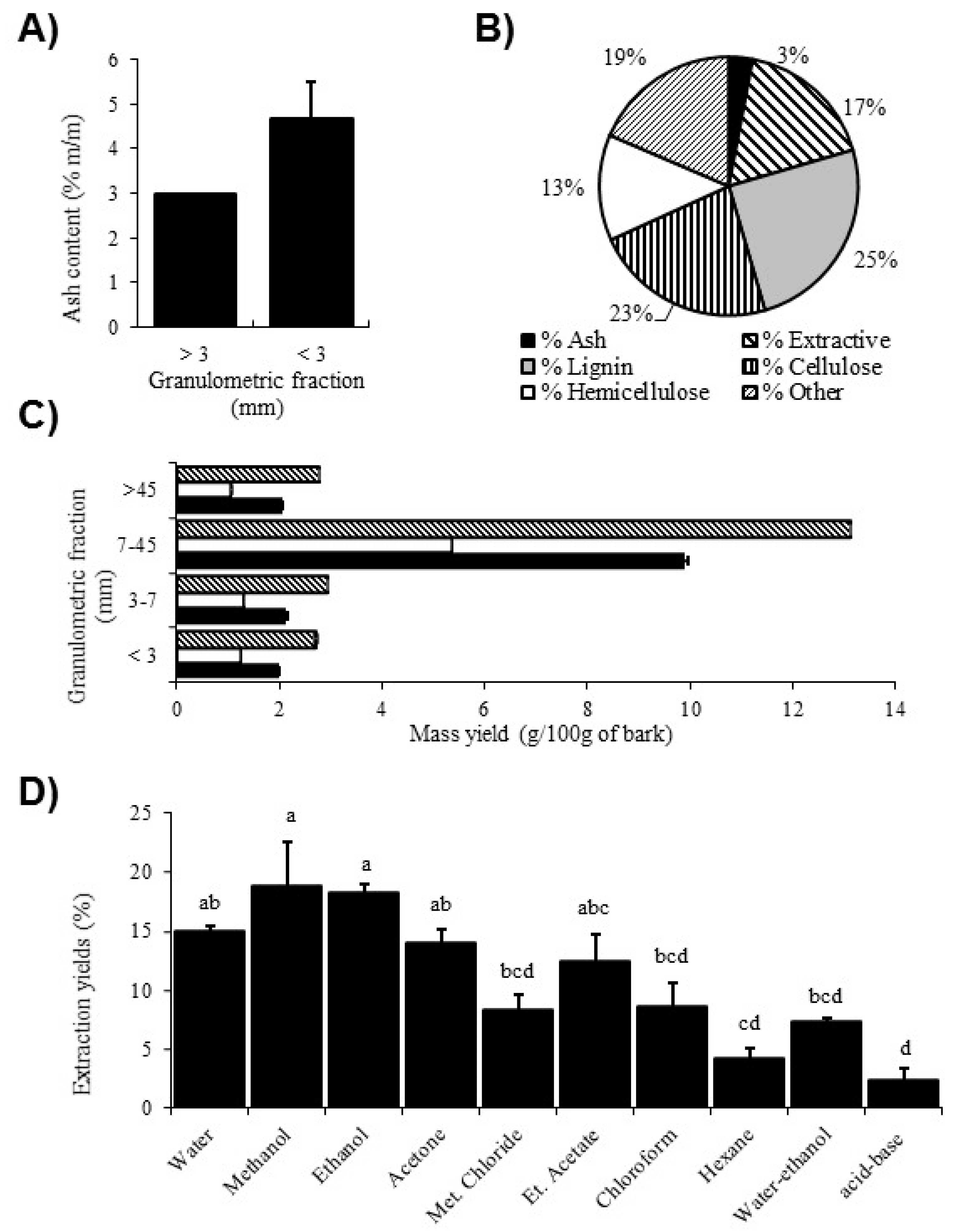
2.1. Determination of Bark Composition and Extractive Yield per Fraction Size

First, the content of quaking aspen bark residues was evaluated. The measure of ash content provides an indication of the undesirable inorganic components present in wood. These components are not extractive material and their abundance can affect the extractive yield. As shown in Figure 1A, the ash content in the fraction size < 3 mm was higher than in the fraction greater than 3 mm, with an average proportion of 4.67% m/m and 2.97%, respectively.
An evaluation of the relative composition of the ash, lignin, cellulose, hemicellulose and extractive contents of bark was also carried out according to National renewable energy laboratory (NREL) protocols on each fraction greater than 3 mm. In Figure 1B, the results show that the bark residues had a significant amount of lignin (25% m/m) and cellulose (23% m/m), a non-negligible amount of extractive material (17% m/m) and, conversely, a low amount of ash (3% m/m).
To identify which fraction size contained the largest proportion of extractives to further optimize the yield, the bark of quaking aspen was sorted into different granulometric fractions (<3, 3–7, 7–45 and >45 mm) before being extracted with three solvents (water, ethanol and water-ethanol). The extractive mass yield (g/100 g) obtained for the different granulometric fractions is summarized in Figure 1C. The results show that the 7–45 mm fraction yielded a greater proportion of extractive materials compared to other fractions, and this result was observed for the three different extraction solvents. The fraction <3 mm generally showed the lowest yield compared to the 3–7 mm and >45 mm fractions.

2.2. Determination of Extractive Yield According to Molecule Polarity

The extraction was carried out with solvents of different polarities (water, methanol, ethanol, acetone, ethyl acetate, chloroform, methyl chloride, hexane, water-ethanol and acid-base extraction), in order to extract most of the specialized metabolites. The results in Figure 1D indicate that the solvents generating the highest yields were polar solvents with methanol being the highest, followed by ethanol and water. Methanol and ethanol extractions produced a significantly higher yield than the less polar solvents (chloroform, methylene chloride and hexane) and the multistage extraction method (water-ethanol and acid-base extraction; p < 0.05). Based on the physicochemical properties of alkaloids, the acid-base extraction is specifically used to obtain extracts enriched with alkaloids. However, the acid-base extracts displayed a significantly lower yield compared to the other extraction methods.

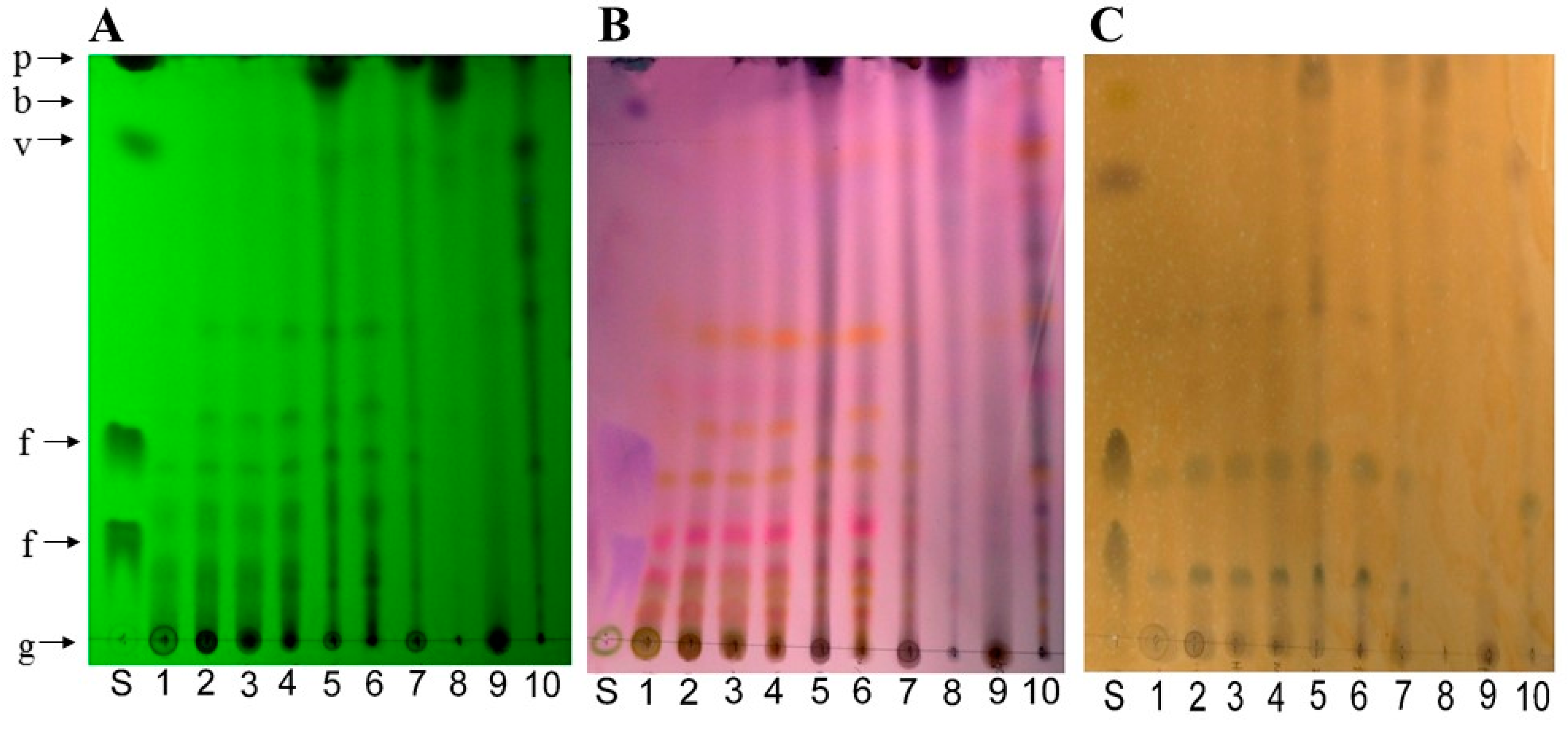
2.3. Thin Layer Chromatography (TLC) Analysis

Initially, the chemical profile of Quaking Aspen bark extracts was determined using thin layer chromatography coupled with different revelation methods. Compounds of the same family have, in the majority of cases, similar polarities, and thus may have a comparable retention factor (Rf) value. Also, compounds from the same family can be revealed in the presence of a specific revelation agent. Figure 2 shows the chemical profile of the extracts according to different revelation methods. Overall, polyconjugated compounds were revealed under UV light at 254 nm (Figure 2A). The majority of organic compounds were revealed in different colors using the p-anisaldehyde reagent, a universal stain for nucleophile and oxygenated compounds (Figure 2B); phenolic compounds were revealed in orange color using the FeCl3 reagent (Figure 2C); and the Dragendorff reagent exposed nitrogenous compounds including alkaloids (data not shown) [22].
The three TLC plates in Figure 2 show that the chemical profile was very similar for extracts (1) water, (2) methanol, (3) ethanol, (4) acetone and (6) ethyl acetate. For instance, similitudes between spots at Rf values of ~0.04, 0.10, 0.19, 0.29, 0.38 and 0.52 were apparent. In addition, these extracts had several spots at the bottom of the silica sheet at the same Rf as polyphenol and sugar standards i.e., under an Rf value of ~0.83. Conversely, the methylene chloride (5) and hexane (8) extracts displayed spots at the same level as betulin and piperine standards and a few spots at the bottom of the plate. The methylene chloride (5), chloroform (7), hexane (8) and acid-base (10) extracts had similar profiles with trails rather than well-defined spots. The water-ethanol (9) extract contained few compounds with a larger spot present at the bottom the silica sheet. Also, all extracts showed similar phenolic compounds profile except for the extracts with hexane (8) and water-ethanol (9) (Figure 2C), which contained low amounts of phenolic compounds. For instance, the apparent spots in the other extracts had the same Rf: ~0.12, 0.28 and 0.56. Finally, the use of the Dragendorff reagent did not reveal spots in extracts, but revealed the alkaloid piperine standard, confirming the effectiveness of this revelation method.

2.4. Antimicrobial Assays

The antimicrobial activity of quaking aspen bark extracts was tested in vitro using the broth microdilution method to determine the minimal inhibitory concentration (MIC) and the minimal bactericidal/fungicidal concentration (MBC/MFC). Serial dilutions of concentration ranging from 0.01 mg/mL to 4.44 mg/mL were assayed. The results presented in Table 1 show the antimicrobial activity of different extracts against eight pathogenic bacteria and fungi species. The rating of antimicrobial efficiency of extracts was realized according to a proposed classification from Algiannis et al. (2001) based on MIC value as follows: strong inhibitor with MIC < 0.5 mg/mL, moderate inhibitor with MIC between 0.6–1.5 mg/mL and weak inhibitor with MIC > 1.6 mg/mL [23]. The results obtained using the water extract revealed the highest efficiency against all microorganisms compared to other extracts (Table 1). The water extract showed a moderate efficiency against S. enterica, P. aeruginosa and A. niger with an MIC of 0.83 mg/mL and a weak efficiency against E. coli, E. faecalis and S. aureus (MIC of 1.67 mg/mL) as well as S. cerevisiae (MIC of 2.22 mg/mL). A bactericidal effect against S. aureus, P. aeruginosa and S. cerevirsiae was also observed according to the MBC/MFC values of 1.67, 4.44 and 4.44 mg/mL, respectively (Table 1). The methanol extract showed a weak antimicrobial activity against S. aureus, A. niger and S. cerevisiae strains with MIC values of 4.44, 1.67 and 2.22 mg/mL, respectively. The methanol extract also had bactericidal activity against S. cerevisiae with an MFC value of 4.44 mg/mL (Table 1). Finally, the acid-base extract showed a weak inhibition of E. coli, P. aeruginosa, A. niger and C. albicans with MIC values of 4.44, 2.22, 2.22 and 4.44 mg/mL, respectively (Table 1). However, no bactericidal properties were observed with this acid-base extract. None of the other extracts tested demonstrated considerable antimicrobial activity (Table 1).

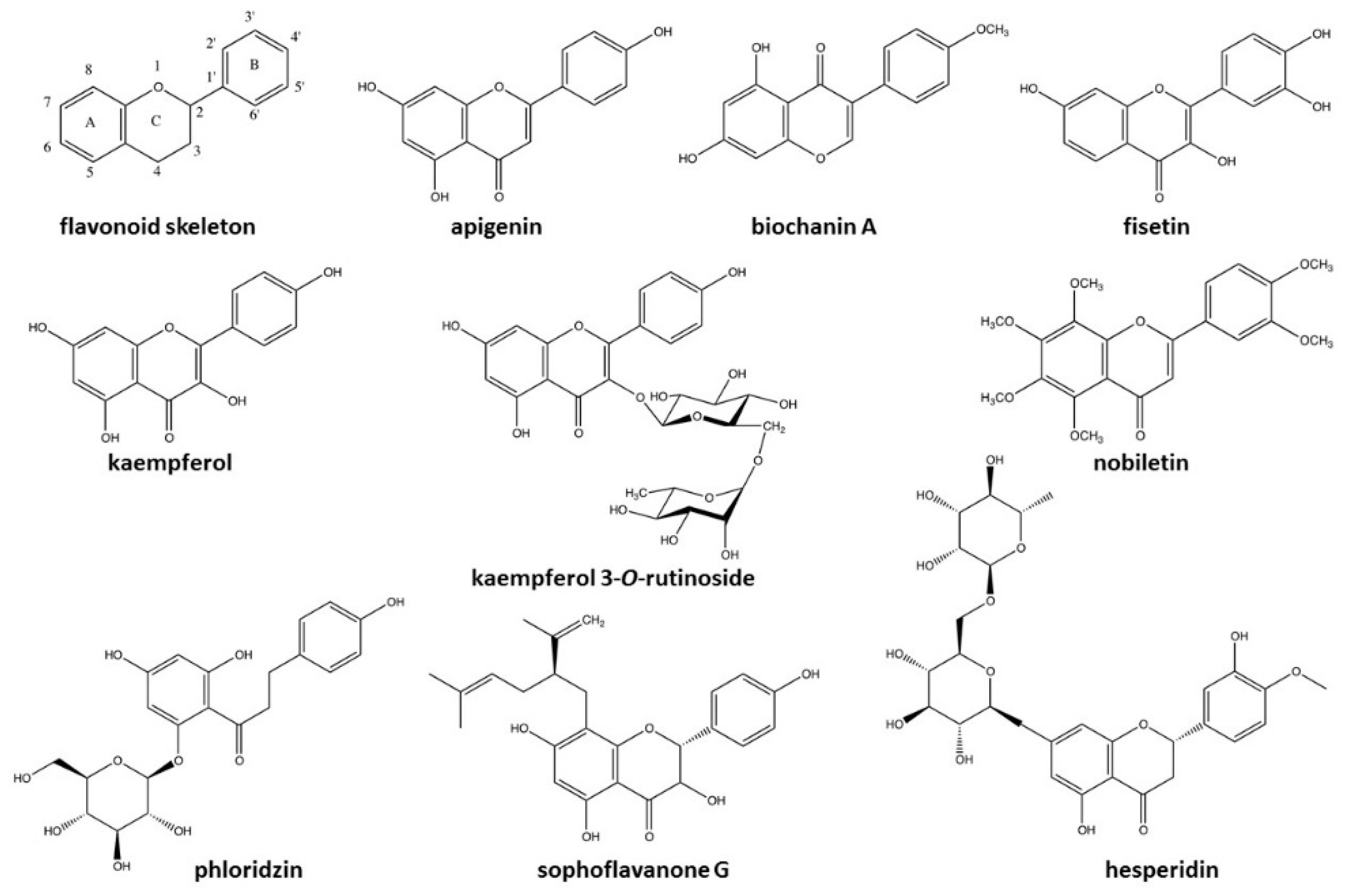
2.5. UPLC-QTOF-MS Characterization

In order to identify compounds present in most effective antimicrobial extracts (namely, those extracted with water and methanol), UPLC-QTOF-MS analyses using positive and negative ionization mode were performed. The masses obtained (showed in Table 2 and Table 3) were compared with exact masses from different libraries using the MZMine 2.0 software [24] to identify potential compounds. A summary of the identified compounds following UPLC-QTOF-MS analysis is presented in Table 2 and Table 3 for the methanol and water extracts, respectively. The exhaustive list for each of the masses obtained is shown in supplementary Tables S1–S4. The percentage area in Table 2 and Table 3 was calculated according to data presented in supplementary tables. In total, 92 different molecules were identified with good confidence, i.e., 39 and 53 molecules in methanol and water extracts, respectively. Molecules were grouped by classes, and other compounds of interest not belonging to the selected families were classified as “others”. In both extracts, polyphenols were most abundant and diversified, followed by glycosylated compounds and phenolic acids. Few terpenoids, alkaloids and sugars were also identified, but these classes were less abundant and diversified. According to the number of compounds identified in Table 2 and Table 3, water was a better solvent to extract a wide variety of compounds compared to methanol.
In each class, compounds were classified according to their percentage area. This value represent the area under the peak of the ionized mass (M ± H) at a specific retention time. It depends largely on the ionization efficiency of the molecule. Thus, at a similar concentration, an easily ionized molecule will appear more abundant then a less ionizable one. However, since similar classes of compounds possess similar chemical feature and ionization potential, we can infer relative abundance based on % area. For example, the polyphenols uvarinol and acacetin in the methanol extract were relatively more abundant compared to other polyphenols with a % area >4%. Considering both extracts, two compounds in particular appeared more abundant: oleamide and 4-prenylresveratrol in water extracts with % areas of 9.61 and 7.26, respectively (Table 3).
Following a literature search on compounds present in both samples, it was possible to target several ones known for their antimicrobial effect. These were grouped and are summarized in Table 4. According to the UPLC-QTOF-MS results (Table 2 and Table 3), all molecules in Table 4 represent 19% and 25.87% of the total % area of the methanol and water extracts, respectively. The grouping of these molecules revealed that the majority of compounds reported for their antimicrobial activity in quaking aspen extracts are polyphenols (eight among the twenty-two identified). In general, the majority of compounds of interest are specific to one extraction solvent, water or methanol, but some were present in both, including catechol, caffeic acid and 4-hydroxybenzaldehyde. Among compounds of interest in the methanol extract, 4-hydroxybenzaldehyde (5.44%), phloridzin (4.76%), and hydroxyanthraquinone (4.05%) were more abundant compared to other compounds based on the % area values (Table 2). For the water extract, kaempferol (5.03%) and its glycosylated form, kaempferol 3-O-rutinoside (3.15%), were the most abundant compounds (% area values, Table 3). There was a higher number of different compounds recognized for their antimicrobial property in the water extract than in the methanol extract. Also, nine among the twenty-three molecules targeted were flavonoids (Figure 3), a subgroup of phenolic compounds. In fact, the molecules illustrated in Figure 3, have a similar core structure: apigenin, biochanin A, fisetin, kaempferol, kaempferol 3-O-rutinoside, nobiletin, phloridzin, sophoraflavanone G, and hesperidin. The basic structure of flavonoids corresponds to fifteen carbon atom skeleton forming two benzene rings and one bridge formed by three carbons that connect the rings together. This bridge generally forms a pyrone ring as illustrated in Figure 3 (flavonoid skeleton).

3. Discussion

3.1. Composition and Extraction Yield Evaluation

The aim of this study was to recover, test and identify specialized metabolites from quaking aspen bark in order to obtain antimicrobial extracts. As the final objective is to use raw bark residues from sawmills to produce natural antimicrobials at an industrial scale, it is relevant to characterize this material to further reach an optimal extraction yield. The results obtained from the extraction of each particle size and corresponding ash content revealed that sieving was relevant for eliminating the fractions with a low mass of extractives (Figure 1A,C). In fact, several studies reported that finest particles often contain soil contaminants, such as sand and fine sediments, that yield no extractive organic compounds. This explain the highest ash content observed from the fraction < 3 mm, which represents the inorganic compound proportion in the bark (Figure 1A). Inorganic material contains minerals such as potassium, calcium and sodium [73,74] and these compounds can be harmful to machinery during extraction processes in addition to being undesirable because they do not provide extractive material [75,76]. Thus, better yields of extractives were obtained with bark of particle sizes greater than 3 mm.
Chemical composition tests carried out according to the NREL protocols allowed us to evaluate the composition of the bark material besides the extractives. Results of the average chemical composition of several tree species of the Quebec territory reported by Browne et al. (2009) showed that the bark contained between 16% to 40% cellulose, 38% to 58% hemicellulose, 40% to 50% lignin and 2% to 25% extractive compounds. Results obtained for bark of quaking aspen in the present study were below the average proportions of hemicellulose and lignin, i.e., 13% and 25%, respectively (Figure 1B). Conversely, the residues were composed of 17% extractive materials and this proportion was relatively high compared to the average of tree species in Quebec [77]. In addition, the bark residues had a proportion of cellulose (23%) within the average content of common trees (16–40%). These results demonstrated the relevance to exploit extractive materials from quaking aspen for biorefinery because their abundance is high relative to others tree species from Quebec and also relative to the lignocellulosic compounds of this species.
The extraction with solvents of different dielectric constants, which influence their polarity, allowed extraction of the majority of extractive molecules present in the quaking aspen sawmill residues. Since the extraction yield was significantly higher with polar solvents (Figure 1D), it suggests that abundant specialized metabolites were highly polar compounds, including phenolic compounds, sugars and other glycosylated compounds.
This hypothesis correlated with the chemical profile obtained following the TLC analysis. The three revelation methods (UV, FeCl3 and p-anisaldehyde) allowed us to determine that a large number of compounds were highly polar by the presence of many spots with a low Rf (Figure 2). The FeCl3 revelation demonstrated that quaking aspen extracts contain many phenolic compounds (Figure 2C). Finally, revelation with the p-anisaldehyde reagent suggests that extracts have compounds similar to sugars based on the presence of spots with the same color and migration as a glucose standard, i.e., green and with an Rf value around 0 (Figure 2B). Furthermore, according to Prado and Meireles (2010), green spots with low Rf values could be associated with flavonoid compounds.

3.2. Antimicrobial Activity

MIC and MBC/MFC results showed that three extracts (water, methanol and acid-base) had antimicrobial properties (Table 1). The water extract followed by the methanol extract yielded a higher efficacy against pathogens. More specifically, the water extract displayed a weak to moderate inhibitory effect on all eight pathogens tested. The larger broad-spectrum antimicrobial activity of this extract could be explained by the diversity of metabolites it contains, as demonstrated by the UPLC-QTOF-MS analysis. Broad-spectrum antimicrobial activity of different plant extracts has been also observed in recent studies [78,79,80]. The results are also consistent with those obtained by [81], who tested the bacteriostatic and bactericidal effects of quaking aspen bud extracts obtain with a methanol extraction against many different pathogens. Quaking aspen extracts had no bacteriostatic effect against Gram-negative bacteria in both studies (E. coli, P. aeruginosa and S. enterica), although Vardar-Ünlü et al. (2008) observed an antimicrobial effect against E. faecalis and C. albicans while no antimicrobial effect was observed on these pathogens in our study. These differences can be explained by the use of different tree parts that can contain different metabolites at different concentrations. The difference in efficacy against C. albicans can also be explained by the difference of the yeast strain used.
Other efforts need to be deployed to maximize efficiency of quaking aspen extracts. In fact, the antimicrobial effect of these extracts was, on average, 800 to 4000 time lower than reference antimicrobial compounds used as active ingredient in drugs and disinfectants. In fact, the MIC range obtained for QAC (reference compound) in this study was 0.651–10.4 μg/mL. Also, recent and older studies reported that the MIC values associated with amphotericin B and cefotaxime, widespread antimicrobials used to treat fungal and bacterial infections, respectively, were between 0.1 and 16 μg/mL against several reference strains [82,83,84,85]. In regards to isolated compounds from plant species that demonstrated strong antimicrobial activity, the gap of MIC values with quaking aspen bark extracts is smaller. For example, MIC values for resveratrol and berberine were between 50 μg/mL and 500 μg/mL [86,87]. Finally, results obtained in this study were in concordance with those obtained for plant extracts in general. For example, Assob et al. (2011) and Khan et al. (2014) evaluated the antimicrobial activity of several plant extracts and the average MIC range was 300–20,000 μg/mL, corresponding to weak or moderate efficiency [79,88]. Isolated compound are generally more effective than extracts. However, an extract with a mix of several compounds can counteract resistance mechanisms through the various mechanisms of action accomplished by the multiple molecules against microorganism targets [89,90,91]. Thus, according to the desired applications, extracts could be relevant in the development of antimicrobial agents.

3.3. Chemical Characterization

The UPLC-QTOF-MS analyses enabled the potential identification of the compounds present in water and methanol extracts. These results (Table 2 and Table 3) coupled with TLC revealed the FeCl3 reagent (Figure 2C), which showed that the water and methanol extraction allowed the recovery of an important variety of phenolic compounds. Interestingly, several studies reviewed the importance of polyphenols and phenolic acids in extracts responsible for antimicrobial activity [13,48,92,93]. These results substantiated the antimicrobial activity obtained in our study with the families of compounds present in water and methanol extracts.
It was possible to observe through the UPLC-QTOF-MS analyses (Table 2 and Table 3) a wide variety of glycosylated compounds. In this case, these glycosylated compounds were, for the vast majority, phenolic compounds bound to a carbohydrate. Depending on the carbohydrate to which they were bound, some of these compounds had different antimicrobial efficiencies. For example, kaempferol, present in methanol extract (Table 2), is recognized for its antimicrobial activity, but kaempferol-hexoside did not seem to have bioactivity against four pathogens (S. aureus, Paeruginosa, E. coli and C. albicans) according to a study by Fattouch et al. (2007) [34]. On the contrary, kaempferol-3-O-rutinoside is a glycosylated compound which appeared to have antimicrobial efficacy against the same four pathogens in previous studies by Panizzi et al. (2002) and Nenaah (2013) [58,60]. These molecules are interesting because their carbohydrate structure serves as a recognition site for cellular specific targets and generally increases their bioactivity [94,95]. Also, these compounds may be of interest in vivo in the presence of enteric enzymes, which have the ability to hydrolyze these structures and remove the glycosylic bond [96,97].
Some terpenoids and alkaloids were also identified in water and methanol extracts, but these compounds are not as diverse as the compound classes mentioned before. However, these are interesting because some compounds of these families are known to have antimicrobial activity, even at low concentration [16,17,98]. In fact, only four alkaloid compounds were identified in both extracts, and these were not very abundant according to the % area observed (Table 2 and Table 3). Firstly, these results corroborate the poor extraction yield obtained with the acid-base method used to concentrate alkaloids (Figure 1D). These results also corroborate those obtained for TLC analysis that showed a low abundance of alkaloid molecules. The characterization of sugars is relevant since they are undesirable compounds for the purpose of this study. Indeed, the carbohydrates can be used as a substrate by the microorganisms and serve as a source of energy for their proliferation. Knowing their retention time (~1.00 min), it would be possible to remove them from the extracts by fractionation and purification methods and induce carbohydrate starvation with respect to the microorganisms [99].
UPLC-QTOF-MS analyses allowed for the identification of molecules associated with antimicrobial activity according to previous studies (see Ref. in Table 4). It has been suggested that these molecules are responsible of the bacteriostatic and bactericidal effects of quaking aspen extracts. In accordance with the UPLC-QTOF-MS results, molecules shown in Table 4 were particularly abundant in the extract. In fact, these represent 19.00% and 25.87% of the total % area for methanol and water extracts, respectively. The higher efficiency of the water extract can be explain by the higher abundance and diversity of antimicrobial compounds in this extract. Based on these results, it was expected we would find an inhibitory effect against pathogens for both extracts given the abundance of potentially antimicrobial compounds they contain. Antimicrobial properties can be related to specific molecules in Table 4, i.e., 4-hydroxybenzaldehyde in methanol extract and kaempferol in water extract, because they seem to be highly abundant in extracts according to % area results in Table 2 and Table 3.
The grouping of compounds recognized for their antimicrobial effectiveness allowed us to establish a potential relationship between their chemical structure and their activity. These revealed that several molecules among all those present are in the flavonoid class (Figure 3). Thereby, there appears to be a close relationship between antimicrobial activity and the flavonoid structure. This relationship corroborates previous findings by Cushnie and Lamb (2005), Rauha et al. (2000) as well as Sohn et al. (2004), and supports the idea of an important antimicrobial efficiency of these molecules [98,100,101].
It remains difficult to attribute the antimicrobial activity to a specific compound in such a complex compound mixture. Synergistic and antagonistic effects may be present which make analyses sophisticated [89,102]. Also, the study of antimicrobial metabolites from natural plant sources remains laborious due to the variability of the compound concentration between extracts from different raw materials. In fact, metabolite profiles can change according to the quaking aspen species collected, harvest location and/or harvest period, etc. [103,104]. Finally, some compounds remained unidentified due to the detection limit of the UPLC-QTOF-MS apparatus, the library used and the limit of available information. Consequently, it is possible that some compounds with high activity may have been discarded.

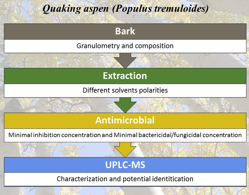
4. Materials and Methods

4.1. Plant Material

The plant material used for this experiment was quaking aspen (Populus tremuloides) bark, obtained from sawmill residues of T.L.T Industry in Ste-Monique, Lac-St-Jean, Quebec, Canada, in winter 2016 and winter 2017. The material was dried after reception at room temperature (~20 °C) to preserve it until extraction.

4.2. Microbial Strains

The bacterial and fungal strains were obtained from the American Type Culture Collection (ATCC) and from the microbiology laboratory of the University of Québec in Trois-Rivières (UQTR). The microorganisms used were Escherichia coli (ATCC 35218), Salmonella enterica (ATCC 10708), Pseudomonas aeruginosa (ATCC 15442), Staphylococcus aureus (ATCC 6538), Enterococcus faecalis (ATCC 29212), Aspergillus niger (ATCC 10535), Candida albicans (from UQTR microbiology laboratory), Saccharomyces cerevisiae (from UQTR microbiology laboratory). The bacterial strains were grown on sterilized Mueller Hinton agar and incubated at 37 °C for 24 h, while the fungal strains were grown on Sabouraud dextrose agar and incubated at 37 °C for 72 h before use.

4.3. Determination of Bark Composition and Extractive Yield per Fraction Size

To determine the general composition and extractive yield of quaking aspen bark residues per fraction size, sieving was first performed. The following sieve mesh sizes were used: 3 mm, 7 mm, 45 mm, to obtain four different fractions: (a) <3 mm, (b) 3–7 mm, (c) 7–45 mm, (d) >45 mm. The bark was then ground in a Wiley Mill crusher at 0.425 mm for further extraction and analysis.
For the general composition, procedures from the National renewable energy laboratory (NREL) were carried out. Specifically, the NREL/TP-510-42620 and NREL/TP-510-42622 protocols were followed on fraction sizes <3 mm and >3 mm to dry and weigh the total biomass, and to calculate ash abundance, respectively. A water and ethanol (85.75% EtOH, 13.7% MeOH, 0.85% ethyl acetate; (Fisher scientific, Ottawa, ON, Canada) extraction using an accelerated solvent extraction apparatus (ASE) (Dionex ASE-350, ThermoFisher scientific, Ottawa, ON, Canada) was carried out for the determination of extractible abundance on fractions >3 mm following protocol NREL/TP-510-42619. Finally, determination of the structural compound abundance was realized on fractions > 3 mm following the NREL/TP-510-42618 protocol with some modifications. Cellulose and hemicellulose were fractioned by acid hydrolysis, and quantification of monomeric sugars was realized by ionic chromatography (Dionex ICS-5000, ThermoFisher scientific, Ottawa, ON, Canada) with a Dionex CarboPac SA-10 column, while lignin was quantified by UV-VIS spectrometry (DR6000, Hach, London, ON, Canada) [105].
To determine which particle size fraction of raw bark residues contained the highest amount of extractives, three aliquots of 100 g of each fraction (3–7 mm, 7–45 mm, >45 mm) was used to perform extraction using an ASE apparatus with three different solvents: (1) distilled water, (2) water-ethanol and (3) ethanol. The yield was determined by measuring the extractive material mass of each fraction.

4.4. Extractive Yield According to Molecular Polarity

Bark residues with a fraction size greater than 3 mm were used to prepare ten aliquots of 10 g of dried bark powder (0.425 mm). Each was extracted with different solvents having distinct polarities: (1) distilled water, (2) methanol (99.9%; HPLC-grade; Fisher scientific), (3) denaturised ethanol, (4) acetone (99.8%; Fisher scientific; certified ACS), (5) methylene chloride (>99%; Acros organics), (6) ethyl acetate (99.6%; Acros organics; certified ACS), (7) chloroform (99.9%. Acros organics), (8) hexane (99.9%; Fisher chemical; HPLC-grade), (9) water-ethanol, and (10) by specific solvents for acid-base extraction method. For samples 3, 4, 5, 6, 7 and 8, the extraction was done with a Soxhlet system for 7 h over six cycles of extraction. For samples 1, 2 and 9, the extraction was performed using ASE. The water extraction was performed for 70 min (6 cycles of 10 min), at 100 °C and at a pressure of 1500 psi, and the ethanol extraction was performed for 45 min (6 cycles of 5 min) at 120 °C and 1500 psi. For the water-ethanol extraction, the matrix was first extracted with water and then extracted with denaturised ethanol with the same parameters as mentioned before according to each extraction solvent. Finally, the acid-base extraction method (liquid-liquid extraction) was performed as described by Yubin (et al. (2014)) [106].
The extraction yield was determined by measuring the dried extractive material mass for each solvent. After that, each extract was suspended in two aliquots, one in DMSO for antimicrobial analysis (99.8%; Fisher scientific; certified ACS) and the other in the respective solvent of extraction for chemistry analysis, both at a concentration of 10 mg/mL. These stock solutions were stored in the fridge at 4 °C until further analysis. All extractions were performed in triplicate.

4.5. Thin Layer Chromatography Method (TLC)

For a rapid visualization of the extract’s chemical profile, TLC analysis was carried out. For this purpose, a TLC plate (silica gel 60 F254 in aluminium, Sigma, Saint-Louis, MO, USA) with a dimension of 14 × 20 cm was used. A solution with standards belonging to different families of compounds such as piperine, betulin, vaniline, ferrulic acid and glucose, all at 1000 ppm, was prepared. Development was realized in a presaturated solvent chamber with CHCl3-MeOH-EtOH (90:9:1). For visualisation purpose, the TLC plate was first observed under UV light at 254 nm and was also sprayed with p-anisaldehyde reagent, following by heating on a hot plate at 105 °C for 5 min. This solution was prepared by following the mixture proposed in Prado and Meireles (2010) [107]. A similar procedure of TLC plate development was further performed but was sprayed with different reagents: FeCl3 (for phenolic compounds revelation) using a modified method based on a protocol from SiliCycle Inc. (2017) by mixing 1.6 g of FeCl3, 50 mL of water and 50 mL of methanol, and secondly with Dragendorff reagent (for alkaloid revelation) following the mixture proposed by Jia and Tian (2009) [22,108].

4.6. Antimicrobial Activity Test by Broth Microdilution Method

To determine the minimum inhibitory concentration (MIC) of extracts, a broth microdilution method described in documents M07-A7 and M38-A2, adapted from the Clinical and Laboratory standard institute (CLSI), were used [109,110].
First, microbial colonies were collected from an agar plate and suspended in a sterilized solution of physiological water (9 g of NaCl in 1 L of distilled water) to obtain a final cell concentration of 0.5 Mc Farland, corresponding to 1.5 × 10⁸ CFU/mL for bacteria, 1.5 × 10⁶ CFU/mL for yeasts and 1.5 × 10⁴ CFU/mL for molds. In a 96-well plate, 100 µL of tested extracts in DMSO solution (10 mg/mL) was deposited in wells of the first column. Then, 50 µL of Mueller Hinton broth was added to each well, and a serial dilution was realized by transferring 50 µL of the first well into the second of the same row and so on to obtain a serial dilution of the extracts ranging between 4.44 to 0.01 mg/mL. Subsequently, 50 µL of the microbial inoculum (0.5 Mc Farland, [110]) was then added to the wells. In each microplate test, a positive control of quaternary ammonium compound (QAC) BTC 6358 solution (1.19 g/L), obtained from the sanitation products company Sani Marc (Victoriaville, QC, Canada), was used. The plate was incubated at 37 °C for 3 h for the bacterial strains and 6 h for fungal strains. After incubation, a solution of INT (2-p-iodophenyl-3-p-nitrophenyl-5-phenyl tetrazolium chloride (Sigma, Saint-Louis, MO, USA) at 2.85 mg/mL was added. This tetrazolium salt, reduced to a formazan red dye by the coenzyme NADH of living cells, served as a cell viability indicator. The plate was incubated a second time to reduce the tetrazolium salt (1 h for the bacterial strains/16 h for the fungal strains). The MIC (minimum inhibitory concentration) was determined in the first well of the serial dilution where no color appeared, which indicates that there is inhibition of microbial proliferation. The non-toxic effect of DMSO at 33% (maximal concentration used during the microdilution test) was evaluated, and the absence of an inhibition effect confirmed the non-toxic effect of the matrix used to dilute the extracts.
To optimise the protocol, screening was performed initially with each bark extract to rapidly determine which ones provided the inhibition of microbial proliferation at maximum concentration (4.44 mg/mL), and only effective extracts were kept for the microdilution analysis. For determination of the minimum bactericidal/fungicidal concentration (MBC/MFC), 100 µL of each well containing extracts with a positive effect on proliferation during the microdilution broth test was plated on the agar surface with the respective growth medium (Mueller Hinton for bacteria and Sabouraud dextrose for fungi). The Petri dishes were incubated at 37 °C for 24 h for the bacterial strains and 72 h for the fungal strains. The MBC and MFC were determined by the lowest concentration that allowed no visible colony on the agar Petri dish, i.e., more than a 99.9% reduction in colonies.

4.7. UPLC-QTOF-MS Method

UPLC-QTOF-MS analyses were carried out externally by the industrial research center of Québec (CRIQ). Briefly, analysis was performed with a Waters Acquity ultra-performance LC system (Waters, Milford, MA, USA). This apparatus is equipped with a binary pump system model (Waters). The separation was achieved using an Ethylene Bridged Hybrid (BEH) C18 column (100 mm × 2.1 mm id. 1.7 mm particle size), also from Waters. The UPLC system was coupled with a Waters QTOF micro mass spectrometer (Waters, Milford, MA, USA) equipped with a z-spray electrospray interface for the MS analysis. The mobile phase used was two eluents: A = 0.2% acetic acid, B = acetonitrile (99.9% HPLC grade). Elution parameters were as follows: solvent flow rate 0.2 mL/min; injection volume 10 µL; proportions of eluent B: 2% (0–1 min); 2–100% (1–30 min); isocratic 100% (30–33 min); 100–2% (33–33.5 min), isocratic 2% (33–40 min). The MS analysis was performed in both positive and negative modes, and the data were acquired from 100 to 1250 m/z without collision. The conditions of the ionization source were established as follows: source temperature 120 °C; cone gas flow rate 50 L/h; desolvation gas flow rate 350 L/h; desolvation gas temperature 200 °C; cone voltage 30 V in negative mode and 70 V in positive mode; capillary voltage 1150 V in negative mode and 1800 V in positive mode. Ultra high purity nitrogen (99%) was used as a nebulizing gas. Data acquisition was carried out with the Masslynx 4.1 software (Waters, Milford, MA, USA). Mass extraction, deconvolution, isotopes and the library search were performed using MZMine 2.0 according to Pluskal et al. (2010) [24]. The molecule identification was determined by the concordance of the m/z values with the theoretical values using different libraries databases: Chemspider, Kegg Compounds Database, LipidMaps Database and the CRIQ database.

4.8. Statistical Analysis

One-way analysis of variance (ANOVA) was performed on data with a 5% level of probability (p < 0.05) using JMP software [24] followed by pairwise mean comparison Tukey’s test was conducted where differences were detected.

5. Conclusions

The aim of this study was to determine the antimicrobial potential of quaking aspen bark residues through the extraction of bioactive metabolites. We found that specialized metabolite extractions are relevant to isolate antimicrobial molecules from bark residues. The extraction yield results as well as MIC and MBC/MFC results identified two promising solvents: water and methanol. The physicochemical analysis of these two extracts identified hundreds of metabolites of interest, among which some had an antimicrobial effect in accordance with previous studies. Analysis of the chemical structure of these compounds revealed possible correlations between flavonoid structure and antimicrobial effect.
Overall, our study supports the relevance of being interested in quaking aspen bark residues to valorize its biomass through these bioactive metabolites. Until now, this species was undervalued because no previous exhaustive study linked the chemical composition of its bark to its antimicrobial activity. However, this study allowed us to confirm the antimicrobial efficacy by the presence of antimicrobial compounds in quaking aspen. This species is also interesting because optimal extraction conditions are consistent with antimicrobial efficacy results. Water, which is the most efficient solvent, is the solvent suitable for extraction since it is non-polluting, inexpensive and the most accessible.

Supplementary Materials

The following are available online at https://www.mdpi.com/1420-3049/23/7/1739/s1, Supplementary tables: Tables S1–S4.

Author Contributions

Conceptualization, J.B.; N.B. and I.D.-P.; Methodology, A.S.-P.; D.B.; J.B.; A.L.; N.B. and I.D.-P.; Validation, A.S.-P.; D.B.; N.B. and I.D.-P.; Formal Analysis, A.S.-P.; D.B.; N.B. and I.D.-P. Investigation, A.S.-P.; D.B.; N.B. and I.D.-P.; Writing-Original Draft Preparation, A.S.-P.; Writing-Review & Editing, A.L.; N.B. and I.D.-P.; Supervision, I.D.-P.; Project Administration, N.B. and I.D.-P.; Funding Acquisition, N.B. and I.D.-P.

Funding

This research was funded by the Consortium de Recherche et innovations en bioprocécés industriels au Québec with Forêt Modèle du Lac-St-Jean and Sani-Marc, grant reference no 2015-033-C17. Also, Mitacs scholarships supported ASP and DB.

Acknowledgments

The authors wish to thank the industrial collaborators and financial institutions for their financial support of this project, including Foret Modele du Lac-St-Jean (FMLSJ), Sani Marc Group, the Coopérative pour la valorisation de la biomasse (CVB), the Consortium de recherche et d’innovation en bioprocédés industriels au Québec (CRIBIQ) and the Conseil de recherches en sciences naturelles et en genie du Canada (CRSNG). Professor Simon Barnabé and Patrick Marchand are acknowledged for providing chemicals and microbiology equipment. The authors wish to thank Stéphanie B. Blais and Josée Doucet for their technical support. Dominic Desrosiers is acknowledged for his scientific support. Finally, the authors wish to thank Pascal Dubé at the Centre de Recherche Industrielle du Québec for the UPLC-QTOF-MS analysis.

Conflicts of Interest

The authors declare no conflict of interest.

References

  1. Cohen, M.L. Epidemiology of drug resistance: Implications for a post-antimicrobial era. Science (Wash.) 1992, 257, 1050–1055. [Google Scholar] [CrossRef]
  2. Gold, H.S.; Moellering, R.C., Jr. Antimicrobial-drug resistance. N. Engl. J. Med. 1996, 335, 1445–1453. [Google Scholar] [CrossRef] [PubMed]
  3. Gurib-Fakim, A. Medicinal plants: Traditions of yesterday and drugs of tomorrow. Mol. Asp. Med. 2006, 27, 1–93. [Google Scholar] [CrossRef] [PubMed]
  4. Rates, S.M.K. Plants as source of drugs. Toxicon 2001, 39, 603–613. [Google Scholar] [CrossRef]
  5. Royer, M.; Houde, R.; Viano, Y.; Stevanovic, T. Non-wood Forest Products Based on Extractives-A New Opportunity for the Canadian Forest Industry Part 1: Hardwood Forest Species. J. Food Res. 2012, 1, 8. [Google Scholar] [CrossRef]
  6. Bouchard, M. Identification D’utilisations pour le Bouleau à Papier et Le peuplier Faux-Tremble; Enseignement Supérieur, Recherche, Science et Technologie: Quebec, QC, Canada, 2013; pp. 1–45. [Google Scholar]
  7. Diouf, P.N.; Stevanovic, T.; Cloutier, A. Antioxidant properties and polyphenol contents of trembling aspen bark extracts. Wood Sci. Technol. 2009, 43, 457–470. [Google Scholar] [CrossRef]
  8. Fernandez, M.P.; Watson, P.A.; Breuil, C. Gas chromatography–mass spectrometry method for the simultaneous determination of wood extractive compounds in quaking aspen. J. Chromatogr. A 2001, 922, 225–233. [Google Scholar] [CrossRef]
  9. Pal, D.; Singh, H.; Kumar, M. A preliminary study on the in vitro antioxidant activity of seeds of Aesculus indica and bark of Populus euphratica. Int. J. Pharm. Pharm. Sci. 2012, 4, 249–250. [Google Scholar]
  10. Cushnie, T.P.T.; Cushnie, B.; Lamb, A.J. Alkaloids: An overview of their antibacterial, antibiotic-enhancing and antivirulence activities. Int. J. Antimicrob. Agents 2014, 44, 377–386. [Google Scholar] [CrossRef] [PubMed]
  11. Lu, J.-J.; Bao, J.-L.; Chen, X.-P.; Huang, M.; Wang, Y.-T. Alkaloids Isolated from Natural Herbs as the Anticancer Agents. Evid.-Based Complement. Altern. Med. 2012, 2012, 12. [Google Scholar] [CrossRef] [PubMed]
  12. Pandey, K.B.; Rizvi, S.I. Plant polyphenols as dietary antioxidants in human health and disease. Oxid. Med. Cell. Longev. 2009, 2, 270–278. [Google Scholar] [CrossRef] [PubMed]
  13. Quideau, S.; Deffieux, D.; Douat-Casassus, C.; Pouységu, L. Plant Polyphenols: Chemical Properties, Biological Activities, and Synthesis. Angew. Chem. Int. Ed. 2011, 50, 586–621. [Google Scholar] [CrossRef] [PubMed]
  14. Lattanzio, V. Phenolic Compounds: Introduction; Springer: Berlin/Heidelberg, Germany, 2013; pp. 1543–1580. [Google Scholar]
  15. Wink, M. Introduction: Biochemistry, physiology and ecological functions of secondary metabolites. Ann. Plant Rev. Biochem. Plant Second. Metabol. Second Ed. 2010, 40, 1–19. [Google Scholar]
  16. Inouye, S.; Takizawa, T.; Yamaguchi, H. Antibacterial activity of essential oils and their major constituents against respiratory tract pathogens by gaseous contact. J. Antimicrob. Chemother. 2001, 47, 565–573. [Google Scholar] [CrossRef] [PubMed] [Green Version]
  17. Trombetta, D.; Castelli, F.; Sarpietro, M.G.; Venuti, V.; Cristani, M.; Daniele, C.; Saija, A.; Mazzanti, G.; Bisignano, G. Mechanisms of Antibacterial Action of Three Monoterpenes. Antimicrob. Agents Chemother. 2005, 49, 2474–2478. [Google Scholar] [CrossRef] [PubMed] [Green Version]
  18. Tholl, D. Biosynthesis and Biological Functions of Terpenoids in Plants. In Biotechnology of Isoprenoids; Schrader, J., Bohlmann, J., Eds.; Springer International Publishing: Cham, Switzerland, 2015; pp. 63–106. [Google Scholar]
  19. Celhay, C.; Mathieu, C.E.; Candy, L.; Vilarem, G.; Rigal, L. Aqueous extraction of polyphenols and antiradicals from wood by-products by a twin-screw extractor: Feasibility study. Comptes R. Chim. 2014, 17, 204–211. [Google Scholar] [CrossRef]
  20. Demirbas, A.; Demirbas, M.F. Biorefineries. In Algae Energy; Springer: Berlin/Heidelberg, Germany, 2010; pp. 159–181. [Google Scholar]
  21. Janowiak, M.K.; Webster, C.R. Promoting ecological sustainability in woody biomass harvesting. J. For. 2010, 108, 16–23. [Google Scholar]
  22. SiliCycle Inc. SiliaPlate™-TLC Visualization Methods. Available online: http://www.silicycle.com/ca/products/thin-layer-chromatography-tlc-plates/siliaplate-tlc-visualization-methods (accessed on 13 July 2018).
  23. Aligiannis, N.; Kalpoutzakis, E.; Mitaku, S.; Chinou, I.B. Composition and antimicrobial activity of the essential oils of two Origanum species. J. Agric. Food Chem. 2001, 49, 4168–4170. [Google Scholar] [CrossRef] [PubMed]
  24. Pluskal, T.; Castillo, S.; Villar-Briones, A.; Oresic, M. MZmine 2: Modular framework for processing, visualizing, and analyzing mass spectrometry-based molecular profile data. BMC Bioinform. 2010, 11, 395. [Google Scholar] [CrossRef] [PubMed]
  25. Abuelsaad, A.S.; Mohamed, I.; Allam, G.; Al-Solumani, A.A. Antimicrobial and immunomodulating activities of hesperidin and ellagic acid against diarrheic Aeromonas hydrophila in a murine model. Life Sci. 2013, 93, 714–722. [Google Scholar] [CrossRef] [PubMed]
  26. Yi, Z.; Yu, Y.; Liang, Y.; Zeng, B. In vitro antioxidant and antimicrobial activities of the extract of Pericarpium Citri Reticulatae of a new Citrus cultivar and its main flavonoids. LWT-Food Sci. Technol. 2008, 41, 597–603. [Google Scholar] [CrossRef]
  27. Kocaçalışkan, I.; Talan, I.; Terzi, I. Antimicrobial activity of catechol and pyrogallol as allelochemicals. Z. für Naturforschung C 2006, 61, 639–642. [Google Scholar] [CrossRef]
  28. Jeong, E.-Y.; Jeon, J.-H.; Lee, C.-H.; Lee, H.-S. Antimicrobial activity of catechol isolated from Diospyros kaki Thunb. roots and its derivatives toward intestinal bacteria. Food Chem. 2009, 115, 1006–1010. [Google Scholar] [CrossRef]
  29. Hwang, J.H.; Choi, H.; Hwang, I.-S.; Kim, A.R.; Woo, E.-R.; Lee, D.G. Synergistic Antibacterial and Antibiofilm Effect Between (+)-Medioresinol and Antibiotics In Vitro. Appl. Biochem. Biotechnol. 2013, 170, 1934–1941. [Google Scholar] [CrossRef] [PubMed]
  30. Hwang, J.H.; Hwang, I.-S.; Liu, Q.-H.; Woo, E.-R.; Lee, D.G. (+)-Medioresinol leads to intracellular ROS accumulation and mitochondria-mediated apoptotic cell death in Candida albicans. Biochimie 2012, 94, 1784–1793. [Google Scholar] [CrossRef] [PubMed]
  31. Cha, J.-D.; Jeong, M.-R.; Jeong, S.-I.; Lee, K.-Y. Antibacterial activity of sophoraflavanone G isolated from the roots of Sophora flavescens. J. Microbiol. Biotechnol. 2007, 17, 858–864. [Google Scholar] [PubMed]
  32. Tsuchiya, H.; Iinuma, M. Reduction of membrane fluidity by antibacterial sophoraflavanone G isolated from Sophora exigua. Phytomedicine 2000, 7, 161–165. [Google Scholar] [CrossRef]
  33. Chi, Y.S.; Jong, H.G.; Son, K.H.; Chang, H.W.; Kang, S.S.; Kim, H.P. Effects of naturally occurring prenylated flavonoids on enzymes metabolizing arachidonic acid: Cyclooxygenases and lipoxygenases. Biochem. Pharmacol. 2001, 62, 1185–1191. [Google Scholar] [CrossRef]
  34. Fattouch, S.; Caboni, P.; Coroneo, V.; Tuberoso, C.I.; Angioni, A.; Dessi, S.; Marzouki, N.; Cabras, P. Antimicrobial activity of Tunisian quince (Cydonia oblonga Miller) pulp and peel polyphenolic extracts. J. Agric. Food Chem. 2007, 55, 963–969. [Google Scholar] [CrossRef] [PubMed]
  35. Kuete, V.; Nguemeving, J.R.; Beng, V.P.; Azebaze, A.G.B.; Etoa, F.-X.; Meyer, M.; Bodo, B.; Nkengfack, A.E. Antimicrobial activity of the methanolic extracts and compounds from Vismia laurentii De Wild (Guttiferae). J. Ethnopharmacol. 2007, 109, 372–379. [Google Scholar] [CrossRef] [PubMed]
  36. He, M.; Wu, T.; Pan, S.; Xu, X. Antimicrobial mechanism of flavonoids against Escherichia coli ATCC 25922 by model membrane study. Appl. Surf. Sci. 2014, 305, 515–521. [Google Scholar] [CrossRef]
  37. Da Costa, M.P.; Bozinis, M.C.V.; Andrade, W.M.; Costa, C.R.; da Silva, A.L.; Alves de Oliveira, C.M.; Kato, L.; Fernandes, O.D.F.L.; Souza, L.K.H.; Silva, M.D.R.R. Antifungal and cytotoxicity activities of the fresh xylem sap of Hymenaea courbaril L. and its major constituent fisetin. BMC Complement. Altern. Med. 2014, 14, 245. [Google Scholar] [CrossRef] [PubMed]
  38. Saxena, G.; McCutcheon, A.R.; Farmer, S.; Towers, G.H.N.; Hancock, R.E.W. Antimicrobial constituents of Rhus glabra. J. Ethnopharmacol. 1994, 42, 95–99. [Google Scholar] [CrossRef]
  39. Kukić, J.; Popović, V.; Petrović, S.; Mucaji, P.; Ćirić, A.; Stojković, D.; Soković, M. Antioxidant and antimicrobial activity of Cynara cardunculus extracts. Food Chem. 2008, 107, 861–868. [Google Scholar] [CrossRef]
  40. Basile, A.; Sorbo, S.; Giordano, S.; Ricciardi, L.; Ferrara, S.; Montesano, D.; Cobianchi, R.C.; Vuotto, M.; Ferrara, L. Antibacterial and allelopathic activity of extract from Castanea sativa leaves. Fitoterapia 2000, 71, S110–S116. [Google Scholar] [CrossRef]
  41. Basile, A.; Giordano, S.; López-Sáez, J.A.; Cobianchi, R.C. Antibacterial activity of pure flavonoids isolated from mosses. Phytochemistry 1999, 52, 1479–1482. [Google Scholar] [CrossRef] [Green Version]
  42. Liu, G.; Liang, J.-C.; Wang, X.-L.; Li, Z.-H.; Wang, W.; Guo, N.; Wu, X.-P.; Shen, F.-G.; Xing, M.-X.; Liu, L.-H. In vitro synergy of biochanin A and ciprofloxacin against clinical isolates of Staphylococcus aureus. Molecules 2011, 16, 6656–6666. [Google Scholar] [CrossRef] [PubMed]
  43. Lou, Z.; Wang, H.; Rao, S.; Sun, J.; Ma, C.; Li, J. p-Coumaric acid kills bacteria through dual damage mechanisms. Food Control 2012, 25, 550–554. [Google Scholar] [CrossRef]
  44. Herald, P.J.; Davidson, P.M. Antibacterial Activity of Selected Hydroxycinnamic Acids. J. Food Sci. 1983, 48, 1378–1379. [Google Scholar] [CrossRef]
  45. Stojković, D.; Petrović, J.; Soković, M.; Glamočlija, J.; Kukić-Marković, J.; Petrović, S. In situ antioxidant and antimicrobial activities of naturally occurring caffeic acid, p-coumaric acid and rutin, using food systems. J. Sci. Food Agric. 2013, 93, 3205–3208. [Google Scholar] [CrossRef] [PubMed]
  46. Merkl, R.; HRádkoVá, I.; FIlIp, V.; ŠMIdRkal, J. Antimicrobial and antioxidant properties of phenolic acids alkyl esters. Czech J. Food Sci. 2010, 28, 275–279. [Google Scholar] [CrossRef] [Green Version]
  47. Lee, O.-H.; Lee, B.-Y. Antioxidant and antimicrobial activities of individual and combined phenolics in Olea europaea leaf extract. Bioresour. Technol. 2010, 101, 3751–3754. [Google Scholar] [CrossRef] [PubMed]
  48. Puupponen-Pimiä, R.; Nohynek, L.; Meier, C.; Kähkönen, M.; Heinonen, M.; Hopia, A.; Oksman-Caldentey, K.M. Antimicrobial properties of phenolic compounds from berries. J. Appl. Microbiol. 2001, 90, 494–507. [Google Scholar] [CrossRef] [PubMed] [Green Version]
  49. Saavedra, M.J.; Borges, A.; Dias, C.; Aires, A.; Bennett, R.N.; Rosa, E.S.; Simões, M. Antimicrobial activity of phenolics and glucosinolate hydrolysis products and their synergy with streptomycin against pathogenic bacteria. Med. Chem. 2010, 6, 174–183. [Google Scholar] [CrossRef] [PubMed]
  50. Vaquero, M.R.; Alberto, M.; de Nadra, M.M. Antibacterial effect of phenolic compounds from different wines. Food Control 2007, 18, 93–101. [Google Scholar] [CrossRef]
  51. Cueva, C.; Moreno-Arribas, M.V.; Martín-Álvarez, P.J.; Bills, G.; Vicente, M.F.; Basilio, A.; Rivas, C.L.; Requena, T.; Rodríguez, J.M.; Bartolomé, B. Antimicrobial activity of phenolic acids against commensal, probiotic and pathogenic bacteria. Res. Microbiol. 2010, 161, 372–382. [Google Scholar] [CrossRef] [PubMed] [Green Version]
  52. Si, W.; Gong, J.; Tsao, R.; Zhou, T.; Yu, H.; Poppe, C.; Johnson, R.; Du, Z. Antimicrobial activity of essential oils and structurally related synthetic food additives towards selected pathogenic and beneficial gut bacteria. J. Appl. Microbiol. 2006, 100, 296–305. [Google Scholar] [CrossRef] [PubMed] [Green Version]
  53. Heleno, S.A.; Ferreira, I.C.; Esteves, A.P.; Ćirić, A.; Glamočlija, J.; Martins, A.; Soković, M.; Queiroz, M.J.R. Antimicrobial and demelanizing activity of Ganoderma lucidum extract, p-hydroxybenzoic and cinnamic acids and their synthetic acetylated glucuronide methyl esters. Food Chem. Toxicol. 2013, 58, 95–100. [Google Scholar] [CrossRef] [PubMed]
  54. Huq, A.; Jamal, J.A.; Stanslas, J. Ethnobotanical, phytochemical, pharmacological, and toxicological aspects of Persicaria hydropiper (L.) delarbre. Evid.-Based Complement. Altern. Med. 2014, 2014. [Google Scholar] [CrossRef] [PubMed]
  55. Duraipandiyan, V.; Indwar, F.; Ignacimuthu, S. Antimicrobial activity of confertifolin from Polygonum hydropiper. Pharm. Biol. 2010, 48, 187–190. [Google Scholar] [CrossRef] [PubMed]
  56. Barreca, D.; Bellocco, E.; Laganà, G.; Ginestra, G.; Bisignano, C. Biochemical and antimicrobial activity of phloretin and its glycosilated derivatives present in apple and kumquat. Food Chem. 2014, 160, 292–297. [Google Scholar] [CrossRef] [PubMed]
  57. Baldisserotto, A.; Malisardi, G.; Scalambra, E.; Andreotti, E.; Romagnoli, C.; Vicentini, C.B.; Manfredini, S.; Vertuani, S. Synthesis, antioxidant and antimicrobial activity of a new phloridzin derivative for dermo-cosmetic applications. Molecules 2012, 17, 13275–13289. [Google Scholar] [CrossRef] [PubMed]
  58. Nenaah, G. Antimicrobial activity of Calotropis procera Ait. (Asclepiadaceae) and isolation of four flavonoid glycosides as the active constituents. World J. Microbiol. Biotechnol. 2013, 29, 1255–1262. [Google Scholar] [CrossRef] [PubMed]
  59. Bisignano, G.; Sanogo, R.; Marino, A.; Aquino, R.; angelo, V.D.; Germano, M.P.; De Pasquale, R.; Pizza, C. Antimicrobial activity of Mitracarpus scaber extract and isolated constituents. Lett. Appl. Microbiol. 2000, 30, 105–108. [Google Scholar] [CrossRef] [PubMed]
  60. Panizzi, L.; Caponi, C.; Catalano, S.; Cioni, P.L.; Morelli, I. In vitro antimicrobial activity of extracts and isolated constituents of Rubus ulmifolius. J. Ethnopharmacol. 2002, 79, 165–168. [Google Scholar] [CrossRef]
  61. Čerňáková, M.; Košťálová, D. Antimicrobial activity of berberine—A constituent ofMahonia aquifolium. Folia Microbiol. 2002, 47, 375–378. [Google Scholar] [CrossRef]
  62. Stermitz, F.R.; Lorenz, P.; Tawara, J.N.; Zenewicz, L.A.; Lewis, K. Synergy in a medicinal plant: Antimicrobial action of berberine potentiated by 5′-methoxyhydnocarpin, a multidrug pump inhibitor. Proc. Natl. Acad. Sci. USA 2000, 97, 1433–1437. [Google Scholar] [CrossRef] [PubMed]
  63. Ojala, T.; Remes, S.; Haansuu, P.; Vuorela, H.; Hiltunen, R.; Haahtela, K.; Vuorela, P. Antimicrobial activity of some coumarin containing herbal plants growing in Finland. J. Ethnopharmacol. 2000, 73, 299–305. [Google Scholar] [CrossRef]
  64. Yao, X.; Zhu, X.; Pan, S.; Fang, Y.; Jiang, F.; Phillips, G.O.; Xu, X. Antimicrobial activity of nobiletin and tangeretin against Pseudomonas. Food Chem. 2012, 132, 1883–1890. [Google Scholar] [CrossRef]
  65. Raybaudi-Massilia, R.M.; Mosqueda-Melgar, J.; Martín-Belloso, O. Antimicrobial activity of malic acid against Listeria monocytogenes, Salmonella Enteritidis and Escherichia coli O157: H7 in apple, pear and melon juices. Food Control 2009, 20, 105–112. [Google Scholar] [CrossRef]
  66. Gadang, V.; Hettiarachchy, N.; Johnson, M.; Owens, C. Evaluation of antibacterial activity of whey protein isolate coating incorporated with nisin, grape seed extract, malic acid, and EDTA on a turkey frankfurter system. J. Food Sci. 2008, 73, M389–M394. [Google Scholar] [CrossRef] [PubMed]
  67. Eswaranandam, S.; Hettiarachchy, N.; Johnson, M. Antimicrobial activity of citric, lactic, malic, or tartaric acids and nisin-incorporated soy protein film against Listeria monocytogenes, Escherichia coli O157: H7, and Salmonella gaminara. J. Food Sci. 2004, 69, FMS79–FMS84. [Google Scholar] [CrossRef]
  68. Wang, J.; Zhao, H.; Kong, W.; Jin, C.; Zhao, Y.; Qu, Y.; Xiao, X. Microcalorimetric assay on the antimicrobial property of five hydroxyanthraquinone derivatives in rhubarb (Rheum palmatum L.) to Bifidobacterium adolescentis. Phytomedicine 2010, 17, 684–689. [Google Scholar] [CrossRef] [PubMed]
  69. Yanwen, W.; Wenyuan, G.; Xiaohe, X.; Yi, L. Calorimetric investigation of the effect of hydroxyanthraquinones in Rheum officinale Baill on Staphylococcus aureus growth. Thermochim. Acta 2005, 429, 167–170. [Google Scholar] [CrossRef]
  70. Friedman, M.; Henika, P.R.; Mandrell, R.E. Antibacterial activities of phenolic benzaldehydes and benzoic acids against Campylobacter jejuni, Escherichia coli, Listeria monocytogenes, and Salmonella enterica. J. Food Prot. 2003, 66, 1811–1821. [Google Scholar] [CrossRef] [PubMed]
  71. Chang, S.-T.; Chen, P.-F.; Chang, S.-C. Antibacterial activity of leaf essential oils and their constituents from Cinnamomum osmophloeum. J. Ethnopharmacol. 2001, 77, 123–127. [Google Scholar] [CrossRef]
  72. Smyth, T.; Ramachandran, V.N.; Smyth, W.F. A study of the antimicrobial activity of selected naturally occurring and synthetic coumarins. Int. J. Antimicrob. Agents 2009, 33, 421–426. [Google Scholar] [CrossRef] [PubMed]
  73. Bridgeman, T.G.; Darvell, L.I.; Jones, J.M.; Williams, P.T.; Fahmi, R.; Bridgwater, A.V.; Barraclough, T.; Shield, I.; Yates, N.; Thain, S.C.; et al. Influence of particle size on the analytical and chemical properties of two energy crops. Fuel 2007, 86, 60–72. [Google Scholar] [CrossRef]
  74. Liu, X.; Bi, X.T. Removal of inorganic constituents from pine bark and switchgrass. Fuel Process. Technol. 2011, 92, 1273–1279. [Google Scholar] [CrossRef]
  75. Miranda, I.; Gominho, J.; Mirra, I.; Pereira, H. Fractioning and chemical characterization of bark of Betula pendula and Eucalyptus globulus. Ind. Crops Prod. 2013, 41, 299–305. [Google Scholar] [CrossRef]
  76. Koppejan, J.; Van Loo, S. The Handbook of Biomass Combustion and Co-Firing; Routledge: Abingdon, UK, 2012. [Google Scholar]
  77. Browne, T.; Paice, M.; Jemaa, N.; Paleologou, M.; Zhang, X.; Champoux, M. Le Bioraffinage Forestier: Possibilité Pour Les Entreprises Québécoises de Pâtes et Papiers; Ministère des Ressources Naturelles et de la Faune, Ed.; Gouvernement du Québec: Québec, QC, Canada, 2009; p. 40.
  78. Mehta, V.V.; Rajesh, G.; Rao, A.; Shenoy, R.; BH, M.P. Antimicrobial Efficacy of Punica granatum mesocarp, Nelumbo nucifera Leaf, Psidium guajava Leaf and Coffea Canephora Extract on Common Oral Pathogens: An In-vitro Study. J. Clin. Diagn. Res. 2014, 8, 65–68. [Google Scholar]
  79. Khan, N.; Abbasi, A.M.; Dastagir, G.; Nazir, A.; Shah, G.M.; Shah, M.M.; Shah, M.H. Ethnobotanical and antimicrobial study of some selected medicinal plants used in Khyber Pakhtunkhwa (KPK) as a potential source to cure infectious diseases. BMC Complement. Altern. Med. 2014, 14, 1–10. [Google Scholar] [CrossRef] [PubMed]
  80. Anja Klančnik, S.P.; Jeršek, B.; Možina, S.S. Evaluation of diffusion and dilution methods to determine the antibacterial activity of plant extracts. J. Microbiol. Methods 2010, 81, 121–126. [Google Scholar] [CrossRef] [PubMed]
  81. Vardar-Ünlü, G.; Silici, S.; Ünlü, M. Composition and in vitro antimicrobial activity of Populus buds and poplar-type propolis. World J. Microbiol. Biotechnol. 2008, 24, 1011–1017. [Google Scholar] [CrossRef]
  82. Dolab, J.G.; Lima, B.; Spaczynska, E.; Kos, J.; Cano, N.H.; Feresin, G.; Tapia, A.; Garibotto, F.; Petenatti, E.; Olivella, M.; et al. The Antimicrobial Activity of Annona emarginata (Schltdl.) H. Rainer and Most Active Isolated Compounds against Clinically Important Bacteria. Molecules 2018, 23, 1187. [Google Scholar] [CrossRef] [PubMed]
  83. European Committee for Antimicrobial Susceptibility Testing (EUCAST) of the European Society of Clinical Microbiology and Infectious Diseases (ESCMID). Determination of minimum inhibitory concentrations (MICs) of antibacterial agents by broth dilution. Clin. Microbiol. Infect. 2003, 9, ix–xv. [Google Scholar]
  84. Aguero, M.B.; Svetaz, L.; Sanchez, M.; Luna, L.; Lima, B.; Lopez, M.L.; Zacchino, S.; Palermo, J.; Wunderlin, D.; Feresin, G.E.; et al. Argentinean Andean propolis associated with the medicinal plant Larrea nitida Cav. (Zygophyllaceae). HPLC-MS and GC-MS characterization and antifungal activity. Food Chem. Toxicol. Int. J. Publ. Br. Ind. Biol. Res. Assoc. 2011, 49, 1970–1978. [Google Scholar] [CrossRef] [PubMed]
  85. Ellis, D. Amphotericin B: Spectrum and resistance. J. Antimicrob. Chemother. 2002, 49 (Suppl. 1), 7–10. [Google Scholar] [CrossRef] [PubMed]
  86. Paulo, L.; Ferreira, S.; Gallardo, E.; Queiroz, J.A.; Domingues, F. Antimicrobial activity and effects of resveratrol on human pathogenic bacteria. World J. Microbiol. Biotechnol. 2010, 26, 1533–1538. [Google Scholar] [CrossRef]
  87. Slobodnikova, L.; Kost’alova, D.; Labudova, D.; Kotulova, D.; Kettmann, V. Antimicrobial activity of Mahonia aquifolium crude extract and its major isolated alkaloids. Phytother. Res. PTR 2004, 18, 674–676. [Google Scholar] [CrossRef] [PubMed]
  88. Assob, J.C.; Kamga, H.L.; Nsagha, D.S.; Njunda, A.L.; Nde, P.F.; Asongalem, E.A.; Njouendou, A.J.; Sandjon, B.; Penlap, V.B. Antimicrobial and toxicological activities of five medicinal plant species from Cameroon traditional medicine. BMC Complement. Altern. Med. 2011, 11, 70. [Google Scholar] [CrossRef] [PubMed]
  89. Wink, M. Evolutionary advantage and molecular modes of action of multi-component mixtures used in phytomedicine. Curr. Drug Metab. 2008, 9, 996–1009. [Google Scholar] [CrossRef] [PubMed]
  90. Butler, M.S.; Buss, A.D. Natural products—The future scaffolds for novel antibiotics? Biochem. Pharmacol. 2006, 71, 919–929. [Google Scholar] [CrossRef] [PubMed]
  91. Farha, M.A.; Brown, E.D. Strategies for target identification of antimicrobial natural products. Nat. Prod. Rep. 2016, 33, 668–680. [Google Scholar] [CrossRef] [PubMed]
  92. Proestos, C.; Boziaris, I.; Nychas, G.-J.; Komaitis, M. Analysis of flavonoids and phenolic acids in Greek aromatic plants: Investigation of their antioxidant capacity and antimicrobial activity. Food Chem. 2006, 95, 664–671. [Google Scholar] [CrossRef]
  93. Taguri, T.; Tanaka, T.; Kouno, I. Antibacterial spectrum of plant polyphenols and extracts depending upon hydroxyphenyl structure. Biol. Pharm. Bull. 2006, 29, 2226–2235. [Google Scholar] [CrossRef] [PubMed]
  94. Méndez, C.; Salas, J.A. Altering the glycosylation pattern of bioactive compounds. Trends Biotechnol. 2001, 19, 449–456. [Google Scholar] [CrossRef]
  95. Fu, X.; Albermann, C.; Jiang, J.; Liao, J.; Zhang, C.; Thorson, J.S. Antibiotic optimization via in vitro glycorandomization. Nat. Biotechnol. 2003, 21, 1467–1469. [Google Scholar] [CrossRef] [PubMed]
  96. Heim, K.E.; Tagliaferro, A.R.; Bobilya, D.J. Flavonoid antioxidants: Chemistry, metabolism and structure-activity relationships. J. Nutr. Biochem. 2002, 13, 572–584. [Google Scholar] [CrossRef]
  97. Gachon, C.M.M.; Langlois-Meurinne, M.; Saindrenan, P. Plant secondary metabolism glycosyltransferases: The emerging functional analysis. Trends Plant Sci. 2005, 10, 542–549. [Google Scholar] [CrossRef] [PubMed]
  98. Cushnie, T.T.; Lamb, A.J. Antimicrobial activity of flavonoids. Int. J. Antimicrob. Agents 2005, 26, 343–356. [Google Scholar] [CrossRef] [PubMed]
  99. Watson, S.P.; Clements, M.O.; Foster, S.J. Characterization of the Starvation-Survival Response of Staphylococcus aureus. J. Bacteriol. 1998, 180, 1750–1758. [Google Scholar] [PubMed]
  100. Rauha, J.-P.; Remes, S.; Heinonen, M.; Hopia, A.; Kähkönen, M.; Kujala, T.; Pihlaja, K.; Vuorela, H.; Vuorela, P. Antimicrobial effects of Finnish plant extracts containing flavonoids and other phenolic compounds. Int. J. Food Microbiol. 2000, 56, 3–12. [Google Scholar] [CrossRef]
  101. Sohn, H.Y.; Son, K.H.; Kwon, C.S.; Kwon, G.S.; Kang, S.S. Antimicrobial and cytotoxic activity of 18 prenylated flavonoids isolated from medicinal plants: Morus alba L. Morus mongolica Schneider, Broussnetia papyrifera (L.) Vent, Sophora flavescens Ait and Echinosophora koreensis Nakai. Phytomedicine 2004, 11, 666–672. [Google Scholar] [CrossRef] [PubMed]
  102. Boik, J.; Kirakosyan, A.; Kaufman, P.B.; Seymour, E.M.; Spelman, K. Interactions of Bioactive Plant Metabolites: Synergism, Antagonism, and Additivity. In Recent Advances in Plant Biotechnology; Springer: Boston, MA, USA, 2009; pp. 213–230. [Google Scholar]
  103. Lindroth, R.; Osier, T.; Barnhill, H.; Wood, S. Effects of genotype and nutrient availability on phytochemistry of trembling aspen (Populus tremuloides Michx.) during leaf senescence. Biochem. Syst. Ecol. 2002, 30, 297–307. [Google Scholar] [CrossRef]
  104. Osier, T.L.; Hwang, S.-Y.; Lindroth, R.L. Within- and between-year variation in early season phytochemistry of quaking aspen (Populus tremuloides Michx.) clones. Biochem. Syst. Ecol. 2000, 28, 197–208. [Google Scholar] [CrossRef]
  105. National Renewable Energy Laboratory Biomass Compositional Analysis Laboratory Procedures. Available online: https://www.nrel.gov/bioenergy/biomass-compositional-analysis.html (accessed on 13 July 2018).
  106. Yubin, J.; Miao, Y.; Bing, W.; Yao, Z. The extraction, separation and purification of alkaloids in the natural medicine. J. Chem. Pharm. Res. 2014, 6, 338–345. [Google Scholar]
  107. Prado, J.M.; Meireles, M.A.A. Supercritical fluid extraction of lemon verbena (Aloysia triphylla): Kinetics, scale-up and chemical composition. In Proceedings of the 12th European Meeting on Supercritical Fluids, Graz, Austria, 9–12 May 2010. [Google Scholar]
  108. Jia, Z.; Tian, C. Quantitative determination of polyethylene glycol with modified Dragendorff reagent method. Desalination 2009, 247, 423–429. [Google Scholar] [CrossRef]
  109. Clinical and Laboratory Standards Institute. Reference method for broth dilution antifungal susceptibility testing filamentous fungi, approved standard. In CLSI Document M38-A2; Clinical and Laboratory Standards Institute: Wayne, PA, USA, 2008. [Google Scholar]
  110. Clinical and Laboratory Standards Institute. Methods for Dilution Antimicrobial Susceptibility Tests for Bacteria That Grow Aerobically; Approved Standard—Ninth Edition. In CLSI Document M07-A9; Clinical and Laboratory Standards Institute: Wayne, PA, USA, 2012. [Google Scholar]
Sample Availability: Samples of the compounds are not available from the authors.
Figure 1. Evaluation of Quaking Aspen bark composition and yield according to different extraction parameters. (A) Ash content (% m/m) of bark according to the granulometric fraction <3 mm or >3 mm; (B) Quaking aspen bark relative composition of extractives, ash and lignocellulosic materials (% m/m); (C) Extraction yields of quaking aspen bark under different granulometric conditions and with different extraction solvents; (D) Mass yield (% of total dry mass) of quaking aspen bark according to different extraction solvents. In graph, columns identified with different letters (a–d) are significantly different using one-way analysis of variance (ANOVA) followed by pairwise mean comparison Tukey’s test where differences were detected (n = 3, p < 0.05).
Molecules 23 01739 g001
Figure 2. TLC plate demonstrating the phytochemical profile of quaking aspen. TLC was executed using three revelation methods: (A) UV light at 254 nm; (B) p-anisaldehyde reagent; (C) iron chloride (FeCl3). The different extracts (1–10) represent quaking aspen extracts with different extraction solvents. From left to right: (1) water, (2) methanol, (3) ethanol, (4) acetone, (5) methylene chloride, (6) ethyl acetate, (7) chloroform, (8) hexane, (9) water-ethanol, (10) acid-base extract. (S) corresponds to a standard mix of glucose (g), ferrulic acid (f), vanillin (v), betulin (b) and piperine (p) with their respective localisation on TLC.
Molecules 23 01739 g002
Figure 3. Chemical structure of flavonoids in the quaking aspen extract with potential antimicrobial activity. These flavonoid compounds were proposed by MZMine 2.0 software following UPLC-QTOF-MS analysis and were specifically selected for their antimicrobial potential according to the literature.
Molecules 23 01739 g003
Table 1. Antimicrobial activity of quaking aspen bark extracts against different strains of microorganisms using the broth microdilution method. The ATCC numbers are E. coli (ATCC 35218), S. enterica (ATCC 10708), P. aeruginosa (ATCC 15442), S. aureus (ATCC 6538), E. faecalis (ATCC 29212), A. niger (ATCC 10535). C. albicans and S. cerevisiae are strains from the microbiology Laboratory at the Université du Québec à Trois-Rivières.
ExtractE. coliS. entericaP. aeruginosaS. aureusE. faecalisA. nigerC. albicansS. cerevisiae
MIC aMBC bMICMBCMICMBCMICMBCMICMBCMICMFCMICMFCMICMFC
Water1.67c0.830.834.441.671.671.670.831.672.224.44
Methanol4.441.672.224.44
Ethanol
Acetone
Methylene chloride
Ethyl acetate4.44
chloroform
Hexane1.67
Water-ethanol
Acid-base4.442.222.224.44
QAC d2.60 e5.21 e0.651 e0.651 e10.4 e10.4 e5.21 e5.21 e2.60 e2.60 e10.4 e10.4 e5.21 e5.21 e2.60 e2.60 e
a MIC, minimum inhibitory concentration. Values given as mg. mL–1 for extracts and as mg. L–1 for QAC; b MBC, minimum bactericidal concentration/MFC, minimum fungicidal concentration. Values given as mg. mL−1 for extracts and as mg. L–1 for QAC; c Not active at maximum concentration (4.44 mg. mL–1); d QAC; quaternary ammonium compound as a positive control; e Concentration expressed in mg. L–1.
Table 2. Summary of the characterization of quaking aspen compounds of interest identified from methanol extracts following UPLC-QTOF-MS analysis.
Compound ClassProposed Compound a% Area bRT c (min)Exact Mass (m/z)
[M + H]+ d[M − H]− d
PolyphenolsUvarinol4.4413.01 573.1666
Acacetin4.3715.73 285.0872
Cyanidin2.227.71288.017
Chamuvaritin1.89.70 451.1318
Hesperidin1.7613.32 301.084
Dimethylquercetin1.576.06 329.1043
Catechol1.225.99 109.0582
Acetylglycitin0.959.09 487.1998
Phenolic acidsDiferuloyquinic acid4.1912.41 543.1303
Coumaric acid1.737.9163.0654
Hydroxybenzoic acid1.139.96137.0445
Coumaroylquinic acid1.055.17337.1691
Caffeic acid0.781.31179.0711
TerpenoidsArbusculin A1.388.15251.16
Confertifolin1.366.57235.1504
Palustradiene0.768.93273.2144
SugarsGalloyl glucose4.825.29 331.1157
D-Ribofuranose1.536.11150.869
Glycosylated compoundsPhloridzin4.7610.93 435.1344
Apigenin-glucoside4.2210.42431.14
Grandidentatin3.899.94423.1767
Coumaric acid glucoside3.456.21325.0998
Naringenin-glucoside1.369.34433.1198
Luteolin-hexoside0.8111.49447.1463
Kaempferol-hexoside0.7510.72461.1678
Kaempferol-hexoside0.719.67447.1667
AlkaloidsDihydrozeatin 3.7510.44222.1829
isoquinoline-1.5-diol3.145.34161.7838
(−)-Hygroline1.547.15144.1
OthersButonate5.4713.95326.9853
4-Hydroxybenzaldehyde5.449.83122.8692
1,2,4-Trimethylbenzene4.1915.79121.5811
Hydroxyanthraquinone4.0510.95225.053
Gluconic acid2.0613.98195.1299
Adipic acid1.655.22146.8463
Nobiletin0.981.45 401.1542
Malic acid0.978.67133.4128
Thiodiacetic acid0.8913.69150.9699
1.4-Naphthoquinone0.831.52159.0468
a Proposed compounds were based on a match of exact mass results with base data in the MZMine 2.0 library using the UPLC-QTOF-MS method. Some compounds were discarded from the list because they did not abide by the following selection criteria: (1) % area above 0.5%; (2) compounds whose exact structure was identified by the library (compounds with a crude formula only were discarded); (3) recognized compounds in literature, mainly for their biological activity; b % area was calculated for each ionization mode analysis; c RT; Retention time; d [M + H]+: exact mass from positive ionization mode analysis. [M – H]: exact mass from negative ionization mode analysis.
Table 3. Summary of the characterization of quaking aspen compounds of interest identified from water extracts following UPLC-QTOF-MS analysis.
Compound ClassProposed Compound a % Area bRT c (min)Exact Mass (m/z)
[M + H]+ d[M − H]− d
Polyphenols4-prenylresveratrol7.2614.4 295.1533
Kaempferol5.0315.6287.0954
Isorhamnetin4.3910.63 315.1832
3-methoxyapigenin3.4915.9301.0837
Cirsimaritin3.1311.46 313.1609
Pinobanksin3.1112.2273.0878
Epirosmanol2.585.08 345.1393
Medioresinol2.197.34 387.1865
Kaempferide2.1214.1 299.187
Kaempferol1.9815.56 285.0911
Catechol1.635.65 109.0384
Sophoraflavanone G1.4510.13 423.1942
5-tricosenylresorcinol1.3910.25 431.1579
Kaempferide1.2115.93 299.0782
Fisetin1.0812.4287.0953
Apigenin0.9213.6271.1084
Biochanin A0.9215.7285.0903
Glepidotin0.6815.23 297.175
Apigenin0.5112.2271.0766
Phenolic acids4-hydroxybenzoic acid2.099.15 137.0339
3-hydroxybenzoic acid1.775.58137.0339
Caffeic acid0.941.18179.0467
4-hydroxyphenylacetic acid0.834.35151.0183
Hydroxycaffeic acid0.671.21195.0708
Vanilic acid0.596.37167.0509
TerpenoidsTrilobolide1.1311.51 521.2712
Phytuberin0.8715.31293.1385
SugarsD-Rhamnose2.641165.0511
Chitobiose1.019.8425.1993
D-Glucose0.651181.0349
Glycosylated compoundsKaempferol 3-O-rutinoside3.1511.08 593.2896
Salicin2.3711.4287.1035
Syringin1.9311.9373.1422
Myricetin 3-O-arabinoside1.745.58 449.1708
Arbutin 1.6512.2 271.0804
Jaceidin 4′-O-glucuronide1.479.67 535.2496
Ferulic acid 4-O-glucoside1.339.93 355.1625
Fisetin 8-C-glucoside1.199.1449.1325
Quercetin-3-O-glucuronide0.89.3 477.251
Gallic acid 4-O-glucoside0.814.4 331.1295
Quercetin-3-O-glucuronide0.6810.16 477.251
AlkaloidsBerberine0.511.9337.1196
OthersOleamide9.6126.6282.2869
Octadecyl3.3823.8254.2584
1-Heptadecanamine2.8926256.276
4-Hydroxybenzaldehyde1.479.6123.0455
4-Hydroxybenzaldehyde1.116.85 121.0385
Coumarin1.037.6147.0472
4-nitrophenol0.9634.9139.9936
Benzoin0.895.1213.1013
Simazine0.761202.0671
Diosgenin0.757.2415.276
1.4-Naphthoquinone0.553.14 157.0664
a Proposed compounds were based on a match of exact mass results with base data in the MZMine 2.0 library using the UPLC-QTOF-MS method. Some compounds were discarded from the list because they did not abide by the following selection criteria: (1) % area above 0.5%; (2) compounds whose exact structure was identified by the library (compounds with a crude formula only were discarded); (3) recognized compounds in literature, mainly for their biological activity; b % area was calculated for each ionization mode analysis; c RT; Retention time; d [M + H]+: exact mass from positive ionization mode analysis. [M − H]: exact mass from negative ionization mode analysis.
Table 4. Examples of compounds with known antimicrobial properties identified in Quaking Aspen bark extracts.
Compound ClassCompound Name aPresence bRef.
Water ExtractMethanol Extract
PolyphenolsHesperidin+[25,26]
Catechol ++[27,28]
Medioresinol++[29,30]
Sophoraflavanone G+[31,32,33]
Kaempherol+++[34,35,36]
Fisetin+[37,38]
Apigenin+[39,40,41]
Biochanin A+[36,42]
Phenolic acidsCoumaric acid+[43,44,45]
Caffeic acid++[46,47,48,49,50]
Vanillic acid+[46,50,51,52]
4-hydroxybenzoic acid++[46,53]
3-hydroxybenzoic acid+[51,52]
TerpenoidsConfertifolin [54,55]
Glycosylated compoundPhloridzin+++[49,56,57]
Kaempferol 3-O-rutinoside++-[58,59,60]
AlkaloidsBerberine+[61,62,63]
Nobiletin+[26,64]
OthersMalic acid+[65,66,67]
Hydroxyanthraquinone+++[67,68,69]
4-hydroxybenzaldehyde++++[70,71]
Coumarin+-[71,72]
a Presented compounds were selected according to their antimicrobial activity demonstrated in recent studies, and this activity has been demonstrated against at least two pathogens; b Relative abundance according to the % area value reported in Table 2 and Table 3. +++: >4%; ++: 2–4%; +: 0.5–2%; −: 0% (absent).

