Article

Flavone Glycosides from Calycotome Villosa Subsp. Intermedia

by
Ali El Antri
1,
Ibtissam Messouri
1,
Rachida Chendid Tlemçani
2,
Mohamed Bouktaib
1,
Rachid El Alami
3,
Brahim El Bali
1 and
Mohammed Lachkar
1,*
1
Laboratoire d´Analyses, d’Essais et d’Environnement (L.A.E.E.), Département de Chimie, Faculté des Sciences “Dhar Mehraz”, Université Sidi Mohamed Ben Abdellah, 30000 Fès, Morocco
2
Département de Biologie, Faculté des Sciences et Techniques, Université Sidi Mohamed Ben Abdellah, 30000 Fès, Morocco
3
Département de Biologie, Faculté des Sciences “Dhar Mehraz”, Université Sidi Mohamed Ben Abdellah, 30000 Fès, Morocco
*
Author to whom correspondence should be addressed.
Molecules 2004, 9(7), 568-573; https://doi.org/10.3390/90700568
Submission received: 3 June 2003 / Revised: 22 December 2003 / Accepted: 15 February 2004 / Published: 30 June 2004

Abstract

:
The known flavonoid chrysin-7-O-(β-D-glycopyranoside) (chrysin glucoside, 1) as a major fraction and a new glycoside flavone, chrysin-7-O-β-D-[(6”- acetyl)glycopyranoside] (2) were isolated from the flowers and leaves of Calycotome Villosa Subsp. Intermedia, They were identified by UV-Vis, IR, 1H-, 13C-NMR and ESI-MS.

Introduction

Calycotome villosa (Poiret) Link Subsp. Intermedia (C.Presl) belongs to the Papillionacea family. It is a 30-150 cm spiny shrub displaying yellow flowers during the spring that grows especially in the north of Africa and Spain [1]. Calycotome Villosa Subsp. Intermedia is not used in the Moroccan folk medicine and to the best of our knowledge, no phytochemical study of this plant has been reported. The first chemical investigation of this species has led to the isolation and structural elucidation of two flavonoid glycosides from the flowers and leaves. Flavonoids are a group of polyphenolic compounds widely distributed throughout the plant kingdom [2,3]. They exhibit varied biological effects and have been identified as antitumor agents, antioxidants, and free radical scavengers [4,5,6,7,8]. Flavonoids occur in nature as aglycones, glucosides and methylated derivatives [9]. In the glucoside form, a least one of the OH groups of the aglycone is glycosylated by one or more saccharides. The glycosidic linkage is normally located in the 3 or 7 position and the carbohydrate can be D-glucose, D-galactose, L-rhamnose and D-xylose.

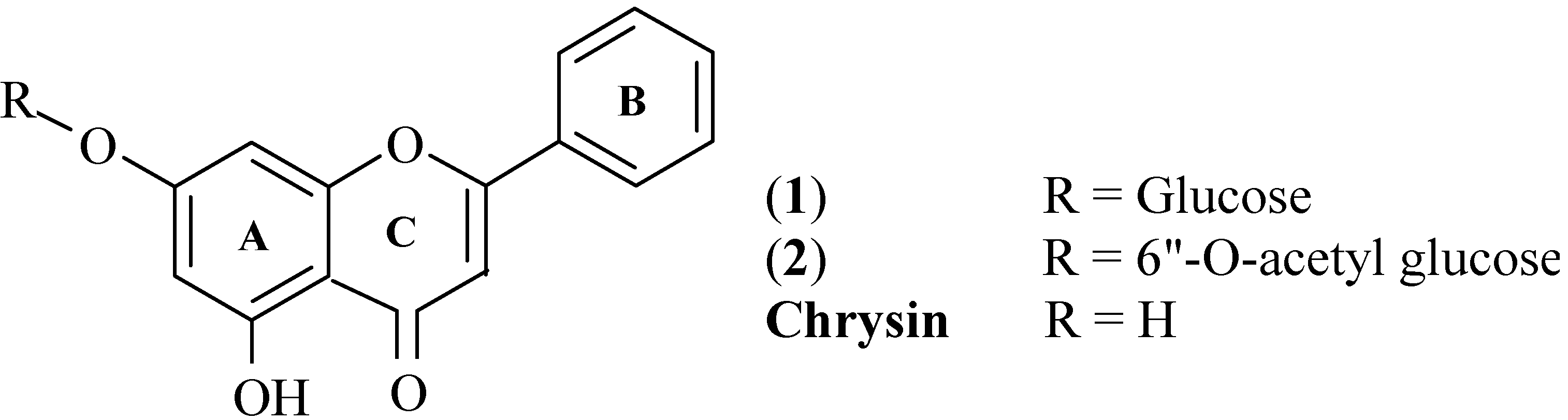
Results and discussion

Two flavone glycosides were isolated from the methanolic extract of Calycotome Villosa Subsp. Intermedia. Compound (1) was obtained as a yellow powder from the cooled methanolic extract. Compound (2) was isolated as a pale yellow powder by repeated column chromatography. Their structures (Scheme 1) were identified as follows.
Scheme 1.
Scheme 1.
Molecules 09 00568 g001
ESI-MS of compound (1), which gave a quasi-molecular ion peak at m/z 417 [M+H]+, and HRMS ESI (m/z 417.1179; calc.: 417.1185), as well as the elemental analysis (found: C 60.56%, H 4.79%; required: C 60.58%, H 4.81%) all indicated that the molecular formula should be C21H20O9. The IR spectrum (KBr) indicated the presence of hydroxyl (3403 cm-1) and carbonyl (1655 cm-1) groups. Bands in the 1650-1050 cm-1 range are typical of a flavone skeleton. The UV spectrum exhibited absorption maxima at 268 nm (band II) and 306 nm (band I) that are characteristic absorption bands of a flavone skeleton [10]. No shift in band I of compound 1 was observed with the addition of AlCl3 and AlCl3/HCl, suggesting formation of a hydroxyl-keto complex at the 5-OH and the absence of an o-dihydroxyl grouping in the B ring [10,11]. As indicated, the ESI mass spectrum of 1 gave a molecular ion peak at m/z 417 [M+H]+. A fragment at m/z 255 [M + H]+ was observed for the aglycone. A loss of 162 mass units from the molecular ion and a signal at δ 60.57 ppm, shown by DEPT to represent a CH2 group, suggested glucose or galactose. The carbohydrate moiety was confirmed to be glucose by acid hydrolysis and TLC comparison with an authentic sample.
The 1H-NMR spectrum exhibited a flavonoid pattern and showed signals at δ 7.04 (1H, s) 6.96 (1H, s) and 6.46 ppm (1H, s) typical of protons at C-3, C-8 and C-6 of a flavone skeleton. Chemical shifts of 8.08 (d, H2’, H6’) and 7.59 (m, H3’, H4’, H5’) suggested that there is no substitution in the B ring of the flavonoid. Thus, the NMR, IR and ESI-MS all suggested that 1 has a chrysin aglycone [12]. The signal at δ 12.79 ppm was assigned to the C-5 hydroxyl. This functionality was confirmed by a batochromic shift in the UV-visible spectrum of 1 after addition of AlCl3. 1H-NMR resonances at δ 3.20 to 3.70 ppm and signals in the 13C-NMR spectrum just below δ 77 ppm indicated the presence of a glucose moiety [13]. The signal at δ 5.13 ppm was assigned to the anomeric proton (H-1”) with a coupling constant (J 6.7 Hz) indicating a β-configuration [11].
Comparison of the 13C-NMR spectrum of compound 1 with that of chrysin aglycone showed that the signal of C-7 was observed to shift upfield slightly (-1.18 ppm), whereas the signals of C-3, C-6 and C-8 were displaced downfield by 2.56, 1.63 and 0.93 ppm respectively. However, the signals of C-4 and C-5 remained unaffected. This indicated that the glucosyl unit was attached to C-7 of the aglycone. DEPT experiments indicated the presence of one methylene carbon (δ 60.58 ppm) and seven quaternary carbons. Based on the above evidence and comparison with the values in literature for analogous compounds [12,14,15], the structure of 1 was established as a known flavonoid 7-O-(β-D-glucopyranosyl)chrysin. Its characteristics are in good agreement with those reported in the literature on a sample isolated from the flowers of Spartium Junceum [16] and a sample synthesised by Dangles et al. [17].
Compound 2 has a molecular formula, C23H22O10, based on the HRMS-ESI (m/z 459.1276; calc.: 459.1291) and elemental analysis (found: C 60.24%, H 4.81%; required: C 60.26%, H 4.80%). Compound 2 also showed characteristic IR absorptions of a flavone skeleton. An absorption band at 1724 cm-1 indicated that compound 2 had an acetyl group. The 1H- and 13C-NMR data of 2 were quite similar to those of 1, except for some differences in the sugar moiety. These NMR data suggested that flavonoid 2 also has a chrysin aglycone. The 1H-NMR spectrum exhibited a flavonoid pattern and showed two meta-coupled doublets at δ 6.50 and 6.85 ppm (1H, J = 1.6 Hz each) represented H-6 and H-8 respectively, and a signal at δ 6.98 ppm (1H, s) typical of the proton at C-3 of a flavone skeleton. The signal at δ 12.74 ppm (1H, s) is characteristic of a C-5 hydroxyl. This functionality was further confirmed by a batochromic shift in the UV spectrum in the presence of AlCl3, as performed for 1 [11]. Furthermore the signals of a glycosidic moiety were visible (3.25 to 4.41 ppm), in particular the anomeric proton, which appeared at δ 5.12 ppm as a doublet with a coupling constant of J = 7.3 Hz indicating a β-linkage of the sugar unit to the aglycone [11]. The 13C-NMR spectrum confirms that 2 is indeed a glycoside of chrysin. The location at C-7 of the sugar moiety was established on the basis of the typical glycosylation shifts that occured with respect to chrysin, particulary the C-7 upfield shift (-1.48 ppm) and the downfield of C-6 (+1.53 ppm) and C-8 (+0.93 ppm) [12]. Morever, the cross peak from H-1” (δ 5.12) to C-7 (δ 162.9) in the HMBC spectrum confirmed that the glucosylation takes place in the 7 position. The UV spectral data of 2 with diagnostic shift reagents confirmed the susbstitution of the sugar unit at position C-7. The chemical shifts of the sugar unit carbons were similar to those of 1. However, the presence of the methyl signal at δ 20.6 ppm, together with the presence of a carbonyl signal at δ 170.2 ppm were observed in 2, indicating the presence of β-D-(6”-acetyl)glucose moiety. The location of the acetyl group on position 6” was established by the downfield shift of C-6” (+2.8 ppm), and the upfield shift of C-5” (-1.0 ppm) with respect to 1. The 1H-COSY and HMBC spectrum confirmed the above assignments. Moreover, the ESI-MS exhibited the fragment ion characteristic of the chrysin aglycone unit at m/z 255. A loss of 204 mass units (C8H14O6) from the molecular ion in the MS confirmed the structure of the sugar moiety. From these results, flavonoid 2 was identified as 7-O-β-D-[(6”-acetyl)glycopyranosyl]chrysin.

Conclusions

The isolation and spectroscopic characterisation of two flavonoids from the leaves and flowers of Calycotome Villosa Subsp. Intermedia was described. Isolation and identification of alkaloids from this plant species are in progress.

Acknowledgements

The authors are grateful to Dr. C. ROLANDO, Equipe Polyphénols, UMR CNRS 8009, Université des Sciences et Technologies de Lille (France), for the NMR spectra and for the MS measurements.

Experimental

General

Melting points were measured in open capillary tubes in a Büchi 530 apparatus and are uncorrected. Microanalyses were performed at the “Service Central d’Analyse du Centre National de la Recherche Scientifique”, Vernaison, France. UV-Vis spectra were obtained on a Varian Cary 3E spectrophotometer, and IR spectra were recorded on a Pye Unicam Perkin-Elmer spectrophotometer. 1H- and 13C-NMR spectra were recorded in DMSO-d6 on a Bruker (Wiessembourg, France) AM 300 spectrometer (300 and 75 MHz, for 1H and 13C NMR respectively) and chemical shifts are given as δ values using TMS as an internal standard. MS data were obtained on a QSTAR Pulsar quadripole time-of-flight (Q-TOF) mass spectrometer (AB/MDS Sciex, Toronto, Canada) equipped with a nanoelectrospray ion source (Protana, Odense, Denmark). Silica gel GF254 was used for TLC. Spots were detected on TLC under UV light. Column chromatography (CC) was carried out on silica gel 60 (70-230 mesh).

Plant material

Calycotome Villosa Subsp. Intermedia aerial parts were collected in January 2001 from Zrireg valley, plateau of Tazzeka, area of Taza, Morocco. The plant was identified by Dr. R. Alami, Faculty of Science, Sidi Mohamed Ben Abdellah University, Fès, Morocco.

Extraction and isolation

The dried plant (30 g) was first extracted with hexane for 24 h and then with MeOH for 24 h using a Soxhlet apparatus. After cooling the methanolic extract, a yellow product precipitated which was filtered to afford 0.30 g of 7-O-(β-D-glucopyranosyl)chrysin (1) as a yellow amorphous powder, mp 213-215°C; UV-visible λmax nm: MeOH 268, 306; AlCl3 282, 325, 384; AlCl3 + HCl 282, 325, 384; IR νmax (KBr, cm-1): 3403, 1655; 1H-NMR: 12.79 (s, HO-C5), 8.08 (d, J = 6.5, H-C2’; H-C6’), 7.59 (m, H-C3’, H-C4’, H-C5’), 7.04 (s, H-C3), 6.86 (d, J = 1.5, H-C8), 6.46 (d, J = 1.5, H-C6), 5.13 (d, J = 6.7, H-C1”), 3.70 (d, J = 10.3, Ha-C6”), 3.45-3.20 (m, H-C2”, H-C3”, H-C4”, H-C5”, Hb-C6”) ppm; 13C-NMR: 182.2 (C-4), 163.7 (C-2), 163.2 (C-7), 161.1 (C-5), 157.1 (C-9), 132.2 (C-4’), 130.6 (C-1’), 129.2 (C-3’, C-5’), 126.5 (C-2’, C-6’), 105.6 (C-3), 105.5 (C-10), 99.9 (C-1”), 99.7 (C-6), 95.0 (C-8), 77.2 (C-5”), 76.4 (C-3”), 73.0 (C-2”), 69.5 (C-4”), 60.6 (C-6”) ppm; HRMS ESI (m/z 417.1179; calc.: 417.1185). ESI-MS m/z: 417 [M + H]+; 255 [aglycone + H]+. Repeated column chromatography on silica gel of the concentrated extract afforded 0.015 g of 7-O-β-D-[(6”-acetyl)glycopyranosyl]chrysin (2) as a pale yellow powder, mp 229-231°C; UV-visible λmax nm: MeOH 269, 305; AlCl3 283, 326, 385; AlCl3 + HCl 283, 326, 385; IR νmax (cm-1): 3407, 1724, 1653; 1H-NMR: 12.74 (s, H-C5), 8.08 (d , J = 6.7 , H-C2’, H-C6’), 7.63-7.60 (m, H-C3’, H-C4’, H-C5’), 6.98 (s, H-C3), 6.85 (d, J = 1.6, H-C8), 6.50 (d, J = 1.6, H-C6), 5.12 (d, J = 7,3, H-C1’’), 4.41 (d, J = 10.1, H-C6’’), 4.12 (d, J = 11.8, H-C6’’), 3.75 (m, H-C5’’), 3.38 (m, H-C2’’, H-C3’’), 3.25 (m, H-C4’’) 2.02 (s, OAc). 13C-NMR: 182.2 (C-4), 170.2 (6”-OCOCH3), 163 .7 (C-2), 162.9 (C-7), 161.1 (C-5), 157.1 (C-9), 132.3 (C-4’), 130.6 (C-1’), 129.2 (C-3’ ,C-5’), 126.5 (C-2’,C-6’), 105.7 (C-3), 105.5 (C-10), 99.7 (C-1’’), 99.6 (C-6), 95.0 (C-8), 76.2 (C-5’’), 73.9 (C-3’’), 73.0 (C2’’), 69.8 (C4’’), 63.4 (C-6’’), 20.6 (6”-OCOCH3). HRMS ESI (m/z 459.1276; calc.: 459.1291). ESI-MS m/z: 459 [M + H]+; 255 [aglycone + H]+.

Acid hydrolysis

Acid hydrolysis of the flavonoid 1 was done according to the method reported in the literature [11,18]. 30 mg of 1 were hydrolysed with 2N HCl in MeOH (30 mL) at 80 °C for 2h. The liberated aglycone was extracted several times with EtOAc and examined by 1H-NMR and MS. The acidic mother liquor was neutralized with Na2CO3, filtered, and evaporated to dryness for examination of the sugar moiety, which proved to be glucose by chromatographic comparison with authentic sample.

References

  1. Greuter, W.; Burdet, H.M.; Long, G. (Eds.) Med-Checklist 4: A Critical Inventory of Vascular Plants of the Circum-mediterranean Countries; Dicotyledones (Lauraceae-Rhamnaceae); C.B. de Genève: Geneva, 1989.
  2. Bohm, B.A. Introduction to Flavonoids; Harwood Academic Publishers: Amsterdam, 1998. [Google Scholar]
  3. Bors, W.; Michel, C.; Stettmaier, K. Flavonoids and other polyphenols. Methods Enzymol. 2001, 335, 166–180. [Google Scholar] [PubMed]
  4. Bors, W.; Heller, W.; Michel, C.; Saran, M. Flavonoids as antioxidants: Determination of radical scavenging efficiencies. Methods Enzymol. 1990, 186, 343–355. [Google Scholar] [PubMed]
  5. Colerige Smith, P.O.; Thomas, P.; Scurr, T.H.; Dormandy, J.A. Causes of various ulceration, a new hypothesis. Br. Med. J. 1980, 296, 1726–1727. [Google Scholar] [CrossRef]
  6. Manthy, J.A.; Buslig, B.S. (Eds.) Flavonoids in the Living System. In Advances in Experimental Medicine and Biology; Plenum: New York, 1998.
  7. Mori, A.; Nishino, C.; Enoki, N.; Tawata, S. Cytotoxicity of plant flavonoids against Hella Cells. Phytochemistry 1988, 27, 1017–1020. [Google Scholar] [CrossRef]
  8. Santos, A.C.; Vyemura, S.A.; Lopes, J.L.; Bazon, J.N.; Mingatto, F.E.; Curtic. Effect of naturally occurring Flavonoids on lipid peroxidation and membrane permeability transition in mitochondria. Free Radic. Biol. Med. 1988, 24, 1455–1461. [Google Scholar]
  9. Middleton, E. The flavonoids. Trends Pharmacol. Sci. 1984, 5, 335–338. [Google Scholar]
  10. Markham, K.R. Techniques of Flavonoids Identification; Academic Press: London, 1982; Chapter 3; pp. 36–51. [Google Scholar]
  11. Mabry, T.J.; Markham, K.R.; Thomas, M.B. The Systematic Identification of Flavonoids; Springer: Berlin, 1970; pp. 35–250. [Google Scholar]
  12. Yang, F.; Li, X.-C.; Wang, H.-Q.; Yang, C.-R. Flavonoid Glycosides from Colebrookea Oppositifolia. Phytochemistry 1996, 42, 867–869. [Google Scholar] [CrossRef]
  13. Markham, K.R.; Ternai, B.; Stanly, R.; Geiger, H.; Mabry, T.J. Carbon-13 NMR studies of flavonoids-III. Tetrahedron 1978, 34, 1389–1397. [Google Scholar] [CrossRef]
  14. Lin, J.-H.; Lin, Y.-T. Flavonoids from the Leaves of Loranthus Kaoi (Chao) Kiu. J. Food Drug Anal. 1999, 7, 185–190. [Google Scholar]
  15. Dai, J.-Q.; Hou, Z.-F.; Zhu, Q.-X.; Yang, L.; Li, Y. Sesquiterpenes and Flavonoids from Serratula Strangulata. J. Chinese. Chem. Soc. 2001, 48, 249–252. [Google Scholar] [CrossRef]
  16. De Rosa, S.; De Stefano, S. Chrysin 7-gentiobioside from the flowers of Spartium Junceum. Phytochemistry 1983, 22, 2323–2324. [Google Scholar] [CrossRef]
  17. Alluis, B.; Dangles, O. Acylated flavone glucosides: Synthesis, conformational investigation, and complexation properties. Helv. Chim. Acta 1999, 82, 2201–2212. [Google Scholar] [CrossRef]
  18. Markham, K.R. Flavones, Flavonols and their glycosides. In Methods in Plant Biochemistry; Harbone, J.B., Ed.; Academic Press: New York, USA, 1986; Vol.1, pp. 197–235. [Google Scholar]
  • Sample availability: A sample of compound 1 is available from the authors.

