Expression of CD40 and CD40L in Gastric Cancer Tissue and Its Clinical Significance

Abstract
:1. Introduction
2. Methods
2.1. Human Gastric Cancer Specimens
2.2. Human Serum Specimens
2.3. Immunohistochemistry
2.4. Measurement of Apoptotic Cells
2.5. Immunostaining Score
- staining intensity,
- number of positive cells,
- score1 (staining intensity) × score2 (number of positive cells).
2.6. Enzyme Immunoassays
2.7. Statistical Analysis
3. Results
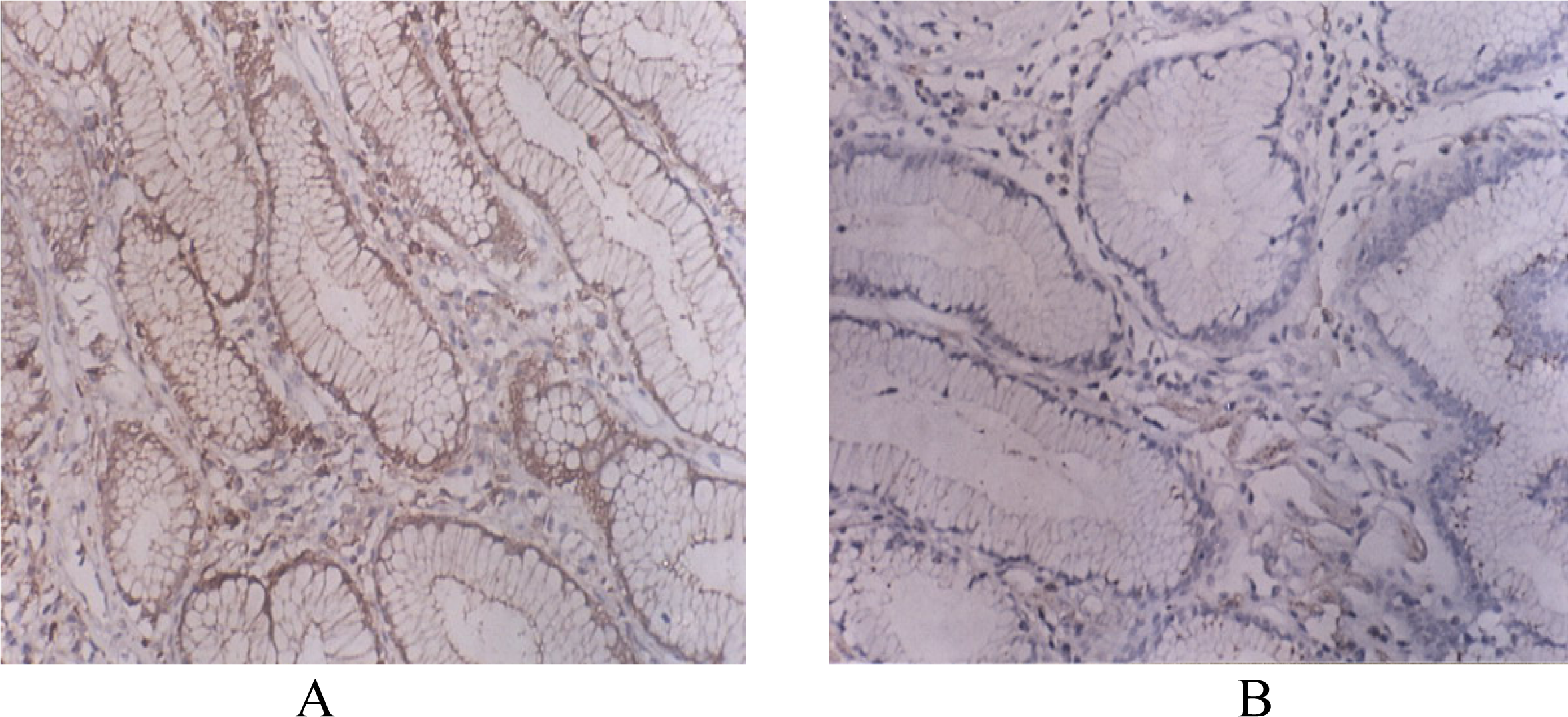
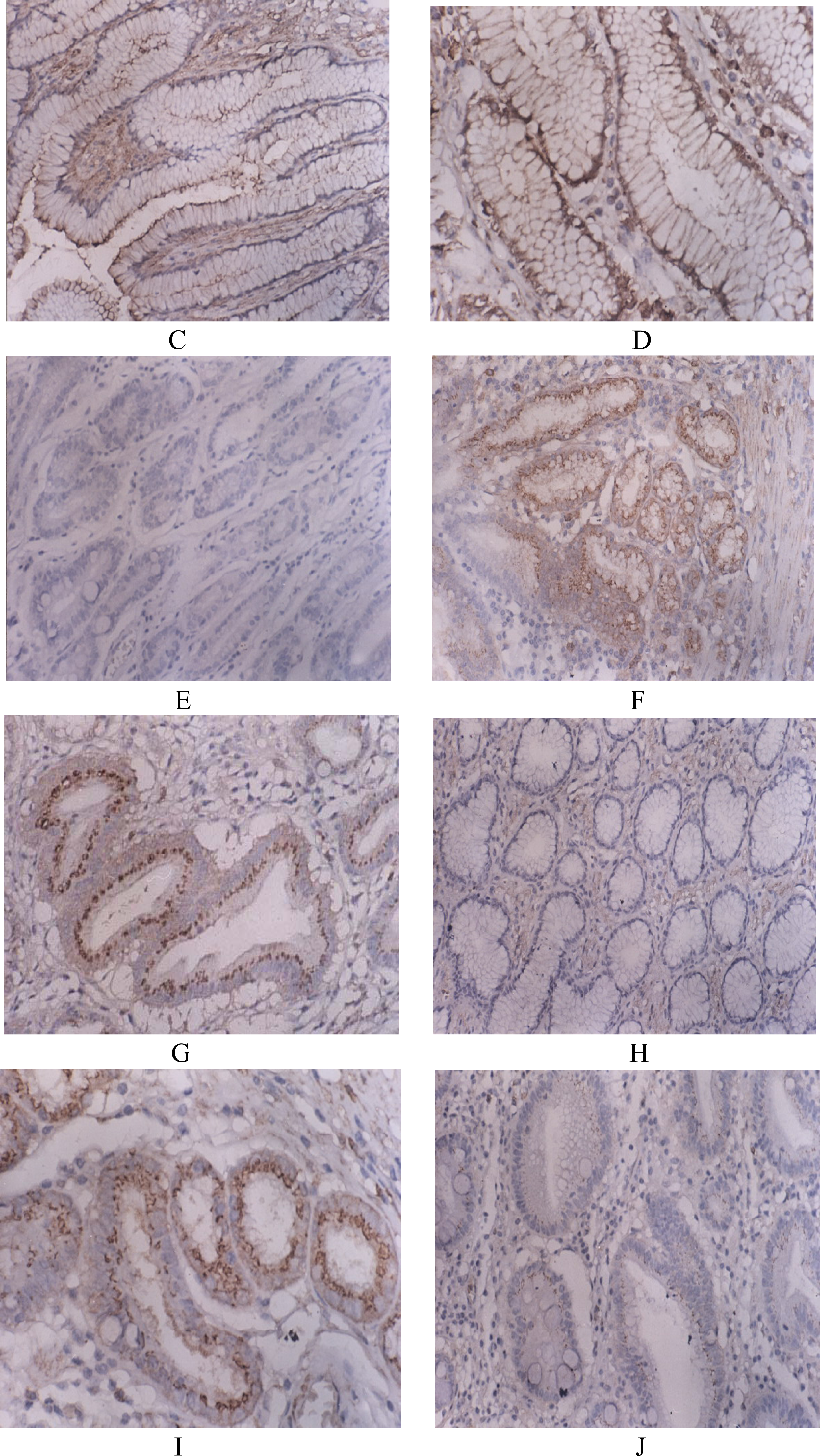
3.1. CD40, CD138, Gelsolin, P53, P65 Expression in Gastric Cancer Tissue and Normal Tissue
3.2. Correlation of CD40, CD138, Gelsolin, P53 and P65 Expression with Clinical Pathological Factors in Patients with Gastric Cancer
3.3. Correlation of CD40 Expression with CD138, Gelsolin, P53 and P65 Expression in Gastric Cancer Tissues
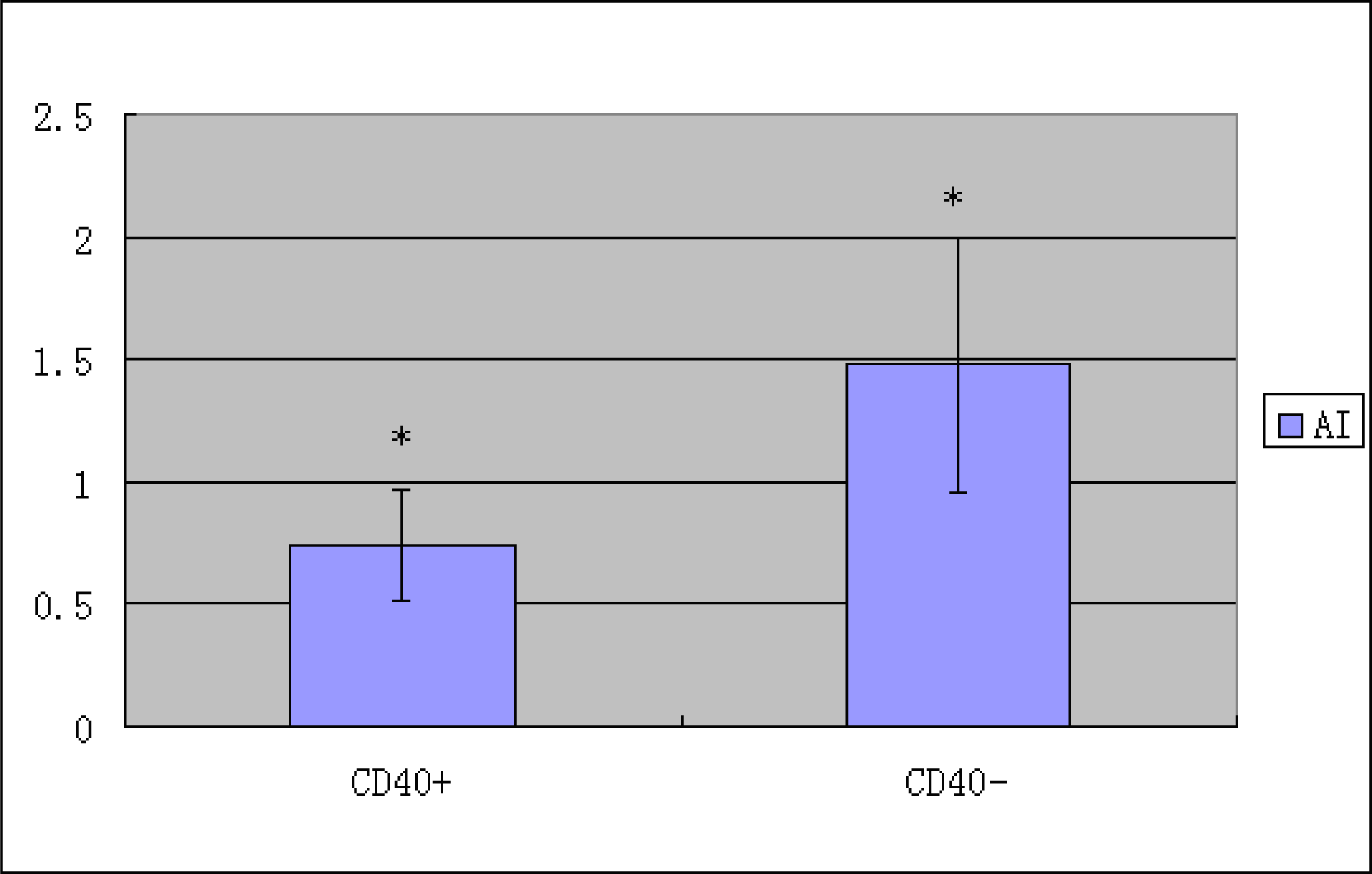
3.4. Correlation of CD40 Expression with Apoptotic Index (AI) in Gastric Cancer Tissues
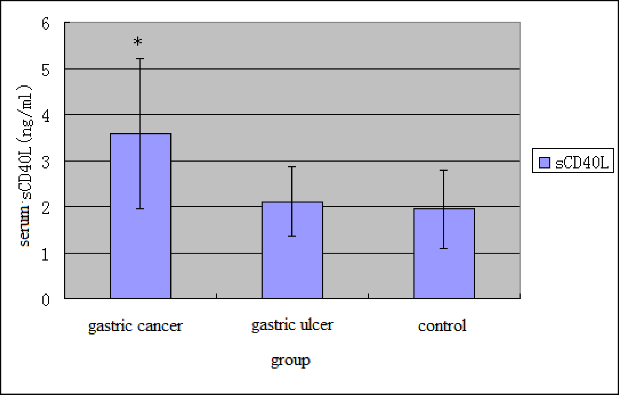
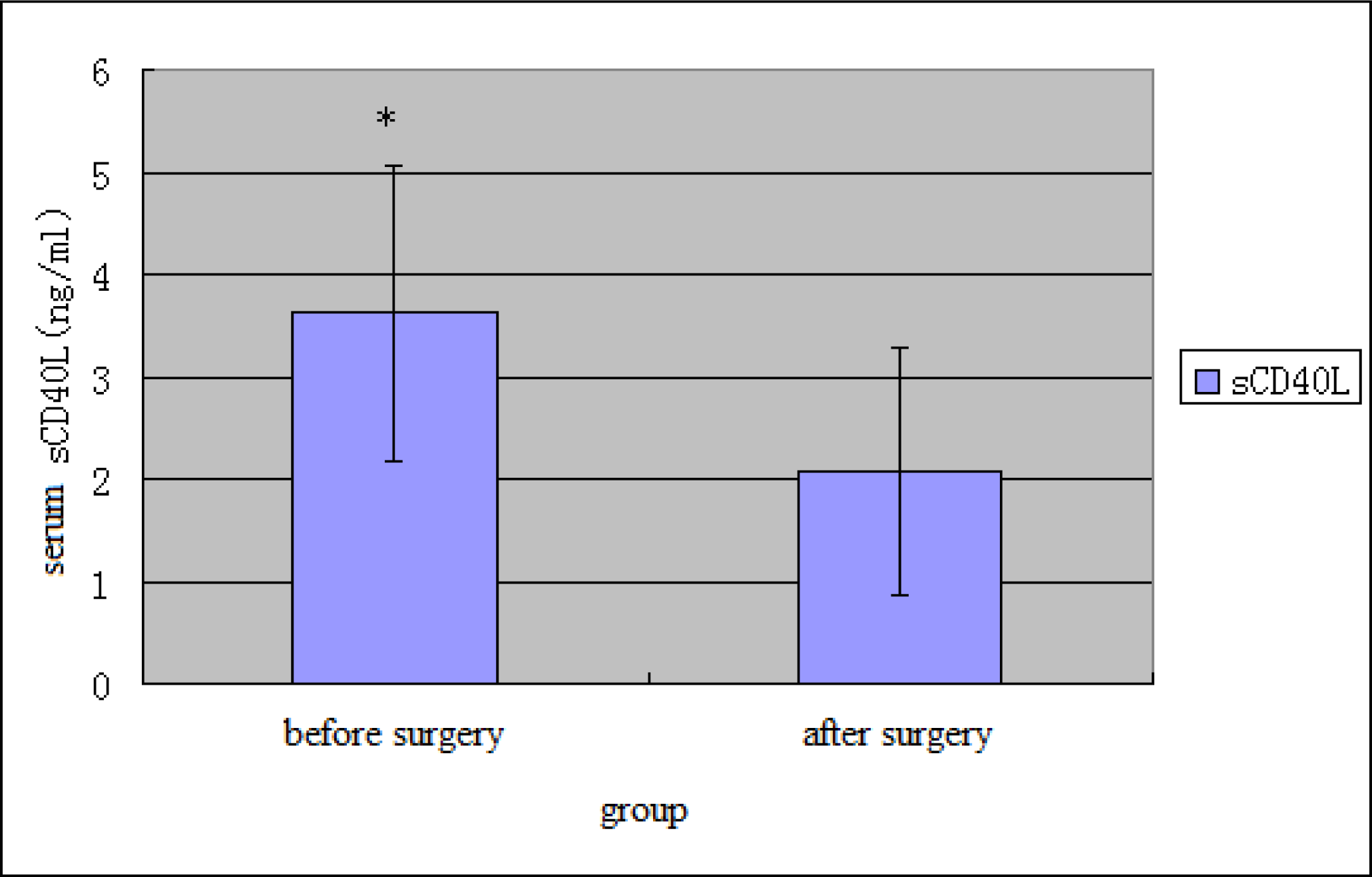
3.5. sCD40L Level in Peripheral Blood
4. Discussion
5. Conclusions
Acknowledgments
Reference
- Hakim, FT; Flomerfelt, FA; Boyiadzis, M; Gress, RE. Aging, immunity and cancer. Curr. Opin. Immunol 2004, 16, 151–156. [Google Scholar]
- Chakrabarti, S; Rizvi, M; Pathak, D; Kirber, MT; Freedman, JE. Hypoxia influences CD40-CD40L mediated inflammation in endothelial and monocytic cells. Immunol. Lett 2009, 122, 170–184. [Google Scholar]
- Chen, D; Adenekan, B; Chen, L; Darracott Vaughan, E; Gerald, W; Feng, Z; Knudsen, BS. Syndecan-1 expression in locally invasive and metastatic prostate cancer. Urology 2004, 63, 402–407. [Google Scholar]
- Gay, F; Estornes, Y; Saurin, J-C; Joly-Pharaboz, M-O; Friederich, E; Scoazec, J-Y; Abello, J. In colon carcinogenesis, the cytoskeletal protein gelsolin is down-regulated during the transition from adenoma to carcinoma. Human Pathol 2008, 39, 1420–1430. [Google Scholar]
- Soong, R; Robbins, PD; Dix, BR; Grieu, F; Lim, B; Knowles, S; Williams, KE; Turbett, GR; House, AK; Iacopetta, BJ. Concordance between p53 protein overexpression and gene mutation in a large series of common human carcinomas. Human Pathol 1996, 27, 1050–1055. [Google Scholar]
- Wu, YG; Wang, L; He, XZ; Xu, HY; Zhou, LC; Zhao, F; Zhang, YY. Expression of CD40 and growth-inhibitory activity of CD40 ligand in colon cancer ex vivo. Cell Immunol 2008, 253, 102–109. [Google Scholar]
- Grammer, AC; Lipsky, PE. CD40-mediated regulation of immune responses by TRAF-dependent and TRAF-independent signaling mechanisms. Adv. Immunol 2000, 76, 61–178. [Google Scholar]
- van Kooten, C; Banchereau, J. CD40-CD40 ligand. J. Leukocyte Biol 2000, 67, 2–17. [Google Scholar]
- Eeva, J; Ropponen, A; Nuutinen, U; Eeva, S-T; Mättö, M; Eray, M; Pelkonen, J. The CD40-induced protection against CD95-mediated apoptosis is associated with a rapid upregulation of anti-apoptotic c-FLIP. Mol. Immunol 2007, 44, 1230–1237. [Google Scholar]
- Hayward, AR; Levy, J; Facchetti, F; Notarangelo, L; Ochs, HD; Etzioni, A; Bonnefoy, JY; Cosyns, M; Weinberg, A. Cholangiopathy and tumors of the pancreas, liver, and biliary tree in boys with X-linked immunodeficiency with hyper-IgM. J. Immunol 1997, 158, 977–983. [Google Scholar]
- Aukrust, P; Muller, F; Ueland, T; Berget, T; Aaser, E; Brunsvig, A; Solum, NO; Forfang, K; Froland, SS; Gullestad, L. Enhanced levels of soluble and membrane-bound CD40 ligand in patients with unstable angina. Circulation 1999, 100, 614–620. [Google Scholar]
- Tao, YF; Nomura, M; Kitabatake, N; Tani, F. Mouse CD40-transfected cell lines cannot exhibit the binding and RANTES-stimulating activity of exogenous heat shock protein 70. Mol. Immunol 2007, 44, 1262–1273. [Google Scholar]
- Li, R; Chen, W-C; Wang, W-P; Tian, W-Y; Zhang, X-G. Optimization of extraction technology of Astragalus polysaccharides by response surface methodology and its effect on CD40. Carbohyd. Polym 2009, 78, 784–788. [Google Scholar]
- Li, R; Chen, W-C; Wang, W-P; Tian, W-Y; Zhang, X-G. Extraction, characterization of Astragalus polysaccharides and its immune modulating activities in rats with gastric cancer. Carbohyd. Polym 2009, 78, 738–742. [Google Scholar]
- Oki, E; Maehara, Y; Tokunaga, E; Kakeji, Y; Sugimachi, K. Reduced expression of p33ING1 and the relationship with p53 expression in human gastric cancer. Cancer Lett 1999, 147, 157–162. [Google Scholar]
- Szkaradkiewicz, A; Majewski, W; Wal, M; Czyżak, M; Majewski, P; Bierła, J; Kuch, A. Epstein-Barr virus (EBV) infection and p53 protein expression in gastric carcinoma. Virus Res 2006, 118, 115–119. [Google Scholar]
- Kim, H-S; Choi, YJ; Baik, SK; Lee, DK; Kwon, SO; Oh, E-S. mRNA expression of syndecan-2 in human colon cancer and polyp tissue: A pilot study. Gastroenterology 2003, 124, A607. [Google Scholar]
- Briggs, CD; Neal, CP; Mann, CD; Steward, WP; Manson, MM; Berry, DP. Prognostic molecular markers in cholangiocarcinoma: A systematic review. Eur. J. Cancer 2009, 45, 33–47. [Google Scholar]


| Group | Gastric Cancer Tissues | Normal Tissues | |
|---|---|---|---|
| Total Cases | 56 | 32 | |
| CD40 expression rate (case) | − | 37 | 30 |
| + | 6 | 2 | |
| ++ | 3 | 0 | |
| +++ | 10 | 0 | |
| * P value | <0.01 | ||
| CD138 expression rate (case) | − | 31 | 1 |
| + | 17 | 8 | |
| ++ | 6 | 9 | |
| +++ | 2 | 14 | |
| * P value | <0.01 | ||
| Gelsolin expression rate (case) | − | 50 | 6 |
| + | 5 | 13 | |
| ++ | 1 | 5 | |
| +++ | 0 | 8 | |
| * P value | <0.05 | ||
| P53 expression rate (case) | − | 17 | 28 |
| + | 11 | 3 | |
| ++ | 16 | 1 | |
| +++ | 12 | 0 | |
| * P value | <0.01 | ||
| P65 expression rate (case) | − | 20 | 25 |
| + | 21 | 4 | |
| ++ | 7 | 3 | |
| +++ | 8 | 0 | |
| * P value | <0.05 | ||
| Clinical pathological factors | Cases (n) | CD40 positive expression rate (%) | P value |
|---|---|---|---|
| Sex | >0.05 | ||
| Man | 37 | 29.7 (11) | |
| Woman | 19 | 42.1 (8) | |
| Age (years) | >0.05 | ||
| ≥50 | 43 | 30.2 (13) | |
| <50 | 13 | 46.2 (6) | |
| Tumor position | >0.05 | ||
| Cardia | 12 | 16.7 (2) | |
| Corpora ventriculi | 24 | 41.7 (10) | |
| Sinuses ventriculi | 20 | 35.1 (7) | |
| Tumor size (cm) | >0.05 | ||
| ≥5 | 31 | 32.3 (10) | |
| <5 | 25 | 36.3 (9) | |
| Tissue differentiation | >0.05 | ||
| High differentiation | 8 | 50 (4) | |
| Middle differentiation | 37 | 24.3 (9) | |
| Low differentiation | 11 | 54.5 (6) | |
| Invasion depth | >0.05 | ||
| Mucous membrane | 5 | 40 (2) | |
| Muscular layer | 28 | 32.1 (9) | |
| Serous membrane from inner to outer | 23 | 34.8 (8) | |
| Blood-vessel invasion | >0.05 | ||
| Yes | 27 | 48.1 (13) | |
| No | 29 | 20.7 (6) | |
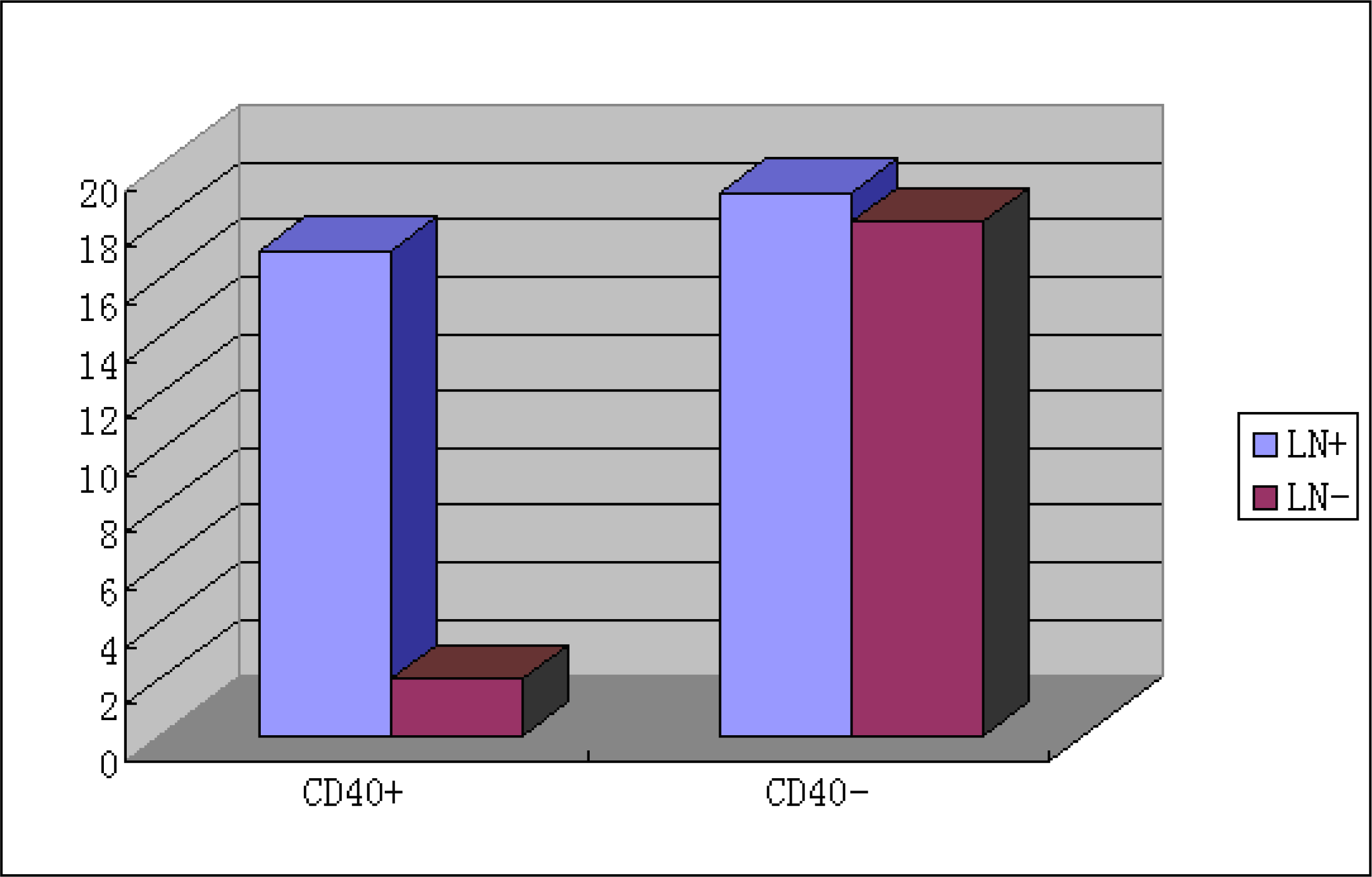
| Lymphatic metastasis | <0.01 | ||
| Yes | 36 | 47.2 (17) | |
| No | 20 | 10 (2) | |
| Distant metastasis | <0.01 | ||
| Yes | 9 | 100 (9) | |
| No | 47 | (21.3) 10 | |
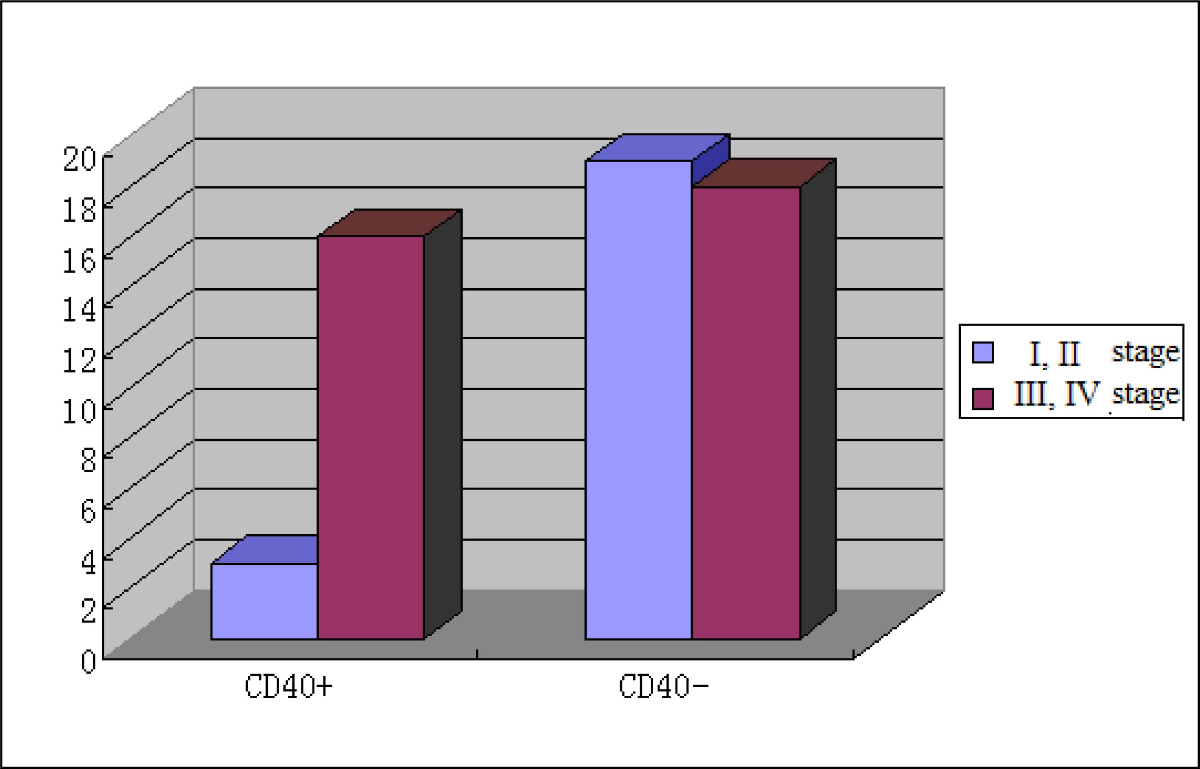
| TNM stage | <0.05 | ||
| I, II | 22 | 13.6 (3) | |
| III, IV | 34 | 47.1 (16) |
| Clinical pathological factors | Cases(n) | CD138 positive expression Rate (%) | P value | Gel positive expression Rate (%) | P value | P53 positive expression Rate (%) | P value | P65 positive expression Rate (%) | P value |
|---|---|---|---|---|---|---|---|---|---|
| Sex | >0.05 | >0.05 | >0.05 | >0.05 | |||||
| Man | 37 | 43.2 (16) | 2.7 (1) | 64.9 (24) | 59.5 (22) | ||||
| Woman | 19 | 47.4 (9) | 26.3 (5) | 78.9 (15) | 73.7 (14) | ||||
| Age (years) | >0.05 | >0.05 | >0.05 | >0.05 | |||||
| ≥50 | 43 | 48.8 (21) | 11.6 (5) | 72.1 (31) | 62.8 (27) | ||||
| <50 | 13 | 30.8 (4) | 7.7 (1) | 61.5 (8) | 69.2 (9) | ||||
| Tumor position | >0.05 | >0.05 | >0.05 | >0.05 | |||||
| Cardia | 12 | 58.3 (7) | 8.3 (1) | 16.7 (2) | 58.3 (7) | ||||
| Corpora ventriculi | 24 | 33.3 (8) | 8.3 (2) | 79.2 (19) | 41.7 (10) | ||||
| Sinuses ventriculi | 20 | 50 (10) | 15 (3) | 90 (18) | 95 (19) | ||||
| Tumor size (cm) | >0.05 | >0.05 | <0.05 | <0.05 | |||||
| ≥5 | 31 | 48.4 (15) | 6.5 (2) | 93.5 (29) | 90.3 (28) | ||||
| <5 | 25 | 40 (10) | 16 (4) | 40 (10 | 32 (8) | ||||
| Tissue | <0.05 | >0.05 | <0.01 | <0.05 | |||||
| High | 8 | 87.5 (7) | 37.5 (3) | 25 (2) | 25 (2) | ||||
| Middle | 37 | 45.9 (17) | 5.4 (2) | 73 (27) | 62.2 (23) | ||||
| Low differentiation | 11 | 9.1 (1) | 9.1 (1) | 90.9 (10) | 100 (11) | ||||
| Invasion depth | <0.05 | <0.05 | >0.05 | <0.01 | |||||
| Mucous membrane | 5 | 100 (5) | 80 (4) | 20 (1) | 20 (1) | ||||
| Muscular layer | 28 | 64.3 (18) | 3.6 (1) | 67.9 (19) | 53.6 (15) | ||||
| Serous membrane from inner to outer | 23 | 8.7 (2) | 4.3 (1) | 82.6 (19) | 87 (20) | ||||
| Blood-vessel invasion | >0.05 | >0.05 | >0.05 | <0.05 | |||||
| Yes | 27 | 37 (10) | 7.4 (2) | 70.4 (19) | 92.6 (25) | ||||
| No | 29 | 51.7 (15) | 13.8 (4) | 69 (20) | 37.9 (11) | ||||
| Lymphatic metastasis | <0.01 | <0.01 | >0.05 | <0.01 | |||||
| Yes | 36 | 19.4 (7) | 2.8 (1) | 72.2 (26) | 88.9 (32) | ||||
| No | 20 | 90 (18) | 25 (5) | 65 (13) | 20 (4) | ||||
| Distant metastasis | <0.01 | >0.05 | >0.05 | >0.01 | |||||
| Yes | 9 | 0 (0) | 0 (0) | 100 (9) | 100 (9) | ||||
| No | 47 | 53.2 (25) | 12.8 (6) | 63.8 (30) | 57.4 (27) | ||||
| TNM stage | <0.05 | >0.05 | >0.05 | <0.01 | |||||
| I, II | 22 | 86.4 (19) | 22.7 (5) | >0.05 | 63.6 (14) | 27.3 (6) | |||
| III, IV | 34 | 17.6 (6) | 2.9 (1) | 73.5 (25) | 88.2 (30) |
| Group | CD138 | Gelsolin | P53 | P65 | ||||
|---|---|---|---|---|---|---|---|---|
| + | − | + | − | + | − | + | − | |
| CD40 positive expression (n = 19) | 4* | 15 | 2 | 17 | 18** | 1 | 19** | 0 |
| CD40 negative expression (n = 37) | 21 | 16 | 4 | 33 | 21 | 16 | 17 | 20 |
| Group | Cases (n) | AI (%)a | P value |
|---|---|---|---|
| gastric cancer tissues | 56 | 1.16 ± 0.37 | |
| CD40 positive cancer tissues | 19 | 0.74 ± 0.23 | |
| CD40 negative cancer tissues | 37 | 1.48 ± 0.52 | <0.01 |
| Clinical pathological factors | Cases (n) | sCD40L (ng/ml)a | P value |
|---|---|---|---|
| Sex | >0.05 | ||
| Man | 32 | 3.48 ± 1.74 | |
| Woman | 13 | 3.65 ± 1.52 | |
| Age (years) | >0.05 | ||
| ≥50 | 36 | 3.39 ± 1.68 | |
| <50 | 9 | 3.73 ± 1.45 | |
| Tumor position | >0.05 | ||
| Cardia | 9 | 3.73 ± 1.45 | |
| Corpora ventriculi | 20 | 3.56 ± 1.64 | |
| Sinuses ventriculi | 16 | 3.44 ± 1.76 | |
| Tumor size (cm) | >0.05 | ||
| ≥5 | 26 | 3.43 ± 1.80 | |
| <5 | 19 | 3.62 ± 1.47 | |
| Tissue differentiation | >0.05 | ||
| High differentiation | 6 | 3.35 ± 1.93 | |
| Middle differentiation | 30 | 3.58 ± 1.69 | |
| Low differentiation | 9 | 3.61 ± 1.56 | |
| Invasion depth | <0.05 | ||
| Mucous membrane | 5 | 2.98 ± 1.19 | |
| Muscular layer | 22 | 3.45 ± 1.80 | |
| Serous membrane from inner to outer | 18 | 4.52 ± 0.66 | |
| Blood-vessel invasion | <0.05 | ||
| Yes | 22 | 4.20 ± 0.98 | |
| No | 23 | 3.16 ± 1.01 | |
| Lymphatic metastasis | <0.01 | ||
| Yes | 30 | 4.17 ± 1.11 | |
| No | 15 | 3.01 ± 0.29 | |
| Distant metastasis | <0.01 | ||
| Yes | 6 | 5.11 ± 0.34 | |
| No | 39 | 3.52 ± 1.36 | |
| TNM stage | <0.05 | ||
| I, II | 17 | 3.05 ± 0.87 | |
| III, IV | 28 | 4.12 ± 1.06 |
© 2009 by the authors; licensee Molecular Diversity Preservation International, Basel, Switzerland. This article is an open-access article distributed under the terms and conditions of the Creative Commons Attribution license (http://creativecommons.org/licenses/by/3.0/).
Share and Cite
Li, R.; Chen, W.-C.; Pang, X.-Q.; Hua, C.; Li, L.; Zhang, X.-G. Expression of CD40 and CD40L in Gastric Cancer Tissue and Its Clinical Significance. Int. J. Mol. Sci. 2009, 10, 3900-3917. https://doi.org/10.3390/ijms10093900
Li R, Chen W-C, Pang X-Q, Hua C, Li L, Zhang X-G. Expression of CD40 and CD40L in Gastric Cancer Tissue and Its Clinical Significance. International Journal of Molecular Sciences. 2009; 10(9):3900-3917. https://doi.org/10.3390/ijms10093900
Chicago/Turabian StyleLi, Rui, Wei-Chang Chen, Xue-Qin Pang, Chen Hua, Ling Li, and Xue-Guang Zhang. 2009. "Expression of CD40 and CD40L in Gastric Cancer Tissue and Its Clinical Significance" International Journal of Molecular Sciences 10, no. 9: 3900-3917. https://doi.org/10.3390/ijms10093900