Article

Triterpenoids from the Roots of Rhaphiolepis indica var. tashiroi and Their Anti-Inflammatory Activity

1
School of Pharmacy, College of Pharmacy, Kaohsiung Medical University, Kaohsiung 80708, Taiwan
2
Graduate Institute of Natural Products, College of Pharmacy, Kaohsiung Medical University, Kaohsiung 80708, Taiwan
3
Graduate Institute of Natural Products, Chang Gung University, Taoyuan 33302, Taiwan
*
Author to whom correspondence should be addressed.
Int. J. Mol. Sci. 2013, 14(5), 8890-8898; https://doi.org/10.3390/ijms14058890
Submission received: 22 February 2013 / Revised: 22 March 2013 / Accepted: 26 March 2013 / Published: 24 April 2013
(This article belongs to the Section Biochemistry)

Abstract

:
Two new triterpenoids, 2α,3β-dihydroxyolean-11,13(18)-dien-19β,28-olide (1) and 3β,5β-dihydroxyglutinol (2), together with eight known compounds (310) were isolated from the roots of Rhaphiolepis indica var. tashiroi (Rosaceae). The structures of 110 were determined by spectroscopic techniques. Among these isolates, 2α,3β-dihydroxyolean-13(18)-en-28-oic acid (9) exhibited inhibitory effect on N-formyl-methionyl-leucyl-phenylalanine (fMLP)-induced superoxide production, with an IC50 value of 16.50 μM.

1. Introduction

Under a screening program of Formosan plants on inhibition of N-formyl-methionyl-leucyl-phenylalanine (fMLP) induced superoxide production assay, a methanolic extract of the roots of Rhaphiolepis indica var. tashiroi (Rosaceae) has shown suppression of N-formyl-methionyl-leucyl-phenylalanine (fMLP)-induced superoxide production with no cytotoxicity against neutrophils. R. indica var. tashiroi is an evergreen shrub or small tree, which is distributed in countries throughout Asia, including India, southern China, the Ryukyus, Korea, and Taiwan [1]. We previously reported six new compounds, including four dibenzofurans, 2-hydroxy-3,4,6-trimethoxydibenzofuran, 2-hydroxy-3,4,9-trimethoxydibenzofuran, 2-hydroxy-3,4,6,9-tetramethoxydibenzofuran, and 1,2-methylenedioxy-3,4,6-trimethoxydibenzofuran, two new biphenyls, 3-hydroxy-2′,5-dimethoxybiphenyl and 2′,3-dihydroxy-5-methoxybiphenyl, and one known 3-hydroxy-5-methoxybiphenyl from the roots of R. indica var. tashiroi [2]. Continuous study from the roots of this plant led to the isolation of two new triterpenoids, one oleanane-type 2α,3β-dihydroxy-olean-11,13(18)-dien-19β,28-olide (1), and one glutinane-type 3β,5β-dihydroxyglutinol (2), together with eight known compounds (310) (Figure 1). This paper describes the structural elucidations and inhibition of superoxide production activity of these isolates.

2. Results and Discussion

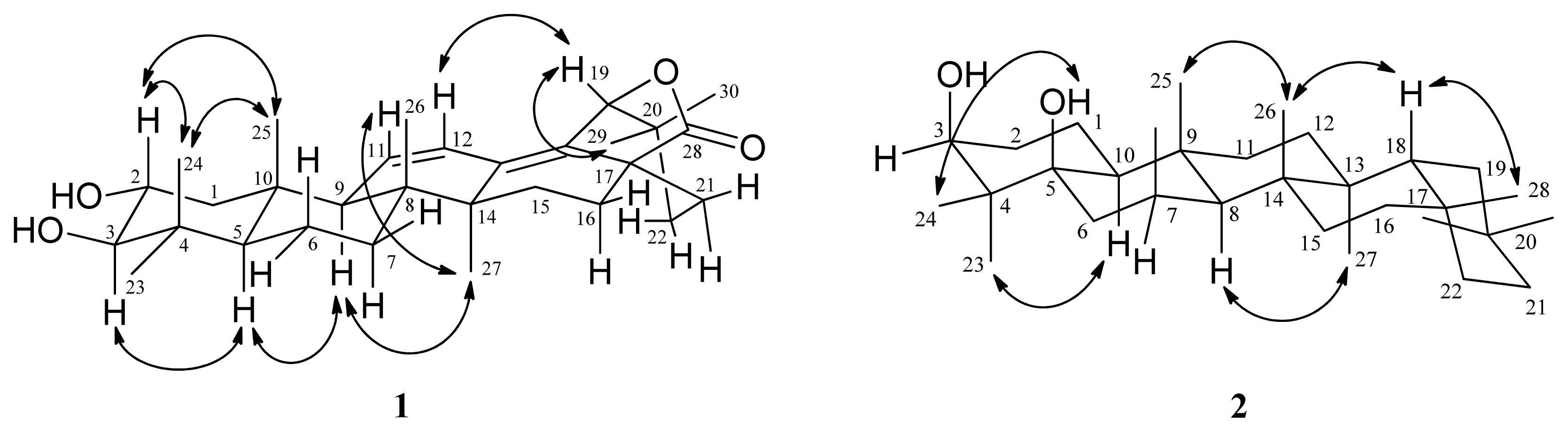
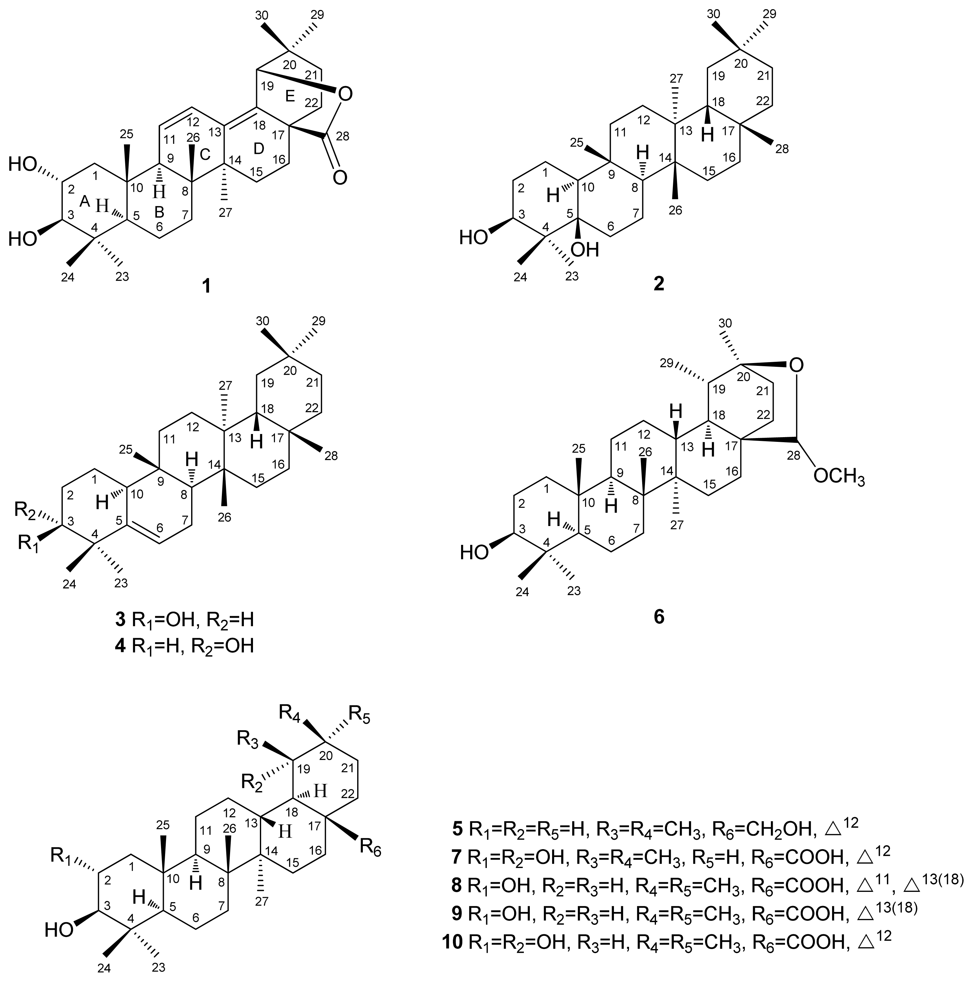
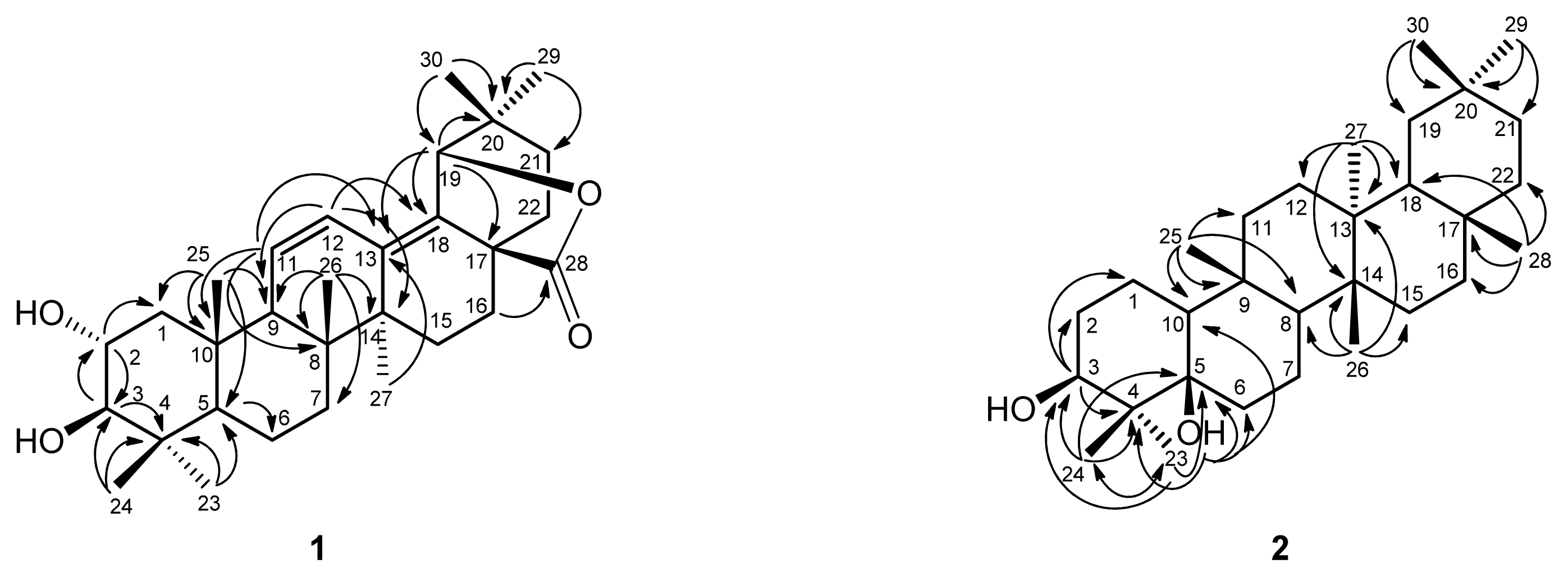
2.1. Structure Elucidation

Compound 1 was obtained as amorphous powder. ESIMS (m/z 491 [M + Na]+) and HRESIMS (m/z 491.3136 [M + Na]+) analysis established the molecular formula of 1 as C30H44O4. The IR absorption bands suggested the presence of hydroxy (3440 cm−1), γ-lactonic carbonyl (1767 cm−1), and olefinic (1630, 1600 cm−1) groups. The 1H NMR spectrum (Table 1) of 1 indicated seven methyl singlets (δH 0.76, 0.82, 0.95, 1.01, 1.02, 1.04, and 1.10), and a pair of cis olefinic protons at δ 5.77 (1H, dd, J = 10.2, 2.0 Hz, H-11) and 6.15 (1H, dd, J = 10.2, 2.8 Hz, H-12). The 13C NMR and DEPT spectra revealed that 1 had 30 carbons. Two tertiary olefinic carbons at δ 123.3 (C-12) and 129.1 (C-11), and two quaternary olefinic carbons at δ 132.9 (C-18) and 134.7 (C-13) indicated the presence of two double bonds. Two oxymethine carbons at δ 68.8 and 83.9, and a γ-lactonic carbonyl carbon at δ 178.1 supported the proposed structure as 1, which was similar to that of camaldulenic acid (8) with oleanane-type skeleton [3]. The major difference was a γ-lactonic carbonyl group in ring E in 1 instead of the COOH group in 8. The observed oxygen-bearing tertiary carbon signal at C-19 (δC 85.0) as well as the HMBC correlations from H-19 (δ 4.72, s) to C-13, C-17, C-18, C-20, and C-21 indicated a five-membered lactone ring between C-17 and C-19 which are also supported by IR absorption and nine degrees of unsaturation. The locations of two double bonds at Δ11 and Δ13(18) were determined by the HMBC correlations from H-11 to C-8, C-10, and C-13, from H-12 to C-9, C-14, and C-18, from H-27 to C-13, and from H-19 to C-13 and C-18 (Figure 2). The relative configurations of 1 were determined through inspection of the NOESY spectrum (Figure 3). The NOESY cross-peaks of H-2/H-25 and H-3/H-5 established the β-orientation of H-2 and α-orientation of H-3 in trans A/B ring junction [4]. Thus, 1 was elucidated as 2α,3β-dihydroxyolean-11,13(18)-dien-19β,28-olide.
Compound 2 was isolated as amorphous powder. The HRESIMS analysis of 2 revealed an [M + Na]+ ion peak at m/z 467.3862 (calcd. 467.3865), which corresponds to the molecular formula C30H52O2, accounting for five degrees of unsaturation. The IR absorption bands showed a hydroxy group at 3400 cm−1. The 1H NMR spectrum (Table 1) revealed eight methyl groups (δH 0.89, 0.94, 0.95, 0.97, 0.99, 1.01, 1.01, and 1.17). The NMR data of 2 resembles to those of glutinol (3) with glutinane-type skeleton. The major differences between 2 and 3 were having a quaternary 5-OH group (δH 1.10; δC 77.4) and a C-6 methylene (δH 1.55, 1.77; δC 34.4) in 2 instead of trisubstituted olefinic signals at C-5 and C-6 in 3. The 13C NMR signal atδC 73.5 and its corresponding proton signal at δH 3.76 (1H, dd, J = 11.2, 4.0 Hz, H-3), were assigned as C-3 and H-3α, respectively [5]. Moreover, the HMBC correlations (Figure 2) from H-3 to C-1, C-2, and C-4, from H-23 to C-3, C-4 C-5 and C-24, from H-24 to C-3, C-4, C-5, and C-23, and from OH-5 to C-4, C-5, C-6, and C-10, suggested that two hydroxy groups were attached to C-3 and C-5, respectively. The relative configurations of 2 were determined through inspection of the NOESY spectrum (Figure 3). The several key NOESY correlations (H-24/OH-5; H-23/H-10; H-8/H-27; H-25/H-26; H-18/H-26; H-18/H-28) suggested that the β-axial orientation of OH-3 and α-orientation of H-10 in trans A/B ring junction (Figure 3). On the basis of these data, 2 was established as 3β,5β-dihydroxyglutinol.
The known compounds, glutinol (3) [6], 5(6)-gluten-3α-ol (4) [7], uvaol (5) [8], 20β,28-epoxy-28α-methoxytaraxasteran-3β-ol (6) [9], tormentic acid (7) [10], camaldulenic acid (8) [3], 2α,3β-dihydroxyolean-13(18)-en-28-oic acid (9) [4], arjunic acid (10) [8] were identified by comparison of their physical and spectroscopic data with values reported in the literatures.

2.2. Inhibition of Superoxide Production Activities

The inhibition of superoxide production of the compounds in vitro was estimated by an assay on inhibitory activity of induction of the N-formyl-methionyl-leucyl-phenylalanine (fMLP)-induced generation of the superoxide anion, an inflammatory mediator produced by neutrophils. The clinical anti-inflammatory agent ibuprofen was used as the positive control. The results are shown in Table 2. The effects of compound 9 (IC50 16.50 ± 0.56 μM) on fMLP-induced superoxide generation was more potent than that of ibuprofen (IC50 27.53 ± 3.58 μM). However, compounds 1, 2, and 8 showed weaker activities than that of ibuprofen with IC50 values of 98.37, 56.08 and 89.42 μM.

3. Experimental Section

3.1. General Experimental Procedures

All melting points were measured on a Yanaco micro-melting apparatus and were uncorrected. Optical rotations were measured on a Jasco P-1020 digital polarimeter. The UV spectra were obtained with a Jasco V-530 UV/VIS spectrophotometer, and IR spectra (KBr or neat) were taken on a Perkin-Elmer System 2000 FT-IR spectrometer. 1D (1H, 13C, DEPT) and 2D (COSY, NOESY, HSQC, HMBC) NMR spectra using CDCl3 or CD3OD as solvent were recorded on Varian Germini 2000-200 (200 MHz for 1H NMR, 50 MHz for 13C NMR), Varian Unity Plus 400 (400 MHz for 1H NMR, 100 MHz for 13C NMR) and Varian VNMRS-600 (600 MHz for 1H NMR, 150 MHz for 13C NMR) spectrometers. Chemical shifts were internally referenced to the solvent signals in CDCl3 (1H, δ 7.26; 13C, δ 77.0) or CD3OD (1H, δ 3.31; 13C, δ 49.0), with TMS as the internal standard. ESIMS were obtained on an API 3000 mass spectrometer (Applied Biosystems) and HRESIMS on a Bruker Daltonics APEX II 30e mass spectrometer. Silica gels (70–230, 230–400 mesh) (Merck) were used for column chromatography (CC), and silica gel 60 F-254 (Merck) was used for analytical and preparative TLC. A spherical C18 100 Å column (20–40 μM) (Silicycle) was used for medium-pressure liquid chromatography.

3.2. Plant Material

The dried roots of R. indica var. tashiroi were collected at Wutai, Pingtung County, Taiwan, in September 2007, and identified by one of the authors (I.-S.C.). A voucher specimen (Chen 6060) was deposited in the Herbarium of the School of Pharmacy, College of Pharmacy, Kaohsiung Medical University, Kaohsiung, Taiwan, China.

3.3. Extraction and Isolation

Dried roots (32.8 kg) of R. indica var. tashiroi were extracted three times with cold MeOH (40 L) to yield a MeOH extract (1.9 kg), which was partitioned using EtOAc–H2O (1:1; 2 L × 3) to produce an EtOAc-soluble fraction (600 g) and an H2O-soluble fraction. The H2O-soluble fraction was partitioned in n-BuOH:H2O (1:1; 3L × 3) to obtain an n-BuOH-soluble fraction (700 g) and an H2O-soluble fraction (400 g). The EtOAc-soluble fraction (100 g) was subjected to silica gel column chromatography (CC) using n-hexane as the primary eluting solvent and gradually increasing the eluent polarity with EtOAc and MeOH to produce 12 fractions (A-1–A-12). Fractions A-5 and A-7 showed anti-inflammatory activity. Fraction A-4 (590 mg) was subjected to MPLC using a mixture of n-hexane:EtOAc (15:1) to obtain seven fractions (A-4-1–A-4-7). Fraction A-4-3 (126 mg) was separated by MPLC using a mixture of n-hexane:CH2Cl2 (1:1) to obtain four fractions (A-4-3-1–A-4-3-4). Fraction A-4-3-1 (25.9 mg) was separated by MPLC using a mixture of n-hexane:CH2Cl2 (2:1) to afford 3 (21.9 mg). Fraction A-4-4 (94.5 mg) was separated by MPLC using a mixture of n-hexane:CH2Cl2 (1:1) to obtain nine fractions (A-4-1–A-4-9). Fraction A-4-4-7 (19.7 mg) was separated by MPLC using a mixture of n-hexane:CH2Cl2 (1:2) to afford 4 (9.5 mg). Fraction A-6 (1.46 g) was subjected to silica gel CC using a mixture of n-hexane:acetone (7:1) as the eluent to yield eight fractions (A-6-1–A-6-8). Fraction A-6-4 (388 mg) was applied to MPLC using a mixture of n-hexane:acetone (10:1) to yield four fractions (A-6-4-1–A-6-4-4). Fraction A-6-4-1 (199 mg) was separated by MPLC (RP-18) using a mixture of acetone:H2O (3:1) to produce 5 (10.3 mg). Fraction A-7 (685 mg) was subjected to MPLC using a mixture of n-hexane:acetone (8:1) to obtain seven fractions (A-7-1–A-7-7). Fraction A-7-6 (77.4 mg) was separated by MPLC using a mixture of CH2Cl2:EtOAc (20:1) to obtain 2 (5 mg), Fraction A-7-7 (54.8 mg) was subjected to MPLC using a mixture of n-hexane:acetone (3:1) to obtain seven fractions (A-7-7-1–A-7-7-7). Fraction A-7-7-3 (13.9 mg) was purified by preparative TLC (RP-18) developed with acetone:H2O (1:6) to yield 6 (2.9 mg). Fraction A-9 (697 mg) was applied to MPLC using a mixture of CH2Cl2:MeOH (50:1) to provide six fractions (A-9-1–A-9-6). Fraction A-9-2 (98.2 mg) was subjected to MPLC (RP-18) using a mixture of acetone:H2O (2:1) to obtain 1 (4.6 mg). Fraction A-9-4 (139.9 mg) was applied to MPLC (RP-18) using a mixture of MeOH:H2O (1:1) to provide 7 (68.2 mg), 8 (9.3 mg), and 9 (74.7 mg). Fraction A-10 (811 mg) was separated by MPLC (RP-18) using a mixture of MeOH:H2O (3:1) to produce 12 fractions (A-10-1–A-10-12). Fraction A-10-12 (23.1 mg) was subjected to MPLC using a mixture of CH2Cl2:MeOH (20:1) to obtain 10 (7.6 mg).
2α,3β-Dihydroxyolean-11,13(18)-dien-19β,28-olide (1): amorphous powder; [ α ] D 24 +49.5 (c 0.09, CHCl3); IR (KBr) νmax cm−1: 3440 (OH), 1767 (γ-lactonic C=O), 1630, 1600; for 1H and 13C NMR spectroscopic data, see Table 1; ESIMS: m/z 491 [M + Na]+; HRESIMS: m/z 491.3136 [M + Na]+ (calcd. for C30H44O4Na, 491.3137).
3β,5β-Dihydroxyglutinol (2) : amorphous powder; [ α ] D 24 +7.6 (c 0.11, CHCl3); IR (KBr) νmax cm−1: 3400 (OH); for 1H and 13C NMR spectroscopic data, see Table 1; ESIMS: m/z 467 [M + Na]+; HRESIMS: m/z 467.3862 [M + Na]+ (calcd. for C30H52O2Na, 467.3865).

3.4. Inhibition of Superoxide Production Assay

3.4.1. Evaluation of O2•− Release by Human Neutrophils

The anti-inflammatory effects of the compounds isolated from the roots of R. indica var. tashiroi were evaluated by measuring the inhibition of superoxide anion production. Superoxide anion production was tested using continuous spectrophotometric analysis of ferricytochrome c reduction by an isolated preparation of human neutrophils.

3.4.2. Preparation of Human Neutrophils

Human neutrophils from the venous blood of healthy [11], adult volunteers (20–28 years old) were isolated using a standard method of dextran sedimentation, before centrifugation in a Ficoll Hypaque gradient and hypotonic lysis of the erythrocytes [12]. The purified neutrophils containing >98% viable cells, as determined by the Trypan blue exclusion method, were resuspended in a Ca2+ (1 mM) Hank’s balanced salt solution (pH 7.4) and maintained at 4 °C until use.

3.4.3. Measurement of O2•− Generation

The assay for measuring O2•− generation was based on the superoxide dismutase (SOD)-inhibitable reduction of ferricytochrome c [13]. Briefly, neutrophils (1 × 10⁶ cells/mL), pretreated with various concentrations of the test compounds for 5 min at 37 °C, were stimulated with fMLP (1 μmol/L) in the presence of ferricytochrome c (0.5 mg/mL). Extracellular O2•− production was assessed using a UV spectrophotometer at 550 nm (Hitachi, UV-3010). The percentage of superoxide inhibition by the test compound was calculated as {[(control − resting) − (compound − resting)]/(control − resting)} × 100. SigmaPlot software was used to determine the IC50 value.

3.4.4. Statistical Analysis

The results are expressed as the means ± SEM, and comparisons were made with the Student’s t test. A probability of 0.05 or less was considered significant.

4. Conclusions

There are about five Rhaphiolepis species in India, southeastern and eastern Asia and one species with three varieties in Taiwan [1]. The leaves of R. umbellata were previously reported to have dibenzofurans [14], biphenyls [1416] as phytoalexin with antifungal activity, and its bark contained flavanol glycosides [17] and procyanidins [18]. Two new triterpenoids were isolated from the roots of R. indica var. tashiroi. To our knowledge, this is the first report of triterpenoids from the Rhaphiolepis plants. The inhibition of superoxide production of the Rhaphiolepis genus has never been examined by other research groups. However, we previously reported anti-inflammatory biphenyls and dibenzofurans from the roots of this Formosan plant [2]. Compound 9 was previously reported to show the anti-inflammatory activity [4]. In this study, 9 showed stronger inhibition of superoxide production (IC50 16.50 ± 0.56 μM) than ibuprofen (IC50 27.53 ± 3.58 μM) suggesting the possiblity of developing a new anti-inflammatory agent. This result implied that the anti-inflammatory mechanism of action is worth studying further.

Acknowledgments

This research was kindly supported by the National Science Council of the Republic of China (NSC 97-2320-B-037-010-MY3) and the Kaohsiung Medical University College of Pharmacy Research Foundation (KMUCOP-102-03).

Conflict of Interest

The authors declare no conflict of interest.

References

  1. Ohashi, H. Rosaceae. In Flora of Taiwan, 2nd ed; Editorial Committee of the Flora of Taiwan, Editorial Committee of the Flora of Taiwan, Department of Botany, National Taiwan University: Taipei, Taiwan, 1993; Volume 3, pp. 69–157. [Google Scholar]
  2. Lin, C.H.; Chang, H.S.; Liao, C.H.; Ou, T.H.; Chen, I.S.; Tsai, I.L. Anti-inflammatory biphenyls and dibenzofurans from Rhaphiolepis indica. J. Nat. Prod 2010, 73, 1628–1631. [Google Scholar]
  3. Begum, S.; Farhat, S.; Siddiqui, B.S. Triterpenoids from the leaves of Eucalyptus camaldulensis var obtusa. J. Nat. Prod 1997, 60, 20–23. [Google Scholar]
  4. Zeng, N.; Shen, Y.; Li, L.Z.; Jiao, W.H.; Gao, P.Y.; Song, S.J.; Chen, W.S.; Lin, H.W. Anti-inflammatory triterpenes from the leaves of Rosa laevigata. J. Nat. Prod 2011, 74, 732–738. [Google Scholar]
  5. Chiu, H.L.; Wu, J.H.; Tung, Y.T.; Lee, T.H.; Chien, S.C.; Kuo, Y.H. Triterpenoids and aromatics from Derris laxiflora. J. Nat. Prod 2008, 71, 1829–1832. [Google Scholar]
  6. Gaind, K.N.; Singla, A.K.; Boar, R.B.; Copsey, D.B. Triterpenoids and sterols of Kalanchoe spathulata. Phytochemistry 1976, 15, 1999–2000. [Google Scholar]
  7. Susidarti, R.A.; Rahmani, M.; Ismail, H.B.M.; Sukari, M.A.; Hin, T.Y.Y.; Lian, G.E.C.; Ali, A.M.; Kulip, J.; Waterman, P.G. A new coumarin and triterpenes from Malaysian Micromelum minutum. Nat. Prod. Res 2006, 20, 145–151. [Google Scholar]
  8. Mahato, S.B.; Kundu, A.P. 13C NMR Spectra of pentacyclic triterpenoids-a compilation and some salient features. Phytochemistry 1994, 37, 1517–1575. [Google Scholar]
  9. Zhao, M.; Zhang, S.; Fu, L.; Li, N.; Bai, J.; Sakai, J.; Wang, L.; Tang, W.; Hasegawa, T.; Ogura, H.; et al. Taraxasterane- and ursane-type triterpenes from Nerium oleander and their biological activities. J. Nat. Prod 2006, 69, 1164–1167. [Google Scholar]
  10. Reher, G.; Budesinsky, M. Triterpenoids from plants of the Sanguisorbeae. Phytochemistry 1992, 31, 3909–3914. [Google Scholar]
  11. Boyum, A. Isolation of mononuclear cells and granulocytes from human blood. Scand. J. Clin. Lab. Invest 1968, 21, 77–89. [Google Scholar]
  12. English, D.; Andersen, B.R. Single-step separation of red blood cells. Granulocytes and mononuclear leukocytes on discontinuous density gradients of Ficoll-Hypaque. J. Immunol. Methods 1974, 5, 249–252. [Google Scholar]
  13. Babior, B.M.; Kipnes, R.S.; Curnutte, J.T. Biological defense mechanisms. The production by leukocytes of superoxide, a potential bactericidal agent. J. Clin. Invest 1973, 52, 741–744. [Google Scholar]
  14. Watanabe, K.; Widyastuti, S.M.; Nonaka, F. Two biphenyl compounds from Rhaphiolepsis umbellata as its phytoalexin. Agric. Biol. Chem 1990, 54, 1861–1862. [Google Scholar]
  15. Widyastuti, S.M.; Nonaka, F.; Watanabe, K.; Maruyama, E.; Sako, N. Accumulation and antifungal spectrum of 4′-methoxyaucuparin as a new phytoalexin in Rhaphiolepsis umbellata Makino. Ann. Phytopath. Soc. Jpn 1991, 57, 232–238. [Google Scholar]
  16. Widyastuti, S.M.; Nonaka, F.; Watanabe, K.; Maruyama, E.; Sako, N. Accumulation and antifungal spectrum of rhaphiolepsin as a second new phytoalexin in Rhaphiolepsis umbellata Makino. Ann. Phytopath. Soc. Jpn 1991, 57, 641–648. [Google Scholar]
  17. Nonaka, G.I.; Ezaki, E.; Hayashi, K.; Nishioka, I. Flavanol glucosides from rhubarb and Rhaphiolepis umbellata. Phytochemistry 1983, 22, 1659–1661. [Google Scholar]
  18. Ezaki-Furuichi, E.; Nonaka, G.I.; Nishioka, I.; Hayashi, K. Isolation and structures of procyanidins (condensed tannins) from Rhaphiolepis umbellata. Agric. Biol. Chem 1986, 50, 2061–2067. [Google Scholar]
Figure 1. Structures of compounds 110.
Ijms 14 08890f1
Figure 2. Key HMBC (↷) connectivities for compounds 1 and 2.
Ijms 14 08890f2
Figure 3. Key NOESY (↷) connectivities for compounds 1 and 2.
Ijms 14 08890f3
Table 1. 1H (400 MHz) and 13C (100 MHz) NMR data of 1 and 2 (CDCl3).
position12


δCδH (J in Hz)δCδH (J in Hz)
141.6 (CH2)0.97, m; 2.24, dd (12.4, 4.4)19.1 (CH2)1.52, m; 1.56, m
268.8 (CH)3.77, ddd (9.6, 4.4, 2.0)30.6 (CH2)1.39, m; 1.78, m
383.9 (CH)3.03, d ( 9.6)73.5 (CH)3.76, dd (11.2, 4.0)
439.2 (C)43.9 (C)
554.9 (CH)0.91, m77.4 (C)
618.1 (CH2)1.44, m34.4 (CH2)1.55, m; 1.77, m
733.0 (CH2)1.39, m; 1.46, m17.2 (CH2)1.45, m; 1.50, m
841.0 (C)52.5 (CH)1.24, m
952.8 (CH)2.14, br s36.8 (C)
1037.9 (C)2.40, m50.9 (CH)1.31, m
11129.1 (CH)5.77, dd (10.2, 2.0)34.9 (CH2)1.18, m; 1.54, m
12123.3 (CH)6.15, dd (10.2, 2.8 )30.3 (CH2)1.31, m; 1.39, m
13134.7 (C)38.2 (C)
1440.6 (C)39.5 (C)
1525.5 (CH2)1.27, m; 1.39, m32.5 (CH2)1.28, m; 1.51, m
1624.2 (CH2)2.33, ddd (14.4, 5.2, 2.6)39.2 (CH2)0.92, m; 1.48, m
1744.0 (C)29.8 (C)
18132.9 (C)42.7 (CH)1.54. m
1985.0 (CH)4.72, s32.8 (CH2)1.28, m; 1.51, m
2035.7 (C)28.2 (C)
2132.6 (CH2)1.45, m; 1.62, m35.3 (CH2)1.18, m; 1.38, m
2234.5 (CH2)1.63, m; 1.83, m35.9 (CH2)1.37, m; 1.56, m
2328.3 (CH3)1.04, s19.2 (CH3)0.97, s
2416.3 (CH3)0.82, s16.5 (CH3)0.89, s
2519.1 (CH3)1.02, s17.1 (CH3)0.95, s
2616.9 (CH3)0.76, s20.4 (CH3)1.01, s
2719.4 (CH3)1.01, s18.7 (CH3)1.01, s
28178.1 (C)32.1 (CH3)1.17, s
2927.8 (CH3)1.10, s35.0 (CH3)0.94, s
3023.3 (CH3)0.95, s31.8 (CH3)0.99, s
OH-51.10, br s check
Table 2. IC50 Values for the isolates of the roots of R. indica var. tashiroi in the inhibition on N-formyl-methionyl-leucyl-phenylalanine (fMLP)-induced superoxide generation in human neutrophils.
CompoundsIC50 (μM)a
2α,3β-dihydroxyolean-11,13(18)-dien-19β,28-olide (1)98.37 ± 6.84
3β,5β-dihydroxyglutinol (2)56.08 ± 0.57
glutinol (3)>100
5(6)-gluten-3α-ol (4)>100
uvaol (5)>100
20β,28-epoxy-28α-methoxytaraxasteran-3β-ol (6)>100
tormentic acid (7)>100
camaldulenic acid (8)89.42 ± 4.26
2α,3β-dihydroxyolean-13(18)-en-28-oic acid (9)16.50 ± 0.56
Ibuprofenb27.53 ± 3.58
aThe IC50 values were calculated from the slopes of the dose–response curves. The values are expressed as the means ± standard errors of the means (SEM) of three independent experiments.
bPositive control.

