Article

The Influence of Environmental Temperatures on Neurotoxicity Induced by Methamphetamine in Male Rats

by
Rymond A. Mohaghegh
1,*,
Jeurel Singleton
2 and
Dolores Shockley
1
1
Meharry Medical College, Department of Pharmacology, 1005 D.B. Todd Blvd Nashville, TN 37208, USA
2
University of Maryland Eastern Shore, Department of Natural Sciences, Princess Anne, MD 21853, USA
*
Author to whom correspondence should be addressed.
Int. J. Mol. Sci. 2002, 3(10), 1095-1104; https://doi.org/10.3390/i3101095
Submission received: 7 June 2002 / Accepted: 1 September 2002 / Published: 31 October 2002

Abstract

:
Methamphetamine (MAMPH) increases core body temperature at room temperature and decreases it in the cold room. MAMPH at doses ≥ 5.0 mg/kg also induces neural toxicity at room temperature, but not in the cold room. We hypothesized that the neural toxicity of the MAMPH is heat related. Thus, the objectives of these experimentswere to investigate the dynamics of heat dissipation and conservation at various ambient temperatures. Forty male Sprague-Dawley rats were divided into four equal groups. Groups 1, 2 and 3 were injected intraperitoneally (i.p.) with saline and one hour later with an equivolume of MAMPH in doses of 2.5, 5.0, or 7.5 mg/kg bwt. Group four was injected with saline/saline. Core body (Tc) and tail skin (Ts) temperatures were recorded with thermistors (YSI series 700) at room temperature (21 ± 1°C) or in a cold room (7 ± 0.5°C) every five minutes for four hours. Tc was used as an index for total body heat, and Ts was used as an index for blood flow to the tail (a measure of heat dissipation /conservation) at various times during the experiment. Analysis of the data (ANOVA and post-hoc) showed that MAMPH at doses of 5.0 and 7.5 mg/kg bwt increased the Tc at room temperature, and decreased the Tc at doses of 2.5, 5.0 and 7.5 mg/kg bwt in the cold room in a dose dependent manner. Analysis of the tail effector mechanism for heat dissipation at room temperature, and for heat conservation in the cold room, demonstrated that Ts does not follow Tc at room temperature, but follows Tc in the cold room. In the cold room, MAMPH treated animals decreased Ts, or probably vasoconstricted the tail as the Tc falls. In contrast, at room temperature, although MAMPH raised the Tc of the animals, there was no evidence for a change in Ts, or no tail vasodilatation. Based on these data, we suggest that MAMPH (i.p.) impair heat dissipation, but not heat conservation. Hence, the accumulated heat in neural tissue may account, in part, for the reported neural toxicity of MAMPH.

Introduction

Methamphetamine (MAMPH) is a central nervous system stimulant with a high potential for abuse and dependency. A number of studies established that repeated low doses or a single large dose of MAMPH produce long-term damage to dopaminergic and serotonergic fibers in various brain regions [1,2,3]. Neurotoxicological studies noted decreases in dopamine levels, loss of dopamine uptake sites [2], reduction in tyrosine hydroxylase activity [4] in the striatum, impairment in dopamine transporter [5] and vesicular dopamine uptake system [6], and impairment of the ionic antiporters at the dopaminergic neurons [7]. Similarly, high doses of MAMPH treatments produce depletions in brain serotonin [8] and decreases in the number of serotonin transporter binding sites [1 and 9].
In rodents, MAMPH and its analogue, amphetamine, simultaneously produce a variety of behavioral effects, many of which are due to their ability to increase activity in mesostriatal and mesolimbic dopamine systems [10,11,12]. These include increases in locomotor activity [13,14,15], stereotypic behavior [14], rearing behavior [13], and hypo- or hypothermia [16,17,18]. We previously showed dissociation between the thermic and motor activity effects of MAMPH [17 and 18]. There are reports indicating that cold environmental temperatures or pharmacological agents that produce hypothermia decrease MAMPH-induced neurotoxicity in rats [19,20,21] and mice [22-24 and 5]. We hypothesize that MAMPH, and probably other psychostimulants, impairs heat dissipation. The accumulated heat may account in part for the described neural toxicity of these drugs. The objectives of these experiments were to study the dynamics of heat dissipation and heat conservation in male rats as a result of various doses of MAMPH injection at cold and room temperatures.

Materials and Methods
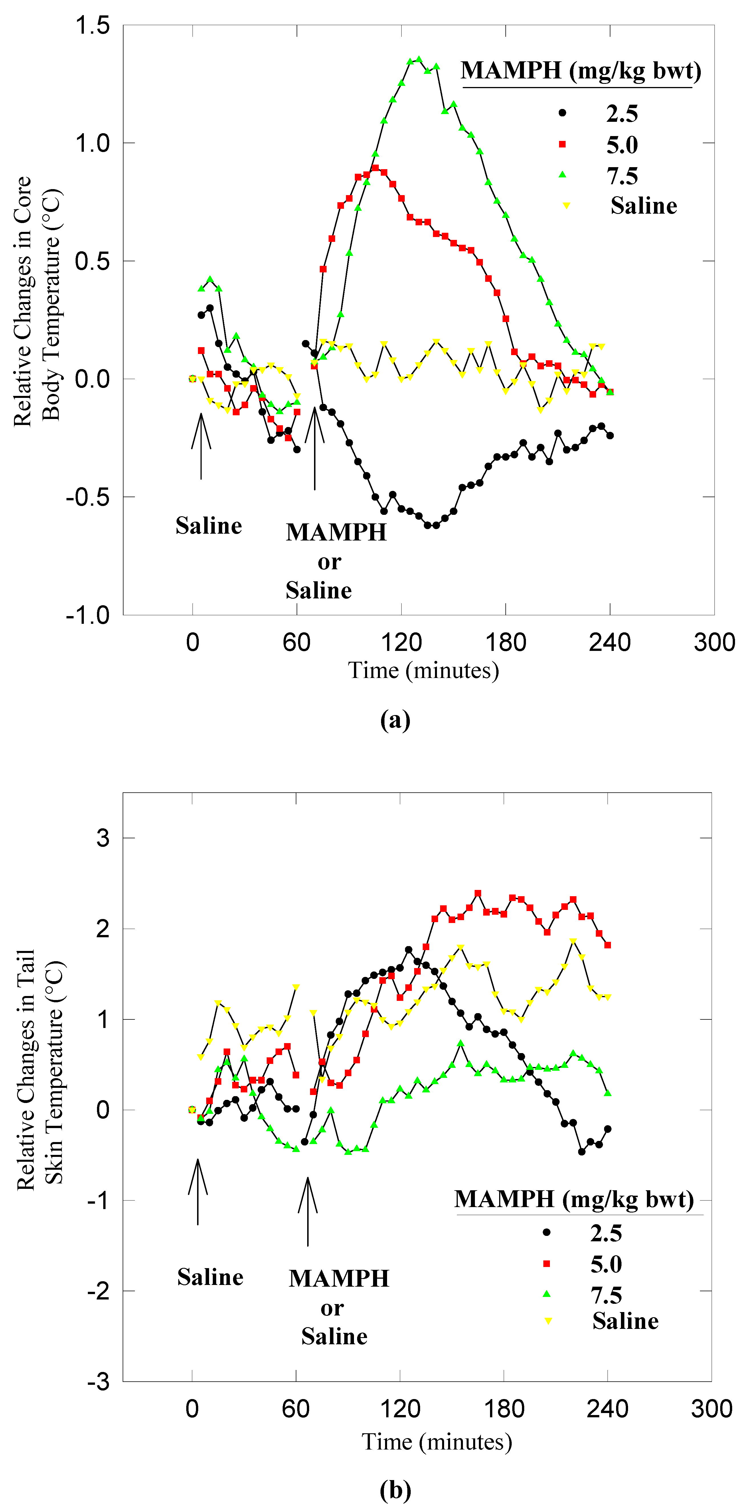
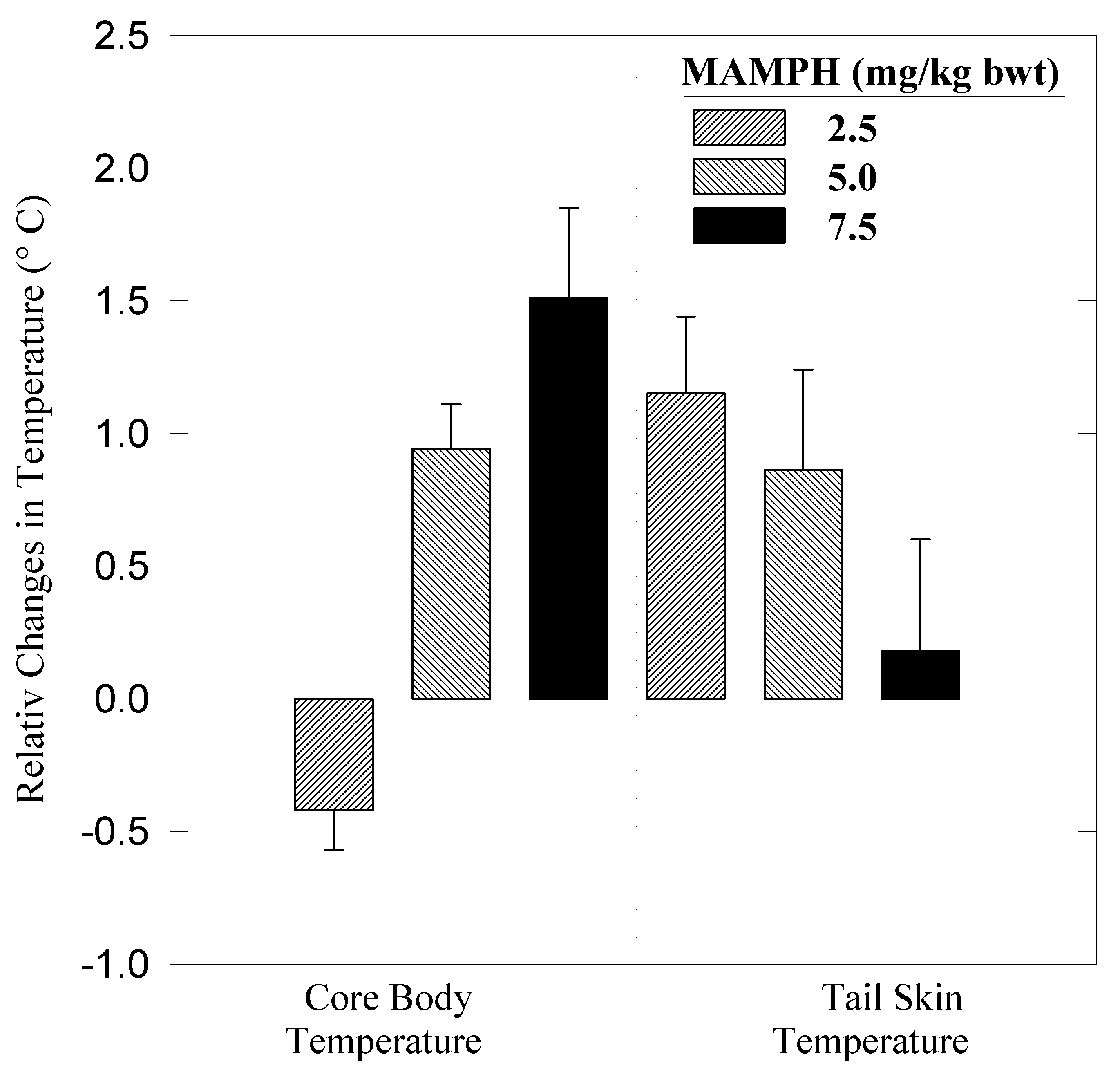
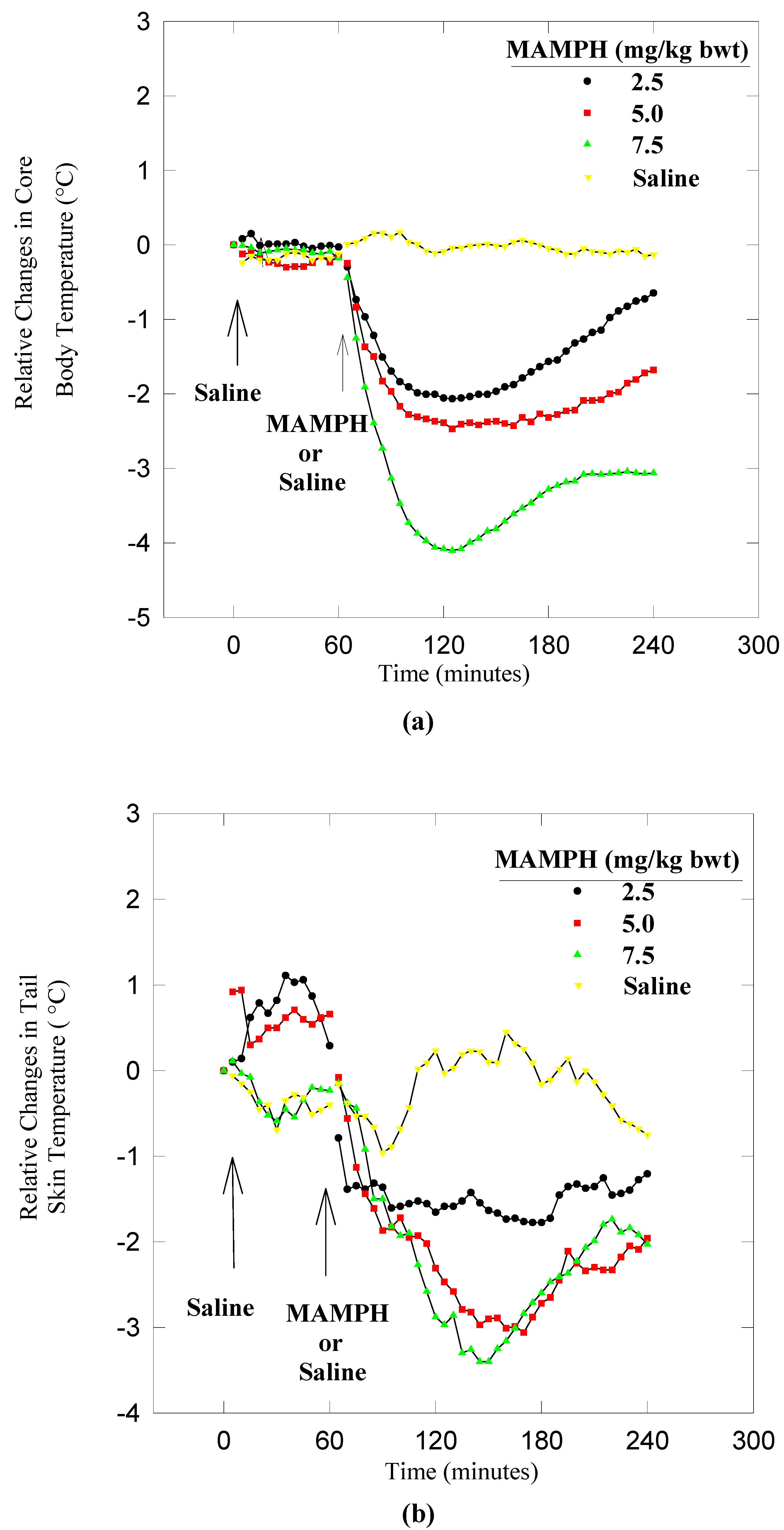
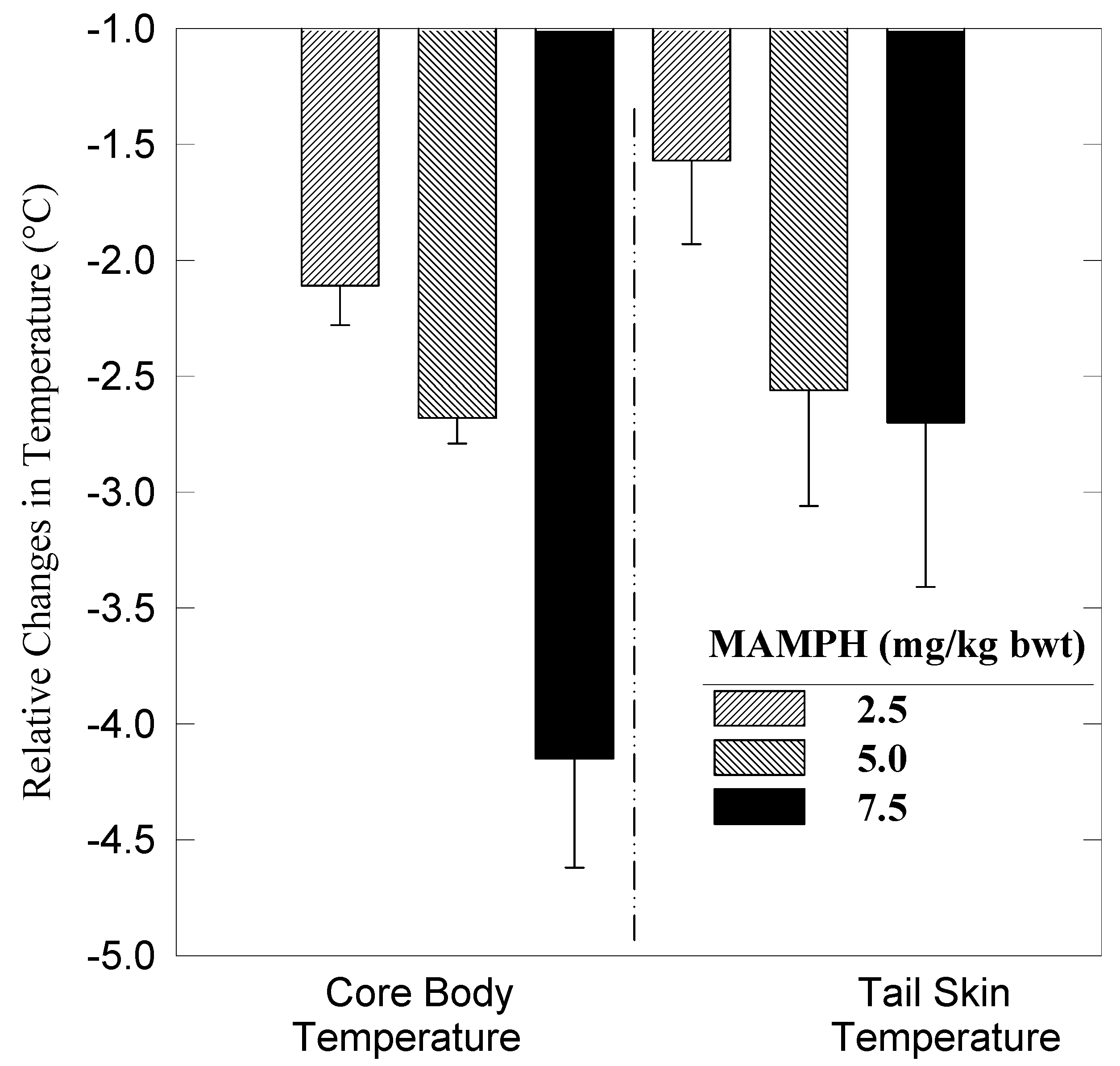
In this study, male Sprague-Dawley rats with body weights of 200-400 grams were used. All animals were housed in the vivarium at the University of Arkansas for Medical Sciences (UAMS) under a photophase of 12 hours (0600 to 1800 hours) and scotophase of 12 hours. Food and water were provided ad libitum. In preparation for experimentation, animals were trained to rectal and tail skin probes and handling for a minimum of seven days. Prior to each experiment, animals received rectal and skin probes for one hour as a probe adjustment period, and held at room temperature (21 ± 1oC) or in the cold room (7 ± 0.5oC) depending upon the type of experiment. Forty male Sprague-Dawley rats were divided into four equal groups. Groups 1, 2 and 3 were injected intraperitoneally (i.p.) with saline and one hour later with an equivolume of MAMPH for doses of 2.5, 5.0, or 7.5 mg/kg per body weight (bwt). Group four was injected with saline/saline as the control. Core body (Tc) and tail skin (Ts) temperatures were recorded with thermistors (YSI series 700) at room temperature (21 ± 1°C) or in a cold room (7 ± 0.5°C) every five minutes for four hours (one hour for saline, and three hours for MAMPH or second saline injection).
Data analysis. Paired and unpaired statistical analysis was used. Tc and Ts of each animal were compared before or after saline or MAMPH treatment. The unpaired analysis (ANOVA, Duncan's Multiple Range test) was performed on control and experimental data from animals treated under identical situations. The paired analysis (ANOVA, Post-hoc "Newman-Keuls" and Student's t-test) was used to compare each individual rat with itself (before and after drug treatment).
All data are presented as relative mean values in order to eliminate, in part, the large variations that appear as a result of the use of absolute values. The changes in Tc and Ts at 7 ± 0.5°C or at 21 ± 1°C were used for analysis of heat conservation or heat dissipation.

Results

Dynamics of heat dissipation

MAMPH at doses of 5.0 and 7.5 mg/kg bwt significantly increased core body temperature (Tc) at room temperature (Figure 1a & Figure 2). At 5.0 mg/kg bwt MAMPH increased Tc from 38.14 ± 0.46oC to 39.04 ± 0.22oC, or by 0.92°C, and at 7.5 mg/kg bwt from 38.45 ± 0.59oC to 39.96 ± 1.18°C, or by 1.51°C. At a dose of 2.5 mg/kg bwt, MAMPH initially increased Tc insignificantly within the first 15 minutes. Following the increase, MAMPH decreased Tc from 38.05 ± 0.46oC to 37.51 ± 0.51oC, or by –0.42°C. One way analysis of variance {ANOVA} of MAMPH treated versus saline controls indicates that the hyperthermic affect of MAMPH is significantly different (p = 0.0001) for all doses of MAMPH. In addition, post-hoc statistical analysis of the data suggested a possible dose-response relationship for the effect of MAMPH on Tc at room temperature. The differences in time (minutes) to reach a maximum response (Tc-max) between doses 2.5, 5.0 and 7.5 mg/kg bwt were not significantly different (p= 0.063). The Tc-max for 2.5 mg/kg bwt was 60.5 ± 13.83, for 5.0 mg/kg bwt was 47.7 ± 24.9, and for 7.5 mg/kg bwt was 70.0 ± 20 minutes.
Statistical analysis of the effector mechanism, tail skin temperature (Ts), for all doses of MAMPH (2.5, 5.0, and 7.5 mg/kg bwt) revealed no significant difference between saline and MAMPH-treated rats (p = 0.9299). However, there was a significant difference in Ts two hours after MAMPH injection at 5.0 mg/kg bwt (p= 0.0001). At this point, the Tc had declined from a maximum of 39.04 ± 0.22°C to 38.26 ± 0.60°C, whereas, Ts had risen from 29.86 ± 1.84 to 32.2 ± 1.10°C. MAMPH at a dose of 7.5 mg/kg bwt, significantly raised Ts in 60% of the rats (N = 6) around 2.5 hours after injection. However, 40% of the animals showed insignificant changes in their Ts and remained this way throughout the duration of the experiment (Figure 1b & Figure 2).
Figure 1. Effects of methamphetamine on a) core body and b) tail skin temperature of male rat at 21 ± 1°C.
Ijms 03 01095 g001
Figure 2. Effects of methamphetamine on core body and tail skin temperature one hour after drug injection at 21 ± 1°C.
Ijms 03 01095 g002

Dynamics of heat conservation

MAMPH at doses of 2.5, 5.0, and 7.5 mg/kg bwt, simultaneously decreased both Tc and Ts significantly in the cold room (Figure 3 and Figure 4). At a dose of 2.5 mg/kg bwt, MAMPH decreased Tc from 38.01 ± 0.34°C to a minimum average value (Tc-min) of 35.98 ± 0.71°C, or by –2.22°C. Following Tc, the Ts decreased from 18.64 ± 2.46°C to 17.11 ± 2.43°C, or by –1.57°C. At a dose of 5.0 mg/kg bwt, MAMPH decreased Tc from 37.97 ± 0.42°C to 35.38 ± 0.82°C, or by –2.68°C, and Ts from 19.22 ± 1.48°C to 16.39 ± 1.36°C, or by –2.56°C. At a dose of 7.5 mg/kg bwt, MAMPH decreased Tc from 38.16 ± 0.51 to 34.01 ± 1.60°C, or by –4.15°C, and Ts from 19.82 ± 1.96°C to 17.07 ± 2.32 °C, or by –2.7°C.
Statistical analysis of the data indicates that the hypothermic affect of MAMPH is significant at all doses (p = 0.0001). Post hoc statistical analysis of the data showed that only the effects of 5.0 and 7.5 mg/kg bwt of MAMPH on Tc were significantly different from each other (Figure 3b). Analysis of the variance and Post-hoc statistical analysis show that the differences in time (minutes) to reach to a minimum response or Tc-min between doses of 2.5, 5.0 and 7.5 mg/kg bwt were not significantly different. The Tc-min for 2.5 mg/kg bwt was 55 ± 15.97, for 5.0-mg/kg bwt was 65 ± 29.32, and for 7.5-mg/kg bwt was 63.50 ± 16.51 minutes.
Figure 3. Effects of methamphetamine on a) core body and b) tail skin temperature at 7± 0.5°C.
Ijms 03 01095 g003
Figure 4. Effects of methamphetamine on core body and tail skin temperature one hour after drug injection at 7± 0.5°C.
Ijms 03 01095 g004

Discussion

Methamphetamine significantly increased Tc at doses of ≥ 5 mg/kg bwt at room temperature, and suppressed it at doses of 2.5 mg/kg bwt at cold environmental temperature. Analysis of the heat exchange or thermo-effectors mechanism as monitored by Ts, revealed that the physiological mechanism underlying the hypo- and hyperthermic effects of MAMPH is different. At room temperature, MAMPH induced a condition similar to hyperthermia demonstrated by Raman et al. [25]. Exposing male rats to a high ambient temperature of 30-40 oC resulted in an increase in Tc but not in Ts or blood flow to the tail [25]. In a normal situation [26,27,28,29], rat vasodilate the vasculature to the tail or it increases Ts to dissipate heat to the environment when it is challenged with a rise in its core body temperature. Methamphetamine at 21 ± 1°C seems to induce hyperthermia. Although, MAMPH induced a significant increase in Tc, the tail did not respond (p> 0.05). Indeed, the heat dissipating mechanism was delayed in response (p< 0.05) by an hour to the rise in Tc at 5.00 mg/kg bwt of MAMPH.
Is the mechanism underlying the hyperthermic effect of MAMPH regulated similarly to fever triggered by pyrogens? It has been shown [30] that pyrogens trigger a rise in Tc “set point” or “set range” to a higher level. The prostaglandin synthetase inhibitors such as indomethacin inhibit this effect of pyrogens. In addition, during fever, the recruitment of the heat gain mechanism and suppression of the heat loss mechanism depends upon the environmental temperature [31]. At room temperature, the heat loss mechanism is suppressed. Thus, the peripheral vascular system that is responsible for heat dissipation does not respond despite a rise in body temperature. At cold ambient temperature, the heat gain mechanism is activated. Thus, the peripheral vasculature was vasoconstricted.
The effect of MAMPH at room temperature seems to be similar to the effect of pyrogens on heat gain/heat conservation. Mohaghegh et al. showed that the hyperthermic effect of MAMPH was blocked by 5.0 mg/kg dose injections of indomethacin [18]. Thus, MAMPH, like pyrogens can increase the “set point” or “set range” of the core body temperature. With a rise in the body's temperature “set point” accompanied with a delay in response of the tail vasculature to dissipate heat, there will be a hyperthermia or chemically induced fever. In contrast to pyrogens, MAMPH at doses of (2.5-mg/kg bwt at cold ambient temperature induced a dose dependent hypothermia. However, this hypothermic effect of MAMPH was unexpectedly associated with a significant (p< 0.05) decrease in Ts. An increase rather a decrease in Ts as previously described [28] was unexpected. In addition, the hypothermic effect of MAMPH was not inhibited by 5.0 or 10 mg/kg bwt of indomethacin [17 and 18]. Previously, it been shown that regulated hypothermia is mediated via prostaglandin E1 system [32]. Thus, probably, MAMPH at 7 ± 0.5°C forced the animal into hypothermia via a profound suppression of metabolic heat production. What may account for this suppression of heat dissipation, despite the hypothermic effect of MAMPH is not clear. It is possible that the hypothermic animal tries to regulate the amount of heat loss by some unknown processes.

Acknowledgement

This research was supported in part by UAMS-GSRF 117-317-1010 & RCMI-G12RR03032 to the senior author.

References and Notes

  1. Ricuarte, G.A.; Schuster, C.R.; Seiden, L.S. Long-term effects of repeated methyl amphetamine administration on dopamine and serotonin neurons in the rat brain: A regional study. Brain Res. 1980, 19, 153–163. [Google Scholar] [CrossRef]
  2. Wagner, G.C.; Ricuarte, G.A.; Seiden, L.S.; Schuster, C.R.; Miller, R.J.; Westley, J. Long-lasting depletions of striatal dopamine and loss of dopamine uptake sites following repeated administration of methamphetamine. Brain Res. 1980, 181, 151–160. [Google Scholar] [CrossRef]
  3. Seiden, L.S. Neurotoxicity of methamphetamine and related drugs. In Psychopharmacology: A Third Generation of Progress; Meltzer, H.Y., Ed.; Raven Press: New York, 1987; pp. 359–366. [Google Scholar]
  4. Kogan, F.J.; Nichols, W.K.; Gibb, J.W. Influence of methamphetamine on nigral and striatal tyrosine hydroxylase activity and on striatal dopamine levels. Eur. J. Pharmacol. 1976, 36, 363–371. [Google Scholar]
  5. Xie, T.; McCann, U.D.; Kim, S. Yuan; Ricaurte, G.A. Effect of temperature on dopamine transporter function and intracellular accumulation of methamphetamine: Implications for methamphetamine-induced dopaminergic neurotoxicity. J. Neurosci. 2000, 20, 7838–7845. [Google Scholar]
  6. Brown, J.M.; Riddle, E.L.; Sandoval, V.; Weston, R.K.; Hanson, J.E.; Crosby, M.J.; Ugarte, Y.V.; Gibb, J.W.; Hanson, G.R.; Fleckenstein, A.E. A single methamphetamine administration rapidly decreases vesicular dopamine uptake. J. Pharmacol. Exp. Ther. 2002, 302, 497–501. [Google Scholar] [CrossRef] [PubMed]
  7. Callahan, B.T.; Cord, B.J.; Yuan, J.; McCann, U.D.; Ricaurte, G.A. Inhibitors of Na (+)/H(+) and Na(+)/Ca(2+) exchange potentiate methamphetamine-induced dopamine neurotoxicity: possible role of ionic dysregulation in methamphetamine neurotoxicity. J. Neurochem. 2001, 77, 1348–62. [Google Scholar]
  8. Bakhit, C.; Gibb, J.W. Methamphetamine-induced depression of tryptophan hydroxylase: Recovery following acute treatment. Eur. J. Pharmacol. 1981, 76, 229–233. [Google Scholar] [CrossRef] [PubMed]
  9. Trulson, M.E.; Trulson, V.M. Effects of chronic methamphetamine administration on tryptophan hydroxylase activity, [3H] serotonin synaptosomal uptake, and serotonin metabolism in rat brain following systemic tryptophan loading. Neuropharmacology 1982, 21, 521–527. [Google Scholar]
  10. Kuczenski, R.; Segal, D.S. In vivo measures of monoamines during amphetamine-induced behaviors in rats. Prog. Neuro-Psychopharmacol. Biol. Psychiatry 1991, 14, S37–S50. [Google Scholar]
  11. Randrup, A.; Munkvad, I. Pharmacology and physiology of stereotyped behavior. J. Psychiat. Res. 1974, 11, 1–10. [Google Scholar] [CrossRef] [PubMed]
  12. Bo, P.; Marchioni, E.; Dollocchio, C.; Albergati, A.; Savoldi, F. Antagonism of EEGgraphic and behavioral effects of methamphetamine by selective receptor blockers (SCH 23390 and Raclopride) in the rabbit. Prog. Neuro-Psychopharmacol. & Biol. Psychit. 1991, 15, 803–815. [Google Scholar] [CrossRef]
  13. Wagstaff, J.D.; Bush, L.G.; Gibb, J.W.; Hanson, G.R. Endogenous neurotensin antagonizes methamphetamine-enhanced dopaminergic activity. Brain Res. 1994, 665, 237–244. [Google Scholar]
  14. Segal, D.S. Kuczenski, RRepeated binge exposures to amphetamine and methamphetamine: Behavioral and neurochemical characterization. J. Pharmacol. Exp. Ther. 1997, 282, 561–573. [Google Scholar]
  15. Itzhak, Y. Modulation of cocaine- and Methamphetamine-induced behavioral sensitization by inhibition of brain nitric oxide synthase. J. Pharmacol. Exp. Ther. 1997, 282, 521–527. [Google Scholar]
  16. Yehuda, S.; Wutman, R.J. Paradoxical effects of D-amphetamine on behavioral thermoregulation: Possible mediation by brain dopamine. J. Pharmacol. Expe. Therapy. 1974, 1990, 118–122. [Google Scholar]
  17. Mohaghegh, R. A. Physiological phenomena underlying the acute thermic effects of methamphetamine on rats in different environmental temperatures. Ph.D. Dissertation, University of Arkansas for Medical Sciences, Little Rock, AR, USA, 1994. [Google Scholar]
  18. Mohaghegh, R.A.; Soulsby, M.E.; Skinner, R.D.; Kennedy, R.H. The interaction between the central and peripheral nervous systems in mediating the thermic effect of methamphetamine. Annals New York Acad. Sci. 1997, 813, 197–203. [Google Scholar] [CrossRef]
  19. Bowyer, J.F.; Tank, A.W.; Newport, G.D.; Slikker, W., Jr.; Ali, S.F.; Holson, R.R. The influence of environmental temperature on the transient effects of methamphetamine on dopamine levels and dopamine release in rat striatum. J. Pharmacol. Exp. Ther. 1992, 260, 817–824. [Google Scholar] [PubMed]
  20. Bowyer, J.F.; Gough, B.; Slikker, W., Jr.; Lipe, G.W.; Newport, G.D.; Holson, R.R. Effects of a cold environment or age on methamphetamine-induced dopamine release in the caudate putamen of female rats. Pharmacol. Biochem. Behav. 1993, 44, 87–98. [Google Scholar] [CrossRef] [PubMed]
  21. Bowyer, J.F.; Davies, D.L.; Schmued, L.; Broening, H.W.; Newport, G.D.; Slikker, W., Jr.; Holson, R.R. Further studies of the role of hyperthermia in methamphetamine neurotoxicity. J. Pharmacol. Exp. Ther. 1994, 268, 1571–1580. [Google Scholar]
  22. Ali, S.F.; Newport, G.D.; Holson, R.R.; Slikker, W., Jr.; Bowyer, J.F. Low environmental temperatures or pharmacologic agents that produce hypothermia decrease methamphetamine neurotoxicity in mice. Brain Res. 1994, 658, 33–38. [Google Scholar] [CrossRef] [PubMed]
  23. Ali, S.F.; Newport, G.D.; Holson, R.R.; Slikker, W., Jr.; Bowyer, J.F. Low environmental temperatures or pharmacologic agents that produce hyperthermia decrease methamphetamine neurotoxicity in mice. Ann. NY Acad. Sci. 1995, 765, 338. [Google Scholar] [CrossRef]
  24. Ali, S.F.; Newport, G.D.; Slikker, W., Jr. Methamphetamine-induced dopaminergic toxicity in mice. Role of environmental and pharmacological agents. Ann. NY Acad. Sci. 1996, 801, 187–198. [Google Scholar] [CrossRef] [PubMed]
  25. Raman, E.R.; Roberts, M.F.; Vanhuyse, V.J. Body temperature control of rat tail blood flow. Am. J. Physiol. 1983, 245, R426–R432. [Google Scholar]
  26. Johansen, R. Heat exchanger through the muskrat tail evidence for vasodilator nerves to the skin. Acta Physiol. Scan. 1962, 55, 160–169. [Google Scholar] [CrossRef]
  27. Rand, R.P.; Burton, A.C.; Ing, T. The tail of the rat in temperature regulation and acclimatization. Can. J. Physiol. Pharmacol. 1965, 43, 257–267. [Google Scholar] [CrossRef]
  28. Gordon, J.G. Relationship between preferred ambient temperature and autonomic thermoregulatory function in rat. Am. J. Physiol. 1987, 252, R1130–R1137. [Google Scholar]
  29. Romanovsky, A.; Ivanov, I. A.; Shimansky, P.Y. Molecular biology of thermoregulation selected contribution: ambient temperature for experiments in rats: a new method for determining the zone of thermal neutrality. J. Appl. Physiol. 2002, 92, 2667–2679. [Google Scholar]
  30. Lipton, J.M.; Welch, J.P.; Clark, W. G. Change in body temperature produced by injecting prostaglandin E1, EGTA and bacterial endotoxins into the PO/AH region and the medulla oblongata of the rat. Experimentia 1973, 29, S06–S08. [Google Scholar]
  31. Stitt, J. T. Fever versus hyperthermia. Fedn. Proc. 1979, 38, 39–43. [Google Scholar]
  32. Shimada, S.G.; Stitt, J.T.; Angelogianni, P. Effects of cold and capsaicin desensitization on prostaglandin E hypothermia in rats. J. Applied Physiol. 1990, 68, 2618–2622. [Google Scholar]

