Article

Preparation and Characterization of a Chloroperoxidase-like Catalytic Antibody

1
College of Life Science and Agriculture, Key Laboratory of Tropic Biological Resources of Ministry of Education, Hainan University, Haikou 570228, P. R. China
2
College of Chemistry, Huazhong Normal University, Wuhan 430079, P. R. China
3
Research Center for Wildlife Animal Diseases, Key Laboratory of Animal Ecology and Conservation Biology, Institute of Zoology, Chinese Academy of Sciences, Beijing 100080, P. R. China
4
College of Life Science, Huazhong Normal University, Wuhan 430079, P. R. China
*
Authors to whom correspondence should be addressed.
Int. J. Mol. Sci. 2007, 8(5), 445-454; https://doi.org/10.3390/i8050445
Submission received: 10 February 2007 / Revised: 12 April 2007 / Accepted: 16 May 2006 / Published: 29 May 2007
(This article belongs to the Section Molecular Recognition)

Abstract

:
The small molecule, meso-tetra(α,α,α,α-o-phenylacetamidophenyl) porphyrin (Mr1147.0) was used as complete antigen to elicit MAb through the immunization and cell fusion techniques. The MAb 1F2 obtained was demonstrated to be very pure by MALDI/TOFMS. The subtype of MAb 1F2 is IgG2a, which has a relative molecular weight of 156,678.8 Da.No significant change in the intensity of absorption peaks in UV and CD spectra was observed over a pH range between 6 and 12. The high stability of the abzyme and the tight binding between Fe porphyrin and antibody were also demonstrated. Vmax, Km, κcat, κcat/Km for abzyme are 5.18 × 10⁻⁸ Ms−1, 1.50 × 10⁻⁸ M, 0.518 s−1, 3.45 × 10⁷ M−1s−1, respectively. The data obtained indicate that catalytic antibody has high catalytic activity. The chloroperoxidase activity of MAb 1F2-Fe porphyrin complex is stable from 10 °C to 60 °C.

1. Introduction

Iodination of free tyrosine and tyrosine in protein is mediated by an enzyme present in thyroid homogenates and subcellular fractions [1,2]. An iodide peroxide has been shown to be present in animal thyroid tissue and to be responsible for the iodination reactions through formation of an oxidized iodinating intermediate [3]. This enzyme is chloroperoxidase (CPO) (EC1.11.1.10). It is a heme protein of Mr.~42,000 Da containing one ferriprotoporphyrin IX per molecule [4]. The enzyme uses H2O2 as the oxidant for the peroxidative formation of a carbon-halogen bond between Cl-, Br-, I- and a suitable halogen acceptor. But it is unable to utilize F for this reaction.
Iodination of tyrosine residues in thymoglobulin, or in its subunits, is one of the essential steps in the formation of thyroxin in the thyroid gland. Iodide can be rapidly bound as 3-iodotyrisine, 3,5- diiodotyrosine, and thyroxin during the chloroperoxidase-catalyzed iodination [5]. Thus, the enzyme plays an important role in the metabolism and physiological function of thyroxin. Deficiency of chloroperoxidase will result in some serious thyroid diseases.
Most of the catalytic antibodies reported to date have been obtained by producing monoclonal antibodies that have been elicited against transition state analogs [69]. In those cases, only the antibody protein, through some amino acid residues of its binding site, is fully responsible for catalysis. In some cases, however, monoclonal antibodies require the presence of a cofactor, such as inorganic cofactors, or metal ions, or metal complexes, to be catalytic. Furthermore, the reaction transition state structure of tyrosine and thyroxin and the detailed catalytic mechanism of chloroperoxidase are not clear, so, it is very difficult to prepare the antibodies with chloroperoxidase activity targeted to transition state analogs.
Based on the initial mechanism of chloroperoxidase [10,11], we undertook the experiments described in this report. meso-Tetra(α,α,α,α-o-phenylacetamidophenyl)porphyrin was used as complete antigen to immunize Balb/c mice. MAb was raised through hybridoma technique, and then the properties of antibody were analysized. The study broadens the scope of antibody-catalyzed reactions and lays a strong foundation for further study on chloroperoxidase mimics.

2. Results and Discussion

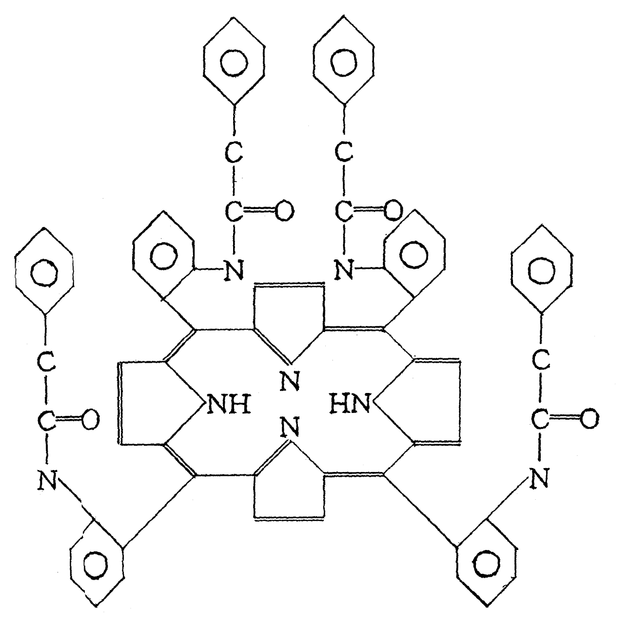
2.1 Preparation of antigen

Synthesized porphyrin (Figure 1) was demonstrated to be the complete antigen by UV spectrum and elemental analysis (C76H58O4N8, Exp/The %: C 79.55/79.56, H 5.18/5.10, N 9.78/9.77, O 5.49/5.57) and MALDI/TOF MS. The relative molecular weight is 1,147.0 Da, which is consistent with the theoretical value of 1,147.3 Da.

2.2 Building of hybridoma cells

Female Balb/c mice were immunized with complete antigen directly. And the splenocytes were fused with Sp2/0 myeloma cells. One line of positive hybridomas, named as 1F2, was obtained by ELISA, screening, and cloning.

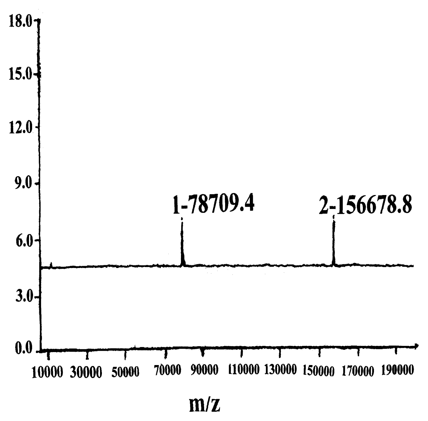
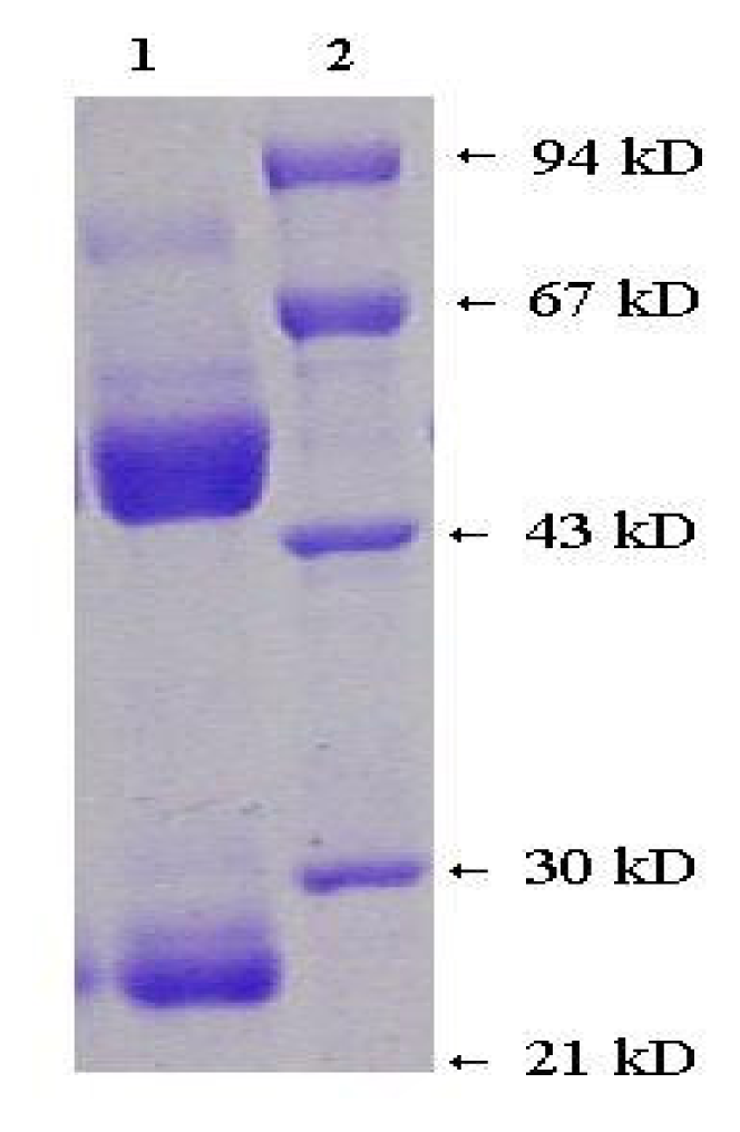
2.3 Antibody properties

The analytical result of antibody-subtype show 1F2 belongs to the IgG2a subtype. The MALDI/TOF MS result for MAb 1F2 show a peak with a mass-to-charge ratio (m/z) of 156678.8 Da that can be assigned as the molecular ion peak of MAb 1F2, namely [M+H]+. The mass spectrum peak with mass-to- charge ratio (m/z) 78709.4 Da can be assigned as the double-charged molecular peak of MAb 1F2 (Figure 2). The prolongated double charge peak confirms the validity of the molecular peak. Additionally, there was no other protein peak, which demonstrates the high purity of MAb 1F2.
After multiple-step purification, the monoclonal antibody 1F2 pretreated with 2.5 % SDS and 10 % β-mercaptoethanol appears only as a light chain band (25,000 ± 1000 Da) and a heavy chain band (55,000 ± 1,000 Da) on SDS-PAGE electrophoresis, which suggests that 1F2 has already been homogenized. The relative molecular weight of the antibody is 160,000 ± 1,000 Da (Figure 3). The dissociation constant of antibody-Fe porphyrin was determined to be 2.084×10⁻¹⁰ M and fluorescence quenching was used to determine the combining ratio of antibody-to-porphyrin, and the result is 1:1 [12].

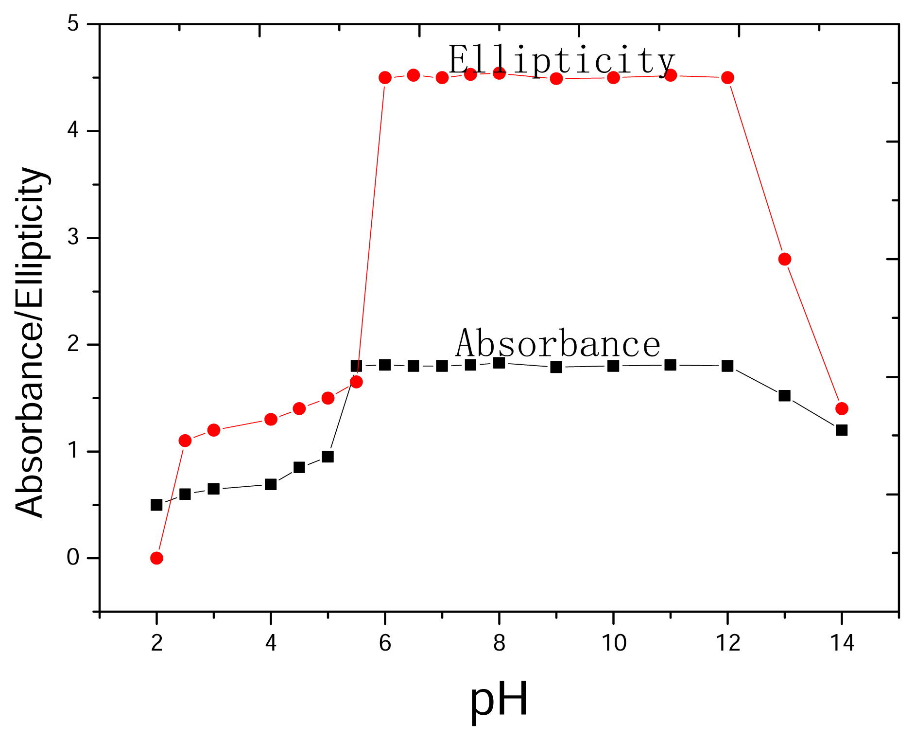
2.4 Dependence on pH

In order to study the dependence of the induced circular dichroism (CD) spectrum on pH, the solution of Fe meso-tetra(α,α,α,α-o-phenylacetamidophenyl)porphyrin with excess antibody 1F2 at several pH values were prepared and both their UV absorbance (at the Soret maximum) and CD ellipticity were measured. The UV absorbance almost remains constant over a broad pH range, viz. between 5.5 and 12. At pH values below 5, absorbance is reduced and the signals are significantly broadened (Figure 4).
The CD spectrum also provides valuable information concerning the antibody combining site. It is unchanged (in both intensity and shape) over a range of six pH units (between 6 and 12). This remarkable stability of the spectrum probably reflect the dominant role of hydrophobic interactions between the antigen - antibody binding site. Apparently, there is no change in the relative orientation of the porphyrin in the antibody combining sites and the distances between the porphyrin and the CDR (complementarily –determining region) residues responsible for the induced CD effect. The similar behavior of both UV and CD spectra indicate that catalytic antibody was remarkably constant over a range of six pH units (Figure 4).

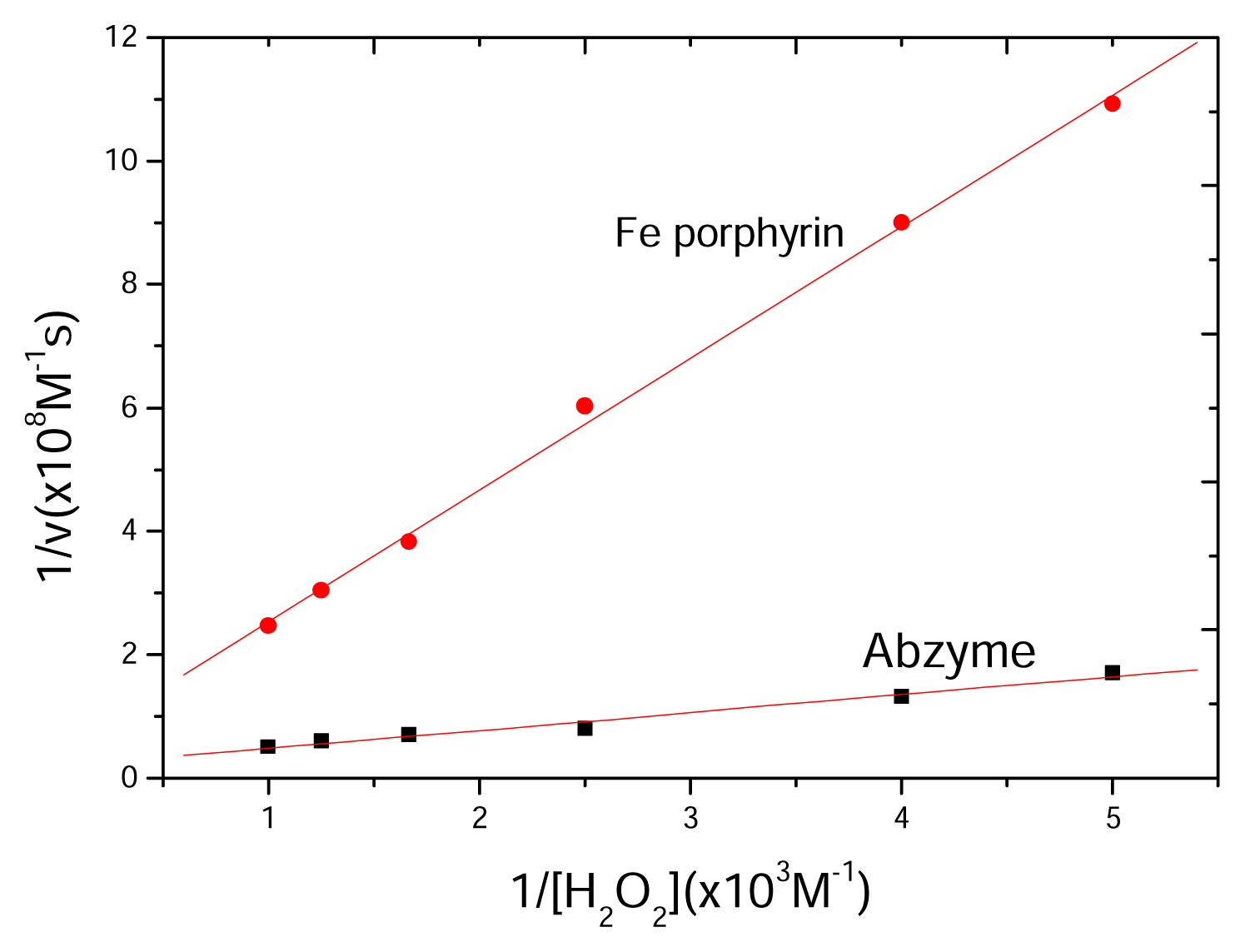
2.5 Kinetics of catalytic reaction

In reactive systems that the concentration of monochlorodimedone was fixed when the concentration of H2O2 varied between 0.2 mM and 1 mM, Catalytic activities of abzyme and Fe porphyrin were determined, respectively. The linear kinetic curves were obtained according to Lineweaver-Burk equation. This shows that the reaction catalyzed by abzyme complies with the kinetic principle of Lineweaver-Burk equation (Figure 5, Table 1).
Fe porphyrin has catalytic activity alone for the chloroperoxidative reaction. MAb 1F2 has no the chloroperoxidase activity alone. But the catalytic activity of Fe porphyrin is significantly increased when their complex is formed. With the addition of corresponding antigen to the reactive system, the reaction catalyzed by the complex of Fe porphyrin and MAb 1F2 is repressed. These results show that the chloroperoxidative reaction catalyzed by the complex of Fe porphyrin and MAb 1F2 actually results from the catalytic action of abzyme, not from the pollution of the chloroperoxidase.

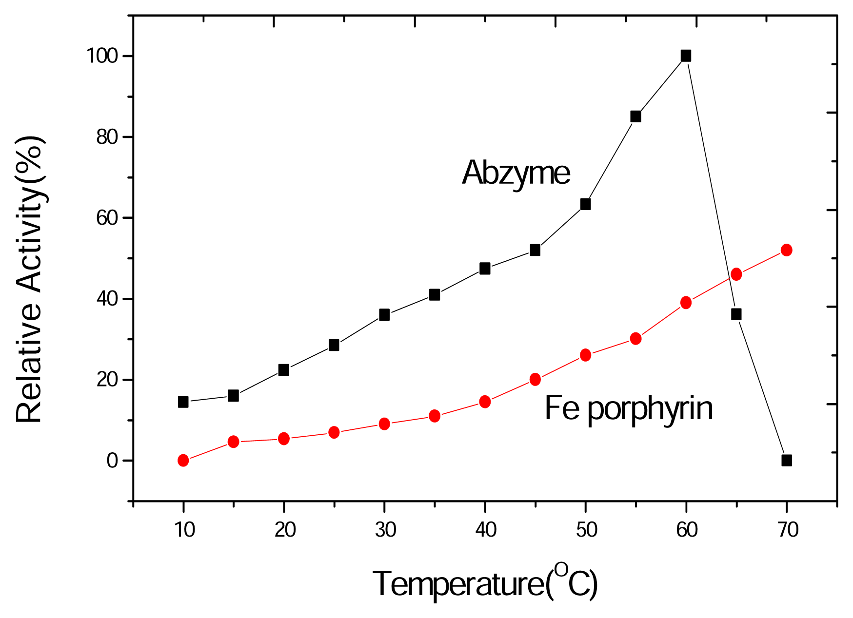
2.6 Thermostability of the chloroperoxidase activity

The chloroperoxidase activity of the MAb 1F2-Fe porphyrin complex was assayed at different temperatures varying between 10 °C and 70 °C and compared with that of Fe porphyrin. With both catalysts, the primary rate of chlorination of monochlorodimedone by H2O2 increases with the temperature below 60 °C. The abzyme activity significantly decrease above 60 °C and almost disappeared at 70 °C. Fe porphyrin still has higher catalytic activity at 70 °C. These results suggest that the denaturation reaction of the abzyme protein may occur at high temperature and cause the change of stereo path structure near the active sites. Thus the catalytic activity of the abzyme rapidly decreases at 70 °C (Figure 6). Compared with the catalytic activity of the free prosthetic group of Fe porphyrin, a conclusion can be drawn that the dissociation of the prosthetic group from MAb 1F2 didn’t happen. The temperature dependence of Fe porphyrin is different from that of abzyme and similar with that of inorganic catalyst.
As seen from these results, the small molecule meso-tetra(α,α,α,α-o-phenylacetamidophenyl) porphyrin could be used as complete antigen to immunize Balb/c mice and induce MAb 1F2. The purity of MAb 1F2 was indicated to be very high by MALDI/TOF MS and SDS-PAGE. The subtype of MAb 1F2 is IgG2a. A relative molecular weight of MAb 1F2 was determined to be 156,678.8 Da by MALDI/TOF MS. The intensities of UV and CD spectra over a pH range (between 6 and 12) almost remain constant, which reveals that the tight binding of Fe porphyrin and MAb 1F2 and the high stability of abzyme. The chloroperoxidase activity of MAb 1F2-Fe porphyrin complex appears thermostable until 60 °C. But these results still show that if a small molecular compound has been rationally designed and has a definite composition of chemistry and complex structure, it is immunogenic even if it has not been linked with carrier. Among the antibodies against porphyrins, only four antibodies obtained have shown, in the presence of the corresponding iron(III)-porphyrin cofactor, a significant peroxides and hem protein activity, Only one antibody was reported to exhibit, in the presence of Mn(III)-tetrakis(p-carboxyvinylphenyl)porphyrin, a weak monooxygenase-like activity [12]. These five catalytic antibodies were elicited against hapten(porphyrin)-carrier(protein). Because hapten is small molecule which has no immunogenic or little immunogenic character, Thomas [12] suggested that aromatic groups should be included in hapten design, and much more immunogenic of hapten. In this paper, the small molecule meso-tetra(α,α,α,α-o-phenylacetamidophenyl) porphyrin was used as complete antigen to elicit chloroperoxidase-like abzyme, because meso-tetra(α,α,α,α-o-phenylacetamidophenyl) porphyrin has eight phenyl groups and higher hydrophobic properties. The experimental results show that it has stronger immungenicity. Having used small molecular porphyrin to elicit chloroperoxidase-like abzyme, a subsequent study aimed at increasing abzyme activity is under way.

3. Experimental section

3.1 Materials and Reagents

6~8 Week old Balb/c mice weighing (20 ± 2) g were supplied by the Laboratory Animal Center of AMMS.HRP-linked goat anti-mouse antibody, mouse monoclonal antibody isotyping reagent kit, BSA, pyrogallic acid, and sinapinic acid were purchased from Sigma-Aldrich (St. Louis, MO). SDS-polyacrylamide gel, Sephadex G-10, Hitrap-protein A were obtained from Amersharm-Pharmacia (Piscataway, NJ). HAT solution, HT solution, and IMDM were supplied by GIBCOBRL(Carlsbad, CA). Polyethylene glycol~4000 was purchased from Merck (Darmstadt, Germany). Cell culture plates (96 well and 24 well) were obtained from Nunc (Roskilde, NJ). All other chemicals used are of analytical grade.

3.2 Antigen Preparation

Meso-Tetra(α,α,α,α-o-phenylacetamidophenyl)porphyrin was synthesized as previously described [4,7]. Briefly, condensation of o-nitrobenzaldehyde and pyrrole followed by reduction of the meso-tetra( o-nitrophenyl)porphyrin led to a satisfactory yield of meso-tetra(o-aminophenyl)porphyrin, H2TamPP. Thin-layer chromatography on silica gel gave excellent separation of four components which, in ratio 1:2:4:1, were presumed to be the four atropisomers in statistical abundance. With Rf values based on their expected polarity and considering the relative amounts of the individual atropisomers, these are provisionally assigned as α,β,α,β-,α, α, β, β-,α, α, α, β-, andα, α, α, α-, with the most polar tetra-α-atropisomer, moving most slowly. Isolation of α,α,α,α-H2TamPP by silica gel column chromatography afforded gram quantities of product. The mixture containing the remaining three atropisomers was re-equilibrated in boiling toluene followed by chromatography to isolate more of the α,α,α,α-atropisomer. Repetition of these steps leads to ultimate conversion of nearly all of the H2TamPP into the desired α,α,α,α-H2TamPP. The four amino atropisomers are rather stable in solution at 25 °C. The amino groups of H2TamPP are also more reactive. The α,α,α,α-atropisomer of H2TamPP could be further modified and the configurationally energy barrier raised through reaction with phenylacetyl chloride forming amide. Phenylacetyl chloride gave 72 % yield of meso-tetra(α,α,α,α-o-phenylacetamidophenyl) porphyrin.

3.3 Preparation and purification of monoclonal antibody against meso-tetra(α,α,α,α-o-phenylacetamidophenyl) porphyrin

MAb was raised against meso-tetra (α,α,α,α-o-phenylacetamidophenyl)porphyrin through the hybridoma technique [13]. Due to antigen’s insolubility, dimethyl sulfoxide (DMSO) was added to help it dissolve. Briefly, six seven-week-old, female Balb/c mice were bled to obtain control serum, and then interpritoreal injections with 0.2 ml suspension of 1 mg/ml solution of the antigen mixed with equal volume of complete Freund’s adjuvant for the initial injection or Freund’s incomplete adjuvant for subsequent injection. The mice were immunized with the same batch of meso-tetra(α,α,α,α-o-phenylacetamidophenyl) porphyrin at two-week intervals for four orders, and then hyper-immunized with the antigen in the absence of adjuvant three days before fusion. Hybridoma was prepared by fusion of immunized mouse splenocytes with mouse myeloma cells (Sp2/0) with ethylene glycol ~4000. ELISA was performed to screen out one line of positive hybridomas, and the positive cell line was cloned 3 times and propagated in vitro and in vivo. After interpritoreal injection with the hybridomas, ascites were gathered and ELISA was performed to measure its antibody titer. The monoclonal antibody 1F2 with higher-titer ascites was purified by 50 % (NH4) 2SO4 fractionation (4 °C, pH 7.2), anion exchange chromatography on DEAE-52 (5 mM Tris, pH 8.0, 20–400 mM NaCl, the antibody eluted in the 100 mM NaCl, pH 7.5) and affinity purification on a HiTrap-protein A column. The subtype of monoclonal antibody 1F2 was determined with mouse monoclonal antibody isotyping reagent kit. The amount of protein was determined as previously described protocol [14]. SDS-PAGE, MALDI/TOF MS was used to check the homogeneity and the purity as previously described [15]. Fluorescence quenching was used to determine the dissociation constant [16].

3.4 Preparation of Fe-Porphyrin

Ferric chloride and meso-tetra(α,α,α,α-o-phenylacetamidophenyl)porphyrin (mole ratio 5:1) were dissolved in dimethylformamide (DMF) and heated to reflux for 30 minutes. The UV spectrum of the reagent mixture was determined. The reduction of the absorbent peaks in Soret band of porphyrin from 4 to 2 suggests that the compound of Fe porphyrin generated. The refluxing reaction continued for 3 hours. And then the solution was cooled and dropped into distilled water. Fe porphyrin was precipitated and filtrated, repeated 3 times. Then, the precipitate was placed in a drier. Minimal DMSO was added to help dissolve some weighted Fe porphyrin, and the solution was diluted with 0.1 M PBS (pH 6.0).

3.5 Dependence on pH

Stock solutions containing Fe meso-tetra (α,α,α,α-o-phenylacetamidophenyl)porphyrin (10−6 M) in 0.1 M PBS (pH 6.0, 20 °C), were prepared.

3.6 Assay of chloroperoxidase activity

The standard assay for chloroperoxidase activity is based on the loss of absorbance at 278 nm accompanying the conversion of 1,1-dimethyl-4-chloro-3,5-cyclohexanedione (monochlorodimedone) [17]. The standard assay mixture contains 300 μmoles H3PO4- K2H2PO4 (pH 2.75), 60 μmoles KCl, 0.6~3.0 μmoles of H2O2, 0.3 μmole monochlorodimedone, 0.1 μM enzyme in a total volume of 3 ml. The reaction was started by the addition of the complex of antibody and Fe porphyrin or Fe porphyrin. incubated at 20 ºC. The change in absorbance at 278 nm as a function of time was continuously recorded on an Uvikon 922 UV spectrophotometer (Kontron). The rate of reaction was linear for 3 to 4 min. Lineweaver-Burk equation was used for determining kinetics of catalytic reaction:
1 / v = K m / V max · 1 / [ S ] + 1 / V max
where, v is the reactive rate; Km is the constant of Michaelis-Menten; [S] is the concentration of substrate; Vmax is the maximal reactive rate.

3.7 Thermostability of chloroperoxidase

The reaction mixture contained 300 μmoles H3PO4- K2H2PO4 (pH 2.75), 60 μmoles KCl, 0.6 μmoles H2O2, 0.3 μmole of monochlorodimedone, 0.1 μM of enzyme in a total volume of 3 ml. When the substrate and buffer was incubated at different temperatures and the catalytic activities were determined at the same time.
Figure 1. The structure of meso-tetra(α,α,α,α-o-phenylacetamidophenyl)porphyrin as complete antigen.
Ijms 08 00445f1
Figure 2. MALDI/TOF MS of MAb 1F2. The voltages of repeller and extractor were 30.0 kV and 9.3 kV. MS signals were obtained through 75 single shots.
Ijms 08 00445f2
Figure 3. SDS-PAGE (T = 12 %, C = 3) of monoclonal antibody 1F2 after affinity chromatography following pretreatment with SDS and β-mercaptoethanol. Lane 1, monoclonal antibody 1F2; Lane 2, protein molecular weight marker, from bottom to top: soybean trypsin inhibitor (21,000 Da), carbonic anhydrase (30,000 Da), ovalbumin (43,000 Da), bovine serum albumin (67,000 Da), and phospholyase b (94,000 Da).
Ijms 08 00445f3
Figure 4. Dependence of UV and CD intensities on pH. Stock solutions containing Fe meso-tetra( α,α,α,α-o-phenylacetamidophenyl) porphyrin (10−6 M) in 0.1 M PBS (pH 6.0), were prepared with excess of MAb 1F2 at 20 °C. The pH was changed by addition of a minimum amount of aqueous NaOH or HCl.
Ijms 08 00445f4
Figure 5. Lineweaver-Burk plot for the relationship between the rate of chlorination and H2O2 concentration. The reaction mixture contains 300 μmoles of PBS, pH 2.75; 60 μmoles KCl; 0.6~3.0 μmoles H2O2; 0.3 μmole monochlorodimedon; 0.1 μM Fe porphyrin-MAb 1F2 (1:1) in a total volume of 3 ml.
Ijms 08 00445f5
Figure 6. Temperature-activity curves for abzyme and Fe porphyrin. The chloroperoxidase activity of the MAb 1F2-Fe porphyrin complex was assayed at different temperatures varying between 10 °C and 70 °C and compared with that of Fe porphyrin. The concentration of monochlorodimedon is 0.3 mM. The concentrations of H2O2 is 0.6 mM. Fe porphyrin (0.1 μM) and abzyme (0.1 μM) were added.
Ijms 08 00445f6
Table 1. Kinetic constants of abzyme and Fe porphyrin.
Vmax (M·s−1)Km (M)κcat (s−1)κcat/Km (M−1·s−1)
Abzyme5.18±0.12×10−81.50±0.03×10−80.518±0.13.45±0.15×10⁷
Fe porphyrin2.44±0.05×10−85.2±0.14×10−80.244±0.084.69±0.18×10⁷

Acknowledgements

We thank Dr. Dacheng Wang and Dr. Junmei Zhou (State Key Laboratory of Biomacromolecule, Beijing, China) for their invaluable help in our CD measurement. We also greatly thank the Fundamental Bureau of CAS for support through a grant (No.KJ951-A1-504-02), and the support from the national natural science foundation of China (No.29701005).

References

  1. Taurog, A.; Potter, G.D.; Chaikoff, I.L. Conversion of inorganic I131 to organic I131 by cell-free preparation of thyroid tissue. J. Biol. Chem 1955, 213, 119–134. [Google Scholar]
  2. DeGroot, L.J.; Carvalho, E. Iodide binding in thyroid cellar fractions. J. Biol. Chem 1960, 235, 1390–1397. [Google Scholar]
  3. Alexander, N.M. Iodide peroxides in rat thyroid and salivary glands and its inhibition by antithyroid compounds. J. Biol. Chem 1959, 234, 1530–1533. [Google Scholar]
  4. Morris, D.R.; Hager, L.P. Chloroperoxidase I. Isolation and properties of the crystalline glycoprotein. J. Biol. Chem 1966, 241, 1763–1768. [Google Scholar]
  5. Taurog, A.; Howells, E.M. Enzymatic iodination of tyrosine and thymoglobulin with chloroperoxidase. J. Biol. Chem 1966, 243, 1329–1339. [Google Scholar]
  6. Lerner, R.A.; Benkovic, S.J.; Schultz, P.G. At the crossroads of chemistry and immunology: catalytic antibodies. Science 1991, 252, 659–667. [Google Scholar]
  7. Patten, P.A.; Gray, N.S.; Yang, P.L.; Marks, C.B.; Wedemnayer, G.J.; Boniface, J.J.; Stevens, R.C.; Schultz, P.G. The immunological evolution of catalysis. Science 1996, 271, 1086–1091. [Google Scholar]
  8. Wentworth, P., Jr; Janda, K.D. Catalytic antibodies: structure and function. Cell. Biochem. Biophys 2001, 35, 63–87. [Google Scholar]
  9. Schultz, P.G. Bring biological solutions to chemical problems. Proc. Natl. Acad. Sci. USA 1998, 95, 14590–14591. [Google Scholar]
  10. Qi, C.; Ding, L.; Liu, L.Y.; Zhang, Y.J.; Zhao, D.Q.; Ni, J.Z. Antigen synthesis, characterization of artificial haloperoxidase. Chin. J. Appl. Chem 2000, 17, 533–535. [Google Scholar]
  11. Collman, J.P.; Gagne, R.R.; Reed, C.A.; Halbert, T.R.; Lang, G.; Robinson, W.T. “Picket fence porphyrins” synthetic models for oxygen binding hemoproteins. J. Am. Chem. Soc 1975, 97, 1427–1439. [Google Scholar]
  12. Thomas, N.R. Hapten design for the generation of catalytic antibodies. Appl. Biochem. Biotech 1994, 47, 345–372. [Google Scholar]
  13. Kohler, G.; Milstein, C. Continuous cultures of fused cells secreting antibody of predefined specificity. Nature 1975, 256, 495–497. [Google Scholar]
  14. Lowry, O.H.; Rosebrough, N.J.; Farr, A.L.; Randall, R.J. Protein measurement with the Folin phenol reagent. J. Biol. Chem 1951, 193, 265–275. [Google Scholar]
  15. Sun, M.Z.; Ding, L.; Qi, C.; Zhao, D.Q.; Ni, J.Z. Application of MALDI/TOF MS on quality of fibrinolytic enzyme and phospholipids A2 from Chinese Agkistrodon blomhoffii Ussrensis Venom. Chin. J. Pharm. Anal 1999, 19, 367–370. [Google Scholar]
  16. Jolley, M.E.; Glaudemans, C.P. The determination of binding constants for binding between carbohydrate ligands and certain proteins. Carbohydr. Res 1974, 33, 377–382. [Google Scholar]
  17. Hallenberg, P.F.; Hager, L.P. Purification of chloroperoxidase from Caldariomyces fumago. Meth. Enzymol 1978, 52, 521–529. [Google Scholar]

