A Comprehensive Review of Glucose Biosensors Based on Nanostructured Metal-Oxides

Abstract
:1. Introduction
2. Electrochemical Principles of Glucose Biosensors
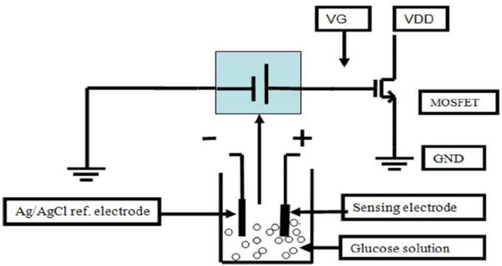
2.1. Potentiometric Glucose Sensor
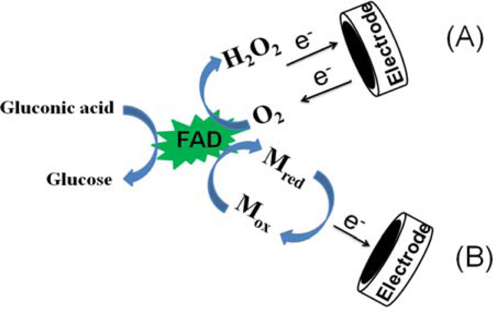
2.2. Amperometric Glucose Sensor
2.3. Impedimetric/Conductometric Glucose Sensor
3. Glucose Sensor Based on Metal-Oxides
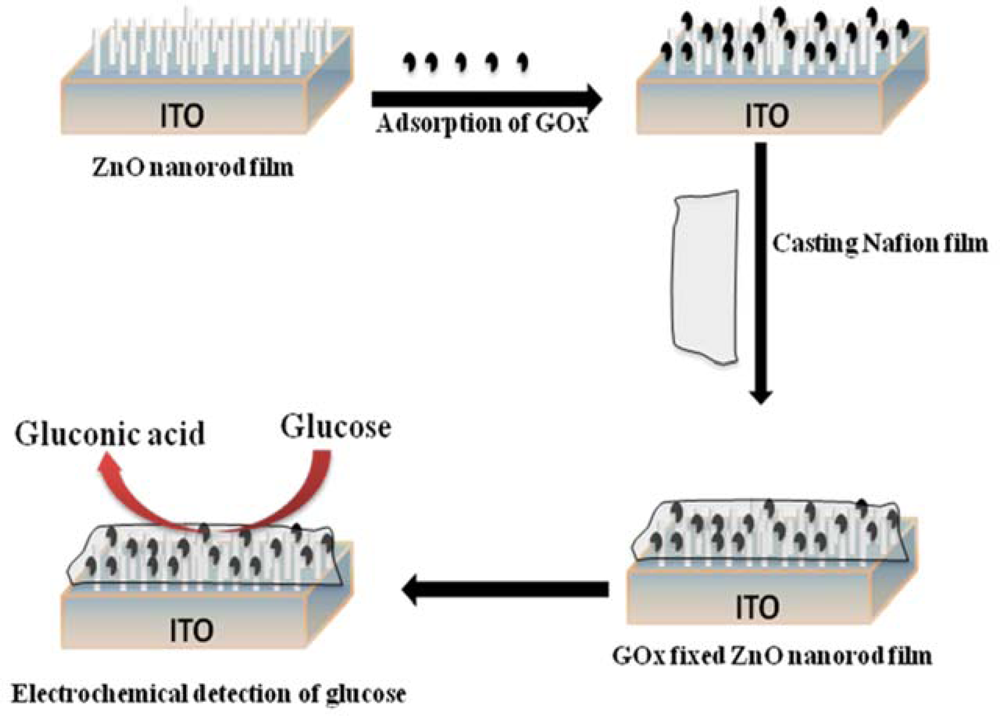
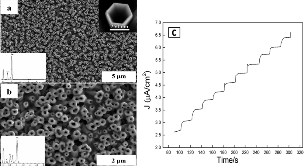
3.1. Zinc Oxide (ZnO) Based Glucose Sensor
3.2. Copper Oxide (CuO/Cu2O) Based Glucose Sensor
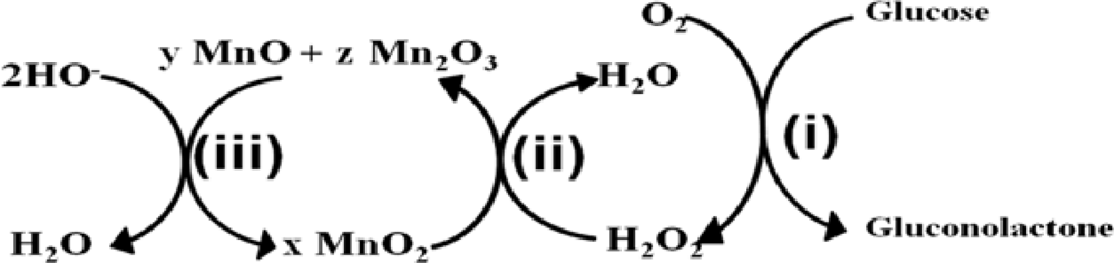
3.3. Manganese Dioxide (MnO2) Based Glucose Sensor
3.4. Titanium Dioxide (TiO2) Based Glucose Sensor
3.5 Cerium Oxide (CeO2) Based Glucose Sensor
3.6. Silicon Dioxide (SiO2) Based Glucose Sensor
3.7. Zirconium Oxide (ZrO2) Based Glucose Sensor
3.8. Other Metal-Oxide Based Glucose Sensors
4. Conclusion: Future Prospective and Challenges
Acknowledgments
References
- Gavin, J.R. The Importance of Monitoring Blood Glucose. In US Endocrine Disease 2007; Touch Briefings: Atlanta, GA, USA, 2007; pp. 1–3. [Google Scholar]
- Wang, J.; Thomas, D.F.; Chen, A. Nonenzymatic Electrochemical Glucose Sensor Based on Nanoporous PtPb Networks. Anal. Chem 2008, 80, 997–1004. [Google Scholar]
- King, H.; Aubert, R.E.; Herman, W.H. Global Burden of Diabetes. 1995–2025: Prevalence, Numerical Estimates, and Projections. Diabetes Care 1998, 21, 1414–1431. [Google Scholar]
- Wild, S.H.; Roglic, G.; Green., A.; Sicree, R.; King, H. Global Prevalence of Diabetes: Estimates for the Year 2000 and Projections for 2030. Diabetes Care 2004, 27, 1047–1053. [Google Scholar]
- Wang, J. Electrochemical Glucose Biosensors. Chem. Rev 2008, 108, 814–825. [Google Scholar]
- Wilson, G.S.; Gifford, R. Biosensors for Real-Time in vivo Measurements. Biosens. Bioelectron 2005, 20, 2388–2403. [Google Scholar]
- Newman, J.D.; Turner, A.P.F. Home Blood Glucose Biosensors: A Commercial Perspective. Biosens. Bioelectron 2005, 20, 2435–2453. [Google Scholar]
- Koschinsky, T; Heinemann, L. Sensors for Glucose Monitoring: Technical and Clinical Aspects. Diabetes Metab Res. Rev 2001, 17, 113–123. [Google Scholar]
- Oliver, N.S.; Toumazou, C.; Cass, A.E.G.; Johnston, D.G. Glucose Sensors: A Review of Current and Emerging Technology. Diabetic Med 2009, 26, 197–210. [Google Scholar]
- Lin, Y.; Lu, F.; Tu, Y.; Ren, Z. Glucose Biosensors Based on Carbon Nanotube Nanoelectrode Ensembles. Nano Lett 2004, 4, 191–195. [Google Scholar]
- Rakow, N.A.; Suslick, K.S. A Colorimetric Sensor Array for Odour Visualization. Nature 2000, 406, 710–712. [Google Scholar]
- Reitz, E.; Jia, W.; Gentile, M.; Wang, Y.; Lei, Y. CuO Nanospheres Based Nonenzymatic Glucose Sensor. Electroanalysis 2008, 20, 2482–2486. [Google Scholar]
- Sachedina, N.; Pickup, J.C. Performance Assessment of The Medtronic-Minimed Continuous Glucose Monitoring System and Its Use for Measurement of Glycaemic Control in Type 1 Diabetes. Diabet. Med 2003, 20, 1012–1015. [Google Scholar]
- Clark, L.C.; Lyons, C. Electrode Systems for Continuous Monitoring in Cardiovascular Surgery. Ann. N. Y. Acad. Sci 2006, 102, 29–45. [Google Scholar]
- Updike, S.J.; Hicks, G.P. The Enzyme Electrode. Nature 1967, 214, 986–988. [Google Scholar]
- Kang, X.H.; Mai, Z.B.; Zou, X.Y.; Cai, P.X.; Mo, J.Y. A Novel Glucose Biosensor Based On Immobilization of Glucose Oxidase in Chitosan on A Glassy Carbon Electrode Modified with Gold-Platinum Alloy Nanoparticles/Multiwall Carbon Nanotubes. Anal. Biochem 2007, 369, 71–79. [Google Scholar]
- Shervedani, R.K.; Mehrjardi, A.H.; Zamiri, N. A Novel Method for Glucose Determination Based On Electrochemical Impedance Spectroscopy Using Glucose Oxidase Self-Assembled Biosensor. Bioelectrochemistry 2006, 69, 201–208. [Google Scholar]
- Caras, S.D.; Petelenz, D.; Janata, J. pH-Based Enzyme Potentiometric Sensors. Part 2. Glucose-Sensitive Field Effect Transistor. Anal. Chem 1985, 57, 1920–1923. [Google Scholar]
- Tang, H.; Chen, J.H.; Yao, S.Z.; Nie, L.H.; Deng, G.H.; Kuang, Y.F. Amperometric Glucose Biosensor Based On Adsorption of Glucose Oxidase at Platinum Nanoparticle-Modified Carbon Nanotube Electrode. Anal. Biochem 2004, 331, 89–97. [Google Scholar]
- Wang, S.G.; Zhang, Q.; Wang, R.L.; Yoon, S.F.; Ahn, J.; Yang, D.J. Multi-Walled Carbon Nanotubes for the Immobilization of Enzyme in Glucose Biosensors. Electrochem. Commun 2003, 5, 800–803. [Google Scholar]
- Tsai, Y.C.; Li, S.C.; Chen, J.M. Cast Thin Film Biosensor Design Based on a Nafion Backbone, a Multiwalled Carbon Nanotube Conduit, and a Glucose Oxidase Function. Langmuir 2005, 21, 3653–3658. [Google Scholar]
- Wang, J. Glucose Biosensors: 40 Years of Advances and Challenges. Electroanalysis 2001, 13, 983–988. [Google Scholar]
- Heller, A.; Feldman, B. Electrochemical Glucose Sensors and Their Applications in Diabetes Management. Chem. Rev 2008, 108, 2482–2505. [Google Scholar]
- Schijgerl, K.; Hitzmann, B.; Jurgens, H.; Kullick, T.; Ulber, R.; Weigal, B. Challenges in Integrating Biosensors and FIA for On-Line Monitoring and Control. Trends Biotechnol. 1996, 14, 21–23. [Google Scholar]
- Proenca, L.; Lopes, M.I.S.; Fonseca, I.; Kokoh, K.B.; Leger, J.-M.; Lamy, C. Electrocatalytic Oxidation of D-sorbitol on Platinum in Acid Medium: Analysis of the Reaction Products. J. Electroanal. Chem 1997, 432, 237–242. [Google Scholar]
- Chen, C.-Y.; Tamiya, E.; Ishihara, K.; Kosugi, Y.; Su, Y.-C.; Nakabayashi, N.; Karube, I. A Biocompatible Needle-Type Glucose Sensor Based on Platinum- Electroplated Carbon Electrode. Appl. Biochem.Biotech 1992, 36, 211–226. [Google Scholar]
- Chou, C.-H.; Chen, J.-C.; Tai, C.-C.; Sun, I.-W.; Zen, J.-M. A Nonenzymatic Glucose Sensor Using Nanoporous Platinum Electrodes Prepared by Electrochemical Alloying/Dealloying in a Water-Insensitive Zinc Chloride-1-Ethyl-3 Methylimidazolium Chloride Ionic Liquid. Electroanalysis 2008, 20, 771–775. [Google Scholar]
- Kurniawan, F.; Tsakova, V.; Mirsky, V.M. Gold Nanoparticles in Nonenzymatic Electrochemical Detection of Sugars. Electroanalysis 2006, 18, 1937–1942. [Google Scholar]
- Feng, D.; Wang, F.; Chen, Z. Electrochemical Glucose Sensor Based On One-Step Construction of Gold Nanoparticle–Chitosan Composite film. Sens. Actuat. B-Chem 2009, 138, 539–544. [Google Scholar]
- Tominaga, M.; Nagashima, M.; Nishiyama, K.; Taniguchi, I. Surface Poisoning During Electrocatalytic Monosaccharide Oxidation Reactions at Gold Electrodes in Alkaline Medium. Electrochem. Commun 2007, 9, 1892–1898. [Google Scholar]
- Liua, Y.; Teng, H.; Hou, H.; You, T. Nonenzymatic Glucose Sensor Based On Renewable Electrospun Ni Nanoparticle-Loaded Carbon Nanofiber Paste Electrode. Biosens. Bioelectron 2009, 24, 3329–3334. [Google Scholar]
- Lu, L.-M.; Zhang, L.; Qu, F.-L.; Lu, H.-X.; Zhang, X.-B.; Wu, Z.-S.; Huana, S.-Y.; Wang, Q.-A.; Shen, G.-L; Yu, R.-Q. A Nano-Ni Based Ultrasensitive Nonenzymatic Electrochemical Sensor for Glucose: Enhancing Sensitivity Through a Nanowire Array Strategy. Biosens. Bioelectron 2009, 25, 218–223. [Google Scholar]
- Grace, A.N.; Pandian, K. Synthesis of Gold and Platinum Nanoparticles Using Tetraaniline as Reducing and Phase Transfer Agent—A Brief Study and Their Role in the Electrocatalytic Oxidation of Glucose. J. Phys. Chem. Sol 2007, 68, 2278–2285. [Google Scholar]
- Holt-Hindle, P.; Nigro, S.; Asmussen, M.; Chen, A. Amperometric Glucose Sensor Based on Platinum–Iridium Nanomaterials. Electrochem. Commun 2008, 10, 1438–1441. [Google Scholar]
- Belousov, V.M.; Vasylyev, M.A.; Lyashenko, L.V.; Vilkova, N.Y; Nieuwenhuys, B.E. The Low-Temperature Reduction of Pd-doped Transition Metal Oxide Surfaces with Hydrogen. J. Chem. Eng 2003, 91, 143–150. [Google Scholar]
- Xiao, F.; Zhao, F.; Mei, D.; Mo, Z.; Zeng, B. Nonenzymatic Glucose Sensor Based On Ultrasonic-Electrodeposition of Bimetallic PtM (M = Ru, Pd and Au) Nanoparticles on Carbon Nanotubes–Ionic Liquid Composite Film. Biosens. Bioelectron 2009, 24, 3481–3486. [Google Scholar]
- Cui, H.-F.; Ye, J.-S.; Liu, X.; Zhang, W.-D.; Sheu, F.-S. Pt–Pb Alloy Nanoparticle/Carbon Nanotube Nanocomposite: A Strong Electrocatalyst for Glucose Oxidation. Nanotechnology 2006, 17, 2334–2339. [Google Scholar]
- Meng, L.; Jin, J.; Yang, G.; Lu, T.; Zhang, H.; Cai, C. Nonenzymatic Electrochemical Detection of Glucose Based on Palladium-Single-Walled Carbon Nanotube Hybrid Nanostructures. Anal. Chem 2009, 81, 7271–7280. [Google Scholar]
- Zhu, H.; Lu, X.; Li, M.; Shao, Y.; Zhu, Z. Nonenzymatic Glucose Voltammetric Sensor Based On Gold Nanoparticles/Carbon Nanotubes/Ionic Liquid Nanocomposites. Talanta 2009, 79, 1446–1453. [Google Scholar]
- Wang, H.; Zhou, C.; Liang, J.; Yu, H.; Peng, F.; Yang, J. High Sensitivity Glucose Biosensor Based on Pt Eelectrodeposition onto Low-density Aligned Carbon Nanotubes. Int. J. Electrochem. Sci 2008, 3, 1258–1267. [Google Scholar]
- Wang, H.; Zhou, C.; Liang, J.; Yu, H.; Peng, F. An Enhanced Glucose Biosensor Modified by Pt/sulfonated- MWCNTs with Layer by Layer Technique. Int. J. Electrochem. Sci 2008, 3, 1180–1185. [Google Scholar]
- Xie, J.; Wang, S.; Aryasomayajula, L; Varadan, V.K. Platinum Decorated Carbon Nanotubes for Highly Sensitive Amperometric Glucose Sensing. Nanotechnology 2007, 18, 065503–065512. [Google Scholar]
- Sun, Y.; Buck, H.; Mallouk, T.E. Combinatorial Discovery of Alloy Electrocatalysts for Amperometric Glucose Sensors. Anal. Chem 2001, 73, 1599–1604. [Google Scholar]
- Liu, A. Towards Development of Chemosensors and Biosensors with Metal-Oxide-Based Nanowires or Nanotubes. Biosens. Bioelectron 2008, 24, 167–177. [Google Scholar]
- Zhai, T.; Fang, X.; Liao, M.; Xu, X.; Zeng, H.; Yoshio, B.; Golberg, D. A Comprehensive Review of One-Dimensional Metal-Oxide Nanostructure Photodetectors. Sensors 2009, 9, 6504–6529. [Google Scholar]
- Huang, J.; Wan, Q. Gas Sensors Based on Semiconducting Metal Oxide One-Dimensional Nanostructures. Sensors 2009, 9, 9903–9924. [Google Scholar]
- Ahammad, A.J.S.; Lee, J.-J.; Rahman, M. A. Electrochemical Sensors Based on Carbon Nanotubes. Sensors 2009, 9, 2289–2319. [Google Scholar]
- Wang, Y.; Xu, H.; Zhang, J.; Li, G. Electrochemical Sensors for Clinic Analysis. Sensors 2008, 8, 2043–2081. [Google Scholar]
- Morikawa, M.; Kimizuka, N.; Yoshihara, M.; Endo, T. New Colorimetric Detection of Glucose by Means of Electron-Accepting Indicators: Ligand Substitution of [Fe(acac)3−n(phen)n]n+ Complexes Triggered by Electron Transfer from Glucose Oxidase. Chem. Eur. J 2002, 8, 5580–5584. [Google Scholar]
- Miwa, Y.; Nishizawa, M.; Matsue, T.; Uchida, I. A Conductometric Glucose Sensor Based on a Twin-Microband Electrode Coated with a Polyaniline Thin Film. Bull. Chem. Soc. Jp 1994, 67, 2864–2866. [Google Scholar]
- Mansouri, S.; Schultz, J.S. A Miniature Optical Glucose Sensor Based on Affinity Binding. Nature Biotech 1984, 2, 885–890. [Google Scholar]
- Pickup, J.C.; Hussain, F.; Evans, N.D.; Rolinski, O.J.; Birch, D.J.S. Fluorescence-Based Glucose Sensors. Biosens. Bioelectron 2005, 20, 2555–2565. [Google Scholar]
- Weiss, S. Fluorescence Spectroscopy of Single Molecules. Science 1999, 283, 1676–1683. [Google Scholar]
- Ali, S.M.U.; Nur, O.; Willander, M.; Danielsson, B. Glucose Detection with a Commercial MOSFET Using a ZnO Nanowires Extended Gate. IEEE Trans. Nanotechnol 2009, 8, 678–683. [Google Scholar]
- Pohanka, M.; SkládaL, P. Electrochemical Biosensors-Principles and Applications. J. Appl. Biomed 2008, 6, 57–64. [Google Scholar]
- Seki, A.; Ikeda, S.-I.; Kubo, I.; Karube, I. Biosensors Based on Light-Addressable Potentiometric Sensors for Urea, Penicillin and Glucose. Anal. Chim. Acta 1998, 373, 9–13. [Google Scholar]
- Luo, X.-L.; Xu, J.-J.; Zhao, W.; Chen, H.-Y. Glucose Biosensor Based on ENFET Doped with SiO2 Nanoparticles. Sens. Actuator B-Chem 2004, 97, 249–255. [Google Scholar]
- Shoji, E.; Freund, M.S. Potentiometric Saccharide Detection Based on the pKa Changes of Poly(aniline boronic acid). J. Am. Chem. Soc 2002, 124, 12486–12493. [Google Scholar]
- Shoji, E.; Freund, M.S. Potentiometric Sensors Based on the Inductive Effect on the pKa of Poly(aniline): A Nonenzymatic Glucose Sensor. J. Am. Chem. Soc 2001, 123, 3383–3384. [Google Scholar]
- Wang, J. Amperometric Biosensors for Clinical and Therapeutic Drug Monitoring: A Review. J. Pharm. Biomed. Anal 1999, 19, 47–53. [Google Scholar]
- Park, S.; Boo, H.; Chung, T.D. Electrochemical Non-Enzymatic Glucose Sensors. Anal. Chim. Acta 2006, 556, 46–57. [Google Scholar]
- Immobilized Enzyme in Analytical Chemistry. In Advances In Biochemical Engineering/Biotechnology; Carr, P.W.; Browers, L.D. (Eds.) Springer Berlin: Heidelberg, Germany, 1980; pp. 89–129.
- Cass, A.E.G.; Davis, G.; Francis, G.D.; Hill, H.A.O.; Aston, W.J.; Higgins, I.J.; Plotkin, E.V.; Scott, L.D.L.; Turner, A.P.F. Ferrocene-Mediated Enzyme Electrode for Amperometric Determination of Glucose. Anal. Chem 1984, 56, 667–671. [Google Scholar]
- Liu, S.; Ju, H. Reagentless Glucose Biosensor Based On Direct Electron Transfer of Glucose Oxidase Immobilized on Colloidal Gold Modified Carbon Paste Electrode. Biosens. Bioelectron 2003, 19, 177–183. [Google Scholar]
- Yi, X.; Patolsky, F.; Katz, E.; Hainfeld, J.F.; Willner, I. Plugging into Enzymes: Nanowiring of Redox Enzymes by a Gold Nanoparticle. Science 2003, 299, 1877–1881. [Google Scholar]
- Basu, S.; Kang, W.P.; Davidson, J.L.; Choi, B.K.; Bonds, A.B.; Cliffel, D.E. Electrochemical Sensing Using Nanodiamond Microprobe. Diam. Relat. Mat 2006, 15, 269–274. [Google Scholar]
- Yang, H.; Zhu, Y. Size Dependence of SiO2 Particles Enhanced Glucose Biosensor. Talanta 2006, 68, 569–574. [Google Scholar]
- Zang, J.; Li, C.M.; Cui, X.; Wang, J.; Sun, X.; Chang, H.D.; Sun, Q. Tailoring Zinc Oxide Nanowires for High Performance Amperometric Glucose Sensor. Electroanalysis 2007, 19, 1008–1014. [Google Scholar]
- Liu, X.W.; Hu, Q.; Wu, Q.; Zhang, W.; Fang, Z.; Xie, Q. Aligned ZnO Nanorods: A Useful Film to Fabricate Amperometric Glucose Biosensor. Colloid Surf. B-Biointerfaces 2009, 74, 154–158. [Google Scholar]
- Bai, Y.; Sun, Y.; Sun, C. Pt–Pb Nanowire Array Electrode for Enzyme-Free Glucose Detection. Biosens. Bioelectron 2008, 24, 579–585. [Google Scholar]
- Salimi, A.; Roushani, M. Non-enzymatic Glucose Detection Free of Ascorbic Acid Interference Using Nickel Powder and Nafion Sol-Gel Dispersed Renewable Carbon Ceramic Electrode. Electrochem. Commun 2005, 7, 879–887. [Google Scholar]
- Wang, J.; Thomas, D.F.; Chen, A. Nonenzymatic Electrochemical Glucose Sensor Based on Nanoporous PtPb Networks. Anal. Chem 2008, 80, 997–1004. [Google Scholar]
- Li, Y.; Song, Y.-Y.; Yang, C.; Xia, X.-H. Hydrogen bubble Dynamic Template Synthesis of Porous Gold for Nonenzymatic Electrochemical Detection of Glucose. Electrochem. Commun 2007, 9, 981–988. [Google Scholar]
- Safavi, A.; Maleki, N.; Farjami, E. Fabrication of a Glucose Sensor Based On a Novel Nanocomposite Electrode. Biosens. Bioelectron 2009, 24, 1655–1660. [Google Scholar]
- You, T.; Niwa, O.; Tomita, M.; Ando, H.; Suzuki, M.; Hirono, S. Characterization and Electrochemical Properties of Highly Dispersed Copper Oxide/Hydroxide Nanoparticles in Graphite-like Carbon Films Prepared by RF Sputtering Method. Electrochem. Commun 2002, 4, 468–471. [Google Scholar]
- Guan, J.G.; Miao, Y.Q.; Zhang, Q.J. Impedemetric Biosensor. J. Biosci. Bioeng 2004, 97, 219–226. [Google Scholar]
- Fuchs, K.; Kaatze, U. Molecular Dynamics Of Carbohydrate Aqueous Solutions. Dielectric Relaxation as a Function of Glucose and Fructose Concentration. J. Phys. Chem., Sect. B 2001, 105, 2036–2042. [Google Scholar]
- Caduff, A.; Hirt, E.; Feldman, Y.; Ali, Z.; Heinemann, L. First Human Experiments with a Novel Non-Invasive, Non-Optical Continuous Glucose Monitoring System. Biosens. Bioelectron 2003, 19, 209–217. [Google Scholar]
- Shervedani, R.K.; Mehrjardi, A.H.; Zamiri, N. A Novel Method for Glucose Determination Based On Electrochemical Impedance Spectroscopy Using Glucose Oxidase Self-Assembled Biosensor. Bioelectrochemistry 2006, 69, 201–208. [Google Scholar]
- Forzani, E.S.; Zhang, H.; Nagahara, L.A.; Amlani, I.; Tsui, R.; Tao, N. A Conducting Polymer Nanojunction Sensor for Glucose Detection. Nano Lett 2004, 9, 1785–1788. [Google Scholar]
- Besteman, K.; Lee, J.-O.; Wiertz, F.G.M.; Heering, H.A.; Dekker, C. Enzyme-Coated Carbon Nanotubes as Single-Molecule Biosensors. Nano Lett 2003, 3, 727–730. [Google Scholar]
- Ansari, S.G.; Ansari, Z.A.; Wahab, R.; Kim, Y.-S.; Khang, G.; Shin, H.-S. Glucose Sensor Based On Nano-Baskets of Tin Oxide Templated in Porous Alumina By Plasma Enhanced CVD. Biosens. Bioelectron 2008, 23, 1838–1842. [Google Scholar]
- Yang, K.; She, G.-W.; Wang, H.; Ou, X.-M.; Zhang, X.-H.; Lee, C.-S.; Lee, S.-T. ZnO Nanotube Arrays as Biosensors for Glucose. J. Phys. Chem. C 2009, 113, 20169–20172. [Google Scholar]
- Zhang, F.F; Wang, X.; Ai, S.; Sun, Z.; Wan, Q.; Zhu, Z.; Xian, Y.; Jin, L.; Yamamoto, K. Immobilization of Uricase on ZnO Nanorods for a Reagentless Uric Acid Biosensor. Anal. Chimi. Acta 2004, 519, 155–160. [Google Scholar]
- Zhao, J.; Wu, D.; Zhi, J. A Novel Tyrosinase Biosensor Based On Biofunctional ZnO Nanorod Microarrays on the Nanocrystalline Diamond Electrode for Detection of Phenolic Compounds. Bioelectrochemistry 2009, 75, 44–49. [Google Scholar]
- Rodriguez, J.A.; Jirsak, T.; Dvorak, J.; Sambasivan, S.; Fischer, D. Reaction of NO2 with Zn and ZnO: Photoemission, XANES, and Density Functional Studies on the Formation of NO3. J. Phys. Chem. B 2000, 104, 319–328. [Google Scholar]
- Tian, Z.R.; Voigt, J.A.; Liu, J.; Mckenzie, B.; Mcdermott, M. J. Biomimetic Arrays of Oriented Helical ZnO Nanorods and Columns. J. Am. Chem. Soc 2002, 124, 12954–12955. [Google Scholar]
- Wei, A.; Suna, X.W.; Wang, J.X.; Lei, Y.; Cai, X.P.; Li, C.M.; Dong, Z.L.; Huang, W. Enzymatic Glucose Biosensor Based On ZnO Nanorod Array Grown by Hydrothermal Decomposition. Appl. Phys. Lett 2006, 89. [Google Scholar]
- Kang, B.S.; Wang, H.T.; Ren, F.; Pearton, S.J.; Morey, T.E.; Dennis, D.M.; Johnson, J.W.; Rajagopal, P.; Roberts, J.C.; Piner, E.L.; Linthicum, K.J. Enzymatic Glucose Detection Using ZnO Nanorods on the Gate Region of AlGaN/GaN High Electron Mobility Transistors. Appl. Phys. Lett 2007, 91. [Google Scholar]
- Kim, J.S.; Park, W.; Lee, C.-H.; Yi, G-C. ZnO Nanorod Biosensor for Highly Sensitive Detection of Specific Protein Binding. J. Korean Phys. Soc 2006, 49, 1–5. [Google Scholar]
- Zhao, Z.W.; Chen, X.J.; Tay, B.K.; Chen, J.S.; Han, Z.J.; Khor, K.A. A Novel Amperometric Biosensor Based On ZnO: Co Nanoclusters For Biosensing Glucose. Biosens. Bioelectron 2007, 23, 135–139. [Google Scholar]
- Dai, Z.; Shao, G.; Hong, J.; Bao, J.; Shen, J. Immobilization and Direct Electrochemistry of Glucose Oxidase on a Tetragonal Pyramid-Shaped Porous ZnO Nanostructure for a Glucose Biosensor. Biosens. Bioelectron 2009, 24, 1286–1291. [Google Scholar]
- Kong, T.; Chen, Y.; Ye, Y.; Zhang, K.; Wang, Z.; Wang, X. An Amperometric Glucose Biosensor Based On the Immobilization of Glucose Oxidase on the ZnO Nanotubes. Sens. Actuator B-Chem 2009, 138, 344–350. [Google Scholar]
- Wang, J.X.; Sun, X.W.; Wei, A.; Lei, Y.; Cai, X.P.; Li, C.M.; Dong, Z.L. Zinc Oxide Nanocomb Biosensor for Glucose Detection. Appl. Phys. Lett 2006, 88. [Google Scholar]
- Liu, J.; Guo, C.; Li, C.M.; Li, Y.; Chi, Q.; Huang, X.; Liao, L.; Yu, T. Carbon-Decorated ZnO Nanowire Array: A Novel Platform for Direct Electrochemistry of Enzymes and Biosensing Applications. Electrochem. Commun 2009, 11, 202–205. [Google Scholar]
- Lin, Y.; Lu, F.; Tu, Y.; Ren, Z. Glucose biosensors Based on Carbon Nanotube Nanoelectrode Ensembles. Nano Lett 2004, 4, 191–195. [Google Scholar]
- Sun, W.; Gao, R.; Jiao, K. Electrochemistry and Electrocatalysis of Hemoglobin in Nafion/nano-CaCO3 Film on a New Ionic Liquid BPPF6 Modified Carbon Paste Electrode. J. Phys. Chem. B 2007, 111, 4560–4567. [Google Scholar]
- Sheng, Q.; Luo, K.; Li, L.; Zheng, J. Direct Electrochemistry of Glucose Oxidase Immobilized on NdPO4 Nanoparticles/Chitosan Composite Film on Glassy Carbon Electrodes and Its Biosensing Application. Bioelectrochemistry 2009, 74, 246–253. [Google Scholar]
- Bordonaba, V.L.; Terry, V. Development of a Glucose Biosensor for Rapid Assessment of Strawberry Quality: Relationship between Biosensor Response and Fruit Composition. J. Agric. Food Chem 2009, 57, 8220–8226. [Google Scholar]
- Wang, Y.-T.; Yu, L.; Zhu, Z.-Q.; Zhang, J.; Zhu, J.-Z.; Fan, C.-H. Improved Enzyme Immobilization for Enhanced Bioelectrocatalytic Activity of Glucose Sensor. Sens. Actuator B-Chem 2009, 136, 332–337. [Google Scholar]
- Koschinsky, T.; Heinemann, L. Sensors for Glucose Monitoring: Technical and Clinical Aspects. Diabetes Metab Res Rev 2001, 17, 113–123. [Google Scholar]
- Bantle, J.P.; Thomas, W. Glucose Measurement in Patients with Diabetes Mellitus with Dermal Interstitial Fluid. J. Lab. Clin. Med 1997, 130, 436–441. [Google Scholar]
- Bolinder, J.; Hagstrom, E.; Ungerstedt, U.; Arner, P. Microdialysis of Subcutaneous Adipose Tissue in Vivo for Continuous Glucose Monitoring in Man. Scand J. Clin. Lab. Invest 1989, 49, 465–474. [Google Scholar]
- Asif, M.H.; Ali, S.M.U.; Nur, O.; Willander, M.; Brannmark, C.; Stralfors, P.; Englund, U.H.; Elinder, F.; Danielsson, B. Functionalised ZnO-nanorod-based Selective Electrochemical Sensor for Intracellular Glucose. Biosens. Bioelectron 2010, in press. [Google Scholar] [CrossRef]
- Mho, S.I.; Johnson, D.C. Electrocatalytic Response of Carbohydrates at Copper-Alloy Electrodes. J. Electroanal. Chem 2001, 500, 524–532. [Google Scholar]
- Salimi, A.; Roushani, M. Non-enzymatic Glucose Detection Free of Ascorbic Acid Interference Using Nickel Powder and Nafion Sol–Gel Dispersed Renewable Carbon Ceramic Electrode. Electrochem. Commun 2005, 7, 879–887. [Google Scholar]
- Jiang, X.; Herricks, T.; Xia, Y. CuO Nanowires Can Be Synthesized by Heating Copper Substrates in Air. Nano Lett 2002, 2, 1333–1338. [Google Scholar]
- Wang, D.; Song, C.; Hu, Z.; Fu, X. Fabrication of Hollow Spheres and Thin Films of Nickel Hydroxide and Nickel Oxide with Hierarchical Structures. J. Phys. Chem. B 2005, 109, 1125–1129. [Google Scholar]
- Gao, X.P.; Bao, J.L.; Pan, G.L.; Zhu, H.Y.; Huang, P.X.; Wu, F.; Song, D.Y. Preparation and Electrochemical Performance of Polycrystalline and Single Crystalline CuO Nanorods as Anode Materials for Li Ion Battery. J. Phys. Chem. B 2004, 108, 5547–5551. [Google Scholar]
- Zhang, J.; Liu, J.; Peng, Q.; Wang, X.; Li, Y. Nearly Monodisperse Cu2O and CuO Nanospheres: Preparation and Applications for Sensitive Gas Sensors. Chem. Mater 2006, 18, 867–871. [Google Scholar]
- Rakhshani, A.E.; Makdisi, Y.; Mathew, X. Deep Energy Levels and Photoelectrical Properties of Thin Cuprous Oxide Films. Thin Solid Films 1996, 288, 69–75. [Google Scholar]
- McAuley, C.B; Du, Y.; Wildgoose, G.G.; Compton, R.G. The use of Copper(II) Oxide Nanorod Bundles for the Non-Enzymatic Voltammetric Sensing of Carbohydrates and Hydrogen Peroxide. Sens. Actuat. B-Chem 2008, 135, 230–235. [Google Scholar]
- Wang, W.; Li, Z.; Zheng, W.; Yang, J.; Zhang, H.; Wang, C. Electrospun Palladium (IV)-doped Copper Oxide Composite Nanofibers for Non-Enzymatic Glucose Sensors. Electrochem. Commun 2009, 11, 1811–1814. [Google Scholar]
- Yeo, I.H.; Johnson, D.C. Anodic Response of Glucose at Copper-Based Alloy Electrodes. J. Electroanal. Chem 2000, 484, 157–163. [Google Scholar]
- Wang, W.; Zhang, L.; Tong, S.; Li, X.; Song, W. Three-Dimensional Network Films of Electrospun Copper Oxide Nanofibers for Glucose Determination. Biosens. Bioelectron 2009, 25, 708–714. [Google Scholar]
- Zhuang, Z.; Su, X.; Yuan, H.; Sun, Q.; Xiao, D.; Choi, M.M.F. An improved Sensitivity Non-Enzymatic Glucose Sensor Based on a CuO Nanowire Modified Cu Electrode. Analyst 2008, 133, 126–132. [Google Scholar]
- Auley, C.B.M.; Wildgoose, G.G.; Compton, R.G.; Shao, L.D.; Green, M.L.H. Copper Oxide Nanoparticle Impurities Are Responsible for the Electroanalytical Detection of Glucose Seen Using Multiwalled Carbon Nanotubes. Sens. Actuat. B-Chem 2008, 132, 356–360. [Google Scholar]
- Zhang, X.; Wang, G.; Zhang, W.; Wei, Y.; Fang, B. Fixure-reduce Method for the Synthesis of Cu2O/MWCNTs Nanocomposites and Its Application as Enzyme-Free Glucose Sensor. Biosens. Bioelectron 2009, 24, 3395–3398. [Google Scholar]
- Jiang, L.-C.; Zhang, W.-D. A Highly Sensitive Nonenzymatic Glucose Sensor Based on CuO Nanoparticles-Modified Carbon Nanotube Electrode. Biosens. Bioelectron 2010, 25, 1402–1407. [Google Scholar]
- Panzner, G.; Egert, B.; Schmidt, H.P. The Stability of CuO and Cu2O Surfaces During Argon Sputtering Studied by XPS and AES. Surf. Sci 1984, 151, 400–408. [Google Scholar]
- Luque, G.L.; Rodriguez, M.C.; Rivas, G.A. Glucose Biosensors Based on the Immobilization of Copper Oxide and Glucose Oxidase within a Carbon Paste Matrix. Talanta 2005, 66, 467–471. [Google Scholar]
- Umar, A.; Rahman, M.M.; Al-Hajry, A.; Hahn, Y.-B. Enzymatic Glucose Biosensor Based on Flower-Shaped Copper Oxide Nanostructures Composed of Thin Nanosheets. Electrochem. Commun 2009, 11, 278–281. [Google Scholar]
- Karyakin, A.A.; Karyakina, E.E.; Gorton, L. Amperometric Biosensor for Glutamate Using Prussian Blue-Based “Artificial Peroxidase” as a Transducer for Hydrogen Peroxide. Anal. Chem 2000, 72, 1720–1723. [Google Scholar]
- Hoshi, T.; Saiki, H.; Kuwazawa, S.; Tsuchiya, C.; Chen, Q.; Anzai, J.-I. Selective Permeation of Hydrogen Peroxide through Polyelectrolyte Multilayer Films and Its Use for Amperometric Biosensors. Anal. Chem 2001, 73, 5310–5315. [Google Scholar]
- Muguruma, H.; Hiratsuka, A.; Karube, I. Thin-Film Glucose Biosensor Based on Plasma-Polymerized Film: Simple Design for Mass Production. Anal. Chem 2000, 72, 2671–2675. [Google Scholar]
- Xu, J.-J.; Yu, Z.-H.; Chen, H.-Y. Glucose Biosensors Prepared by Electropolymerization of p-Chlorophenylamine with and without Nafion. Anal. Chim. Acta 2002, 463, 239–247. [Google Scholar]
- Pandey, P.C.; Upadhyay, S.; Shukla, N.K.; Sharma, S. Studies on the Electrochemical Performance of Glucose Biosensor Based on Ferrocene Encapsulated ORMOSIL and Glucose Oxidase Modified Graphite Paste Electrode. Biosens. Bioelectron 2003, 18, 1257–1268. [Google Scholar]
- Krikstopaitis, K.; Kulys, J.; Tetianec, L. Bioelectrocatalytical Glucose Oxidation with Phenoxazine Modified Glucose Oxidase. Electrochem.Commun 2004, 6, 331–336. [Google Scholar]
- Elizabeth, A.H.H.; Gooding, J.J.; Hall, C.E. Redox Enzyme Linked Electrochemical Sensors: Theory Meets Practice. Mikrochim. Acta 1995, 121, 119–145. [Google Scholar]
- Luo, X.-L.; Xu, J.-J.; Zhao, W.; Chen, H.-Y. Ascorbic Acid Sensor Based on Ion-Sensitive Field-Effect Transistor Modified with MnO2 Nanoparticles. Electrochem. Commun 2004, 6, 1169–1173. [Google Scholar]
- Kim, D.W.; Hong, Y.K.; Lee, N-S.; Kim, C.S.; Jeon, B.K.; Kang, B.M. Seperation of Magnesium Isotopes by Chemical Exchange with Manganese(IV) Oxide. J. Nucl. Sci. Technol 2001, 38, 780–784. [Google Scholar]
- Russo, F.; Johnson, C.J.; Johnson, C.J.; McKenzie, D.; Aiken, J.M.; Pedersen, J.A. Pathogenic Prion Protein Is Degraded by a Manganese Oxide Mineral Found in Soils. J. Gen. Virol 2009, 90, 275–280. [Google Scholar]
- Turkusic, E.; Kalcher, K.; Schachl, K.; Komersova, A.; Bartos, M.; Moderegger, H.; Svancara, I.; Vytras, K. Amperometric Determination of Glucose with an MnO2 and Glucose Oxidase Bulk-Modified Screen-Printed Carbon Ink Biosensor. Anal.Lett. 2001, 34, 2633–2647. [Google Scholar]
- Xu, J.-J.; Feng, J.-J.; Zhong, X.; Chen, H.-Y. Low-Potential Detection of Glucose with a Biosensor Based on the Immobilization of Glucose Oxidase on Polymer/Manganese Oxide Layered Nanocomposite. Electroanalysis 2008, 20, 507–512. [Google Scholar]
- Xu, J.-J.; Luo, X.-L.; Du, Y.; Chen, H.-Y. Application of MnO2 Nanoparticles as an Eliminator of Ascorbate Interference to Amperometric Glucose Biosensors. Electrochem. Commun 2004, 6, 1169–1173. [Google Scholar]
- Chen, J.; Zhang, W.-D.; Ye, J.-S. Nonenzymatic Electrochemical Glucose Sensor Based on MnO2/MWNTs Nanocomposites. Electrochem. Commun 2008, 10, 1268–1271. [Google Scholar]
- Mathur, S.; Erdemc, A.; Cavelius, C.; Barth, S.; Altmayer, J. Amplified Electrochemical DNA-Sensing of Nanostructured Metal Oxide Filmsdeposited on Disposable Graphite Electrodes Functionalized by Chemical Vapor Deposition. Sens. Actuat. B-Chem 2009, 136, 432–437. [Google Scholar]
- Durrant, J.R. Protein Adsorption on Nanocrystalline TiO2 Film: An Immobilization Strategy for Bioanalytical Devices. Anal. Chem 1998, 70, 5111–5113. [Google Scholar]
- Seo, D-W.; Sarker, S.; Deb Nath, N.C.; Choi, S-W.; Ahammad, A.J.S.; Lee, J.-J.; Kim, W.-G. Synthesis of a Novel Imidazolium –Based Electrolytes and Application for Dye-Sensitized Solar Cell. Electrochim. Acta 2010, 55, 1483–1488. [Google Scholar]
- Tian, M.; Wu, G.; Adams, B.; Wen, J.; Chen, A. Kinetics of Photoelectrocatalytic Degradation of Nitrophenols on Nanostructured TiO2 Electrodes. J. Phys. Chem. C 2008, 112, 825–831. [Google Scholar]
- Lucarelli, L.; Nadtochenko, V.; Kiwi, J. Environmental Photochemistry: Quantitative Adsorption and FTIR Studies during the TiO2-Photocatalyzed Degradation of Orange II. Langmuir 2000, 16, 1102–1108. [Google Scholar]
- Kavan, L.; Rathousky, J.; Gra1tzel, M.; Shklover, V.; Zukal, A. Surfactant-Templated TiO2 (Anatase): Characteristic Features of Lithium Insertion Electrochemistry in Organized Nanostructures. J. Phys. Chem. B 2000, 104, 12012–12020. [Google Scholar]
- Shen, Q.; You, S.-K.; Park, S.-G.; Jiang, H.; Guo, D.; Chen, B.; Wang, X. Electrochemical Biosensing for Cancer Cells Based on TiO2/CNT Nanocomposites Modified Electrodes. Electroanalysis 2008, 20, 2526–2530. [Google Scholar]
- Kurokawa, Y.; Sano, T.; Ohta, H.; Nakagawa, Y. Immobilization of Enzyme onto Cellulose-Titanium Oxide Composite Fiber. Biotechnol. Bioeneng 1993, 42, 394–397. [Google Scholar]
- Kennedy, J.F.; Kay, I.M. Hydrous Titanium Oxides—/New Supports for the Simple Immobilization of Enzymes. J. Chem. Soc. Perkin Trans 1976, 1, 329–335. [Google Scholar]
- Lev, O.; Tsionsky, M.; Rabinovich, L.; Glezer, V.; Sampath, S.; Pankratov, I.; Gun, J. Organically Modified Sol-Gel Sensor. Anal. Chem 1995, 67, 22A–30A. [Google Scholar]
- Gill, I. Bio-doped Nanocomposite Polymers: Sol-Gel Bioencapsulates. Chem. Mater 2001, 13, 3404–3421. [Google Scholar]
- Walcarius, A. Electrochemical Applications of Silica-Based Organic-Inorganic Hybrid Materials. Chem. Mater 2001, 13, 3351–3372. [Google Scholar]
- Choi, H.N.; Kim, M.A.; Lee, W.-Y. Amperometric Glucose Biosensor Based on Sol–Gel-Derived Metal Oxide/Nafion Composite Films. Anal.Chim. Acta 2005, 537, 179–187. [Google Scholar]
- Guo, B.; Liu, Z.; Hong, L.; Jiang, H. Sol-gel Derived Photocatalytic Porous TiO2 Thin Films. Surf. Coat. Technol 2005, 198, 24–29. [Google Scholar]
- Shu, X.; Chen, Y.; Yuan, H.; Gao, S.; Xiao, D. H2O2 Sensor Based on the Room-Temperature Phosphorescence of Nano TiO2/SiO2 Composite. Anal. Chem 2007, 79, 3695–3702. [Google Scholar]
- Xu, X.; Zhao, J.; Jiang, D.; Kong, J.; Liu, B.; Deng, J. TiO2 Sol-Gel Derived Amperometric Biosensor for H2O2 on the Electropolymerized Phenazine Methosulfate Modified Electrode. Anal. Bioanal. Chem 2002, 374, 1261–1266. [Google Scholar]
- Wang, R.; Ruan, C.; Kanayeva, D.; Lassiter, K.; Li, Y. TiO2 Nanowire Bundle Microelectrode Based Impedance Immunosensor for Rapid and Sensitive Detection of Listeria monocytogenes. Nano Lett 2008, 8, 2625–2631. [Google Scholar]
- Yuan, S.; Hu, S. Characterization and Electrochemical Studies of Nafion/nano-TiO2 Film Modified Electrodes. Electrochim. Acta 2004, 49, 4287–4293. [Google Scholar]
- Yang, D.-H.; Takahara, N.; Lee, S.-W.; Kunitake, T. Fabrication of Glucose-Sensitive TiO2 Ultrathin Films by Molecular Imprinting and Selective Detection of Monosaccharides. Sens. Actuat. B-Chem 2008, 130, 379–385. [Google Scholar]
- Viticoli, M.; Curulli, A.; Cusma, A.; Kaciulis, S.; Nunziante, S.; Pandolfi, L.; Valentini, F.; Padeletti, G. Third-Generation Biosensors Based on TiO2 Nanostructured Films. Mater. Sci. Eng. C 2006, 26, 947–951. [Google Scholar]
- Bao, S.-J.; Li, C.M.; Zang, J.-F.; Cui, X.-Q.; Qiao, Y.; Guo, J. New Nanostructured TiO2 for Direct Electrochemistry and Glucose Sensor Applications. Adv. Funct. Mater 2008, 18, 591–599. [Google Scholar]
- Russell, R.J.; Pishko, M.V.; Gefrides, C.C.; McShane, M.J.; Cote, G.L. A Fluorescence-Based Glucose Biosensor Using Concanavalin A and Dextran Encapsulated in a Poly(ethylene glycol) Hydrogel. Anal. Chem 1999, 71, 3126–3132. [Google Scholar]
- Grant, S.A.; Glass, R.S. A Sol–Gel Based Fiber Optic Sensor for Local Blood pH Measurement. Sens. Actuat.-B- Chem 1997, 45, 35–42. [Google Scholar]
- Gulcev, M.D.; Goring, G.L.G.; Rakic, M.; Brennan, J.D. Reagentless pH Based Biosensing Using a Fluorescently-Labeled Dextran Co-Entrapped with a Hydrolytic Enzyme in Sol–Gel Derived Nanocomposite Films. Anal. Chim. Acta 2002, 457, 47–59. [Google Scholar]
- Doong, R.; Shih, H.-M. Glutamate Optical Biosensor Based on the Immobilization of Glutamate Dehydrogenase in Titanium Dioxide Sol–Gel Matrix. Biosens. Bioelectron 2006, 22, 185–191. [Google Scholar]
- Yang, X.; Zhoua, Z.; Xiao, D.; Choi, M.M.F. A Fluorescent Glucose Biosensor Based on Immobilized Glucose Oxidase on Bamboo Inner Shell Membrane. Biosens. Bioelectron 2006, 21, 1613–1620. [Google Scholar]
- Yonzon, C.R.; Haynes, C.L.; Zhang, X.; Walsh, J.T.; Duyne, R.P.V. A Glucose Biosensor Based on Surface-Enhanced Raman Scattering: Improved Partition Layer, Temporal Stability, Reversibility, and Resistance to Serum Protein Interference. Anal. Chem 2004, 76, 78–85. [Google Scholar]
- Doong, R.; Shih, H.-M. Array-Based Titanium Dioxide Biosensors for Ratiometric Determination of Glucose, Glutamate and Urea. Biosens. Bioelectron 2010, 25, 1439–1446. [Google Scholar]
- Feng, K.-J.; Yang, Y.-H.; Wang, Z.-J.; Jiang, J.-H.; Shen, G.-L.; Yu, R.-Q. A Nano-Porous CeO2/Chitosan Composite Film as the Immobilization Matrix for Colorectal Cancer DNA Sequence-Selective Electrochemical Biosensor. Talanta 2006, 70, 561–565. [Google Scholar]
- Tarnuzzer, R.W.; Colon, J.; Patil, S.; Seal, S. Vacancy Engineered Ceria Nanostructures for Protection from Radiation-Induced Cellular Damage. Nano Lett 2005, 5, 2573–2577. [Google Scholar]
- Zhang, W.; Yang, T.; Zhuang, X.; Guo, Z.; Jiao, K. An Ionic Liquid Supported CeO2 Nanoshuttles–Carbon Nanotubes Composite as a Platform for Impedance DNA Hybridization Sensing. Biosens. Bioelectron 2009, 24, 2417–2422. [Google Scholar]
- Gojova, A.; Lee, J.-T.; Jung, H.S.; Guo, B.; Barakat, A.I.; Kennedy, I.M. Effect of Cerium Oxide Nanoparticles on Inflammation in Vascular Endothelial cells. Inhal. Toxicol 2009, 21, 123–130. [Google Scholar]
- Njagi, J.; Ispas, C.; Andreescu, S. Mixed Ceria-Based Metal Oxides Biosensor for Operation in Oxygen Restrictive Environments. Anal. Chem 2008, 80, 7266–7274. [Google Scholar]
- Ansari, A.A.; Solanki, P.R.; Malhotra, B.D. Hydrogen Peroxide Sensor Based on Horseradish Peroxidase Immobilized Nanostructured Cerium Oxide Film. J. Biotechnol 2009, 142, 179–184. [Google Scholar]
- Ansari, A.A.; Kaushik, A.; Solanki, P.R.; Malhotra, B.D. Sol–gel Derived Nanoporous Cerium Oxide Film for Application to Cholesterol Biosensor. Electrochem. Commun 2008, 10, 1246–1249. [Google Scholar]
- Solanki, P.R.; Dhand, C.; Kaushik, A.; Ansari, A.A.; Sood, K.N.; Malhotra, B.D. Nanostructured Cerium Oxide Film for Triglyceride Sensor. Sens. Actuat. B-Chem 2009, 141, 551–556. [Google Scholar]
- Malhotra, B.D.; Kaushik, A. Metal Oxide–Chitosan Based Nanocomposite for Cholesterol Biosensor. Thin Solid Films 2009, 518, 614–620. [Google Scholar]
- Ispas, C.; Njagi, J.; Cates, M.; Andreescu, S. Electrochemical Studies of Ceria as Electrode Material for Sensing and Biosensing Applications. J. Electrochem. Soc 2008, 155, F169–F176. [Google Scholar]
- Rodriguez, J.A.; Liu, S.M.P.; Hrbek, J.; Evans, J.; Pérez, M. Activity of CeOx and TiOx Nanoparticles Grown on Au(111) in the Water-Gas Shift Reaction. Science 2007, 318, 1757–1759. [Google Scholar]
- Saha, S.; Arya, S.K.; Singh, S.P.; Sreenivas, K.; Malhotra, B.D.; Gupta, V. Nanoporous Cerium Oxide Thin Film for Glucose Biosensor. Biosens. Bioelectron 2009, 24, 2040–2045. [Google Scholar]
- Chettibi, S.; Wojcieszak, R.; Boudjennad, E.H.; Belloni, J.; Bettahar, M.M.; Keghouche, N. Ni–Ce Intermetallic Phases in CeO2-supported Nickel Catalysts Synthesized by G-Radiolysis. Catalysis Today 2006, 113, 157–165. [Google Scholar]
- Sigler, P.; Masters, B.J. The Hydrogen Peroxide-induced Ce*(III)-Ce(IV) Exchange System. J. Am. Chem. Soc 1957, 79, 6353–6357. [Google Scholar]
- Ansari, A.A.; Solanki, P.R.; Malhotra, B.D. Sol-gel Derived Nanostructured Cerium Oxide Film for Glucose Sensor. Appl. Phys. Lett 2008, 92. [Google Scholar]
- Wu, Q.; Zhang, F.; Xiao, P.; Tao, H.; Wang, X.; Hu, Z. Great Influence of Anions for Controllable Synthesis of CeO2 Nanostructures: From Nanorods to Nanocubes. J. Phys. Chem. C 2008, 112, 17076–17080. [Google Scholar]
- Zhou, H.-P.; Zhang, Y.-W.; Mai, H.-X.; Sun, X.; Liu, Q.; Song, W.-G.; Yan, C.-H. Spontaneous Organization of Uniform CeO2 Nanoflowers by 3D Oriented Attachment in Hot Surfactant Solutions Monitored with an In Situ Electrical Conductance Technique. Chem. Eur. J 2008, 14, 3380–3390. [Google Scholar]
- Manesh, K.M; Kim, J,H.; Santhosh, P.; Gopalan, A.I.; Lee, K.P.; Kang, H.D. Fabrication of a Gold Nanoparticles Decorated Carbon Nanotubes Based Novel Modified Electrode for the Electrochemical Detection of Glucose. J. Nanosci. Nanotechnol 2007, 7, 3365–3372. [Google Scholar]
- Ragupathy, D.; Gopalan, A.I.; Lee, K.P. Synergistic Contributions of Multiwall Carbon Nanotubes and Gold Nanoparticles in a Chitosan–Ionic Liquid Matrix towards Improved Performance for a Glucose Sensor. Electrochem Commun 2009, 11, 397–401. [Google Scholar]
- Gopalan, A.I.; Lee., K.P.; Manesh, K.M.; Santhosh, P.; Kim, J.H.; Kang, J.S. Electrochemical Determination of Dopamine and Ascorbic Acid at a Novel Gold Nanoparticles Distributed Poly(4-aminothiophenol) Modified Electrode. Talanta 2007, 71, 1774–1781. [Google Scholar]
- Kang, X.; Wang, J.; Tang, Z.; Wu, H.; Lin, Y. Direct Electrochemistry and Electrocatalysis of Horseradish Peroxidase Immobilized in Hybrid Organic–Inorganic Film of Chitosan/Sol–Gel/Carbon Nanotubes. Talanta 2009, 78, 120–125. [Google Scholar]
- Wu, S.; Ju, H.; Liu, Y. Conductive Mesocellular Silica–Carbon Nanocomposite Foams for Immobilization, Direct Electrochemistry, and Biosensing of Proteins. Adv. Funct. Mater 2007, 17, 585–592. [Google Scholar]
- Yang, H.; Zhu, Y. Glucose biosensor Based on nano-SiO2 and “unprotected” Pt nanoclusters. Biosens. Bioelectron 2007, 22, 2989–2993. [Google Scholar]
- Li, Y.; Liu, X.; Yuan, H.; Xiao, D. Glucose Biosensor Based on the Room-Temperature Phosphorescence of TiO2/SiO2 Nanocomposite. Biosens. Bioelectron 2009, 24, 3706–3710. [Google Scholar]
- Liang, R.P.; Jiang, J.L.; Qiu, J.D. Preparation of GOD/sol–gel Silica Film on Prussian Blue Modified Electrode for Glucose Biosensor Application. Electroanalysis 2008, 20, 2642–2648. [Google Scholar]
- Zou, Y.; Xiang, C.; Suna, L.-X.; Xu, F. Glucose Biosensor Based on Electrodeposition of Platinum Nanoparticles onto Carbon Nanotubes and Immobilizing Enzyme with Chitosan-SiO2 Sol–Gel. Biosens. Bioelectron 2008, 23, 1010–1016. [Google Scholar]
- Patel, A.C.; Li, S.; Yuan, J.-M.; Wei, Y. In Situ Encapsulation of Horseradish Peroxidase in Electrospun Porous Silica Fibers for Potential Biosensor Applications. Nano Lett 2006, 6, 1042–1046. [Google Scholar]
- Gopalan, A.I.; Lee, K.P.; Ragupathy, D.; Lee, S.H.; Lee, J.W. An Electrochemical Glucose Biosensor Exploiting a Polyaniline Grafted Multiwalled Carbon Nanotube/Perfluorosulfonate Ionomer–Silica Nanocomposites. Biomaterials 2009, 30, 5999–6005. [Google Scholar]
- Qiua, B.; Lina, Z.; Wanga, J.; Chena, Z.; Chena, J.; Chen, G. An Electrochemiluminescent Biosensor for Glucose Based on the Electrochemiluminescence of Luminol on the Nafion/Glucoseoxidase/Poly(nickel(II)tetrasulfo phthalocyanine)/multi-walled Carbon Nanotubes Modified Electrode. Talanta 2009, 78, 76–80. [Google Scholar]
- Jia, J.Z.; Wang, K.; Zhu, Z.J.; Song, H.T.; Xia, X.H. One-step Immobilization of Glucose Oxidase in a Silica Matrix on a Pt Electrode by an Electrochemically Induced Sol-Gel Process. Langmuir 2007, 23, 11896–11900. [Google Scholar]
- Tsai, Y.C.; Li, S.C.; Chen, J.M. Cast Thin Film Biosensor Design Based on a Nafion Backbone, A Multiwalled Carbon Nanotube Conduit, and a Glucose Oxidase Function. Langmuir 2005, 21, 3653–3658. [Google Scholar]
- Klimova, T.; Rojas, M. L.; Castillo, P.; Cuevas, R.; Ramirez, J. Characterization of Al2O3-ZrO2 Mixed Oxide Catalytic Supports Prepared by the Sol-Gel Method. Microporous Mesoporous Mat 1998, 20, 293–306. [Google Scholar]
- He, C.; Liu, J.; Xie, L.; Zhang, Q.; Li, C.; Gui, D.; Zhang, G.; Wu, C. Activity and Thermal Stability Improvements of Glucose Oxidase upon Adsorption on Core-Shell PMMA-BSA Nanoparticles. Langmuir 2009, 25, 13456–13460. [Google Scholar]
- Liu, B.; Cao, Y.; Chen, D.; Kong, J.; Deng, J. Amperometric Biosensor Based on a Nanoporous ZrO2 matrix. Anal. Chim. Acta 2003, 478, 59–66. [Google Scholar]
- Zong, S.; Cao, Y.; Zhou, Y.; Ju, H. Zirconia Nanoparticles Enhanced Grafted Collagen Tri-Helix Scaffold for Unmediated Biosensing of Hydrogen Peroxide. Langmuir 2006, 22, 8915–8919. [Google Scholar]
- Yang, X.; Chen, X.; Yang, L.; Yang, W. Direct Electrochemistry and Electrocatalysis of Horseradish Peroxidase in α-Zirconium Phosphate Nanosheet Film. Bioelectrochemistry 2008, 74, 90–95. [Google Scholar]
- Kim, H-J.; Yoon, S.H.; Choi, H.N.; Lyu, Y.K.; Lee, W.-Y. Amperometric Glucose Biosensor Based on Sol-Gel-Derived Zirconia/Nafion Composite Film as Encapsulation Matrix. Bull. Korean Chem. Soc 2006, 27, 65–70. [Google Scholar]
- Yang, X.; Zhang, Q.; Sun, Y.; Liu, S. Direct Electron Transfer Reactivity of Glucose Oxidase on Electrodes Modified With Zirconium Dioxide Nanoparticles. IEEE Sens. J 2007, 7, 1735–1740. [Google Scholar]
- Li, C.; Liu, Y.; Li, L.; Du, Z.; Xu, S.; Zhang, M.; Yin, X.; Wang, T. A Novel Amperometric Biosensor Based on NiO Hollownanospheres for Biosensing Glucose. Talanta 2008, 77, 455–459. [Google Scholar]
- Park, J.Y.; Kim, Y.H.; Seong, A.; Yoo, Y.J. Amperometric Determination of Glucose Based on Direct Electron Transfer between Glucose Oxidase and Tinoxide. Biotechnol. Bioprocess Eng 2008, 13, 431–435. [Google Scholar]
- Umar, A.; Rahman, M.M.; Hahn, Y.-B. MgO Polyhedral Nanocages and Nanocrystals Based Glucose Biosensor. Electrochem Commun 2009, 11, 1353–1357. [Google Scholar]
- Irhayem, E.A.; Elzanowska, H.; Jhas, A.S.; Skrzynecka, B.; Birss, V. Glucose Detection Based on Electrochemically Formed Ir Oxide Films. J. Electroanal. Chem 2002, 538–539, 153–164. [Google Scholar]
- Zhang, X.; Chan, K.-Y.; Tseung, A.C.C. Electrochemical Oxidation of Glucose by Pt/WO3 Electrode. J. Electroanal. Chem 1995, 386, 24l–243. [Google Scholar]
- Cui, G.; Kim, S.J.; Choi, S.H.; Nam, H.; Cha, G.S. A Disposable Amperometric Sensor Screen Printed on a Nitrocellulose Strip: A Glucose Biosensor Employing Lead Oxide as an Interference-Removing Agent. Anal. Chem 2000, 72, 1925–1929. [Google Scholar]
- Kotzian, P.; Brázdilová, P.; Řezková, S.; Kalcher, K.; Vytřas, K. Amperometric Glucose Biosensor Based on Rhodium Dioxide-Modified Carbon Ink. Electroanalysis 2006, 18, 1499–1504. [Google Scholar]
- Wilson, M.S.; Rauh, R.D. Novel Amperometric Immunosensors Based on Iridium Oxide Matrices. Biosens. Bioelectron 2004, 19, 693–699. [Google Scholar]
- Glezer, V.; Lev, O. Sol-Gel Vanadium Pentaoxide Glucose Biosensor. J. Am. Chem. Soc 1993, 115, 2533–2534. [Google Scholar]
- Sÿljukić, B.; Banks, C.E.; Compton, R.G. Iron Oxide Particles Are the Active Sites for Hydrogen Peroxide Sensing at Multiwalled Carbon Nanotube Modified Electrodes. Nano Lett 2006, 6, 1556–1558. [Google Scholar]
- Kumar, A.S.; Chen, P.-Y.; Chien, S.-H.; Zen, J.-M. Development of an Enzymeless/Mediatorless Glucose Sensor Using Ruthenium Oxide-Prussian Blue Combinative Analogue. Electroanalysis 2005, 17, 210–222. [Google Scholar]
- Carnes, C.L.; Klabunde, K.J. The Catalytic Methanol Synthesis over Nanoparticle Metal Oxide Catalysts. J. Mol. Catal.A: Chem 2003, 194, 227–236. [Google Scholar]
- Biju, V.; Khadar, M.A. Analysis of AC Electrical Properties of Nanocrystalline Nickel Oxide. Mater. Sci. Eng. A 2001, 304–306, 814–817. [Google Scholar]
- Bain, S.-W.; Ma, Z.; Cui, Z.-M.; Zhang, l-S.; Niu, F.; Song, W.-G. Synthesis of Micrometer-Sized Nanostructured Magnesium Oxide and Its High Catalytic Activity in the Claisen-Schmidt Condensation Reaction. J. Phys. Chem. C 2008, 112, 11340–11344. [Google Scholar]
- Yu, J.C.; Xu, A.; Zhang, L.; Song, R.; Wu, L. Synthesis and Characterization of Porous Magnesium Hydroxide and Oxide Nanoplates. J. Phys. Chem. B 2004, 108, 64–70. [Google Scholar]
- Zhan, J.; Bando, Y.; Hu, J.; Golberg, D. Bulk Synthesis of Single-Crystalline Magnesium Oxide Nanotubes. Inorg. Chem 2004, 43, 2462–2464. [Google Scholar]
- Lu, L.; Zhang, L.; Zhang, X.; Wu, Z.; Huan, S.; Shen, G.; Yu, R. A MgO Nanoparticles Composite Matrix-Based Electrochemical Biosensor for Hydrogen Peroxide with High Sensitivity. Electroanalysis 2010, 22, 471–477. [Google Scholar]
- Kotzian, P.; Brázdilova, P.; Kalcher, K.; Vytřas, K. Determination of Hydrogen Peroxide, Glucose and Hypoxanthine using (Bio)Sensors Based on Ruthenium Dioxide-Modified Screen-Printed Electrodes. Anal. Lett 2005, 38, 1099–1113. [Google Scholar]
- Kaushik, A.; Khan, R.; Solanki, P.R.; Pandey, P.; Alam, J.; Ahmad, S.; Malhotra, B.D. Iron Oxide Nanoparticles–Chitosan Composite Based Glucose Biosensor. Biosens Bioelectron 2008, 24, 676–683. [Google Scholar]
- Qiu, J.; Peng, H.; Liang, R. Ferrocene-modified Fe3O4@SiO2 Magnetic Nanoparticles as Building Blocks for Construction of Reagentless Enzyme-Based Biosensors. Electrochem. Commun 2007, 9, 2734–2738. [Google Scholar]
- Poghossian, A.S. Method of Fabrication of ISFET-based Biosensors on an Si–SiO2–Si Structure. Sens. Actuat. B-Chem 1997, 44, 361–364. [Google Scholar]
- Kormos, F.; Sziráki, L.; Tarsiche, I. Potentiometric Biosensor for Urinary Glucose Level Monitoring. LRA 2000, 12, 291–295. [Google Scholar]
- Zhao, Z.; Lei, W.; Zhang, X.; Wang, B.; Jiang, H. ZnO-Based Amperometric Enzyme Biosensors. Sensors 2010, 10, 1216–1231. [Google Scholar]
- Teixeira, M.F.S.; Fatibello-Filho, O.; Ferracin, L.C.; Rocha-Filho, R.C.; Bocchi, N. A λ-MnO-Based Graphite–Epoxy Electrode as Lithium Ion Sensor. Sens. Actuat. B-Chem 2000, 67, 96–100. [Google Scholar]
- Pang, X.; He, D.; Luo, S.; Cai, Q. An Amperometric Glucose Biosensor Fabricated with Pt Nanoparticle-Decorated Carbon Nanotubes/TiO2 Nanotube Arrays Composite. Sens. Actuat. B-Chem 2009, 137, 134–138. [Google Scholar]
- Nakatou, M.; Miura, N. Detection of Propene by Using New-Type Impedancemetric Zirconiabased Sensor Attached with Oxide Sensing-Electrode. Sens. Actuat. B-Chem. 2006, 120, 57–62. [Google Scholar]


| Electrode matrix | Detection techniques | Enzymatic/nonenzymatic | Sensitivity/detection limit (μM) | Response time(s)/applied potential(V) | Ref. |
|---|---|---|---|---|---|
| CuO nanospheres | Amperometric | nonenzymatic | 404.53 μA mM−1 cm−2/1 | −/+0.60 | [12] |
| MOSFET using a ZnO nanowires | Potentiometric | enzymatic | −/∼10−3 | − | [54] |
| n-type silicon substrates covered with SiO2 and/or Al2O3 | Potentiometric | enzymatic | 13 mV mM−1/− | − | [56] |
| ENFET doped with SiO2 nanoparticles | Potentiometric | enzymatic | 48 mV pH−1 in the pH range of 2–12/25 | 300/− | [57] |
| ZnO nanowire | Amperometric | enzymatic | 26.3 mA mA−1 cm−2/0.7 | 10/+0.8 | [68] |
| ZnO nanorods | Amperometric | enzymatic | −/3 | <5/−0.1 | [69] |
| Nano-basket SnO2 templated in porous Al2O3 | Conductmetric | enzymatic | −/in the range of 5 × 10³–2 × 10⁴ | − | [82] |
| ZnO nanotube | Amperometric | enzymaitc | 30.85 μA mM−1 cm−2/10 | <6/+0.8 | [83] |
| ZnO nanorod | Amperometric | enzymatic | 23.1 μA mM−1 cm−2/10 | <5/+0.8 | [88] |
| ZnO:Co nanocluster | Amperometric | enzymatic | 13.3 μA mM−1 cm−2/20 | 8/0.55 | [91] |
| pyramid-shaped porous ZnO | Amperometric | enzymatic | −/10 | −/−0.50 | [92] |
| ZnO nanotube | Amperometric | enzymatic | 21.7 μA mM−1 cm−2/1 | 3/+0.8 | [93] |
| ZnO nanocomb | Amperometric | enzymatic | 15.33 μA mM−1 cm−2/20 | <10/+0.8 | [94] |
| C-decorated ZnO nanowire | Amperometric | enzymatic | 237.8 μA mM−1 cm−2/0.2 | ∼5/−0.45 | [95] |
| MWNTs/ZnO nanoparticle | Amperometric | enzymatic | 50.2mA cm−2 M−1/0.25 | 6/−0.1 | [100] |
| Pd (IV)-doped CuO oxide nanofiber | Amperometric | nonenzymatic | 1061.4 μA mM−1 cm−2/1.9 × 10−2 | 1/+0.3 | [113] |
| CuO nanofibre | Amperometric | nonenzymaic | 431.3 μA mM−1 cm−2/− | ∼1/+0.4 | [115] |
| CuO nanowire | Amperometric | nonenzymatic | 0.49 μA μmol−1 dm−3/0.049 | −/+0.33 | [116] |
| Cu2O/MWCNTs nanocomposites | Amperometric | nonenzymatic | 6.53 μA μmol−1 L−1/0.05 | −/−0.2 | [118] |
| MWNTs/CuO nanoparticle | Amperometric | nonenzymatic | 2596 μA mM−1 cm−2/0.2 | ∼1/+0.4 | [119] |
| flower-shaped CuO | Amperometric | enzymatic | 47.19 μA mM−1 cm−2/1.37 | <5/+0.58 | [119] |
| MnO2 | Amperometric | enzymatic | −/0.472 | −/+0.48 | [133] |
| MnO2/MWNTs nanocomposite | Amperometric | nonenzymatic | 33.19 μA mM−1/28 × 103 | −/+0.3 | [136] |
| TiO2 nanofilm | Amperometric | enzymatic | −/∼1 | few second/−0.45 | [156] |
| Nanostructured TiO2/CNT | Amperometric | enzymatic | 0.3 μA mmol−1/− | <10/−0.45 | [155] |
| Array-based TiO2 | Optical | enzymatic | −/3.1–7.8 | − | [164] |
| Nanostructured CeO2 | Amperometric | enzymatic | 0.00287 μA mg−1 dL−1 cm−2/12.0 | − | [179] |
| SiO2–Carbon Nanocomposite | Amperometric | enzymatic | −/34 | −/−0.4 | [186] |
| Nano-SiO2 and “unprotected” Pt nanoclusters | Amperometric | enzymatic | 3.85 μA mM−1/1.5 | −/+0.6 | [187] |
| TiO2/SiO2 nanocomposite | Phosphorescence | enzymatic | −/1.2 × 10−4 | − | [188] |
| CNT/perfluorosulfonate ionomer–SiO2 nanocomposite | Amperometric | enzymatic | 5.01 μA mM−1/0.1 | ∼6/+0.2 | [192] |
| ZrO2 nanoparticle | Amperometric | enzymatic | − | −/+0.4 | [202] |
| NiO hollow nanospheres | Amperometric | enzymatic | 3.43μA Mm−1/47 | ∼8/+0.35 | [203] |
| MgO polyhedral nanocages and nanocrystals | Amperometric | enzymatic | 31.6 μA μM−1 cm−2/6.83 × 10−2 ± 0.02 | <5/+0.58 | [205] |
| Nitrocellulose, NC/PbO2 | Amperometric | enzymatic | 0.183 μA mM−1 / − | −/+0.7 | [208] |
| RhO2 modified carbon Ink | Amperometric | enzymatic | 64 μA mM−1 cm−2/1.11 | 28/−0.2 | [209] |
| RuOx –prussian blue | Amperometric | nonenzymatic | 6.2 μA mM−1 cm−2/40 | − | [213] |
| RuO2 modified Screen printed electrode | Amperometric | enzymatic | −/0.611 | −/+0.5 | [220] |
| Fe3O4 nanoparticle/Chitosan | Amperometric | enzymatic | 9.3 μA mM−1 cm−2/500 | ∼5/− | [221] |
| Ferrocene-modified Fe3O4@SiO2 magnetic nanoparticles | Amperometric | enzymatic | −/3.2 | −/+0.35 | [222] |
| Si–SiO2–Si | Potentiometric | enzymatic | 12 mV decade−1 in human urine/− | 90 | [223] |
| SnO2 film | Potentiometric | enzymatic | 50 ± 2 ΔmV ΔpC−1/− | ∼300 | [224] |
| Metal-oxides | IEP | Availability of enzymatic/nonenzymatic sensor | Compatibility with CNT | Compatibility with conducting polymer, metal nanoparticle | Application for other biosensors | Reference |
|---|---|---|---|---|---|---|
| ZnO | 9.5 | available/ N/A | available | N/A, Co | H2O2, gas, cholesterol | [84,88,91,100,225] |
| CuO | 6.5 | available/available | available | N/A, Pd(IV) | H2O2, carbohydrates, gas | [112,113,118,119] |
| MnO2 | 4–5 | available/available | available | available, N/A | ascorbic acid, H2O2, Li+ | [133,134,135,226] |
| TiO2 | 3.9–8.2 | available/N/A | available | available, Pt | H2O2, DNA hybridization, gas | [137,157,227] |
| CeO2 | ∼9 | available/ N/A | N/A | N/A | DNA hybridization, H2O2 | [167,170–172,176] |
| SiO2 | 1.7–3.5 | available/N/A | available | N/A | H2O2, biomolecules, urea, penicillin | [56,186,187,191] |
| ZrO2 | 4.15 | available/ N/A | N/A | N/A | H2O2, gas | [196,200,228] |
© 2010 by the authors; licensee MDPI, Basel, Switzerland. This article is an Open Access article distributed under the terms and conditions of the Creative Commons Attribution license ( http://creativecommons.org/licenses/by/3.0/).
Share and Cite
Rahman, M.M.; Ahammad, A.J.S.; Jin, J.-H.; Ahn, S.J.; Lee, J.-J. A Comprehensive Review of Glucose Biosensors Based on Nanostructured Metal-Oxides. Sensors 2010, 10, 4855-4886. https://doi.org/10.3390/s100504855
Rahman MM, Ahammad AJS, Jin J-H, Ahn SJ, Lee J-J. A Comprehensive Review of Glucose Biosensors Based on Nanostructured Metal-Oxides. Sensors. 2010; 10(5):4855-4886. https://doi.org/10.3390/s100504855
Chicago/Turabian StyleRahman, Md. Mahbubur, A. J. Saleh Ahammad, Joon-Hyung Jin, Sang Jung Ahn, and Jae-Joon Lee. 2010. "A Comprehensive Review of Glucose Biosensors Based on Nanostructured Metal-Oxides" Sensors 10, no. 5: 4855-4886. https://doi.org/10.3390/s100504855
APA StyleRahman, M. M., Ahammad, A. J. S., Jin, J. -H., Ahn, S. J., & Lee, J. -J. (2010). A Comprehensive Review of Glucose Biosensors Based on Nanostructured Metal-Oxides. Sensors, 10(5), 4855-4886. https://doi.org/10.3390/s100504855