
The Convenience of Polydopamine in Designing SERS Biosensors with a Sustainable Prospect for Medical Application
 and
and
Abstract
:1. Introduction
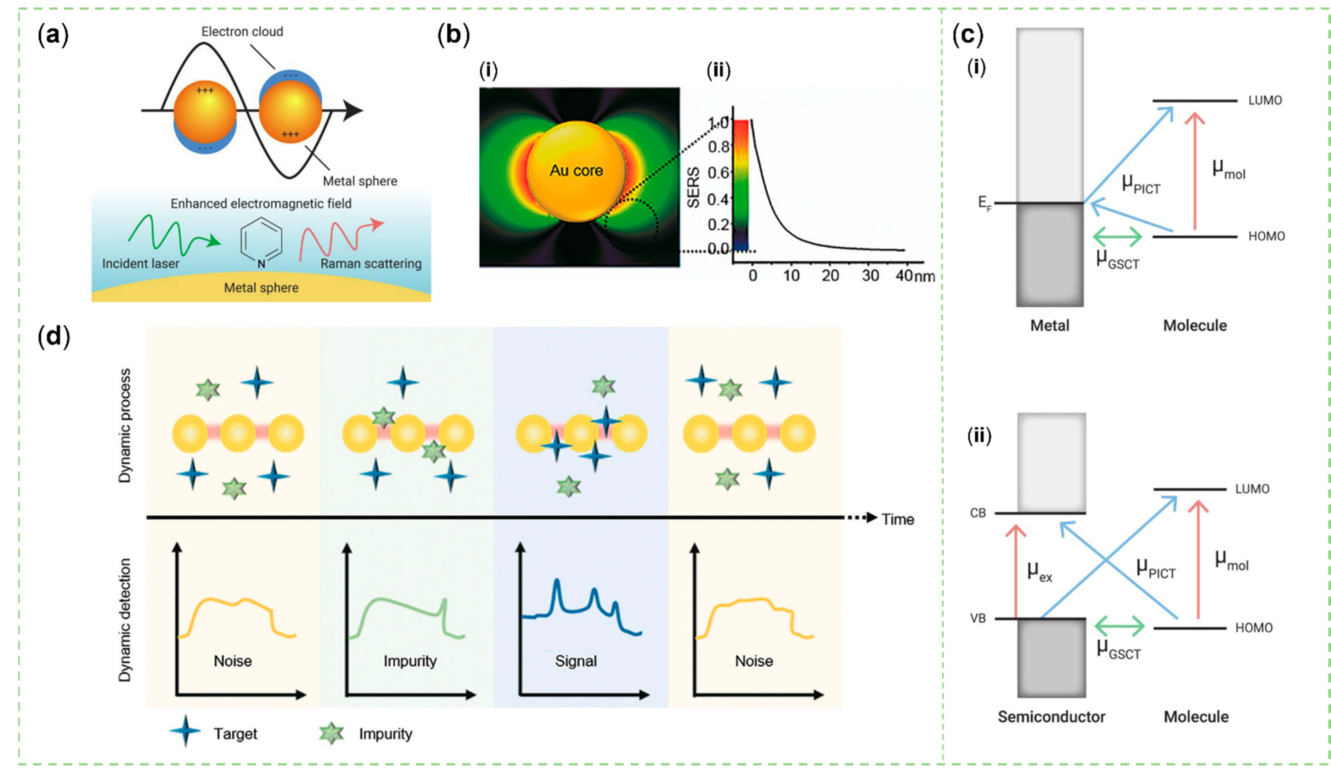
2. Mechanism of SERS
3. Synthesis and Molecular Structure of PDA
4. Guideline for Designing SERS Substrates Using PDA
4.1. Introduction of Metal or Semiconductor Materials
4.1.1. Adhesive Property
4.1.2. Reductive Property
4.1.3. Charge Transfer
4.2. Confining Signal Reporters
4.3. Identify Target Analytes
4.4. Enhancing the Practicality of Sensors
5. Biomedical Application
5.1. Detection of Nucleic Acid
5.2. Detection of Protein
5.3. Detection of Other Biological Indexes
6. Conclusions and Perspectives
Author Contributions
Funding
Institutional Review Board Statement
Informed Consent Statement
Data Availability Statement
Conflicts of Interest
References
- Gilbertson, L.M.; Zimmerman, J.B.; Plata, D.L.; Hutchison, J.E.; Anastas, P.T. Designing nanomaterials to maximize performance and minimize undesirable implications guided by the Principles of Green Chemistry. Chem. Soc. Rev. 2015, 44, 5758–5777. [Google Scholar] [CrossRef] [PubMed]
- Bartolucci, C.; Scognamiglio, V.; Antonacci, A.; Fraceto, L.F. What makes nanotechnologies applied to agriculture green? Nano Today 2022, 43, 101389. [Google Scholar] [CrossRef]
- Han, X.X.; Rodriguez, R.S.; Haynes, C.L.; Ozaki, Y.; Zhao, B. Surface-enhanced Raman spectroscopy. Nat. Rev. Methods Prim. 2022, 1, 87. [Google Scholar] [CrossRef]
- Langer, J.; Jimenez de Aberasturi, D.; Aizpurua, J.; Alvarez-Puebla, R.A.; Auguié, B.; Baumberg, J.J.; Bazan, G.C.; Bell, S.E.J.; Boisen, A.; Brolo, A.G.; et al. Present and Future of Surface-Enhanced Raman Scattering. ACS Nano 2020, 14, 28–117. [Google Scholar] [CrossRef] [PubMed]
- Zong, C.; Xu, M.; Xu, L.J.; Wei, T.; Ma, X.; Zheng, X.S.; Hu, R.; Ren, B. Surface-Enhanced Raman Spectroscopy for Bioanalysis: Reliability and Challenges. Chem. Rev. 2018, 118, 4946–4980. [Google Scholar] [CrossRef]
- Ouyang, L.; Hu, Y.; Zhu, L.; Cheng, G.J.; Irudayaraj, J. A reusable laser wrapped graphene-Ag array based SERS sensor for trace detection of genomic DNA methylation. Biosens. Bioelectron. 2017, 92, 755–762. [Google Scholar] [CrossRef]
- Zhou, B.; Ou, W.; Zhao, C.; Shen, J.; Zhang, G.; Tang, X.; Deng, Z.; Zhu, G.; Li, Y.Y.; Lu, J. Insertable and reusable SERS sensors for rapid on-site quality control of fish and meat products. Chem. Eng. J. 2021, 426, 130733. [Google Scholar] [CrossRef]
- He, Q.; Han, Y.; Huang, Y.; Gao, J.; Gao, Y.; Han, L.; Zhang, Y. Reusable dual-enhancement SERS sensor based on graphene and hybrid nanostructures for ultrasensitive lead (||) detection. Sens. Actuators B 2021, 341, 130031. [Google Scholar] [CrossRef]
- Du, Y.; Liu, H.; Chen, Y.; Tian, Y.; Zhang, X.; Gu, C.; Jiang, T.; Zhou, J. Recyclable label-free SERS-based immunoassay of PSA in human serum mediated by enhanced photocatalysis arising from Ag nanoparticles and external magnetic field. Appl. Surf. Sci. 2020, 528, 146953. [Google Scholar] [CrossRef]
- Min, K.; Choi, K.S.; Jeon, W.J.; Lee, D.K.; Oh, S.; Lee, J.; Choi, J.-Y.; Yu, H.K. Hierarchical Ag nanostructures on Sn-doped indium oxide nano-branches: Super-hydrophobic surface for surface-enhanced Raman scattering. RSC Adv. 2018, 8, 12927–12932. [Google Scholar] [CrossRef]
- Li, C.; Huang, Y.; Li, X.; Zhang, Y.; Chen, Q.; Ye, Z.; Alqarni, Z.; Bell, S.E.J.; Xu, Y. Towards practical and sustainable SERS: A review of recent developments in the construction of multifunctional enhancing substrates. J. Mater. Chem. C 2021, 9, 11517–11552. [Google Scholar] [CrossRef]
- Gong, T.; Das, C.M.; Yin, M.J.; Lv, T.R.; Singh, N.M.; Soehartono, A.M.; Singh, G.; An, Q.F.; Yong, K.T. Development of SERS tags for human diseases screening and detection. Coord. Chem. Rev. 2022, 470, 214711. [Google Scholar] [CrossRef]
- Sun, J.; Ma, Q.; Xue, D.; Shan, W.; Liu, R.; Dong, B.; Zhang, J.; Wang, Z.; Shao, B. Polymer/inorganic nanohybrids: An attractive materials for analysis and sensing. TrAC Trends Anal. Chem. 2021, 140, 116273. [Google Scholar] [CrossRef]
- Lan, L.; Ping, J.; Xiong, J.; Ying, Y. Sustainable Natural Bio-Origin Materials for Future Flexible Devices. Adv. Sci. 2022, 9, 2200560. [Google Scholar] [CrossRef]
- Lee, H.; Dellatore, S.M.; Miller, W.M.; Messersmith, P.B. Mussel-inspired surface chemistry for multifunctional coatings. Science 2007, 318, 426–430. [Google Scholar] [CrossRef]
- Liu, Y.; Ai, K.; Lu, L. Polydopamine and Its Derivative Materials: Synthesis and Promising Applications in Energy, Environmental, and Biomedical Fields. Chem. Rev. 2014, 114, 5057–5115. [Google Scholar] [CrossRef]
- Ryu, J.H.; Messersmith, P.B.; Lee, H. Polydopamine Surface Chemistry: A Decade of Discovery. Acs Appl. Mater. Interfaces 2018, 10, 7523–7540. [Google Scholar] [CrossRef]
- Raman, C.V.; Krishnan, K.S. A New Type of Secondary Radiation. Nature 1928, 121, 501–502. [Google Scholar] [CrossRef]
- Cialla-May, D.; Schmitt, M.; Popp, J. Theoretical principles of Raman spectroscopy. Phys. Sci. Rev. 2019, 4, 6. [Google Scholar] [CrossRef]
- Kneipp, J.; Kneipp, H.; Kneipp, K. SERS—A single-molecule and nanoscale tool for bioanalytics. Chem. Soc. Rev. 2008, 37, 1052–1060. [Google Scholar] [CrossRef]
- Fleischmann, M.; Hendra, P.J.; McQuillan, A.J. Raman spectra of pyridine adsorbed at a silver electrode. Chem. Phys. Lett. 1974, 26, 163–166. [Google Scholar] [CrossRef]
- Jeanmaire, D.L.; Van Duyne, R.P. Surface raman spectroelectrochemistry: Part I. Heterocyclic, aromatic, and aliphatic amines adsorbed on the anodized silver electrode. J. Electroanal. Chem. Interfacial Electrochem. 1977, 84, 1–20. [Google Scholar] [CrossRef]
- Cong, S.; Liu, X.; Jiang, Y.; Zhang, W.; Zhao, Z. Surface Enhanced Raman Scattering Revealed by Interfacial Charge-Transfer Transitions. Innovation 2020, 1, 100051. [Google Scholar] [CrossRef] [PubMed]
- Viets, C.; Hill, W. Laser power effects in SERS spectroscopy at thin metal films. J. Phys. Chem. B 2001, 105, 6330–6336. [Google Scholar] [CrossRef]
- Xia, M. 2D Materials-Coated Plasmonic Structures for SERS Applications. Coatings 2018, 8, 137. [Google Scholar] [CrossRef]
- Zhang, X.; Si, S.; Zhang, X.; Wu, W.; Xiao, X.; Jiang, C. Improved thermal stability of graphene-veiled noble metal nanoarrays as recyclable SERS substrates. Acs Appl. Mater. Interfaces 2017, 9, 40726–40733. [Google Scholar] [CrossRef]
- Chi, H.; Wang, C.; Wang, Z.; Zhu, H.; Mesias, V.S.D.; Dai, X.; Chen, Q.; Liu, W.; Huang, J. Highly reusable nanoporous silver sheet for sensitive SERS detection of pesticides. Analyst 2020, 145, 5158–5165. [Google Scholar] [CrossRef]
- Wang, C.; Mu, X.; Huo, J.; Zhang, B.; Zhang, K. Highly-efficient SERS detection for E. coli using a microfluidic chip with integrated NaYF4: Yb, Er@ SiO2@ Au under near-infrared laser excitation. Microsyst. Technol. 2021, 27, 3285–3291. [Google Scholar] [CrossRef]
- Li, L.; Jiang, R.; Shan, B.; Lu, Y.; Zheng, C.; Li, M. Near-infrared II plasmonic porous cubic nanoshells for in vivo noninvasive SERS visualization of sub-millimeter microtumors. Nat. Commun. 2022, 13, 5249. [Google Scholar] [CrossRef]
- Hang, Y.; Boryczka, J.; Wu, N. Visible-light and near-infrared fluorescence and surface-enhanced Raman scattering point-of-care sensing and bio-imaging: A review. Chem. Soc. Rev. 2022, 51, 329–375. [Google Scholar] [CrossRef]
- Cao, Y.; Zhang, J.; Yang, Y.; Huang, Z.; Long, N.V.; Fu, C. Engineering of SERS Substrates Based on Noble Metal Nanomaterials for Chemical and Biomedical Applications. Appl. Spectrosc. Rev. 2015, 50, 499–525. [Google Scholar] [CrossRef]
- Guillot, N.; de la Chapelle, M.L. The electromagnetic effect in surface enhanced Raman scattering: Enhancement optimization using precisely controlled nanostructures. J. Quant. Spectrosc. Radiat. Transf. 2012, 113, 2321–2333. [Google Scholar] [CrossRef]
- Nitzan, A.; Brus, L. Theoretical model for enhanced photochemistry on rough surfaces. J. Chem. Phys. 1981, 75, 2205–2214. [Google Scholar] [CrossRef]
- Stiles, P.L.; Dieringer, J.A.; Shah, N.C.; Van Duyne, R.P. Surface-enhanced Raman spectroscopy. Annu. Rev. Anal. Chem. 2008, 1, 601–626. [Google Scholar] [CrossRef] [PubMed]
- Willets, K.A.; Van Duyne, R.P. Localized surface plasmon resonance spectroscopy and sensing. Annu. Rev. Phys. Chem. 2007, 58, 267–297. [Google Scholar] [CrossRef] [PubMed]
- Hergert, W.; Wriedt, T. Mie theory: A review. In The Mie Theory: Basics and Applications, 2nd ed.; Springer: Berlin/Heidelberg, Germany, 2012; Volume 169, pp. 53–71. [Google Scholar]
- Alessandri, I.; Lombardi, J.R. Enhanced Raman Scattering with Dielectrics. Chem. Rev. 2016, 116, 14921–14981. [Google Scholar] [CrossRef]
- Alessandri, I. Enhancing Raman scattering without plasmons: Unprecedented sensitivity achieved by TiO2 shell-based resonators. J. Am. Chem. Soc. 2013, 135, 5541–5544. [Google Scholar] [CrossRef]
- Shin, H.Y.; Shim, E.L.; Choi, Y.J.; Park, J.H.; Yoon, S. Giant enhancement of the RMaman response due to one-dimensional ZnO nanostructures. Nanoscale 2014, 6, 14622–14626. [Google Scholar] [CrossRef]
- Cao, L.; Laim, L.; Valenzuela, P.D.; Nabet, B.; Spanier, J.E. On the Raman scattering from semiconducting nanowires. J. Raman Spectrosc. 2007, 38, 697–703. [Google Scholar] [CrossRef]
- Cao, L.; Nabet, B.; Spanier, J.E. Enhanced Raman scattering from individual semiconductor nanocones and nanowires. Phys. Rev. Lett. 2006, 96, 157402. [Google Scholar] [CrossRef]
- Saikin, S.K.; Olivares-Amaya, R.; Rappoport, D.; Stopa, M.; Aspuru-Guzik, A. On the chemical bonding effects in the Raman response: Benzenethiol adsorbed on silver clusters. Phys. Chem. Chem. Phys. 2009, 11, 9401–9411. [Google Scholar] [CrossRef]
- Liang, X.; Liang, B.; Pan, Z.; Lang, X.; Zhang, Y.; Wang, G.; Yin, P.; Guo, L. Tuning plasmonic and chemical enhancement for SERS detection on graphene-based Au hybrids. Nanoscale 2015, 7, 20188–20196. [Google Scholar] [CrossRef] [PubMed]
- Jensen, L.; Aikens, C.M.; Schatz, G.C. Electronic structure methods for studying surface-enhanced Raman scattering. Chem. Soc. Rev. 2008, 37, 1061–1073. [Google Scholar] [CrossRef] [PubMed]
- Cheng, W.; Zeng, X.; Chen, H.; Li, Z.; Zeng, W.; Mei, L.; Zhao, Y. Versatile Polydopamine Platforms: Synthesis and Promising Applications for Surface Modification and Advanced Nanomedicine. ACS Nano 2019, 13, 8537–8565. [Google Scholar] [CrossRef] [PubMed]
- Hong, S.; Na, Y.S.; Choi, S.; Song, I.T.; Kim, W.Y.; Lee, H. Non-Covalent Self-Assembly and Covalent Polymerization Co-Contribute to Polydopamine Formation. Adv. Funct. Mater. 2012, 22, 4711–4717. [Google Scholar] [CrossRef]
- Alfieri, M.L.; Weil, T.; Ng, D.Y.W.; Ball, V. Polydopamine at biological interfaces. Adv. Colloid Interface Sci. 2022, 305, 102689. [Google Scholar] [CrossRef] [PubMed]
- Liebscher, J. Chemistry of Polydopamine—Scope, Variation, and Limitation. Eur. J. Org. Chem. 2019, 31–32, 4976–4994. [Google Scholar] [CrossRef]
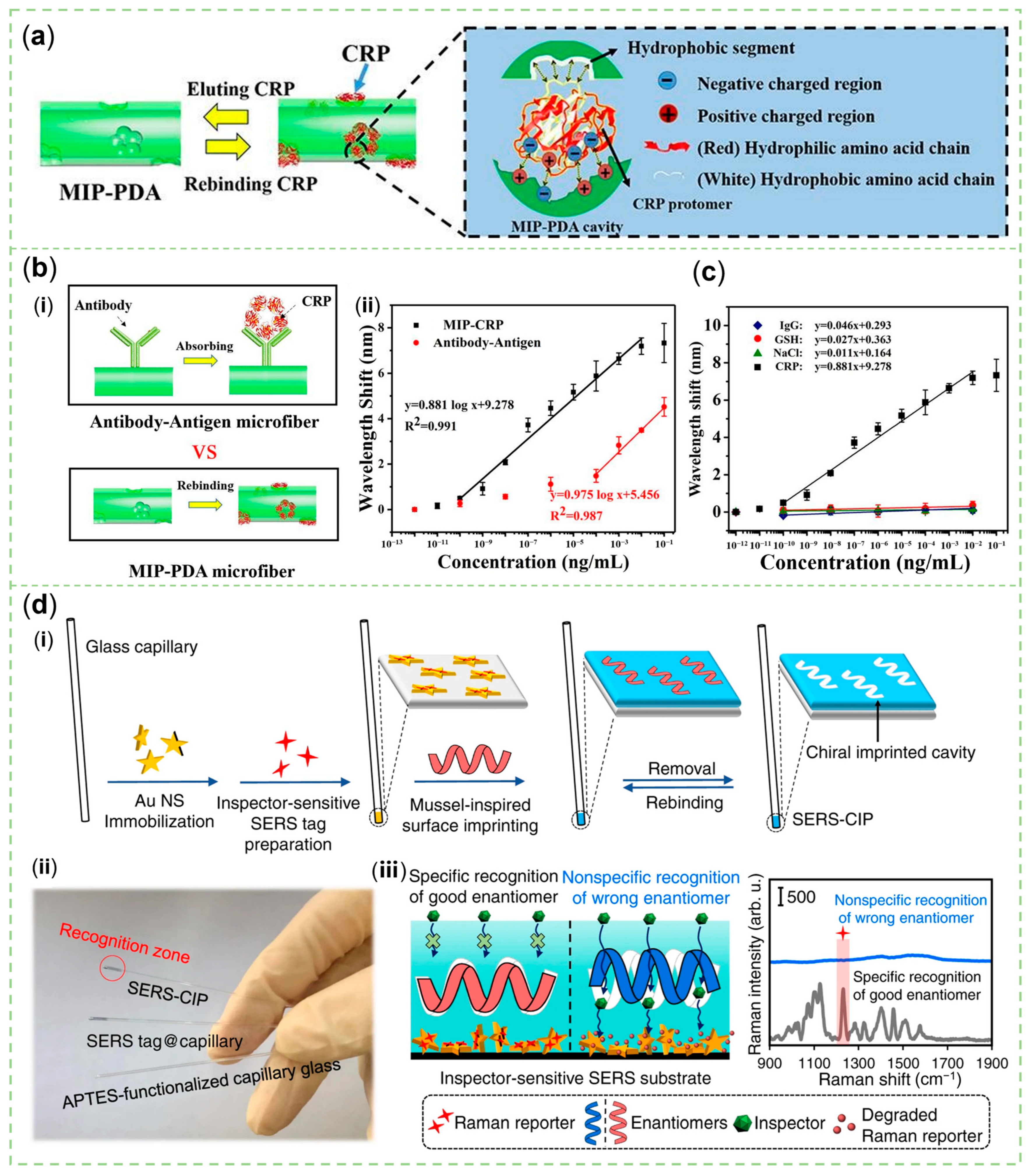
- Liu, X.; Lin, W.; Xiao, P.; Yang, M.; Sun, L.P.; Zhang, Y.; Xue, W.; Guan, B.O. Polydopamine-based molecular imprinted optic microfiber sensor enhanced by template-mediated molecular rearrangement for ultra-sensitive C-reactive protein detection. Chem. Eng. J. 2020, 387, 124074. [Google Scholar] [CrossRef]
- Alfieri, M.L.; Micillo, R.; Panzella, L.; Crescenzi, O.; Oscurato, S.L.; Maddalena, P.; Napolitano, A.; Ball, V.; d’Ischia, M. Structural Basis of Polydopamine Film Formation: Probing 5,6-Dihydroxyindole-Based Eumelanin Type Units and the Porphyrin Issue. ACS Appl. Mater. Interfaces 2018, 10, 7670–7680. [Google Scholar] [CrossRef]
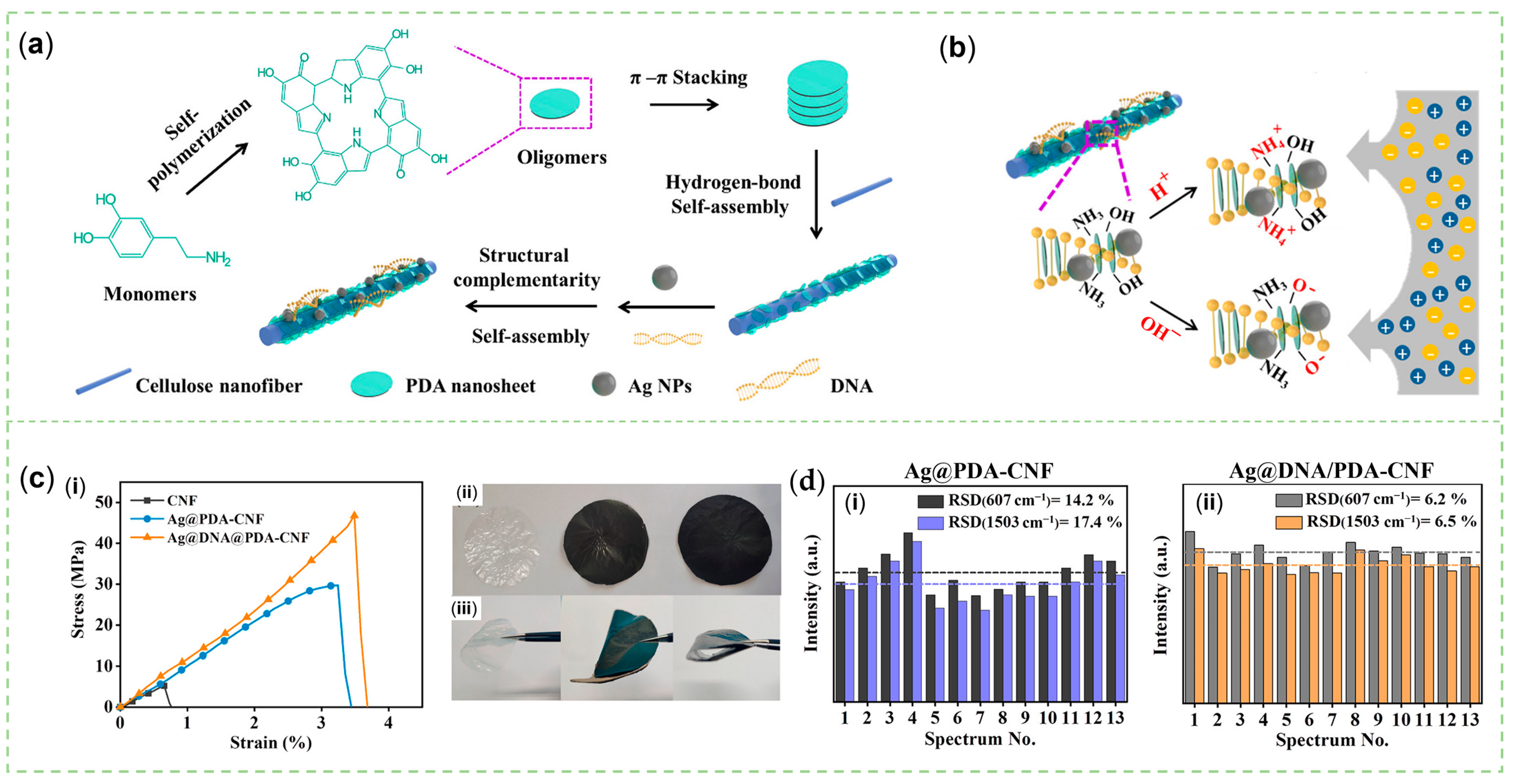
- Xu, X.Y.; Hu, X.M.; Fu, F.Y.; Liu, L.; Liu, X.D. DNA-Induced Assembly of Silver Nanoparticle Decorated Cellulose Nanofiber: A Flexible Surface-Enhanced Raman Spectroscopy Substrate for the Selective Charge Molecular Detection and Wipe Test of Pesticide Residues in Fruits. ACS Sustain. Chem. Eng. 2021, 9, 5217–5229. [Google Scholar] [CrossRef]
- Lee, H.A.; Park, E.; Lee, H. Polydopamine and Its Derivative Surface Chemistry in Material Science: A Focused Review for Studies at KAIST. Adv. Mater. 2020, 32, 1907505. [Google Scholar] [CrossRef] [PubMed]
- Xu, L.Q.; Yang, W.J.; Neoh, K.G.; Kang, E.T.; Fu, G.D. Dopamine-Induced Reduction and Functionalization of Graphene Oxide Nanosheets. Macromolecules 2010, 43, 8336–8339. [Google Scholar] [CrossRef]
- Badillo-Ramirez, I.; Saniger, J.M.; Popp, J.; Cialla-May, D. SERS characterization of dopamine and in situ dopamine polymerization on silver nanoparticles. Phys. Chem. Chem. Phys. 2021, 23, 12158–12170. [Google Scholar] [CrossRef] [PubMed]
- Alfieri, M.L.; Panzella, L.; Oscurato, S.L.; Salvatore, M.; Avolio, R.; Errico, M.E.; Maddalena, P.; Napolitano, A.; D’Ischia, M. The Chemistry of Polydopamine Film Formation: The Amine-Quinone Interplay. Biomimetics 2018, 3, 26. [Google Scholar] [CrossRef] [PubMed]
- Lee, K.; Park, M.; Malollari, K.G.; Shin, J.; Winkler, S.M.; Zheng, Y.; Park, J.H.; Grigoropoulos, C.P.; Messersmith, P.B. Laser-induced graphitization of polydopamine leads to enhanced mechanical performance while preserving multifunctionality. Nat. Commun. 2020, 11, 4848. [Google Scholar] [CrossRef] [PubMed]
- Sun, C.; Zhang, L.; Zhang, R.; Gao, M.; Zhang, X. Facilely synthesized polydopamine encapsulated surface-enhanced Raman scattering (SERS) probes for multiplex tumor associated cell surface antigen detection using SERS imaging. RSC Adv. 2015, 5, 72369–72372. [Google Scholar] [CrossRef]
- Lin, J.S.; Tian, X.D.; Li, G.; Zhang, F.L.; Wang, Y.; Li, J.F. Advanced plasmonic technologies for multi-scale biomedical imaging. Chem. Soc. Rev. 2022, 51, 9445–9468. [Google Scholar] [CrossRef]
- Zheng, Z.; Cong, S.; Gong, W.; Xuan, J.; Li, G.; Lu, W.; Geng, F.; Zhao, Z. Semiconductor SERS enhancement enabled by oxygen incorporation. Nat. Commun. 2017, 8, 1993. [Google Scholar] [CrossRef]
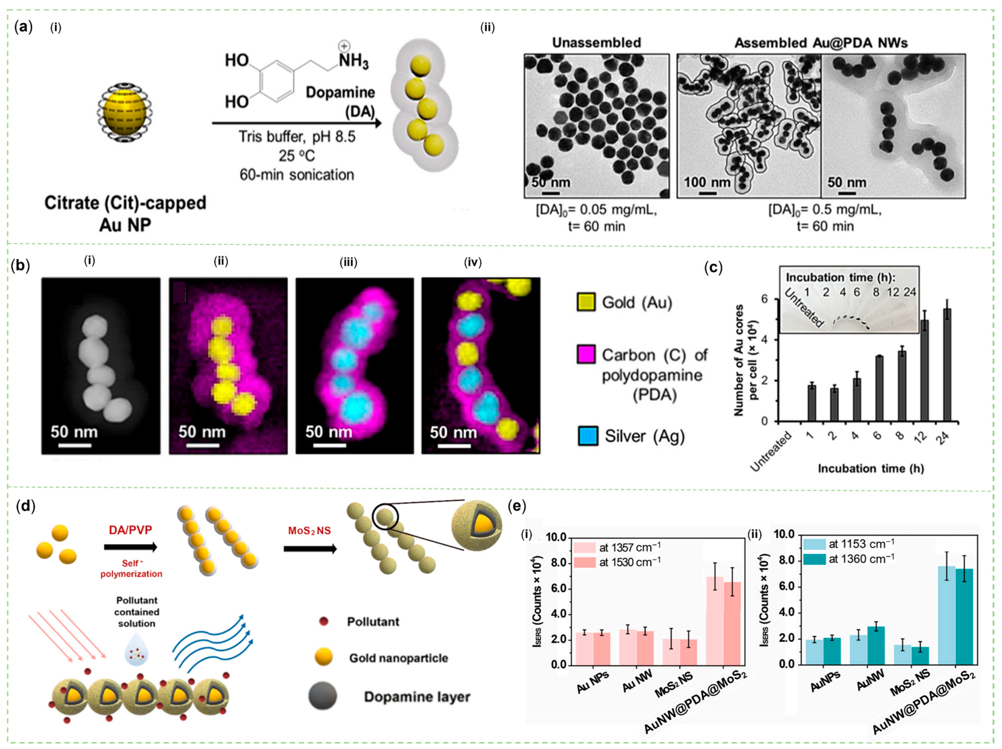
- Chen, D.; Zhu, X.; Huang, J.; Wang, G.; Zhao, Y.; Chen, F.; Wei, J.; Song, Z.; Zhao, Y. Polydopamine@Gold Nanowaxberry Enabling Improved SERS Sensing of Pesticides, Pollutants, and Explosives in Complex Samples. Anal. Chem. 2018, 90, 9048–9054. [Google Scholar] [CrossRef]
- Zhang, Z.; Si, T.; Liu, J.; Han, K.; Zhou, G. Controllable synthesis of AgNWs@PDA@AgNPs core-shell nanocobs based on a mussel-inspired polydopamine for highly sensitive SERS detection. RSC Adv. 2018, 8, 27349–27358. [Google Scholar] [CrossRef]
- Chen, M.; Zhang, L.; Yang, B.; Gao, M.; Zhang, X. Facile synthesis of terminal-alkyne bioorthogonal molecules for live -cell surface-enhanced Raman scattering imaging through Au-core and silver/dopamine-shell nanotags. Anal. Bioanal. Chem. 2018, 410, 2203–2210. [Google Scholar] [CrossRef] [PubMed]
- Tegegne, W.A.; Mekonnen, M.L.; Beyene, A.B.; Su, W.N.; Hwang, B.J. Sensitive and reliable detection of deoxynivalenol mycotoxin in pig feed by surface enhanced Raman spectroscopy on silver nanocubes@polydopamine substrate. Spectrochim. Acta Part A 2020, 229, 117940. [Google Scholar] [CrossRef] [PubMed]
- Yilmaz, M. Silver-Nanoparticle-Decorated Gold Nanorod Arrays via Bioinspired Polydopamine Coating as Surface-Enhanced Raman Spectroscopy (SERS) Platforms. Coatings 2019, 9, 198. [Google Scholar] [CrossRef]
- Choi, C.K.K.; Chiu, Y.T.E.; Zhuo, X.; Liu, Y.; Pak, C.Y.; Liu, X.; Tse, Y.L.S.; Wang, J.; Choi, C.H.J. Dopamine-Mediated Assembly of Citrate-Capped Plasmonic Nanoparticles into Stable Core–Shell Nanoworms for Intracellular Applications. ACS Nano 2019, 13, 5864–5884. [Google Scholar] [CrossRef] [PubMed]
- Yuan, H.; Yu, S.; Kim, M.; Lee, J.E.; Kang, H.; Jang, D.; Ramasamy, M.S.; Kim, D.H. Dopamine-mediated self-assembled anisotropic Au nanoworms conjugated with MoS2 nanosheets for SERS-based sensing. Sens. Actuators B 2022, 371, 132453. [Google Scholar] [CrossRef]
- Park, B.; Dang, T.V.; Yoo, J.; Tran, T.D.; Ghoreishian, S.M.; Lee, G.H.; Il Kim, M.; Huh, Y.S. Silver nanoparticle-coated polydopamine-copper hybrid nanoflowers as ultrasensitive surface-enhanced Raman spectroscopy probes for detecting thiol-containing molecules. Sens. Actuators B 2022, 369, 132246. [Google Scholar] [CrossRef]
- Achadu, O.J.; Nwaji, N.; Lee, D.; Lee, J.; Akinoglu, E.M.; Giersig, M.; Park, E.Y. 3D hierarchically porous magnetic molybdenum trioxide@gold nanospheres as a nanogap-enhanced Raman scattering biosensor for SARS-CoV-2. Nanoscale Adv. 2022, 4, 871–883. [Google Scholar] [CrossRef] [PubMed]
- Wang, Y.; Shang, B.; Liu, M.; Shi, F.; Peng, B.; Deng, Z. Hollow polydopamine colloidal composite particles: Structure tuning, functionalization and applications. J. Colloid Interface Sci. 2018, 513, 43–52. [Google Scholar] [CrossRef] [PubMed]
- Xia, Y.; Liu, Y.; Hu, X.; Zhao, F.; Zeng, B. Dual-Mode Electrochemical Competitive Immunosensor Based on Cd2+/Au/Polydopamine/Ti3C2 Composite and Copper-Based Metal–Organic Framework for 17β-Estradiol Detection. ACS Sens. 2022, 7, 3077–3084. [Google Scholar] [CrossRef] [PubMed]
- Yan, T.; Wu, T.; Wei, S.; Wang, H.; Sun, M.; Yan, L.; Wei, Q.; Ju, H. Photoelectrochemical competitive immunosensor for 17β-estradiol detection based on ZnIn2S4@NH2-MIL-125(Ti) amplified by PDA NS/Mn:ZnCdS. Biosens. Bioelectron. 2020, 148, 111739. [Google Scholar] [CrossRef]
- Zhao, M.; Zhang, X.; Deng, C. Rational synthesis of novel recyclable Fe3O4@MOF nanocomposites for enzymatic digestion. Chem. Commun. 2015, 51, 8116–8119. [Google Scholar] [CrossRef] [PubMed]
- Li, H.; Wang, X.; Wang, Z.; Wang, Y.; Dai, J.; Gao, L.; Wei, M.; Yan, Y.; Li, C. A polydopamine-based molecularly imprinted polymer on nanoparticles of type SiO2@rGO@Ag for the detection of lambda-cyhalothrin via SERS. Microchim. Acta 2018, 185, 1–10. [Google Scholar]
- Xue, Y.; Shao, J.; Sui, G.Q.; Ma, Y.Q.; Li, H.J. Rapid detection of orange II dyes in water with SERS imprinted sensor based on PDA-modified MOFs@Ag. J. Environ. Chem. Eng. 2021, 9, 106317. [Google Scholar] [CrossRef]
- Cheng, D.S.; Zhang, Y.L.; Yan, C.W.; Deng, Z.M.; Tang, X.N.; Cai, G.M.; Wang, X. Polydopamine-assisted in situ growth of three-dimensional ZnO/Ag nanocomposites on PET films for SERS and catalytic properties. J. Mol. Liq. 2021, 338, 116639. [Google Scholar] [CrossRef]
- Zhou, Y.; Zhou, J.; Wang, F.; Yang, H. Polydopamine-based functional composite particles for tumor cell targeting and dual-mode cellular imaging. Talanta 2018, 181, 248–257. [Google Scholar] [CrossRef]
- Chin, H.K.; Lin, P.Y.; Chen, J.D.; Kirankumar, R.; Wen, Z.H.; Hsieh, S.C. Polydopamine-Mediated Ag and ZnO as an Active and Recyclable SERS Substrate for Rhodamine B with Significantly Improved Enhancement Factor and Efficient Photocatalytic Degradation. Appl. Sci. 2021, 11, 4914. [Google Scholar] [CrossRef]
- Zhang, L.Z.; Liu, J.; Zhou, G.W.; Zhang, Z.L. Controllable In-Situ Growth of Silver Nanoparticles on Filter Paper for Flexible and Highly Sensitive SERS Sensors for Malachite Green Residue Detection. Nanomaterials 2020, 10, 826. [Google Scholar] [CrossRef]
- Jiao, W.; Chen, C.; You, W.; Zhang, J.; Liu, J.; Che, R. Yolk–Shell Fe/Fe4N@Pd/C Magnetic Nanocomposite as an Efficient Recyclable ORR Electrocatalyst and SERS Substrate. Small 2019, 15, 1805032. [Google Scholar] [CrossRef]
- Wang, D.; Bao, L.P.; Li, H.J.; Guo, X.Y.; Liu, W.Z.; Wang, X.Y.; Hou, X.M.; He, B. Polydopamine stabilizes silver nanoparticles as a SERS substrate for efficient detection of myocardial infarction. Nanoscale 2022, 14, 6212–6219. [Google Scholar] [CrossRef]
- Wang, C.D.; Wang, X.; Li, C.; Xu, X.H.; Ye, W.C.; Qiu, G.Y.; Wang, D.G. Silver mirror films deposited on well plates for SERS detection of multi-analytes: Aiming at 96-well technology. Talanta 2021, 222, 121544. [Google Scholar] [CrossRef]
- Yu, W.; Lin, X.; Duan, N.; Wang, Z.; Wu, S. A fluorescence and surface-enhanced Raman scattering dual-mode aptasensor for sensitive detection of deoxynivalenol based on gold nanoclusters and silver nanoparticles modified metal-polydopamine framework. Anal. Chim. Acta 2023, 1244, 340846. [Google Scholar] [CrossRef] [PubMed]
- Shen, W.; Lin, X.; Jiang, C.; Li, C.; Lin, H.; Huang, J.; Wang, S.; Liu, G.; Yan, X.; Zhong, Q.; et al. Reliable Quantitative SERS Analysis Facilitated by Core–Shell Nanoparticles with Embedded Internal Standards. Angew. Chem. Int. Ed. 2015, 54, 7308–7312. [Google Scholar] [CrossRef] [PubMed]
- Yin, Y.; Mei, R.; Wang, Y.; Zhao, X.; Yu, Q.; Liu, W.; Chen, L. Silica-Coated, Waxberry-like Surface-Enhanced Raman Resonant Scattering Tag-Pair with Near-Infrared Raman Dye Encoding: Toward In Vivo Duplexing Detection. Anal. Chem. 2020, 92, 14814–14821. [Google Scholar] [CrossRef] [PubMed]
- Zhou, J.; Xiong, Q.; Ma, J.; Ren, J.; Messersmith, P.B.; Chen, P.; Duan, H. Polydopamine-Enabled Approach toward Tailored Plasmonic Nanogapped Nanoparticles: From Nanogap Engineering to Multifunctionality. ACS Nano 2016, 10, 11066–11075. [Google Scholar] [CrossRef] [PubMed]
- Hu, W.; Xia, L.; Hu, Y.; Li, G. Honeycomb-like ReS2/polydopamine/Ag plasmonic composite assembled membrane substrate for rapid surface-enhanced Raman scattering analysis of 6-benzylaminopurine and alternariol in food. Sens. Actuators B 2023, 380, 133339. [Google Scholar] [CrossRef]
- Yang, L.; Yin, D.; Shen, Y.; Yang, M.; Li, X.; Han, X.; Jiang, X.; Zhao, B. Mesoporous semiconducting TiO2 with rich active sites as a remarkable substrate for surface-enhanced Raman scattering. Phys. Chem. Chem. Phys. 2017, 19, 18731–18738. [Google Scholar] [CrossRef]
- Rajaraman, T.S.; Parikh, S.P.; Gandhi, V.G. Black TiO2: A review of its properties and conflicting trends. Chem. Eng. J. 2020, 389, 123918. [Google Scholar] [CrossRef]
- Wang, J.H.; Yang, Y.H.; Li, H.; Gao, J.; He, P.; Bian, L.; Dong, F.Q.; He, Y. Stable and tunable plasmon resonance of molybdenum oxide nanosheets from the ultraviolet to the near-infrared region for ultrasensitive surface-enhanced Raman analysis. Chem. Sci. 2019, 10, 6330–6335. [Google Scholar] [CrossRef]
- Gu, L.J.; Ma, C.L.; Zhang, X.H.; Zhang, W.; Cong, S.; Zhao, Z.G. Populating surface-trapped electrons towards SERS enhancement of W18O49 nanowires. Chem. Commun. 2018, 54, 6332–6335. [Google Scholar] [CrossRef]
- Wang, Y.; Liu, J.; Ozaki, Y.; Xu, Z.; Zhao, B. Effect of TiO2 on Altering Direction of Interfacial Charge Transfer in a TiO2-Ag-MPY-FePc System by SERS. Angew. Chem. Int. Ed. 2019, 58, 8172–8176. [Google Scholar] [CrossRef]
- Yang, S.; Yao, J.; Quan, Y.; Hu, M.; Su, R.; Gao, M.; Han, D.; Yang, J. Monitoring the charge-transfer process in a Nd-doped semiconductor based on photoluminescence and SERS technology. Light Sci. Appl. 2020, 9, 117. [Google Scholar] [CrossRef] [PubMed]
- Wang, X.; Shi, W.; Wang, S.; Zhao, H.; Lin, J.; Yang, Z.; Chen, M.; Guo, L. Two-Dimensional Amorphous TiO2 Nanosheets Enabling High-Efficiency Photoinduced Charge Transfer for Excellent SERS Activity. J. Am. Chem. Soc. 2019, 141, 5856–5862. [Google Scholar] [CrossRef] [PubMed]
- Guo, F.; Chen, J.; Zhao, J.; Chen, Z.; Xia, D.; Zhan, Z.; Wang, Q. Z-scheme heterojunction g-C3N4@PDA/BiOBr with biomimetic polydopamine as electron transfer mediators for enhanced visible-light driven degradation of sulfamethoxazole. Chem. Eng. J. 2020, 386, 124014. [Google Scholar] [CrossRef]
- Xie, A.; Zhang, K.; Wu, F.; Wang, N.; Wang, Y.; Wang, M. Polydopamine nanofilms as visible light-harvesting interfaces for palladium nanocrystal catalyzed coupling reactions. Catal. Sci. Technol. 2016, 6, 1764–1771. [Google Scholar] [CrossRef]
- Yu, Z.; Li, F.; Yang, Q.; Shi, H.; Chen, Q.; Xu, M. Nature-Mimic Method To Fabricate Polydopamine/Graphitic Carbon Nitride for Enhancing Photocatalytic Degradation Performance. ACS Sustain. Chem. Eng. 2017, 5, 7840–7850. [Google Scholar] [CrossRef]
- Cheng, S.; Qi, M.; Li, W.; Sun, W.; Li, M.; Lin, J.; Bai, X.; Sun, Y.; Dong, B.; Wang, L. Dual-Responsive Nanocomposites for Synergistic Antibacterial Therapies Facilitating Bacteria-Infected Wound Healing. Adv. Healthc. Mater. 2022, 12, 2202652. [Google Scholar] [CrossRef]
- Kim, J.H.; Lee, M.; Park, C.B. Polydopamine as a Biomimetic Electron Gate for Artificial Photosynthesis. Angew. Chem. Int. Ed. 2014, 53, 6364–6368. [Google Scholar] [CrossRef]
- Lin, P.Y.; He, G.; Chen, J.; Dwivedi, A.K.; Hsieh, S. Monitoring the photoinduced surface catalytic coupling reaction and environmental exhaust fumes with an Ag/PDA/CuO modified 3D glass microfiber platform. J. Ind. Eng. Chem. 2020, 82, 424–432. [Google Scholar] [CrossRef]
- Akin, M.S.; Yilmaz, M.; Babur, E.; Ozdemir, B.; Erdogan, H.; Tamer, U.; Demirel, G. Large area uniform deposition of silver nanoparticles through bio-inspired polydopamine coating on silicon nanowire arrays for practical SERS applications. J. Mater. Chem. B 2014, 2, 4894–4900. [Google Scholar] [CrossRef]
- Lin, Q.; Yang, Y.; Ma, Y.; Zhang, R.; Wang, J.; Chen, X.; Shao, Z. Bandgap Engineered Polypyrrole–Polydopamine Hybrid with Intrinsic Raman and Photoacoustic Imaging Contrasts. Nano Lett. 2018, 18, 7485–7493. [Google Scholar] [CrossRef]
- Lin, X.; Wang, Y.; Wang, L.; Lu, Y.; Li, J.; Lu, D.; Zhou, T.; Huang, Z.; Huang, J.; Huang, H.; et al. Interference-free and high precision biosensor based on surface enhanced Raman spectroscopy integrated with surface molecularly imprinted polymer technology for tumor biomarker detection in human blood. Biosens. Bioelectron. 2019, 143, 111599. [Google Scholar] [CrossRef] [PubMed]
- Li, J.; Liu, F.G.; Ye, J. Boosting the Brightness of Thiolated Surface-Enhanced Raman Scattering Nanoprobes by Maximal Utilization of the Three- Dimensional Volume of Electromagnetic Fields. J. Phys. Chem. Lett. 2022, 13, 6496–6502. [Google Scholar] [CrossRef] [PubMed]
- Yang, L.; Zhang, D.; Wang, M.; Yang, Y. Effects of solvent polarity on the novel excited-state intramolecular thiol proton transfer and photophysical property compared with the oxygen proton transfer. Spectrochim. Acta Part A 2023, 293, 122475. [Google Scholar] [CrossRef] [PubMed]
- Fang, P.P.; Lu, X.; Liu, H.; Tong, Y. Applications of shell-isolated nanoparticles in surface-enhanced Raman spectroscopy and fluorescence. TrAC Trends Anal. Chem. 2015, 66, 103–117. [Google Scholar] [CrossRef]
- Zhang, Y.; Qian, J.; Wang, D.; Wang, Y.; He, S. Multifunctional gold nanorods with ultrahigh stability and tunability for in vivo fluorescence imaging, SERS detection, and photodynamic therapy. Angew. Chem. Int. Ed. 2013, 52, 1148–1151. [Google Scholar] [CrossRef]
- Li, Y.; Hao, Z.; Cao, H.; Wei, S.; Jiao, T.; Wang, M. Study on annealed graphene oxide nano-sheets for improving the surface enhanced fluorescence of silver nanoparticles. Opt. Laser Technol. 2023, 160, 109054. [Google Scholar] [CrossRef]
- Wu, Z.; Sun, D.W.; Pu, H.; Wei, Q. A dual signal-on biosensor based on dual-gated locked mesoporous silica nanoparticles for the detection of Aflatoxin B1. Talanta 2023, 253, 124027. [Google Scholar] [CrossRef]
- Cao, Y.; Han, S.; Zhang, H.; Wang, J.; Jiang, Q.Y.; Zhou, Y.; Yu, Y.J.; Wang, J.; Chen, F.; Ng, D.K. Detection of cell-surface sialic acids and photodynamic eradication of cancer cells using dye-modified polydopamine-coated gold nanobipyramids. J. Mater. Chem. B 2021, 9, 5780–5784. [Google Scholar] [CrossRef]
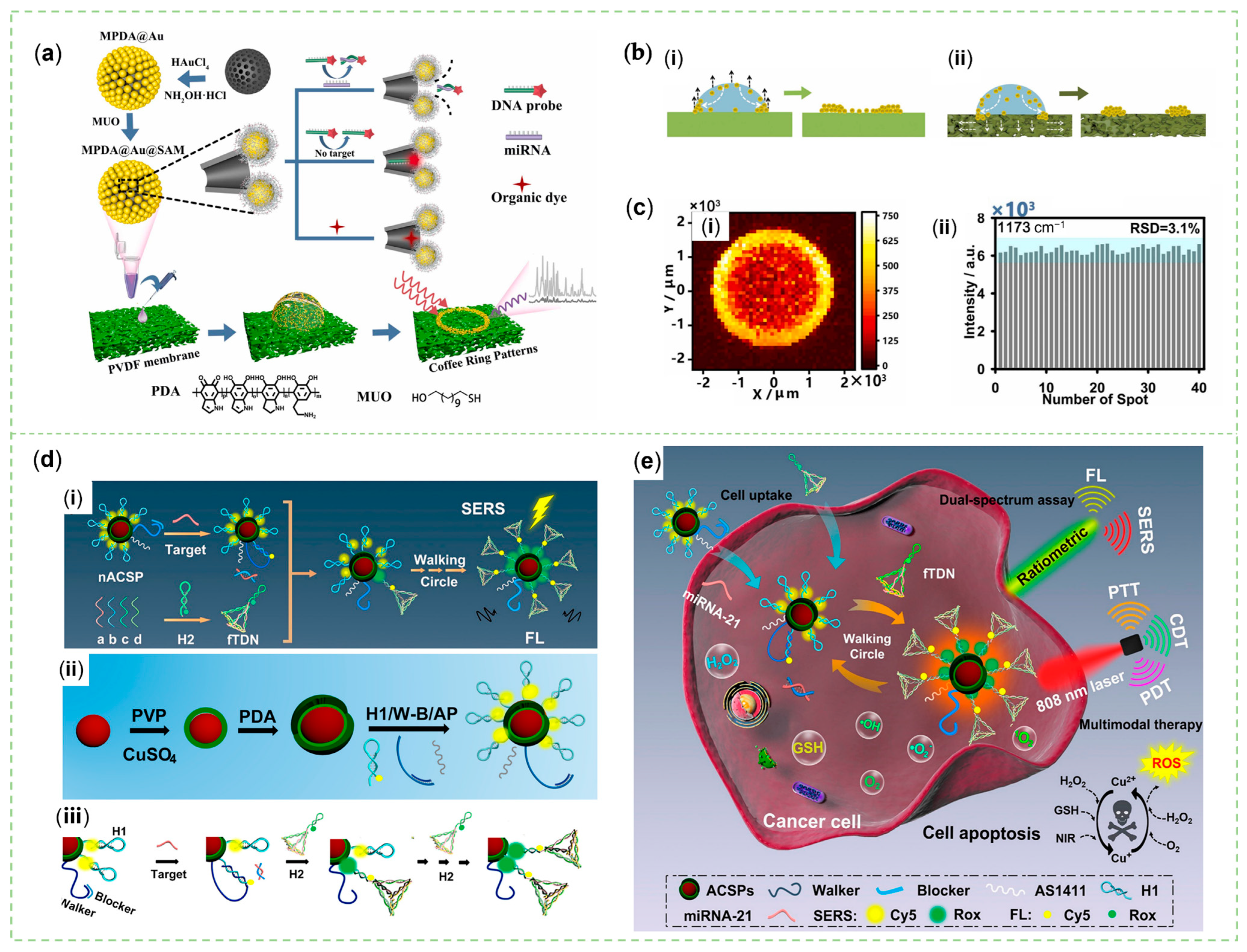
- He, P.; Han, W.H.; Bi, C.; Song, W.L.; Niu, S.Y.; Zhou, H.; Zhang, X.R. Many Birds, One Stone: A Smart Nanodevice for Ratiometric Dual-Spectrum Assay of Intracellular MicroRNA and Multimodal Synergetic Cancer Therapy. ACS Nano 2021, 15, 6961–6976. [Google Scholar] [CrossRef]
- Xu, G.; Hou, J.; Zhao, Y.; Bao, J.; Yang, M.; Fa, H.; Yang, Y.; Li, L.; Huo, D.; Hou, C. Dual-signal aptamer sensor based on polydopamine-gold nanoparticles and exonuclease I for ultrasensitive malathion detection. Sens. Actuators B 2019, 287, 428–436. [Google Scholar] [CrossRef]
- Achadu, O.J.; Abe, F.; Hossain, F.; Nasrin, F.; Yamazaki, M.; Suzuki, T.; Park, E.Y. Sulfur-doped carbon dots@polydopamine-functionalized magnetic silver nanocubes for dual-modality detection of norovirus. Biosens. Bioelectron. 2021, 193, 113540. [Google Scholar] [CrossRef] [PubMed]
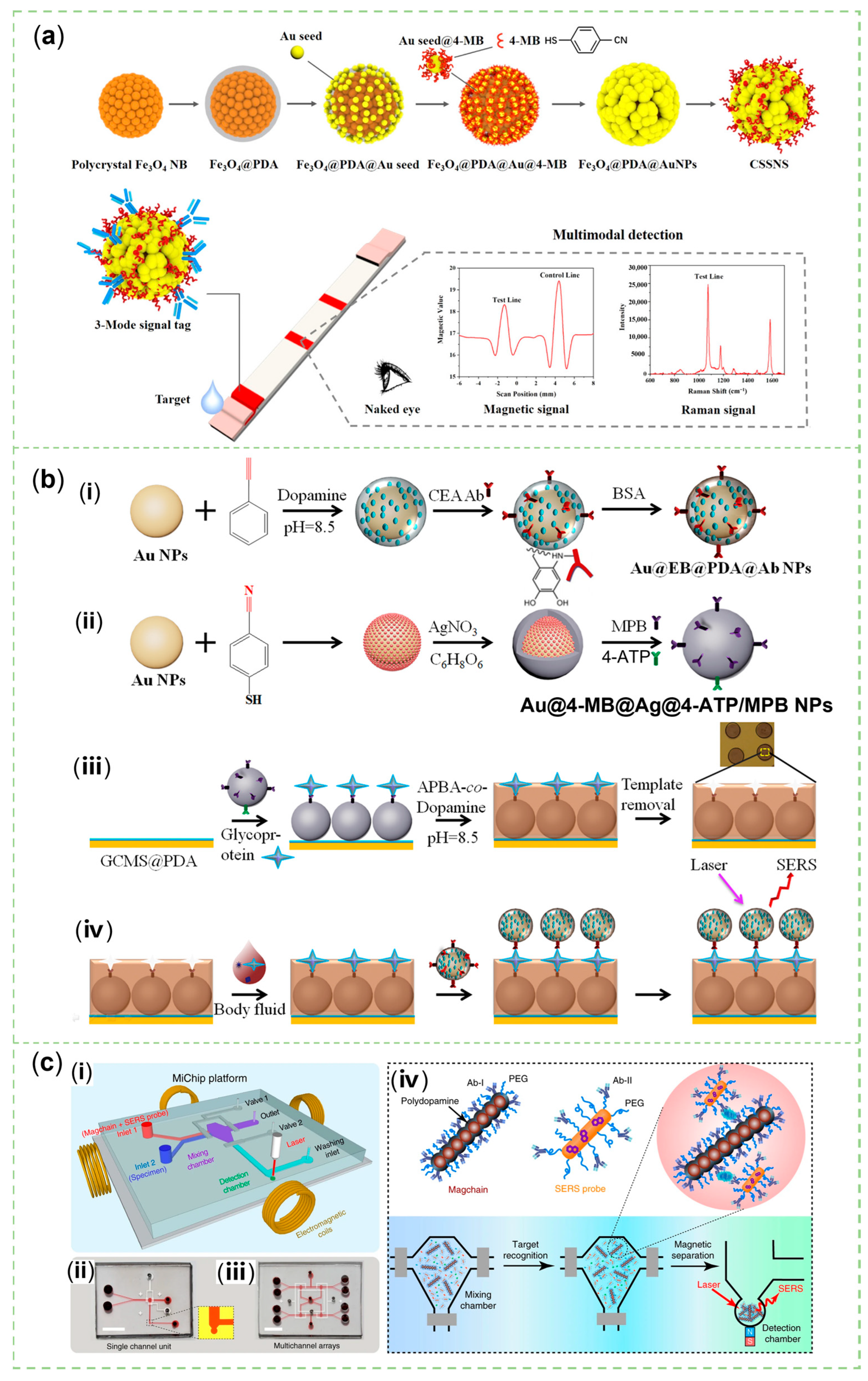
- Xiong, Q.; Lim, C.Y.; Ren, J.; Zhou, J.; Pu, K.; Chan-Park, M.B.; Mao, H.; Lam, Y.C.; Duan, H. Magnetic nanochain integrated microfluidic biochips. Nat. Commun. 2018, 9, 1743. [Google Scholar] [CrossRef] [PubMed]
- Jiang, N.; Hu, Y.; Wei, W.; Zhu, T.; Yang, K.; Zhu, G.; Yu, M. Detection of microRNA using a polydopamine mediated bimetallic SERS substrate and a re-circulated enzymatic amplification system. Microchim. Acta 2019, 186, 1–9. [Google Scholar] [CrossRef] [PubMed]
- Li, T.D.; Zhang, R.; Chen, H.; Huang, Z.P.; Ye, X.; Wang, H.; Deng, A.M.; Kong, J.L. An ultrasensitive polydopamine bi-functionalized SERS immunoassay for exosome-based diagnosis and classification of pancreatic cancer. Chem. Sci. 2018, 9, 5372–5382. [Google Scholar] [CrossRef]
- Wang, Y.L.; Li, Q.Y.; Zhang, R.; Tang, K.Q.; Ding, C.F.; Yu, S.N. SERS-based immunocapture and detection of pathogenic bacteria using a boronic acid-functionalized polydopamine-coated Au@Ag nanoprobe. Microchim. Acta 2020, 187, 290. [Google Scholar] [CrossRef]
- Arabi, M.; Ostovan, A.; Zhang, Z.Y.; Wang, Y.Q.; Mei, R.C.; Fu, L.W.; Wang, X.Y.; Ma, J.P.; Chen, L.X. Label-free SERS detection of Raman-Inactive protein biomarkers by Raman reporter indicator: Toward ultrasensitivity and universality. Biosens. Bioelectron. 2021, 174, 112825. [Google Scholar] [CrossRef]
- Yang, Y.Y.; Li, Y.T.; Li, X.J.; Zhang, L.; Kouadio Fodjo, E.; Han, S. Controllable in situ fabrication of portable AuNP/mussel-inspired polydopamine molecularly imprinted SERS substrate for selective enrichment and recognition of phthalate plasticizers. Chem. Eng. J. 2020, 402, 125179. [Google Scholar] [CrossRef]
- Chen, W.; Fu, M.; Zhu, X.; Liu, Q. Protein recognition by polydopamine-based molecularly imprinted hollow spheres. Biosens. Bioelectron. 2019, 142, 111492. [Google Scholar] [CrossRef]
- Arabi, M.; Ostovan, A.; Wang, Y.Q.; Mei, R.C.; Fu, L.W.; Li, J.H.; Wang, X.Y.; Chen, L.X. Chiral molecular imprinting-based SERS detection strategy for absolute enantiomeric discrimination. Nat. Commun. 2022, 13, 5757. [Google Scholar] [CrossRef]
- Kong, H.J.; Sun, X.P.; Yang, L.; Liu, X.L.; Yang, H.F.; Jin, R.H. Polydopamine/Silver Substrates Stemmed from Chiral Silica for SERS Differentiation of Amino Acid Enantiomers. Acs Appl. Mater. Interfaces 2020, 12, 29868–29875. [Google Scholar] [CrossRef]
- Yang, Y.Y.; Li, Y.T.; Zhai, W.L.; Li, X.J.; Li, D.; Lin, H.L.; Han, S. Electrokinetic Preseparation and Molecularly Imprinted Trapping for Highly Selective SERS Detection of Charged Phthalate Plasticizers. Anal. Chem. 2021, 93, 946–955. [Google Scholar] [CrossRef] [PubMed]
- Yu, B.; Liu, J.; Liu, S.; Zhou, F. Pdop layer exhibiting zwitterionicity: A simple electrochemical interface for governing ion permeability. Chem. Commun. 2010, 46, 5900–5902. [Google Scholar] [CrossRef] [PubMed]
- Nayak, S.; Blumenfeld, N.R.; Laksanasopin, T.; Sia, S.K. Point-of-Care Diagnostics: Recent Developments in a Connected Age. Anal. Chem. 2017, 89, 102–123. [Google Scholar] [CrossRef] [PubMed]
- Liu, X.; Liu, X.; Rong, P.; Liu, D. Recent advances in background-free Raman scattering for bioanalysis. TrAC Trends Anal. Chem. 2020, 123, 115765. [Google Scholar] [CrossRef]
- Linh, V.T.N.; Yim, S.G.; Mun, C.; Yang, J.Y.; Lee, S.; Yoo, Y.W.; Sung, D.K.; Lee, Y.I.; Kim, D.H.; Park, S.G.; et al. Bioinspired plasmonic nanoflower-decorated microneedle for label-free intradermal sensing. Appl. Surf. Sci. 2021, 551, 149411. [Google Scholar] [CrossRef]
- Renard, D.; Tian, S.; Ahmadivand, A.; DeSantis, C.J.; Clark, B.D.; Nordlander, P.; Halas, N.J. Polydopamine-Stabilized Aluminum Nanocrystals: Aqueous Stability and Benzo[a]pyrene Detection. ACS Nano 2019, 13, 3117–3124. [Google Scholar] [CrossRef]
- Chang, Y.L.; Su, C.J.; Lu, L.C.; Wan, D.H. Aluminum Plasmonic Nanoclusters for Paper-Based Surface- Enhanced Raman Spectroscopy. Anal. Chem. 2022, 94, 16319–16327. [Google Scholar] [CrossRef]
- Liu, J.; Si, T.; Zhang, L.; Zhang, Z. Mussel-Inspired Fabrication of SERS Swabs for Highly Sensitive and Conformal Rapid Detection of Thiram Bactericides. Nanomaterials 2019, 9, 1331. [Google Scholar] [CrossRef]
- Li, H.J.; Wang, J.F.; Fang, H.Q.; Xu, H.D.; Yu, H.C.; Zhou, T.Y.; Liu, C.B.; Che, G.B.; Wang, D.D. Hydrophilic modification of PVDF-based SERS imprinted membrane for the selective detection of L-tyrosine. J. Environ. Manag. 2022, 304, 114260. [Google Scholar] [CrossRef]
- Wan, M.H.; Zhao, H.D.; Wang, Z.H.; Zhao, Y.B.; Sun, L. Preparation of Ag@PDA@SiO2 electrospinning nanofibrous membranes for direct bacteria SERS detection and antimicrobial activities. Mater. Res. Express 2020, 7, 095012. [Google Scholar] [CrossRef]
- Zhang, Z.L.; Si, T.T.; Liu, J.; Zhou, G.W. In-Situ Grown Silver Nanoparticles on Nonwoven Fabrics Based on Mussel-Inspired Polydopamine for Highly Sensitive SERS Carbaryl Pesticides Detection. Nanomaterials 2019, 9, 384. [Google Scholar] [CrossRef] [PubMed]
- Liu, J.; Si, T.; Zhang, Z. Mussel-inspired immobilization of silver nanoparticles toward sponge for rapid swabbing extraction and SERS detection of trace inorganic explosives. Talanta 2019, 204, 189–197. [Google Scholar] [CrossRef] [PubMed]
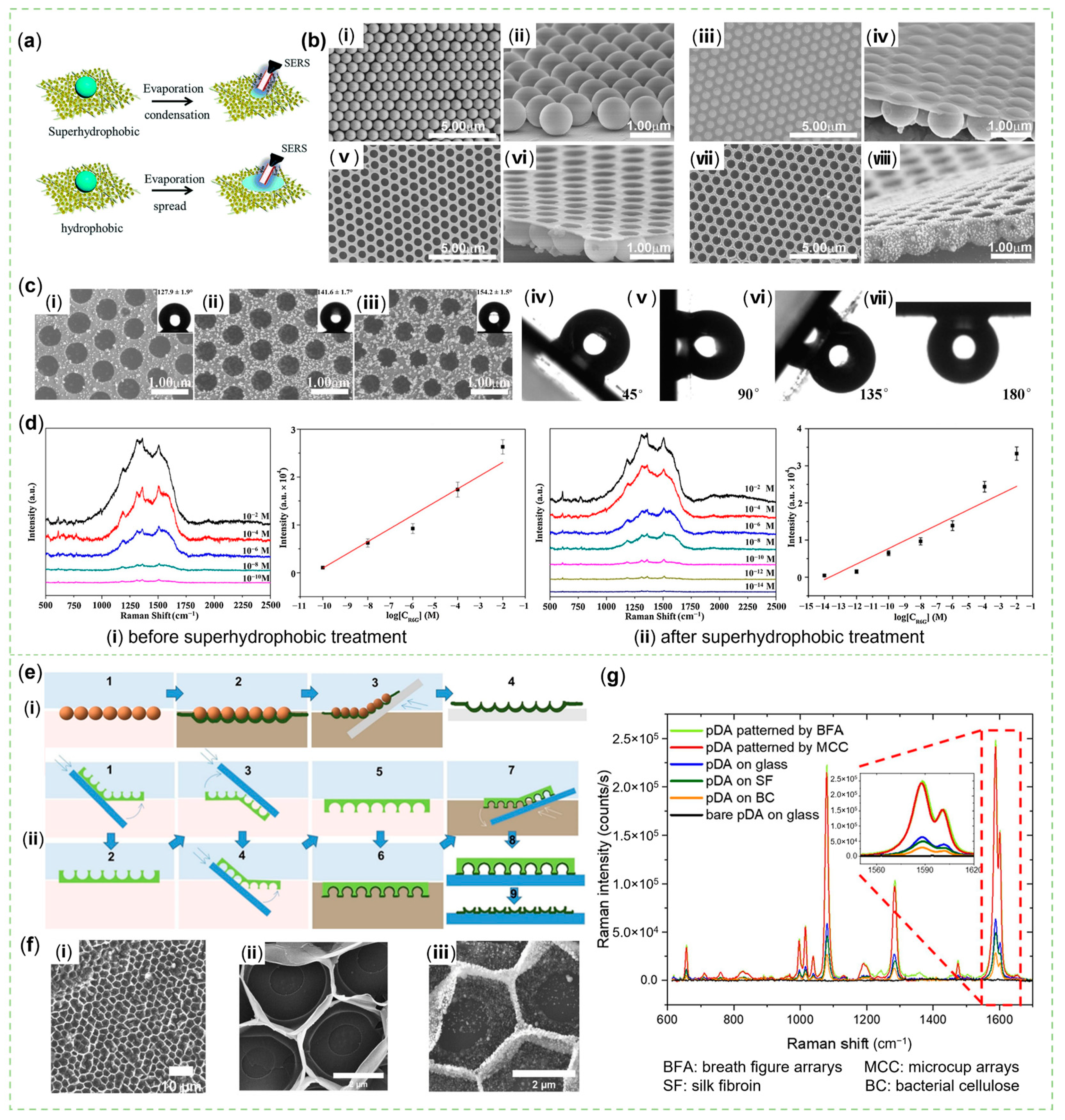
- Shang, B.; Wang, Y.B.; Yang, P.; Peng, B.; Deng, Z.W. Synthesis of superhydrophobic polydopamine-Ag microbowl/nanoparticle array substrates for highly sensitive, durable and reproducible surface-enhanced Raman scattering detection. Sens. Actuators B 2018, 255, 995–1005. [Google Scholar] [CrossRef]
- Kozma, E.; Andicsova, A.E.; Siskova, A.O.; Tullii, G.; Galeotti, F. Biomimetic design of functional plasmonic surfaces based on polydopamine. Appl. Surf. Sci. 2022, 591, 153135. [Google Scholar] [CrossRef]
- Dong, J.C.; Wang, T.C.; Xu, E.Z.; Bai, F.; Liu, J.; Zhang, Z.L. Flexible Hydrophobic CFP@PDA@AuNPs Stripes for Highly Sensitive SERS Detection of Methylene Blue Residue. Nanomaterials 2022, 12, 2163. [Google Scholar] [CrossRef]
- Liu, Y.; Li, Z.; Yin, Z.; Zhang, H.; Gao, Y.; Huo, G.; Wu, A.; Zeng, L. Amplified Photoacoustic Signal and Enhanced Photothermal Conversion of Polydopamine-Coated Gold Nanobipyramids for Phototheranostics and Synergistic Chemotherapy. ACS Appl. Mater. Interfaces 2020, 12, 14866–14875. [Google Scholar] [CrossRef]
- Liu, X.; Liao, G.; Zou, L.; Zheng, Y.; Yang, X.; Wang, Q.; Geng, X.; Li, S.; Liu, Y.; Wang, K. Construction of Bio/Nanointerfaces: Stable Gold Nanoparticle Bioconjugates in Complex Systems. Acs Appl. Mater. Interfaces 2019, 11, 40817–40825. [Google Scholar] [CrossRef]
- Chang, N.; Wang, D.L.; Liu, B.; He, D.; Wu, H.; Zhao, X.W. Stable Plasmonic Coloration of Versatile Surfaces via Colloidal Monolayer Transfer Printing. Adv. Eng. Mater. 2019, 21, 1900313. [Google Scholar] [CrossRef]
- Poulichet, V.; Morel, M.; Rudiuk, S.; Baigl, D. Liquid-liquid coffee-ring effect. J. Colloid Interface Sci. 2020, 573, 370–375. [Google Scholar] [CrossRef]
- Mampallil, D.; Eral, H.B. A review on suppression and utilization of the coffee-ring effect. Adv. Colloid Interface Sci. 2018, 252, 38–54. [Google Scholar] [CrossRef]
- Liang, X.; Zhang, H.; Xu, C.; Cao, D.; Gao, Q.; Cheng, S. Condensation effect-induced improved sensitivity for SERS trace detection on a superhydrophobic plasmonic nanofibrous mat. RSC Adv. 2017, 7, 44492–44498. [Google Scholar] [CrossRef]
- Luo, X.; Pan, R.; Cai, M.; Liu, W.; Chen, C.; Jiang, G.; Hu, X.; Zhang, H.; Zhong, M. Atto-Molar Raman detection on patterned superhydrophilic-superhydrophobic platform via localizable evaporation enrichment. Sens. Actuators B 2021, 326, 128826. [Google Scholar] [CrossRef]
- Bhushan, B.; Jung, Y.C. Natural and biomimetic artificial surfaces for superhydrophobicity, self-cleaning, low adhesion, and drag reduction. Prog. Mater. Sci. 2011, 56, 1–108. [Google Scholar] [CrossRef]
- Wang, B.; Liang, W.; Guo, Z.; Liu, W. Biomimetic super-lyophobic and super-lyophilic materials applied for oil/water separation: A new strategy beyond nature. Chem. Soc. Rev. 2015, 44, 336–361. [Google Scholar] [CrossRef] [PubMed]
- Kuang, M.; Wang, J.; Jiang, L. Bio-inspired photonic crystals with superwettability. Chem. Soc. Rev. 2016, 45, 6833–6854. [Google Scholar] [CrossRef] [PubMed]
- Yu, X.; Zhong, Q.Z.; Yang, H.C.; Wan, L.S.; Xu, Z.K. Mussel-Inspired Modification of Honeycomb Structured Films for Superhydrophobic Surfaces with Tunable Water Adhesion. J. Phys. Chem. C 2015, 119, 3667–3673. [Google Scholar] [CrossRef]
- Coy, E.; Iatsunskyi, I.; Colmenares, J.C.; Kim, Y.; Mrówczyński, R. Polydopamine Films with 2D-like Layered Structure and High Mechanical Resilience. ACS Appl. Mater. Interfaces 2021, 13, 23113–23120. [Google Scholar] [CrossRef]
- Marchesi D’Alvise, T.; Harvey, S.; Hueske, L.; Szelwicka, J.; Veith, L.; Knowles, T.P.J.; Kubiczek, D.; Flaig, C.; Port, F.; Gottschalk, K.-E.; et al. Ultrathin Polydopamine Films with Phospholipid Nanodiscs Containing a Glycophorin A Domain. Adv. Funct. Mater. 2020, 30, 2000378. [Google Scholar] [CrossRef]
- Lane, L.A.; Qian, X.; Nie, S. SERS Nanoparticles in Medicine: From Label-Free Detection to Spectroscopic Tagging. Chem. Rev. 2015, 115, 10489–10529. [Google Scholar] [CrossRef]
- Song, L.; Chen, J.; Xu, B.B.; Huang, Y. Flexible plasmonic biosensors for healthcare monitoring: Progress and prospects. ACS Nano 2021, 15, 18822–18847. [Google Scholar] [CrossRef]
- Wang, Y.; Li, B.; Tian, T.; Liu, Y.; Zhang, J.; Qian, K. Advanced on-site and in vitro signal amplification biosensors for biomolecule analysis. TrAC Trends Anal. Chem. 2022, 149, 116565. [Google Scholar] [CrossRef]
- Meng, X.; Yang, F.; Dong, H.; Dou, L.; Zhang, X. Recent advances in optical imaging of biomarkers in vivo. Nano Today 2021, 38, 101156. [Google Scholar] [CrossRef]
- Lee, H.K.; Lee, Y.H.; Koh, C.S.L.; Phan-Quang, G.C.; Han, X.; Lay, C.L.; Sim, H.Y.F.; Kao, Y.C.; An, Q.; Ling, X.Y. Designing surface-enhanced Raman scattering (SERS) platforms beyond hotspot engineering: Emerging opportunities in analyte manipulations and hybrid materials. Chem. Soc. Rev. 2019, 48, 731–756. [Google Scholar] [CrossRef] [PubMed]
- Ding, Q.; Wang, J.; Chen, X.; Liu, H.; Li, Q.; Wang, Y.; Yang, S. Quantitative and Sensitive SERS Platform with Analyte Enrichment and Filtration Function. Nano Lett. 2020, 20, 7304–7312. [Google Scholar] [CrossRef]
- Fang, Y.; Seong, N.H.; Dlott, D.D. Measurement of the Distribution of Site Enhancements in Surface-Enhanced Raman Scattering. Science 2008, 321, 388–392. [Google Scholar] [CrossRef]
- Liu, B.; Liu, X.; Shi, S.; Huang, R.; Su, R.; Qi, W.; He, Z. Design and mechanisms of antifouling materials for surface plasmon resonance sensors. Acta Biomater. 2016, 40, 100–118. [Google Scholar] [CrossRef]
- Zhou, M.Z.; Wang, Z.Q.; Xia, D.Q.; Xie, X.Y.; Chen, Y.H.; Xing, Y.X.; Cai, K.Y.; Zhang, J.X. Hybrid nanoassembly with two-tier host-guest architecture and regioselective enrichment capacity for repetitive SERS detection. Sens. Actuators B 2022, 369, 132359. [Google Scholar] [CrossRef]
- Zhai, J.; Li, X.; Zhang, J.; Pan, H.; Peng, Q.; Gan, H.; Su, S.; Yuwen, L.; Song, C. SERS/electrochemical dual-mode biosensor based on multi-functionalized molybdenum disulfide nanosheet probes and SERS-active Ag nanorods array electrodes for reliable detection of cancer-related miRNA. Sens. Actuators B 2022, 368, 132245. [Google Scholar] [CrossRef]
- Yuwen, L.; Sun, Y.; Tan, G.; Xiu, W.; Zhang, Y.; Weng, L.; Teng, Z.; Wang, L. MoS2@polydopamine-Ag nanosheets with enhanced antibacterial activity for effective treatment of Staphylococcus aureus biofilms and wound infection. Nanoscale 2018, 10, 16711–16720. [Google Scholar] [CrossRef]
- Wu, Y.; He, Y.; Yang, X.; Yuan, R.; Chai, Y. A novel recyclable surface-enhanced Raman spectroscopy platform with duplex-specific nuclease signal amplification for ultrasensitive analysis of microRNA 155. Sens. Actuators B 2018, 275, 260–266. [Google Scholar] [CrossRef]
- Lu, D.; Xia, J.; Deng, Z.; Cao, X.W. Detection of squamous cell carcinoma antigen in cervical cancer by surface-enhanced Raman scattering-based immunoassay. Anal. Methods 2019, 11, 2809–2818. [Google Scholar] [CrossRef]
- Wang, J.J.; Xu, C.X.; Lei, M.L.; Ma, Y.; Wang, X.X.; Wang, R.; Sun, J.L.; Wang, R. Microcavity-based SERS chip for ultrasensitive immune detection of cardiac biomarkers. Microchem. J. 2021, 171, 106875. [Google Scholar] [CrossRef]
- Huang, Z.; Zhang, R.; Chen, H.; Weng, W.; Lin, Q.; Deng, D.; Li, Z.; Kong, J. Sensitive polydopamine bi-functionalized SERS immunoassay for microalbuminuria detection. Biosens. Bioelectron. 2019, 142, 111542. [Google Scholar] [CrossRef] [PubMed]
- Li, X.; Li, B.; Hong, J.; Zhou, X. Highly selective determination of acid phosphatase in biological samples using a biomimetic recognition-based SERS sensor. Sens. Actuators B 2018, 276, 421–428. [Google Scholar] [CrossRef]
- Liu, X.; Wang, K.; Cao, B.; Shen, L.; Ke, X.; Cui, D.; Zhong, C.; Li, W. Multifunctional Nano-Sunflowers with Color-Magnetic-Raman Properties for Multimodal Lateral Flow Immunoassay. Anal. Chem. 2021, 93, 3626–3634. [Google Scholar] [CrossRef]
- Xia, J.; Liu, Y.F.; Ran, M.L.; Lu, W.B.; Bi, L.Y.; Wang, Q.; Lu, D.; Cao, X.W. The simultaneous detection of the squamous cell carcinoma antigen and cancer antigen 125 in the cervical cancer serum using nano-Ag polydopamine nanospheres in an SERS-based lateral flow immunoassay. RSC Adv. 2020, 10, 29156–29170. [Google Scholar] [CrossRef]
- Cheng, N.; Du, D.; Wang, X.; Liu, D.; Xu, W.; Luo, Y.; Lin, Y. Recent Advances in Biosensors for Detecting Cancer-Derived Exosomes. Trends Biotechnol. 2019, 37, 1236–1254. [Google Scholar] [CrossRef]
- Hong, W.E.; Hsu, I.L.; Huang, S.Y.; Lee, C.W.; Ko, H.; Tsai, P.J.; Shieh, D.B.; Huang, C.C. Assembled growth of 3D Fe3O4@Au nanoparticles for efficient photothermal ablation and SERS detection of microorganisms. J. Mater. Chem. B 2018, 6, 5689–5697. [Google Scholar] [CrossRef]
- D’Autréaux, B.; Toledano, M.B. ROS as signalling molecules: Mechanisms that generate specificity in ROS homeostasis. Nat. Rev. Mol. Cell Biol. 2007, 8, 813–824. [Google Scholar] [CrossRef]
- Sun, Y.; Sun, X.; Li, X.; Li, W.; Li, C.; Zhou, Y.; Wang, L.; Dong, B. A versatile nanocomposite based on nanoceria for antibacterial enhancement and protection from aPDT-aggravated inflammation via modulation of macrophage polarization. Biomaterials 2021, 268, 120614. [Google Scholar] [CrossRef]
- Winterbourn, C.C. Reconciling the chemistry and biology of reactive oxygen species. Nat. Chem. Biol. 2008, 4, 278–286. [Google Scholar] [CrossRef] [PubMed]
- Murphy, M.P.; Holmgren, A.; Larsson, N.-G.; Halliwell, B.; Chang, C.J.; Kalyanaraman, B.; Rhee, S.G.; Thornalley, P.J.; Partridge, L.; Gems, D.; et al. Unraveling the Biological Roles of Reactive Oxygen Species. Cell Metab. 2011, 13, 361–366. [Google Scholar] [CrossRef] [PubMed]
- Kumar, S.; Kumar, A.; Kim, G.H.; Rhim, W.K.; Hartman, K.L.; Nam, J.M. Myoglobin and Polydopamine-Engineered Raman Nanoprobes for Detecting, Imaging, and Monitoring Reactive Oxygen Species in Biological Samples and Living Cells. Small 2017, 13, 1701584. [Google Scholar] [CrossRef] [PubMed]
- Cheng, D.S.; Bai, X.; He, M.T.; Wu, J.H.; Yang, H.J.; Ran, J.H.; Cai, G.M.; Wang, X. Polydopamine-assisted immobilization of Ag@AuNPs on cotton fabrics for sensitive and responsive SERS detection. Cellulose 2019, 26, 4191–4204. [Google Scholar] [CrossRef]
- Sieste, S.; Mack, T.; Synatschke, C.V.; Schilling, C.; Meyer zu Reckendorf, C.; Pendi, L.; Harvey, S.; Ruggeri, F.S.; Knowles, T.P.J.; Meier, C.; et al. Water-Dispersible Polydopamine-Coated Nanofibers for Stimulation of Neuronal Growth and Adhesion. Adv. Healthc. Mater. 2018, 7, 1701485. [Google Scholar] [CrossRef] [PubMed]


| Properties | Materials | Ref. |
|---|---|---|
| biocompatibility | Ag/PDA/ZnO-filter paper | [77] |
| Au-HA *-PDA-PLGA *-microneedles | [126] | |
| inoxidizability | Ag nanocubes@PDA | [63] |
| Fe/Fe4N@Pd/C | [79] | |
| PDA@Ag-anti-cTn Ⅰ * | [80] | |
| Al nanocrystals@PDA | [127] | |
| Al NPs@PDA on cellulose paper | [128] | |
| antifouling property | Au@EB *@PDA@Ab@BSA | [102] |
| Magchains *@PDA@PEG@Ab | [113] | |
| uniformity | Capillary glass@Au NSs@MIP | [117] |
| flexibility and uniformity | PET/PDA/ZnO/Ag | [75] |
| filter paper@PDA@Ag NPs | [78] | |
| cotton swab@PDA@Ag NPs | [129] | |
| W18O49@Ag/PDA@PVDF-MIP membranes | [130] | |
| Ag@PDA@SiO2 nanofibrous membranes | [131] | |
| non-woven fabrics@PDA@Ag NPs | [132] | |
| polyurethane sponge@PDA@Ag NPs | [133] | |
| mechanical stability and uniformity | Ag@DNA/PDA-CNF | [51] |
| Au-HA-PDA-PLGA-microneedles | [126] | |
| hydrophobic or superhydrophobic | PDA-Ag microbowl array | [134] |
| PDA film patterned by microcup or breath figure arrays | [135] | |
| cellulose filter paper@PDA@Ag NPs | [136] |
| Different Design | Material | Method | Targets | Linear Detection Range | LOD | Ref. |
|---|---|---|---|---|---|---|
| Colloidal solutions | ACE2 *-mag-MoO3-PDA@Au-4-MBA | SERS | SARS-CoV-2 spike protein | 10 fg mL−1–1 ng mL−1 | 4.5 fg mL−1 (in PBS) | [68] |
| 9.7 fg mL−1 (in whole-cell lysate media) | ||||||
| PDA@Ag-anti-cTn Ⅰ * | SERS | cTn Ⅰ | - | 0.01 ng mL−1 (in PBS) | [80] | |
| - | 0.025 ng mL−1 (in human serum) | |||||
| Au@Cu2−xS@PDA | FL | miRNA-21 | 1 pM–10 nM | 0.11 pM | [110] | |
| SERS | 10 aM–1 nM (in vitro) | 4.95 aM | ||||
| 0.29 fM–9.30 pM (in living cells) | 0.11 fM | |||||
| Au@PDA@Ag | SERS | miRNA-31 | 0.6–1.8 fM | 0.2 fM | [114] | |
| Fe3O4@PDA/Pt-TB/S1 and AuNFs-modified S2 | SERS | miRNA 155 | 1 fM–10 μM | 0.28 fM | [161] | |
| PDR*@Au NPs and Au nanocages@4-MBA | SERS | SCCA | 10 pg mL−1–1 μg mL−1 | 7.16 pg mL−1 (in PBS) | [162] | |
| 8.03 pg mL−1 (in peripheral blood) | ||||||
| Modified chips | Au@EB *@PDA@Ab NPs and GCMS *@PDA-Au@MB@Ag@4-ATP */MPB * NPs-MIP circular array | SERS | CEA * | 0.1 pg mL−1–10 μg mL−1 | 0.064 pg mL−1 | [102] |
| S-agCDs@PDA-MNPs-Ag NCs and a single-layer graphene substrate | SERS | NoV * | 1 fg mL−1–10 ng mL−1 | 0.1 fg mL−1 (in PBS) | [112] | |
| 0.95 fg mL−1 (in 10% human serum) | ||||||
| 10–106 RNA copies mL−1 (clinical NoV detection) | 10 RNA copies mL−1 | |||||
| FL | 10 fg mL−1–10 ng mL−1 | 5.8 fg mL−1 (in PBS) | ||||
| 6.5 fg mL−1 (in 10% human serum) | ||||||
| 102–106 RNA copies mL−1 (clinical NoV detection) | 80 RNA copies mL−1 | |||||
| Au@Ag/4-ATP@PDA@Ab and PDA-modified glass ship | SERS | migration inhibitory factor on exosome | 5.44 × 10²–2.72 × 10⁴ particles/mL | one exosome in a 2 μL (9 × 10⁻¹⁹ mol L−1) | [115] | |
| glypican-1 | 5.44 × 10²–2.72 × 10⁴ particles/mL | 9 × 10⁻¹⁹ mol L−1 | ||||
| epidermal growth factor receptor | 5.44 × 10²–2.72 × 10⁴ particles/mL | 9 × 10⁻¹⁹ mol L−1 | ||||
| CD63 | 2.72 × 10³–2.72 × 10⁴ particles/mL | 4.5 × 10⁻¹⁸ mol L−1 | ||||
| EpCAM | 5.44 × 10²–2.72 × 10⁴ particles/mL | 9 × 10⁻¹⁹ mol L−1 | ||||
| Capillary glass@Au NSs@MIP | SERS | trypsin enzyme | 0.01–1000 μg L−1 | 4.1 × 10⁻³ μg L−1 | [117] | |
| pepsin | 1 × 10⁻³–1000 μg L−1 | 0.6 × 10⁻³ μg L−1 | ||||
| BSA | 0.4 × 10⁻³ μg L−1 | |||||
| hemoglobin | 0.4 × 10⁻³ μg L−1 | |||||
| MPDA@Au-SAM and PVDF | SERS | miRNA-21 | 1 pM–10 μM | 308.5 fM | [158] | |
| MPA@MB-P2 And AgNRs array electrode | SERS | miRNA-106a | 100 fM–100 nM | 67.44 fM | [159] | |
| EC | 433.34 fM | |||||
| Au-PS-PDA-Si chip | SERS | cTn Ⅰ | 0.01–100 ng mL−1 | 3.16 pg mL−1 | [163] | |
| creatine kinase isoenzyme MB | 4.27 pg mL−1 | |||||
| Au@Ag/4-MPY*@BSA@PDA@Ab and PDA-modified glass chip | SERS | albumin | 10–300 mg/L | 0.2mg/L | [164] | |
| Au@MPB PDA-MIPs glass slide | SERS | acid phosphatase | 18.2–1.82 × 10⁶ pM (1 ng mL−1–100 μg mL−1) | 1.82 pM (0.1 ng mL−1) | [165] | |
| horseradish peroxidase | 1 ng mL−1–100 μg mL−1 | - | ||||
| transferrin | 0.1 ng mL−1–10 μg mL−1 | - | ||||
| Microfluidic devices | Au NRs and MiChip * | SERS | prostate-specific antigen | 0.1–100 ng mL−1 | 10 pg mL−1 | [113] |
| CEA | ||||||
| α-fetoprotein | ||||||
| Escherichia coli O157:H7 | 100–104 CFU μL−1 | - | ||||
| Staphylococcus aureus | ||||||
| Au NPs@NTP@Ag and MiChip | prostate-specific antigen | 0–1 pg mL−1 | 0.2 pg mL−1 | |||
| Lateral Flow devices | Fe3O4@PDA@AuNPs | colorimetry | HCG * | 0–500 mIU mL−1 | 10 mIU mL−1 | [166] |
| magnetic signal | 0–500 mIU mL−1 | 1.2 mIU mL−1 | ||||
| SERS | 0–50 mIU mL−1 | 0.2 mIU mL−1 | ||||
| PDA@Ag-NPs | SERS | SCCA * | 10 pg mL−1–10 μg mL−1 | 7.156 pg mL−1 (in PBS) | [167] | |
| 8.093 pg mL−1 (in human serum) | ||||||
| cancer antigen 125 | 7.182 pg mL−1 (in PBS) | |||||
| 7.370 pg mL−1 (in human serum) |
Disclaimer/Publisher’s Note: The statements, opinions and data contained in all publications are solely those of the individual author(s) and contributor(s) and not of MDPI and/or the editor(s). MDPI and/or the editor(s) disclaim responsibility for any injury to people or property resulting from any ideas, methods, instructions or products referred to in the content. |
© 2023 by the authors. Licensee MDPI, Basel, Switzerland. This article is an open access article distributed under the terms and conditions of the Creative Commons Attribution (CC BY) license (https://creativecommons.org/licenses/by/4.0/).
Share and Cite
Tian, L.; Chen, C.; Gong, J.; Han, Q.; Shi, Y.; Li, M.; Cheng, L.; Wang, L.; Dong, B. The Convenience of Polydopamine in Designing SERS Biosensors with a Sustainable Prospect for Medical Application. Sensors 2023, 23, 4641. https://doi.org/10.3390/s23104641
Tian L, Chen C, Gong J, Han Q, Shi Y, Li M, Cheng L, Wang L, Dong B. The Convenience of Polydopamine in Designing SERS Biosensors with a Sustainable Prospect for Medical Application. Sensors. 2023; 23(10):4641. https://doi.org/10.3390/s23104641
Chicago/Turabian StyleTian, Lulu, Cong Chen, Jing Gong, Qi Han, Yujia Shi, Meiqi Li, Liang Cheng, Lin Wang, and Biao Dong. 2023. "The Convenience of Polydopamine in Designing SERS Biosensors with a Sustainable Prospect for Medical Application" Sensors 23, no. 10: 4641. https://doi.org/10.3390/s23104641