
Using the Photo–Piezoelectric Effect of AuPt@BaTiO3 Oxidase Mimetics for Colorimetric Detection of GSH in Serum
School of Materials Science and Engineering, Ocean University of China, 266100 Qingdao, China
*
Authors to whom correspondence should be addressed.
Sensors 2024, 24(7), 2242; https://doi.org/10.3390/s24072242
Submission received: 25 January 2024
/
Revised: 19 March 2024
/
Accepted: 30 March 2024
/
Published: 31 March 2024
(This article belongs to the Special Issue Editorial Board Members' Collection Series: Biosensors Based on Nanotechnology: Fabrication, Structure and Application)
Abstract
:Nanozymes possess major advantages in catalysis and biosensing compared with natural nanozymes. In this study, the AuPt@BaTiO3 bimetallic alloy Schottky junction is prepared to act as oxidase mimetics, and its photo−piezoelectric effect is investigated. The synergy between the photo−piezoelectric effect and the local surface plasmon resonance enhances the directional migration and separation of photogenerated electrons, as well as hot electrons induced by the AuPt bimetallic alloy. This synergy significantly improves the oxidase−like activity. A GSH colorimetric detection platform is developed based on this fading principle. Leveraging the photo−piezoelectric effect allows for highly sensitive detection with a low detection limit (0.225 μM) and reduces the detection time from 10 min to 3 min. The high recovery rate (ranging from 99.91% to 101.8%) in actual serum detection suggests promising potential for practical applications. The development of bimetallic alloy heterojunctions presents new opportunities for creating efficient nanozymes.
1. Introduction
The molecular composition of serum can provide insights into the overall health of an individual. Notably, glutathione (GSH), a potent antioxidant in the human body, plays a crucial role in trapping and neutralizing highly active free radicals [1]. Abnormal levels of GSH have been linked to various diseases, including cancer [2] and Parkinson’s disease [3], making it a valuable biomarker. Current GSH detection methods encompass electrochemical techniques [4], fluorescence spectroscopy [5], and surface−enhanced Raman spectroscopy [6]. Hence, for comprehensive health monitoring, it is imperative to determine GSH concentrations accurately and swiftly.
While natural nanozymes boast remarkable catalytic activity, their applications are often hampered by strict storage prerequisites, prohibitive costs, and limited stability. Consequently, nanozymes have garnered increasing attention. Various enzymatic nanomaterials, such as Fe3O4 and CuO (serving as peroxidase nanozymes), and MnO2 and Ag2O (acting as oxidase nanozymes) [7], have been developed. Owing to their cost−effectiveness and stability, they have found applications in fields like food safety and environmental monitoring. Transitioning from natural nanozymes to potent nano−nanozymes can substantially enhance biodetection’s efficacy, a burgeoning trend in bioanalysis. However, many nanozymes are vulnerable to external perturbations, often exhibiting suboptimal catalytic activity. This underscores the need for developing nanozymes that marry high catalytic activity with selectivity.
BaTiO3 stands out as a premier piezoelectric material, boasting exceptional chemical and structural robustness [8]. The piezoelectric effects, triggered by mechanical energy or ultrasonic waves, can influence carrier migration and band bending [9]. Concurrently, the local surface plasmon resonance (LSPR) effect inherent to noble metal nanoparticles has been the subject of extensive research [10,11,12]. Given gold’s (Au) stability, its alloys, particularly with platinum (Pt), exhibit enhanced stability and co−catalytic prowess [13]. It is been discerned that the integration of the piezoelectric and LSPR effects can facilitate more efficient photogenerated carrier separation [14]. The juxtaposition of semiconductor materials and noble metals, resulting in a Schottky barrier, is adept at curbing electron−hole pair recombination [15]. This sets the stage for the intriguing proposition of developing bimetallic alloy nanozymes, particularly exploring their photo−piezoelectric impact on catalytic enhancement.
In our current study, we synthesized an AuPt@BaTiO3 bimetallic alloy Schottky junction, leveraging it as oxidase mimetics and delving into its photo−piezoelectric characteristics. The interplay between the photo−piezoelectric phenomenon and the LSPR of the bimetallic alloy bolstered the oxidase−like activity significantly. Using the AuPt@BaTiO3 sea−urchin microspheres (SUMs), we realized swift and precise GSH detection. Remarkably, the photo−piezoelectric effect slashed the detection duration from 10 min to a mere 3 min. Crafting such bimetallic alloy heterojunctions, with an emphasis on their photo−piezoelectric attributes, offers promising avenues for nano−enzyme development.
2. Materials and Methods
2.1. Reagents and Materials
All reagents were not purified further before use. Methylbenzene, hydrochloric acid (HCl), lysine (Lys), arginine (Arg), titanium tetrachloride (TiCl4), tetrabutyltitanate, gold (III) chloride (HAuCl4·3H2O), leucine (Leu), cysteine (Cys), proline (Pro), glycine (Gly), barium hydroxide octahydrate (Ba(OH)2·8H2O), formic acid, ethylene diamine tetraacetic disodium (EDTA−2Na), chloroplatinic acid (H2PtCl6), 3,3′,5,5′−tetramethylbenzidine (TMB, ≥99%), L−histidine (L−His), glutathione (GSH), glutamic acid (Glu), and fetal bovine serum were purchased from Macklin Biochemical Co., Ltd. (Shanghai, China). Sodium borohydride (NaBH4), disodium hydrogen phosphate (Na2HPO4), p−Benzoquinone (p−BQ), isopropanol (IPA), potassium nitrate (KNO3), citric acid, ascorbic acid (AA), and sodium nitrate (NaNO3) were purchased from Sinopharm Chemical Reagents Co., Ltd. (Shanghai, China).
2.2. Instruments
X-ray diffraction patterns (XRD) were obtained using a Rigaku Smartlab 3 KW X-ray diffractometer with Cu Kα radiation in the 2θ range from 10° to 80° at a scan rate of 10°/min. Scanning electron microscopy (SEM) images were captured using a ZEISS Sigma 300. X-ray photoelectron spectroscopy (XPS) analysis was performed on the Thermo Scientific K−Alpha (USA). A Bruker EMXplus−6/1 was used to record the electron paramagnetic resonance (EPR) spectra. Transmission electron microscopy (TEM) images were acquired from JEOL JEM 2100 (operational voltage of 200 kV). Absorbance was determined by a UV−8000 ultraviolet−visible (UV−vis) spectrophotometer. The pH value of the buffer solution was measured using a PHS−3D pH meter.
2.3. Synthesis of TiO2
Firstly, TiO2 microspheres were synthesized by following the literature−reported procedure [16]. Specifically, 10 mL tetrabutyltitanate was added to 3 mL concentrated hydrochloric acid and stirred for 15 min. 2 mL TiCl4 (2 M) aqueous solution was dropped into the mixture within 10 min. Then, 50 mL of methylbenzene was added to the uniform mixture and stirred for 2 h. The mixture was then placed in a Teflon−lined autoclave at 150 °C for 4 h. Finally, it was washed several times with deionized water and ethanol and dried at 60 °C for 6 h.
2.4. Synthesis of BaTiO3 SUMs
Firstly, 0.32 g TiO2 were dispersed in 40 mL ultra−pure water and mixed with 40 mL 2.52 g Ba(OH)2·8H2O aqueous solution. The mixture was transferred to 100 mL Teflon−lined autoclave after stirring for 30 min and heated at 160 °C for 6 h. To remove any particles (if formed) of barium carbonate byproducts after hydrothermal conversion while the suspension is still hot (~60 °C), 10 mL of formic acid is added and precipitated at room temperature for 15 min. The precipitate was washed several times with deionized water and ethanol and dried at 60 °C for 6 h.
2.5. Synthesis of AuPt@BaTiO3 SUMs
Initially, 0.2 g BaTiO3 was dispersed in 60 mL of ultra−pure water. Then, H2PtCl6 (2 mM) and HAuCl4·3H2O (2 mM) were added to the solution and stirred for 2 h. During the stirring, 2 mL NaBH4 solution (0.1 mol L−1) was added and the agitation continued for 4 h. The suspension was observed to change from pale yellow to gray−black. The precipitate was collected by centrifugal separation and washed three times with deionized water and ethanol. Finally, it was dried at 60 °C for 6 h. Au@BaTiO3 SUMs and Pt@BaTiO3 SUMs were prepared by adding HAuCl4·3H2O and H2PtCl6, respectively. The remaining steps were the same as the above process.
2.6. Catalytic Mechanism of Oxidase−like Activity
To study the reactive oxygen species (ROS) of AuPt@BaTiO3 SUMs oxidase−like activity, a series of comparative experiments were carried out with different ROS scavengers and the removal of dissolved oxygen in water. ROS scavengers L−histidine, p−Benzoquinone (p−BQ), isopropanol (IPA), and EDTA−2Na were added to capture singlet oxygen (1O2), superoxide radicals (O2•−), hydroxyl radicals (·OH) and hole (h+), respectively. Specifically, the AuPt@BaTiO3 SUMs solution (0.28 mg mL−1) and scavenger (5 mM) were added to the Na2HPO4−CA buffer (0.5 mM TMB, pH = 4.0, 0.2 M). Then, the solution was reacted at 20 °C for 10 min and the absorbance was measured. Furthermore, the Na2HPO4−CA buffer (0.5 mM TMB, pH = 4.0, 0.2 M) was bubbled with N2 for 10 min to remove dissolved oxygen. Then, AuPt@BaTiO3 SUMs solution (0.28 mg mL−1) was added to the buffer solution, and the absorbance was measured after reaction at 20 °C for 10 min. The mechanism of the AuPt@BaTiO3 SUMs Photo−piezoelectric enhanced oxidase−like activity was investigated by comparative experiments. The AuPt@BaTiO3 SUMs solution (0.28 mg mL−1) was added to Na2HPO4−CA buffer (pH = 4.0, 0.2 M) containing 0.5 mM TMB. Then, the reaction was performed for 1 min under different conditions (dark, light, ultrasound, light−ultrasound), and the absorbance was measured.
2.7. Photo−Piezoelectric Enhanced AuPt@BaTiO3 SUMs Oxidase−like Activity
TMB was used as a colorimetric substrate to demonstrate the oxidase−like activity of AuPt@BaTiO3 SUMs. Usually, AuPt@BaTiO3 SUMs (0.28 mg mL−1) were added to the Na2HPO4−CA buffer solution (0.5 mM TMB, pH = 4.0, 0.2 M). The absorbance of the solution was measured after reaction for 1 min under different conditions.
2.8. Kinetic Analysis
The concentration of TMB changed from 0.1 mM to 1.0 mM, and the absorbance curve of 60 s reaction was recorded at 20 °C. The initial reaction rate (V) of different substrate concentrations was obtained by Equation (1) [17]:
2.9. Colorimetric Detection of GSH
GSH solution of varying concentrations was added under optimal reaction conditions. The absorbance was measured at 20 °C for 10 min (after exposure to light−ultrasound for 3 min). The selectivity of the AuPt@BaTiO3 SUMs colorimetric platform was assessed using interfering materials as controls.
The selectivity of colorimetric detection was investigated, using interference substances as controls. Among them, the concentrations of L−His, Gly, Leu, Arg, Lys, Pro, Glu, K+, Na+, Cys, mixture, and AA were 80 μM.
Fetal bovine serum was selected as the real sample for the detection of GSH content. (1) The fetal bovine serum was diluted 1000 times. Then, GSH concentration was detected by the method described above.
3. Results
3.1. Characterization of the AuPt@BaTiO3 SUMs
BaTiO3 SUMs were fabricated via the hydrothermal synthesis of a TiO2 precursor. Their surface was subsequently modified with AuPt alloy nanoparticles using the chemical reduction method, as depicted in Figure S1. In Figure 1a,b, the synthesized 3D TiO2 and BaTiO3 SUMs exhibit a diameter of about 1 μm. The shape of the BaTiO3 SUMs, formed through the diffuse−reaction mechanism, remained almost unchanged from that of the precursor TiO2 [20]. Furthermore, the width of the BaTiO3 nanorods ranged between 5–40 nm, as seen in Figure 1c. In Figure 1d, we observed uniformly dispersed AuPt alloy nanoparticles on AuPt@BaTiO3 SUMs. TEM images, represented in Figure 1j,k, further confirmed the presence of these dispersed AuPt nanoparticles with diameters ranging from 5–25 nm. The HRTEM image in Figure 1j indicated a lattice spacing of 0.23 nm for the AuPt alloy nanoparticles, corresponding to the AuPt alloy (111) planes [21]. A bimetallic nanoparticle was prominently seen, beautifully adorned on the BaTiO3 nanorods. EDX analysis, shown in Figure S2, along with element mapping (Figure 1e–i,l–p, and Figure S3), further corroborated the uniform distribution of AuPt alloy nanoparticles on the BaTiO3 SUMs’ surface.
XRD diffraction patterns of the obtained product are presented in Figure S4. The TiO2 sample exhibited diffraction peaks at 2θ values of 26.5°, 33.7°, 36.0°, 37.6°, 51.5°, 54.4°, 61.5°, 62.8°, and 65.5°. These correspond to the (110), (101), (111), (210), (211), (220), (002), (310), and (301) crystal planes of the tetragonal rutile phase of TiO2 (JCPDS 21−1276) [22]. Moreover, sharp diffraction peaks observed in the 2θ range from 10° to 80° align perfectly with the structural crystals of the tetra system barium perovskite titanate (JCPDS 05−0626) [23]. Peaks observed at approximately 38.8°, 45.0°, and 65.7° can be attributed to the (111), (200), and (220) planes of the AuPt alloy, signaling the formation of the AuPt alloy [24].
The elemental chemical states in the AuPt@BaTiO3 SUMs were analyzed using XPS. Figure 2a reveals the presence of five elements: O, Ba, Ti, Au, and Pt in the sample. The high−resolution XPS spectrum for O 1s in the AuPt@BaTiO3 SUMs, displayed in Figure 2b, showcases the O 1s signal at 529.30 eV, devoid of any satellite peaks, indicative of the high crystallinity of the AuPt@BaTiO3 SUMs [25]. In Figure 2c, two peaks at 778.29 eV and 793.58 eV are associated with Ba 3d5/2 and Ba 3d3/2 in the perovskite structure, respectively. Another pair of peaks at 779.64 eV and 795.10 eV relate to Ba atoms in non−perovskite structures [26]. The deconvoluted Ti 2p spectrum, as shown in Figure 2d, highlights two predominant peaks: Ti 2p3/2 at 457.87 eV and Ti 2p1/2 at 463.59 eV, typically denoting Ti4+ [27]. The high−resolution map of Au 4f, displayed in Figure 2e, indicates that Au° (at 82.88 and 87.89 eV) and Au3+ (at 86.55 and 90.42 eV) correspond to Au 4f7/2 and Au 4f5/2, respectively [28]. The deconvoluted Pt 4f spectrum in Figure 2f shows main peaks at 70.10 eV and 73.48 eV, designated for Pt 4f7/2 and Pt 4f5/2, respectively [29]. The electron interaction within the alloy is evident by the negative shift in the Pt 4f peaks (from 75.8 eV to 72.5 eV) when compared to monometallic Pt, further confirming the formation of the AuPt alloy. These collective findings affirm the successful synthesis of AuPt@BaTiO3 SUMs.
3.2. Catalytic Mechanism of Oxidase−like Activity
The oxidase−like activity was examined under varying parameters: nano−enzyme concentration, pH value, temperature, and reaction time. Optimal conditions were observed when the pH was set to 4 (Figure S5), the reaction temperature was 20 °C (Figure S6), the reaction time was 10 min (Figure S7), and the AuPt@BaTiO3 SUMs concentration was 0.28 mg mL−1 (Figure S8). Further, under light−ultrasound conditions, the ideal illumination time for the oxidase mimetics of AuPt@BaTiO3 SUMs was determined to be 3 min (Figure S9).
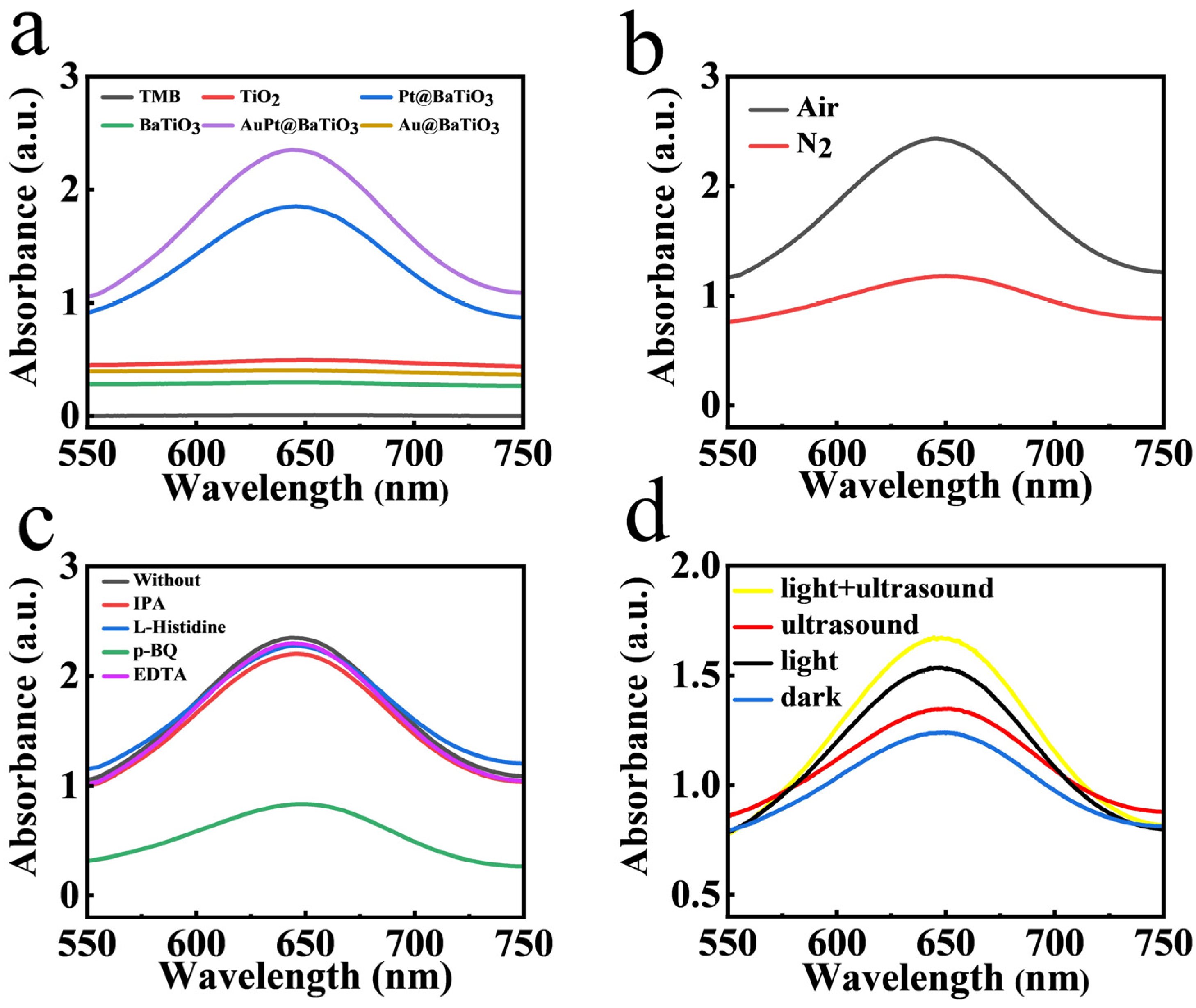
Figure 3a illustrates that the absorption peak of AuPt@BaTiO3 SUMs was markedly higher than that of Au@BaTiO3 SUMs and Pt@BaTiO3 SUMs. This establishes that the AuPt@BaTiO3 bimetallic alloy Schottky junction substantially augmented the oxidase−like activity.
The oxidase−like activity mechanism of AuPt@BaTiO3 SUMs was explored in different atmospheres. According to Figure 3b, the absorbance post N2 bubbling was significantly reduced compared to that in air. This underscores the dependence of oxidase−like activity on dissolved oxygen. It is postulated that the dissolved oxygen may transform into reactive oxygen species (ROS) during oxidation, producing O2•−, ·OH, 1O2, and h+. To further this understanding, scavengers like 5 mM p−BQ, IPA, L−histidine, and EDTA were introduced to counteract O2•−, ·OH, 1O2, and h+, respectively [30]. Figure 3c shows that, except for HQ which drastically decreased absorbance, the other scavengers had minimal impact. This infers a pivotal role for O2•− in the catalytic oxidation process.
To delve deeper into the catalytic mechanism, electron paramagnetic resonance (EPR) tests were executed using 5, 5−dimethyl−1−pyrroline N−oxide (DMPO). Figure S10 reveals a characteristic peak associated with O2•−. Notably, with prolonged illumination time, this signal’s intensity increased, corroborating the notion that light considerably elevates the O2•− quantity.
Figure 3d highlights that the absorption peak intensity was amplified under either ultrasound or light conditions compared to darkness. Intriguingly, a combination of light–ultrasound further intensified this peak. While it is recognized that BaTiO3 SUMs generate a polarization charge under ultrasonic cavitation’s mechanical stress with suboptimal catalytic efficiency [31], the combined light–ultrasound stimulation significantly boosts this efficiency. This suggests a synergistic enhancement in oxidase−like activity due to combined light–ultrasound action.
As illustrated in Scheme 1, the augmented oxidase−like activity stems from the photo−piezoelectric effect of the AuPt@BaTiO3 bimetallic alloy Schottky junction. The main factors contributing to this increase are:
1. Formation of the Bimetallic Alloy Schottky Junction: AuPt alloy nanoparticles were integrated onto BaTiO3 SUMs, establishing a bimetallic alloy Schottky junction. With BaTiO3 SUMs’s work function at 4.8 eV and the AuPt alloy’s work function lying between that of Au (5.1 eV) and Pt (5.65 eV) [32,33,34], AuPt@BaTiO3 SUMs create an optimal Schottky barrier between Au@BaTiO3 SUMs and Pt@BaTiO3 SUMs. As illustrated in Scheme 1a, Pt@BaTiO3 SUMs only allow a limited number of photoexcited electrons to traverse the elevated Schottky barrier Φ3. The relatively low barrier height Φ1 of Au@BaTiO3 SUMs also allowed the backflow of photogenerated electrons, resulting in the recombination of charge carriers [35]. Conversely, the lower barrier height Φ2 in AuPt@BaTiO3 SUMs promotes more substantial charge separation, facilitating the transfer of photogenerated electrons [36].
2. Positive Polarization Charge Under Ultrasound: The polarization charge that arises at the interface under ultrasound induces a downward bend in the BaTiO3 SUMs’ energy band [31]. This reduced energy band height expedites the migration of photogenerated electrons to AuPt alloy nanoparticles [14,37]. Concurrently, the energy threshold for hot electrons to cross this barrier is diminished, bolstering the efficient separation of hot electrons that arise due to the excitation of larger AuPt alloy nanoparticles [38,39]. Moreover, the more compact AuPt alloy nanoparticles act as electron sinks, capturing excited electrons, which accelerates surface electron migration, thereby amplifying enzyme activity [40].
3. Synergistic Effect of Au and Pt Alloying: The amalgamation of Au and Pt offers a potent synergistic effect, primarily driven by the alteration of the d−band center and kinetics. The alloying triggers a downward shift of Pt’s d−band center, which attenuates the binding between Pt and oxygen, thereby enhancing catalytic activity [41,42].
In summary, factors like the Schottky junction, LSPR effect, and piezoelectric effect collectively enhance photosensitive carrier separation. This optimized separation facilitates the increased production of O2•−, culminating in heightened oxidase activity.
3.3. Steady−State Kinetics
Steady−state kinetic experiments were carried out using varying concentrations of TMB, ranging from 0.1 to 1.0 mM, to examine the oxidase−like activity of AuPt@BaTiO3 SUMs (Table S1). The Michaelis constant (Km) and the maximum reaction velocity (Vmax) were derived from the Michaelis–Menten curve (as depicted in Figure S11b) and the double reciprocal (Lineweaver–Burk) plot (presented in Figure S11c). The obtained values were 0.1218 mM for Km and 3.16 × 10⁻⁷ M s−1 for Vmax, respectively. It is well−established in enzymology that a lower Km value signifies a greater substrate affinity, while a higher Vmax points towards a rapid enzymatic response. A comparison with data from previously reported studies, as presented in Table 1, reveals that the oxidase mimetics of AuPt@BaTiO3 SUMs demonstrate superior affinity towards the substrate TMB. Moreover, the reaction rate was found to be more rapid. Consequently, this underscores the impressive catalytic efficiency of the AuPt@BaTiO3 SUMs.
3.4. GSH Detection
As an antioxidant, GSH effectively consumes O2•− and reduces oxTMB to TMB via a single electron transfer process [50]. The absorbance at 652 nm was observed to decrease progressively with increasing GSH concentration (Figure 4a,c). The standard curve was plotted based on the relationship between the peak absorption intensity and GSH concentration. In the dark, the linear equation is: Abs = −0.0206C[GSH] + 2.284 (R2 = 0.9978) (Figure 4b). Under light−ultrasound conditions, the linear equation is Abs = −0.0228C[GSH] + 2.159 (R2 = 0.9914) (Figure 4d). The reaction times were 10 min in the dark and 3 min under light–ultrasound. The LOD was determined to be 0.415 μM in the dark and 0.225 μM under light−ultrasound, both of which are lower than the values reported in previous studies (Table 2).
Possible disruptors in human serum were examined to assess the system’s selectivity. The results (Figure 5) indicated that Cys did interfere with the system’s selectivity to a degree, but the interference from other disruptors was minimal. Given that the Cys concentration in human serum is significantly lower than the GSH concentration [58,59], this interference is limited. Thus, the colorimetric detection of GSH demonstrated excellent selectivity.
Furthermore, stability is a crucial factor when evaluating potential applications. The oxidase−like activity of AuPt@BaTiO3 SUMs was tested every 5 min, revealing impressive short−term stability, with a relative standard deviation (RSD) of 1.46% after 11 cycles (Figure S12a). Simultaneously, the absorbance measured every alternate day displayed minor fluctuations in catalytic performance over 15 days, boasting an RSD of 0.63%, indicative of commendable long−term stability (Figure S12b). To evaluate repeatability, five batches of AuPt@BaTiO3 SUMs oxidase mimetics were prepared under identical experimental conditions. The nearly consistent absorbance results across batches suggest excellent reproducibility (Figure S13).
To assess the accuracy of the colorimetric determination of GSH in serum, the GSH content in real samples was evaluated using the standard addition method [60]. The recovery rate for fetal bovine serum ranged from 99.91% to 101.8%, with an RSD of less than 1.06%. These results suggest that this method holds promise for detecting GSH in real−world settings (Table 3).
4. Conclusions
In conclusion, we successfully prepared AuPt@BaTiO3 SUMs, which showcased outstanding oxidase−like activity to serve as oxidase mimetics for the colorimetric detection of GSH. The combined effects of the photo−piezoelectric phenomenon and LSPR facilitated the directed migration and separation of photogenerated electrons, enhanced by the AuPt bimetallic alloy. This photo−piezoelectric influence significantly boosted oxidase−like activity, leading to high−performance colorimetric GSH detection. Remarkably, our detection platform’s superior catalytic activity allowed for a short detection time of just 3 min, ensuring high sensitivity with a linear range of 0.5–50 μM and an impressive LOD of 0.225 μM for GSH detection. Furthermore, this sensing approach demonstrated commendable analytical performance in intricate samples, boasting a reliable recovery rate of 99.91–101.8% in real−world sample testing. Overall, this research introduces a novel perspective on the strategic design of highly efficient nanozymes leveraging the photo−piezoelectric effect.
Supplementary Materials
The following supporting information can be downloaded at: https://www.mdpi.com/article/10.3390/s24072242/s1, Figure S1: Fabrication process of the AuPt@BaTiO3 SUMs; Figure S2: EDX photo of the prepared AuPt@BaTiO3 SUMs; Figure S3: EDS mapping of the AuPt@BaTiO3 SUMs; Figure S4: XRD patterns of the TiO2, BaTiO3 SUMs and the AuPt@BaTiO3 SUMs; Figure S5: The oxidase−like activity of AuPt@BaTiO3 SUMs depends on pH. Reaction condition: 0.28 mg mL−1 AuPt@BaTiO3 SUMs, 0.2 M Na2HPO4−CA buffer, 0.5 mM TMB, 10 min, 20 °C; Figure S6: The oxidase−like activity of AuPt@BaTiO3 SUMs depends on temperature. Reaction condition: 0.28 mg mL−1 AuPt@BaTiO3 SUMs, 0.2 M Na2HPO4−CA buffer (pH=4.0), 0.5 mM TMB, 10 min; Figure S7: Time−dependent absorbance changes of TMB oxidation at 652 nm. Reaction condition: 0.28 mg mL−1 AuPt@BaTiO3 SUMs, 0.2 M Na2HPO4−CA buffer (pH=4.0), 0.5 mM TMB, 20 °C; Figure S8: The concentration effects on the oxidase−like activity of the AuPt@BaTiO3 SUMs. Reaction condition: 0.2 M Na2HPO4−CA buffer (pH=4.0), 0.5 mM TMB, 10 min, 20 °C; Figure S9: Time−dependent absorbance changes of TMB oxidation at 652 nm. Reaction condition: 0.28 mg mL−1 AuPt@BaTiO3 SUMs, 0.2 M Na2HPO4−CA buffer (pH=4.0), Hg lamp and ultrasound, 0.5 mM TMB, 20 °C; Figure S10: DMPO−EPR spin−trapping spectra for O2•−; Figure S11: Steady−state kinetics of TMB oxidation using the AuPt@BaTiO3 SUMs: (a) Typical absorbance spectra of different reaction systems for 1 min. (b) Michaelis−Menten curves for different TMB concentrations. (c) Lineweaver−Burk plot for different TMB concentrations; Figure S12: (a) The short−term stability of the AuPt@BaTiO3 SUMs. (b) The long−term storage stability of the AuPt@BaTiO3 SUMs; Figure S13: The reproducibility of the AuPt@BaTiO3 SUMs; Table S1: Data of catalytic kinetic parameters.
Author Contributions
Y.L.: Conceptualization, Investigation, Methodology, Data curation, Visualization, Writing—original draft. Y.H.: Investigation, Data curation, Visualization. B.Z.: Data curation, Visualization. Y.M.: Funding acquisition, Writing—review and editing, Supervision. R.X.: Data curation, Visualization. M.Z.: Conceptualization, Funding acquisition, Writing—review and editing, Supervision. H.C.: Conceptualization, Supervision. All authors have read and agreed to the published version of the manuscript.
Funding
This research was funded by the National Natural Science Foundation of China (52072353, 22106152); Fundamental Research Funds for the Central University (202042009, 202113030); Fellowship of China Postdoctoral Science Foundation (2020M672146); Natural Science Foundation of Shandong Province (ZR2021QB059).
Institutional Review Board Statement
Not applicable.
Informed Consent Statement
Not applicable.
Data Availability Statement
Data are contained within the article.
Conflicts of Interest
The authors declare no conflicts of interest.
References
- Zhang, L.L.; Wang, J.; Zhao, C.L.; Zhou, F.J.; Yao, C.; Song, C. Ultra−fast colorimetric detection of glutathione by magnetic Fe NPs with peroxidase−like activity. Sens. Actuators B Chem. 2022, 361, 131750. [Google Scholar] [CrossRef]
- Estrela, J.M.; Ortega, A.; Obrador, E. Glutathione in cancer biology and therapy. Crit. Rev. Clin. Lab. Sci. 2006, 43, 143–181. [Google Scholar] [CrossRef] [PubMed]
- Li, Y.; Li, P.; Chen, Y.; Wu, Y.; Wei, J. Interfacial deposition of Ag nanozyme on metal−polyphenol nanosphere for SERS detection of cellular glutathione. Biosens. Bioelectron. 2023, 228, 115200. [Google Scholar] [CrossRef] [PubMed]
- Xu, Z.; Li, P.; Chen, H.; Zhu, X.; Zhang, Y.; Liu, M.; Yao, S. Picomolar glutathione detection based on the dual−signal self−calibration electrochemical sensor of ferrocene−functionalized copper metal−organic framework via solid−state electrochemistry of cuprous chloride. J. Colloid Interface Sci. 2022, 628, 798–806. [Google Scholar] [CrossRef] [PubMed]
- Xu, Y.X.; Yan, J.; Zhu, Y.; Chen, H.Y.; Wu, C.Y.; Zhu, X.H.; Zhang, Y.Y.; Li, H.T.; Liu, M.L.; Yao, S.Z. Self−Cascade Nanoenzyme of Cupric Oxide Nanoparticles (CuO NPs) Induced In Situ Catalysis Formation of Polyelectrolyte as Template for the Synthesis of Near−Infrared Fluorescent Silver Nanoclusters and the Application in Glutathione Detection and Bioimaging. Anal. Chem. 2022, 94, 14642–14651. [Google Scholar] [CrossRef] [PubMed]
- Wang, C.; Gao, Y.; Hu, S.; Zhu, A.; Ying, Y.; Guo, X.; Wu, Y.; Wen, Y.; Yang, H. MnO(2) coated Au nanoparticles advance SERS detection of cellular glutathione. Biosens. Bioelectron. 2022, 215, 114388. [Google Scholar] [CrossRef]
- Xu, X.Y.; Sun, Q.J.; Ma, Y.M.; Jiang, X.X.; Niu, N.; Chen, L.G. Synthesis of KCl−doped lignin carbon dots nanoenzymes for colorimetric sensing glutathione in human serum. Sens. Actuators B Chem. 2022, 364, 131881. [Google Scholar] [CrossRef]
- Zheng, H.Q.; Chen, J.; Que, M.D.; Yang, T.; Liu, Z.K.; Cai, W.H.; Yang, L.F.; Liu, X.W.; Li, Y.J.; Yang, X.F.; et al. Highly efficient piezoelectric field enhanced photocatalytic performance via in situ formation of BaTiO3 on Ti3C2Tx for phenolic compound degradation. Inorg. Chem. Front. 2022, 9, 4201–4215. [Google Scholar] [CrossRef]
- Zhao, Y.; Wang, S.; Ding, Y.; Zhang, Z.; Huang, T.; Zhang, Y.; Wan, X.; Wang, Z.L.; Li, L. Piezotronic Effect−Augmented Cu2–xO–BaTiO3 Sonosensitizers for Multifunctional Cancer Dynamic Therapy. ACS Nano. 2022, 16, 9304–9316. [Google Scholar] [CrossRef]
- An, X.; Kays, J.C.; Lightcap, I.V.; Ouyang, T.; Dennis, A.M.; Reinhard, B.M. Wavelength−Dependent Bifunctional Plasmonic Photocatalysis in Au/Chalcopyrite Hybrid Nanostructures. ACS Nano. 2022, 16, 6813–6824. [Google Scholar] [CrossRef]
- Sun, Y.; Hu, J.Y.; Zhang, Y. Visible light assisted trace gaseous NO sensor with anti−humidity ability via LSPR enhancement effect. Sens. Actuators B Chem. 2022, 367, 132032. [Google Scholar] [CrossRef]
- Hu, J.Y.; Liu, X.; Zhang, J.W.; Gu, X.; Zhang, Y. Plasmon−activated NO2 sensor based on Au@MoS2 core−shell nanoparticles with heightened sensitivity and full recoverability. Sens. Actuators B Chem. 2023, 382, 133505. [Google Scholar] [CrossRef]
- Song, Y.J.; Wang, H.; Wang, Z.T.; Guo, B.B.; Jing, K.G.; Li, Y.J.; Wu, L. Selective Photocatalytic Synthesis of Haloanilines from Halonitrobenzenes over Multifunctional AuPt/Monolayer Titanate Nanosheet. ACS Catal. 2018, 8, 9656–9664. [Google Scholar] [CrossRef]
- Zheng, W.X.; Tang, Y.F.; Liu, Z.W.; Xing, G.X.; Zhao, K. Enhanced charge carrier separation by bi−piezoelectric effects based on pine needle−like BaTiO3/ZnO continuous nanofibers. J. Mater. Chem. A 2022, 10, 13544–13555. [Google Scholar] [CrossRef]
- Cheng, S.S.; Luo, Y.; Zhang, J.; Shi, R.; Wei, S.T.; Dong, K.J.; Liu, X.M.; Wu, S.L.; Wang, H.B. The highly effective therapy of ovarian cancer by Bismuth−doped oxygen−deficient BaTiO3 with enhanced sono−piezocatalytic effects. Chem. Eng. J. 2022, 442, 136380. [Google Scholar] [CrossRef]
- He, X.; Liu, J.Y.; Zhu, M.H.; Guo, Y.; Ren, Z.Q.; Li, X. Preparation of hierarchical rutile TiO2 microspheres as scattering centers for efficient dye−sensitized solar cells. Electrochim. Acta 2017, 255, 187–194. [Google Scholar] [CrossRef]
- Tan, X.F.; Yang, Q.L.; Sun, X.M.; Sun, P.; Li, H. PdIr aerogels with boosted peroxidase−like activity for a sensitive total antioxidant capacity colorimetric bioassay. ACS Appl. Mater. Interfaces 2022, 14, 10047–10054. [Google Scholar] [CrossRef] [PubMed]
- Dong, L.; Li, R.Y.; Wang, L.Q.; Lan, X.F.; Sun, H.T.; Zhao, Y.; Wang, L.G. Green synthesis of platinum nanoclusters using lentinan for sensitively colorimetric detection of glucose. Int. J. Biol. Macromol. 2021, 172, 289–298. [Google Scholar] [CrossRef]
- Jiang, B.; Duan, D.M.; Gao, L.Z.; Zhou, M.J.; Fan, K.L.; Tang, Y.; Xi, J.Q.; Bi, Y.H.; Tong, Z.; Gao, G.F.; et al. Standardized assays for determining the catalytic activity and kinetics of peroxidase−like nanozymes. Nat. Protoc. 2018, 13, 1506–1520. [Google Scholar] [CrossRef]
- Hu, M.Z. −C.; Kurian, V.; Payzant, E.A.; Rawn, C.J.; Hunt, R.D. Wet−chemical synthesis of monodispersed barium titanate particles—Hydrothermal conversion of TiO2 microspheres to nanocrystalline BaTiO3. Powder Technology 2000, 110, 2–14. [Google Scholar] [CrossRef]
- Dutta, A.; Ouyang, J. Enhanced electrocatalytic performance on polymer−stabilized graphene decorated with alloy nanoparticles for ethanol oxidation reaction in alkaline media. Appl. Catal. B: Environ. 2014, 158–159, 119–128. [Google Scholar] [CrossRef]
- Kumar, P.; Thakur, U.K.; Alam, K.; Kar, P.; Kisslinger, R.; Zeng, S.; Patel, S.; Shankar, K. Arrays of TiO2 nanorods embedded with fluorine doped carbon nitride quantum dots (CNFQDs) for visible light driven water splitting. Carbon 2018, 137, 174–187. [Google Scholar] [CrossRef]
- Peng, F.; Yin, R.; Liao, Y.H.; Xie, X.; Sun, J.L.; Xia, D.H.; He, C. Kinetics and mechanisms of enhanced degradation of ibuprofen by piezo−catalytic activation of persulfate. Chem. Eng. J. 2020, 392, 123818. [Google Scholar] [CrossRef]
- Xu, J.B.; Zhao, T.S.; Liang, Z.X.; Zhu, L.D. Facile preparation of AuPt alloy nanoparticles from organometallic complex precursor. Chem. Mater. 2008, 20, 1688–1690. [Google Scholar] [CrossRef]
- Liu, Q.; Zhai, D.; Xiao, Z.D.; Tang, C.; Sun, Q.W.; Bowen, C.R.; Luo, H.; Zhang, D. Piezo−photoelectronic coupling effect of BaTiO3@TiO2 nanowires for highly concentrated dye degradation. Nano Energy 2022, 92, 106702. [Google Scholar] [CrossRef]
- Zhou, X.F.; Wu, S.H.; Li, C.B.; Yan, F.; Bai, H.R.; Shen, B.; Zeng, H.R.; Zhai, J.W. Piezophototronic effect in enhancing charge carrier separation and transfer in ZnO/BaTiO3 heterostructures for high−efficiency catalytic oxidation. Nano Energy 2019, 66, 104127. [Google Scholar] [CrossRef]
- Cao, G.Q.; Deskins, N.A.; Yi, N. Carbon monoxide oxidation over copper and nitrogen modified titanium dioxide. Appl. Catal. B Environ. 2021, 285, 119748. [Google Scholar] [CrossRef]
- Xu, S.; Guo, L.; Sun, Q.; Wang, Z.L. Piezotronic effect enhanced plasmonic photocatalysis by AuNPs/BaTiO3 heterostructures. 2019, 29, 1808737. 29. [CrossRef]
- Jimenez−Calvo, P.; Munoz−Batista, M.J.; Isaacs, M.; Ramnarain, V.; Ihiawakrim, D.; Li, X.Y.; Munoz−Marquez, M.A.; Teobaldi, G.; Kociak, M.; Paineau, E. A compact photoreactor for automated H2 photoproduction: Revisiting the (Pd, Pt, Au)/TiO2 (P25) Schottky junctions. Chem. Eng. J. 2023, 459, 141514. [Google Scholar] [CrossRef]
- Zhu, X.; Tang, J.; Ouyang, X.L.; Liao, Y.B.; Feng, H.P.; Yu, J.F.; Chen, L.; Lu, Y.T.; Yi, Y.Y.; Tang, L. Multifunctional MnCo@C yolk−shell nanozymes with smartphone platform for rapid colorimetric analysis of total antioxidant capacity and phenolic compounds. Biosens. Bioelectron. 2022, 216, 114652. [Google Scholar] [CrossRef]
- Zhang, Y.; Wang, S.; Zhao, Y.; Ding, Y.; Zhang, Z.; Jiang, T.; Wang, Z.L.; Li, L. Piezo−phototronic effect boosted catalysis in plasmonic bimetallic ZnO heterostructure with guided fermi level alignment. Mater. Today Nano 2022, 18, 100177. [Google Scholar] [CrossRef]
- Zheng, S.; Ding, B.; Qian, X.; Yang, Y.; Mao, L.; Zheng, S.; Zhang, J. High efficiency degradation of tetracycline and rhodamine B using Z−type BaTiO3/γ−Bi2O3 heterojunction. Sep. Purif. Technol. 2021, 278, 119666. [Google Scholar] [CrossRef]
- Aksoy, M.; Korkut, S.E.; Metin, O. AuPt alloy nanoparticles supported on graphitic carbon nitride: In situ synthesis and superb catalytic performance in the light−assisted hydrolytic dehydrogenation of ammonia borane. Appl. Surf. Sci. 2022, 602, 154286. [Google Scholar] [CrossRef]
- Liu, Y.P.; Zhu, L.Y.; Feng, P.; Dang, C.C.; Li, M.; Lu, H.L.; Gao, L.M. Bimetallic AuPt alloy nanoparticles decorated on ZnO nanowires towards efficient and selective H2S gas sensing. Sens. Actuators B Chem. 2022, 367, 132024. [Google Scholar] [CrossRef]
- Shiraishi, Y.; Toi, S.; Ichikawa, S.; Hirai, T. Photocatalytic NH3 Splitting on TiO2 Particles Decorated with Pt−Au Bimetallic Alloy Nanoparticles. ACS Appl. Nano Mater. 2020, 3, 1612–1620. [Google Scholar] [CrossRef]
- Li, X.J.; Chen, L.; Wang, J.F.; Zhang, J.Y.; Zhao, C.R.; Lin, H.J.; Wu, Y.; He, Y.M. Novel platinum−bismuth alloy loaded KTa0.5Nb0.5O3 composite photocatalyst for effective nitrogen−to−ammonium conversion. J. Colloid Interface Sci. 2022, 618, 362–374. [Google Scholar] [CrossRef] [PubMed]
- Chen, P.; Ni, J.M.; Liang, Y.M.; Yang, B.Q.; Jia, F.F.; Song, S.X. Piezo−Photocatalytic Reduction of Au(I) by Defect−Rich MoS2 Nanoflowers for Efficient Gold Recovery from a Thiosulfate Solution. ACS Sustain. Chem. Eng. 2021, 9, 589–598. [Google Scholar] [CrossRef]
- Xiang, D.; Liu, Z.; Wu, M.; Liu, H.; Zhang, X.; Wang, Z.; Wang, Z.L.; Li, L. Enhanced Piezo−Photoelectric Catalysis with Oriented Carrier Migration in Asymmetric Au−ZnO Nanorod Array. Small 2020, 16, 201907603. [Google Scholar] [CrossRef]
- Raji, R.; Gopchandran, K.G. Plasmonic photocatalytic activity of ZnO:Au nanostructures: Tailoring the plasmon absorption and interfacial charge transfer mechanism. J. Hazard. Mater. 2019, 368, 345–357. [Google Scholar] [CrossRef] [PubMed]
- Xian, T.; Sun, X.; Di, L.; Sun, C.; Li, H.; Ma, C.; Yang, H. Enhancing the piezo−photocatalytic tetracycline degradation activity of BiOBr by the decoration of AuPt alloy nanoparaticles: Degradation pathways and mechanism investigation. Appl. Surf. Sci. 2023, 638, 158136. [Google Scholar] [CrossRef]
- Xu, Z.H.; Kibria, M.G.; AlOtaibi, B.; Duchesne, P.N.; Besteiro, L.V.; Gao, Y.; Zhang, Q.Z.; Mi, Z.T.; Zhang, P.; Govorov, A.O.; et al. Towards enhancing photocatalytic hydrogen generation: Which is more important, alloy synergistic effect or plasmonic effect? Appl. Catal. B Environ. 2018, 221, 77–85. [Google Scholar] [CrossRef]
- Xie, Y.X.; Yang, Y.; Muller, D.A.; Abruna, H.D.; Dimitrov, N.; Fang, J.Y. Enhanced ORR Kinetics on Au−Doped Pt−Cu Porous Films in Alkaline Media. ACS Catal. 2020, 10, 9967–9976. [Google Scholar] [CrossRef]
- Qiao, F.M.; Chen, L.J.; Li, X.N.; Li, L.F.; Ai, S.Y. Peroxidase−like activity of manganese selenide nanoparticles and its analytical application for visual detection of hydrogen peroxide and glucose. Sens. Actuators B Chem. 2014, 193, 255–262. [Google Scholar] [CrossRef]
- Gupta, P.K.; Son, S.E.; Venkatesan, J.; Seong, G.H. Cauliflower−like platinum nanostructures mediated photothermal and colorimetric dual−readout biosensor for sensitive cholesterol detection. Sens. Actuators B Chem. 2023, 386, 133741. [Google Scholar] [CrossRef]
- Ali, S.; Sikdar, S.; Basak, S.; Mondal, M.; Mallick, K.; Salman Haydar, M.; Ghosh, S.; Nath Roy, M. Assemble multi−enzyme mimic tandem Mn3O4@ g−C3N4 for augment ROS elimination and label free detection. Chem. Eng. J. 2023, 463, 142355. [Google Scholar] [CrossRef]
- Shen, Y.; Gao, X.; Chen, H.; Wei, Y.; Yang, H.; Gu, Y. Ultrathin C3N4 nanosheets−based oxidase−like 2D fluorescence nanozyme for dual−mode detection of organophosphorus pesticides. J. Hazard. Mater. 2023, 451, 131171. [Google Scholar] [CrossRef] [PubMed]
- Yang, Q.Y.; Wan, C.Q.; Wang, Y.X.; Shen, X.F.; Pang, Y.H. Bismuth−based metal−organic framework peroxidase−mimic nanozyme: Preparation and mechanism for colorimetric−converted ultra−trace electrochemical sensing of chromium ion. J. Hazard. Mater. 2023, 451, 131148. [Google Scholar] [CrossRef] [PubMed]
- Xu, W.; Jiao, L.; Yan, H.; Wu, Y.; Chen, L.; Gu, W.; Du, D.; Lin, Y.; Zhu, C. Glucose Oxidase−Integrated Metal−Organic Framework Hybrids as Biomimetic Cascade Nanozymes for Ultrasensitive Glucose Biosensing. ACS Appl. Mater. Interfaces 2019, 11, 22096–22101. [Google Scholar] [CrossRef] [PubMed]
- Chen, Y.; Xia, Y.; Liu, Y.; Tang, Y.; Zhao, F.; Zeng, B. Colorimetric and electrochemical detection platforms for tetracycline based on surface molecularly imprinted polyionic liquid on Mn3O4 nanozyme. Biosens. Bioelectron. 2022, 216, 114650. [Google Scholar] [CrossRef] [PubMed]
- Fu, G.L.; Sanjay, S.T.; Zhou, W.; Brekken, R.A.; Kirken, R.A.; Li, X.J. Exploration of Nanoparticle−Mediated Photothermal Effect of TMB−H2O2 Colorimetric System and Its Application in a Visual Quantitative Photothermal Immunoassay. Anal. Chem. 2018, 90, 5930–5937. [Google Scholar] [CrossRef]
- Peng, D.; Yang, Y.; Que, M.; Ding, Y.; Wu, M.; Deng, X.; He, Q.; Ma, X.; Li, X.; Qiu, H. Partially oxidized MoS2 nanosheets with high water−solubility to enhance the peroxidase−mimic activity for sensitive detection of glutathione. Anal. Chim. Acta 2023, 1250, 340968. [Google Scholar] [CrossRef]
- Bian, B.; Liu, Q.Y.; Yu, S.T. Peroxidase mimetic activity of porphyrin modified ZnFe2O4/reduced graphene oxide and its application for colorimetric detection of H2O2 and glutathione. Colloid. Surface B 2019, 181, 567–575. [Google Scholar] [CrossRef] [PubMed]
- Xiang, S.; Long, X.C.; Tu, Q.X.; Feng, J.; Zhang, X.H.; Feng, G.W.; Lei, L. Self−assembled, hemin−functionalized peptide nanotubes: An innovative strategy for detecting glutathione and glucose molecules with peroxidase−like activity. Nano Converg. 2023, 10, 1647. [Google Scholar] [CrossRef]
- Dou, Y.; Yang, R.; Xiao, Y.; Wu, J.; Qu, L.B.; Sun, Y.Q.; Li, Z.H. Teaching a fluorophore new tricks: Exploiting the light−driven organic oxidase nanozyme properties of thiazolothiazole for highly sensitive biomedical detection. Sens. Actuators B Chem. 2022, 354, 131226. [Google Scholar] [CrossRef]
- Narang, J.; Chauhan, N.; Jain, P.; Pundir, C.S. Silver nanoparticles/multiwalled carbon nanotube/polyaniline film for amperometric glutathione biosensor. Int. J. Biol. Macromol. 2012, 50, 672–678. [Google Scholar] [CrossRef]
- Wang, D.; Meng, Y.T.; Zhang, Y.; Wang, Q.; Lu, W.J.; Shuang, S.M.; Dong, C. A specific discriminating GSH from Cys/Hcy fluorescence nanosensor: The carbon dots−MnO2 nanocomposites. Sens. Actuators B Chem. 2022, 367, 132135. [Google Scholar] [CrossRef]
- Li, Y.; Zhang, L.B.; Zhang, Z.; Liu, Y.; Chen, J.; Liu, J.; Du, P.Y.; Guo, H.X.; Lu, X.Q. MnO2 Nanospheres Assisted by Cysteine Combined with MnO2 Nanosheets as a Fluorescence Resonance Energy Transfer System for “Switch−on” Detection of Glutathione. Anal. Chem. 2021, 93, 9621–9627. [Google Scholar] [CrossRef]
- Yang, W.; Weng, C.Y.; Li, X.Y.; He, H.L.; Fei, J.W.; Xu, W.; Yan, X.Q.; Zhu, W.Y.; Zhang, H.S.; Zhou, X.M. A sensitive colorimetric sensor based on one−pot preparation of h−Fe3O4@ppy with high peroxidase−like activity for determination of glutathione and H2O2. Sens. Actuators B Chem. 2021, 338, 129844. [Google Scholar] [CrossRef]
- Jin, P.; Niu, X.Y.; Gao, Z.X.; Xue, X.Q.; Zhang, F.; Cheng, W.; Ren, C.L.; Du, H.Y.; Manyande, A.; Chen, H.L. Ultrafine platinum nanoparticles supported on covalent organic frameworks as stable and reusable oxidase−like catalysts for cellular glutathione detection. ACS Appl. Nano Mater. 2021, 4, 5834–5841. [Google Scholar] [CrossRef]
- Thulasinathan, B.; Ganesan, V.; Manickam, P.; Kumar, P.; Govarthanan, M.; Chinnathambi, S.; Alagarsamy, A. Simultaneous electrochemical determination of persistent petrogenic organic pollutants based on AgNPs synthesized using carbon dots derived from mushroom. Sci. Total Environ. 2023, 884, 163729. [Google Scholar] [CrossRef]
Figure 1.
SEM image of (a) TiO2, (b–c) BaTiO3 SUMs, and (d) AuPt@BaTiO3 SUMs. (e–i) EDS mapping of the AuPt@BaTiO3 SUMs. (j–k) HRTEM and TEM images of the AuPt@BaTiO3 SUMs and (l–p) EDS mapping of the single AuPt@BaTiO3 SUMs. Inset: schematic illustration of the detailed structure of the AuPt@BaTiO3 SUMs.

Figure 2.
(a) Survey spectra of the AuPt@BaTiO3 SUMs. (b–f) High−resolution XPS spectra for O 1s, Ba 3d, Ti 2p, Au 4f, and Pt 4f.
Figure 2.
(a) Survey spectra of the AuPt@BaTiO3 SUMs. (b–f) High−resolution XPS spectra for O 1s, Ba 3d, Ti 2p, Au 4f, and Pt 4f.
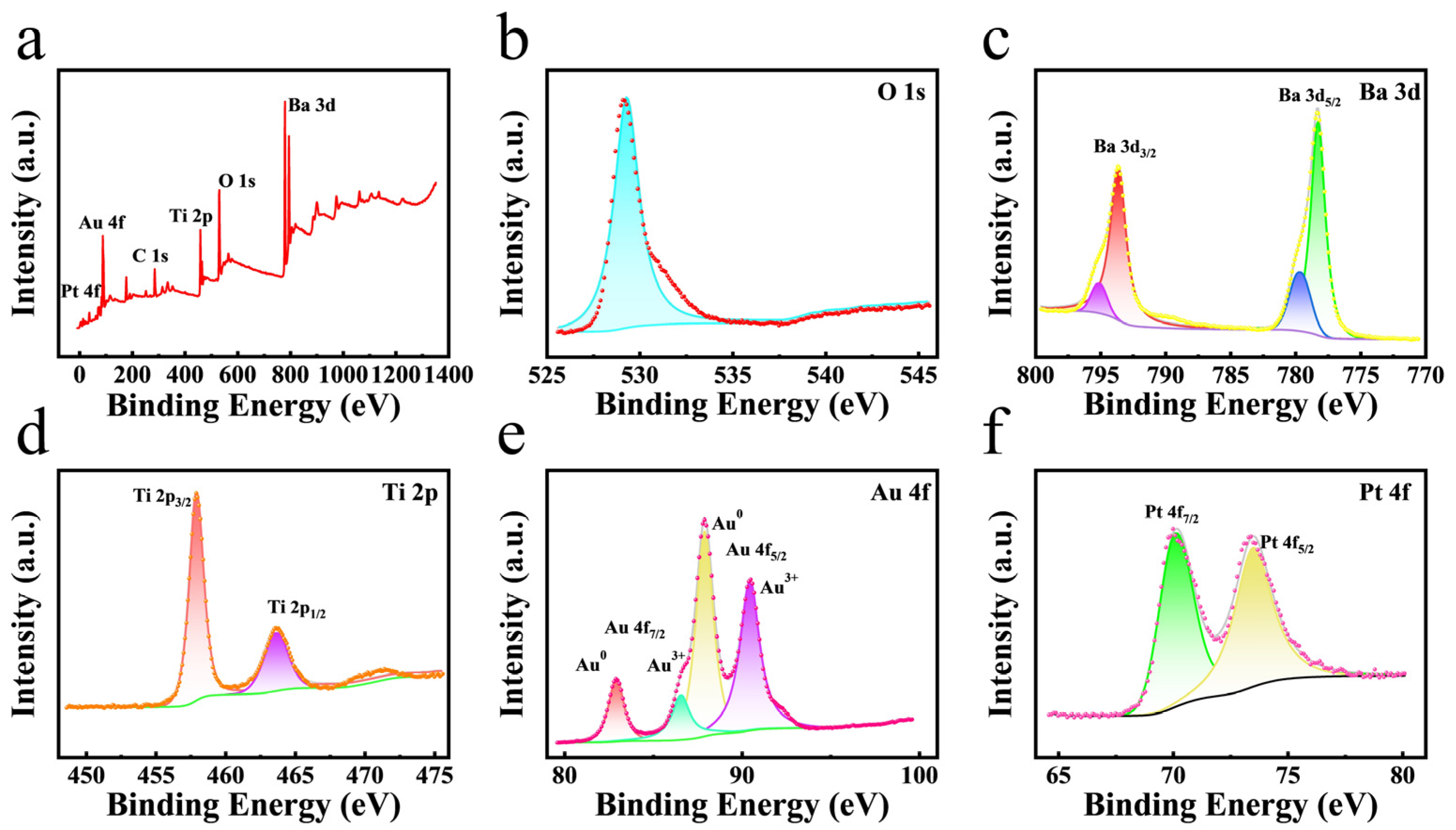
Figure 3.
(a) UV−vis spectra of different reaction systems. (b) UV−vis spectra under different gaseous conditions (Air, N2). (c) UV−vis spectra of different capture agents for the (AuPt@BaTiO3 SUMs + TMB) system. (d) UV−vis spectra of the (AuPt@BaTiO3 SUMs + TMB) system under different conditions (dark, light, ultrasound, light−ultrasound).
Scheme 1.
(a) Schematic illustration of energy−band structures for Au@BaTiO3 SUMs, Pt@BaTiO3 SUMs, and AuPt@BaTiO3 SUMs. (b) The photo−piezoelectric effect enhances the oxidase−like activity of the AuPt@BaTiO3 SUMs.
Scheme 1.
(a) Schematic illustration of energy−band structures for Au@BaTiO3 SUMs, Pt@BaTiO3 SUMs, and AuPt@BaTiO3 SUMs. (b) The photo−piezoelectric effect enhances the oxidase−like activity of the AuPt@BaTiO3 SUMs.

Figure 4.
(a) UV−vis absorbance of the detection system upon the addition of 0.5–80 μM GSH in the dark. (b) The calibration curve corresponds to GSH concentration. Inset: the corresponding photograph of the colored products for reaction with different GSH concentrations. Reaction conditions: 0.5 mM TMB, 0.28 mg mL−1 AuPt@BaTiO3 SUMs, 0.2 M Na2HPO4−CA buffer (pH = 4.0), 10 min and 20 °C. (c) UV−vis absorbance of the detection system upon the addition of 0.5–50 μM GSH under light–ultrasound. (d) The calibration curve corresponds to GSH concentration. Reaction conditions: 0.5 mM TMB, 0.28 mg mL−1 AuPt@BaTiO3 SUMs, 0.2 M Na2HPO4−CA buffer (pH = 4.0), 3 min, and 20 °C.

Figure 5.
Anti−interference experiments of the colorimetric detection of GSH (80 μM GSH, mixture, Cys, AA, Gly, Leu, Arg, Lys, Pro, Glu, L−His, K+, Na+).
Figure 5.
Anti−interference experiments of the colorimetric detection of GSH (80 μM GSH, mixture, Cys, AA, Gly, Leu, Arg, Lys, Pro, Glu, L−His, K+, Na+).


Table 1.
Comparison of catalytic kinetic parameters.
| Materials | Substrate | Vmax (×10−7 M s−1) | Km (mM) | Reference |
|---|---|---|---|---|
| HRP | TMB | 1 | 0.43 | [43] |
| CS@Pt NS | TMB | 6.524 | 0.49 | [44] |
| 3:3−Mn3O4@g−C3N4 | TMB | 1.645 | 0.343 | [45] |
| PtPdNPs@g−C3N4 | TMB | 3.639 | 0.225 | [46] |
| BiO−BDC−NH2 | TMB | 0.581 | 0.41 | [47] |
| Fe−MOF | TMB | 0.56 | 2.6 | [48] |
| DSMIP@Mn3O4 | TMB | 0.092 | 5.1 | [49] |
| AuPt@BaTiO3 SUMs | TMB | 3.16 | 0.1218 | This work |
Table 2.
Comparison of the sensing performance with other methods in the detection of GSH.
| Materials | Methods | Linear Range (μM) | LOD (μM) | Detection Time (min) | Ref. |
|---|---|---|---|---|---|
| ox−MoS2 NSs | colorimetry | 0.5–5 | 0.276 | 60 | [51] |
| Por−ZnFe2O4/rGO | colorimetry | 2–40 | 0.76 | 10 | [52] |
| hemin−PNT | colorimetry | 1–35 | 0.51 | 10 | [53] |
| TTz−Cl2+ | colorimetry | 1–17 | 0.47 | 15 | [54] |
| AgNPs/C–MWCNT/PANI/Au | electrochemistry | 0.3–0.35 | 0.3 | − | [55] |
| CDs−MnO2 NFs | fluorescence | 2–200 | 0.558 | 5 | [56] |
| Cys−MnO2 nanospheres/MnO2 nanosheets | fluorescence | 5–50,150–800 | 2.96 | 15 | [57] |
| AuPt@BaTiO3 SUMs | colorimetry | 0.5–50 | 0.225 | 3 | This work |
Table 3.
Detection of GSH in an actual sample (n = 3).
| Sample | Conditions | Added (μM) | Found (μM) | Recovery (%) | RSD (%) |
|---|---|---|---|---|---|
| fetal bovine serum | light−ultrasound | 10 | 10.18 | 101.8 | 0.37 |
| 30 | 29.97 | 99.91 | 0.39 | ||
| 50 | 50.66 | 101.3 | 1.06 |
Disclaimer/Publisher’s Note: The statements, opinions and data contained in all publications are solely those of the individual author(s) and contributor(s) and not of MDPI and/or the editor(s). MDPI and/or the editor(s) disclaim responsibility for any injury to people or property resulting from any ideas, methods, instructions or products referred to in the content. |
© 2024 by the authors. Licensee MDPI, Basel, Switzerland. This article is an open access article distributed under the terms and conditions of the Creative Commons Attribution (CC BY) license (https://creativecommons.org/licenses/by/4.0/).
Share and Cite
MDPI and ACS Style
Liao, Y.; He, Y.; Zhang, B.; Ma, Y.; Xu, R.; Zhao, M.; Cui, H. Using the Photo–Piezoelectric Effect of AuPt@BaTiO3 Oxidase Mimetics for Colorimetric Detection of GSH in Serum. Sensors 2024, 24, 2242. https://doi.org/10.3390/s24072242
AMA Style
Liao Y, He Y, Zhang B, Ma Y, Xu R, Zhao M, Cui H. Using the Photo–Piezoelectric Effect of AuPt@BaTiO3 Oxidase Mimetics for Colorimetric Detection of GSH in Serum. Sensors. 2024; 24(7):2242. https://doi.org/10.3390/s24072242
Chicago/Turabian StyleLiao, Yiquan, Yichang He, Bin Zhang, Ye Ma, Ruiqi Xu, Minggang Zhao, and Hongzhi Cui. 2024. "Using the Photo–Piezoelectric Effect of AuPt@BaTiO3 Oxidase Mimetics for Colorimetric Detection of GSH in Serum" Sensors 24, no. 7: 2242. https://doi.org/10.3390/s24072242
Note that from the first issue of 2016, this journal uses article numbers instead of page numbers. See further details here.