Article

Using Monoclonal Antibody to Determine Lead Ions with a Localized Surface Plasmon Resonance Fiber-optic Biosensor

Department of Chemical Engineering, National Chung-Cheng University, Chia-Yi 621, Taiwan
*
Author to whom correspondence should be addressed.
Sensors 2008, 8(1), 582-593; https://doi.org/10.3390/s8010582
Submission received: 4 October 2007 / Accepted: 24 January 2008 / Published: 25 January 2008

Abstract

:
A novel reflection-based localized surface plasmon resonance (LSPR) fiber­optic probe has been developed to determine the heavy metal lead ion concentration. Monoclonal antibody as the detecting probe containing massive amino groups to capture Pb(II)-chelate complexes was immobilized onto gold nanoparticle-modified optical fiber (NMAuOF). The optimal immobilizing conditions of monoclonal antibody on to the NMAuOF are 189 μg/mL in pH7.4 PBS for 2 h at 25°C. The absorbability of the functionalized NMAuOF sensor increases to 12.2 % upon changing the Pb(II)-EDTA level from 10 to 100 ppb with a detection limit of 0.27 ppb. The sensor retains 92.7 % of its original activity and gives reproducible results after storage in 5% D-(+)-Trehalose dehydrate solution at 4°C for 35 days. In conclusion, the monoclonal antibody-functionalized NMAuOF sensor shows a promising result for determining the concentration of Pb(II) with high sensitivity.

1. Introduction

Lead is one of the most widely used heavy metals and has a large number of industrial applications, including battery manufacture, paint, gasoline, alloys, radiation shielding, tank lining and piping. Among these, lead in paint and gasoline together accounts for most of the lead now in the environment and leads to serious pollution and human health problems. Elevated levels of lead can be harmful if levels are built up in the body. Over the past several decades, it is found that lead is toxic by ingestion and inhalation, can affect the gut and central nervous system, and can cause anemia [1,2]. Besides, lead overexposure can also cause birth defects, mental retardation, behavioral disorders, and death in fetuses and young children [3,4]. Hence, detection and quantization of lead contamination is an important issue with the increasing requirement of environmental concern and health safety.
Traditionally, the commonly used methods for determination of lead are atomic absorption (AA) spectroscopy [5] and inductively coupled plasma (ICP) optical emission spectrometry, and inductively coupled plasma mass spectrometry (ICPMS) [6,7]. These methods are sensitive and allow discrimination among different metal ions but require a tedious sample pretreatment and sophisticated instrument. Therefore, it is essential to develop a rapid and sensitive screening method to ascertain the presence of lead for in situ environmental monitoring. In the past, a lot of efforts have been devoted to develop rather inexpensive biosensors by using enzyme, peptide, protein, or antibody as recognition elements for the detection of heavy metals [8-12]. Theses recognition elements may be immobilized on a suitable surface or supporter, and its binding or inhibition can be measured by different detection techniques such as ion-sensitive field effect transistors, potentiometric and piezoelectric devices, amperometric electrodes, and optical-based biosensors. Among the recognition elements, monoclonal antibody provides a unique opportunity for distinguishing different metals and a basis for rapid and sensitive immunoassays [13-15]. To increase association with monoclonal antibody, small metal ions were initially conjugated with chelators to form a metal-chelate complex. The metal-chelate complexes represent a distinct class of hapten ligands which was recognized by specific amino acid residues in the antibody's binding-sites to comprise a special subset of metal-binding protein [16,17]. Therefore, the specific binding characteristics of monoclonal antibody will be employed to our new design of biosensor for lead detection.
A novel fiber-based biosensor with extraordinary optic properties of gold nanoparticles (NMAu) has recently been explored [18,19]. The absorption band of gold nanoparticles is obtained when the incident photon frequency is resonant with the collective oscillation of the conduction electrons and is known as the localized surface plasmon resonance (LSPR). The resonance frequency of the LSPR is highly dependent upon the local environment of the nanoparticles [20-22]. Therefore, the optical properties (e.g., absorbance and peak wavelength) of gold nanoparticles are sensitive to the refractive index of the surrounding solvent and the binding events to those functionalized nanoparticles. With a suitable receptor immobilized at the surface of the gold nanoparticles, the resulting LSPR fiber-optic sensor can detect the corresponding analyte even if the analyte is spectroscopically silent in the UV-vis region. Hence, the presence of analytes can be determined directly without the use of labeled molecules.
For this study, a monoclonal antibody-functionalized fiber-based biosensor utilizing the LSPR effect was developed to evaluate the concentration of Pb(II). By using a self-assembling technique, a bioactive layer consisting of the monoclonal antibody was immobilized by covalent coupling onto the gold nanoparticle layer and the optimal conditions of immobilization will be examined. Prior to detecting, Pb(II) was treated with excess chelators (i.e., EDTA) to form Pb(II)-chelate complexes. The monoclonal antibody can exclusively bind with Pb(II)-chelate complex and causes a change in light attenuation. Based on the correlation between binding rate and light attenuation, the concentration of Pb(II) can be determined. Several factors, including stability and binding constant, were investigated as well.

2. Materials and Methods

2.1. Reagents and Materials

Monoclonal Antibody to lead (K55061M) was purchased from Biodesign International Company, USA. Basically, Pb(II)-EDTA was covalently conjugated to keyhole limpet hemocyanin (KLH) and was used as an immunogen. Then, the KLH-Pb(II) conjugate was injected into mouse to produce the monoclonal antibody. Multimode plastic-clad silica optical fiber (model F-MBC) was purchased from Newport (Irvine, CA) with core and cladding diameters of 400 and 430 μm, respectively. The following chemicals, n-hexadecyltrimethylammonium bromide (CTAB, Fluka), sodium borohydride (Lancaster), 3-(mercaptopropyl)-trimethoxysilane (MPTMS, Acros), cystamine dihydrochloride (Sigma), phosphate buffered saline (PBS, Sigma), N-(3-Dimethylaminopropy)-N-ethylcarbodiimide hydrochloride (EDC, Sigma), N-hydroxy- succinimide (NHS, Sigma), ethylenediamine tetraacetic acid (EDTA, Sigma), lead(II) nitrate (Osaka, 99+%), D-(+)-Trehalose dehydrate (Sigma), were used as received. All aqueous solutions were prepared with water purified using a YMDI-100 water purification unit (Yeameei Membrane) with a specific resistance of 18 MΩ cm.

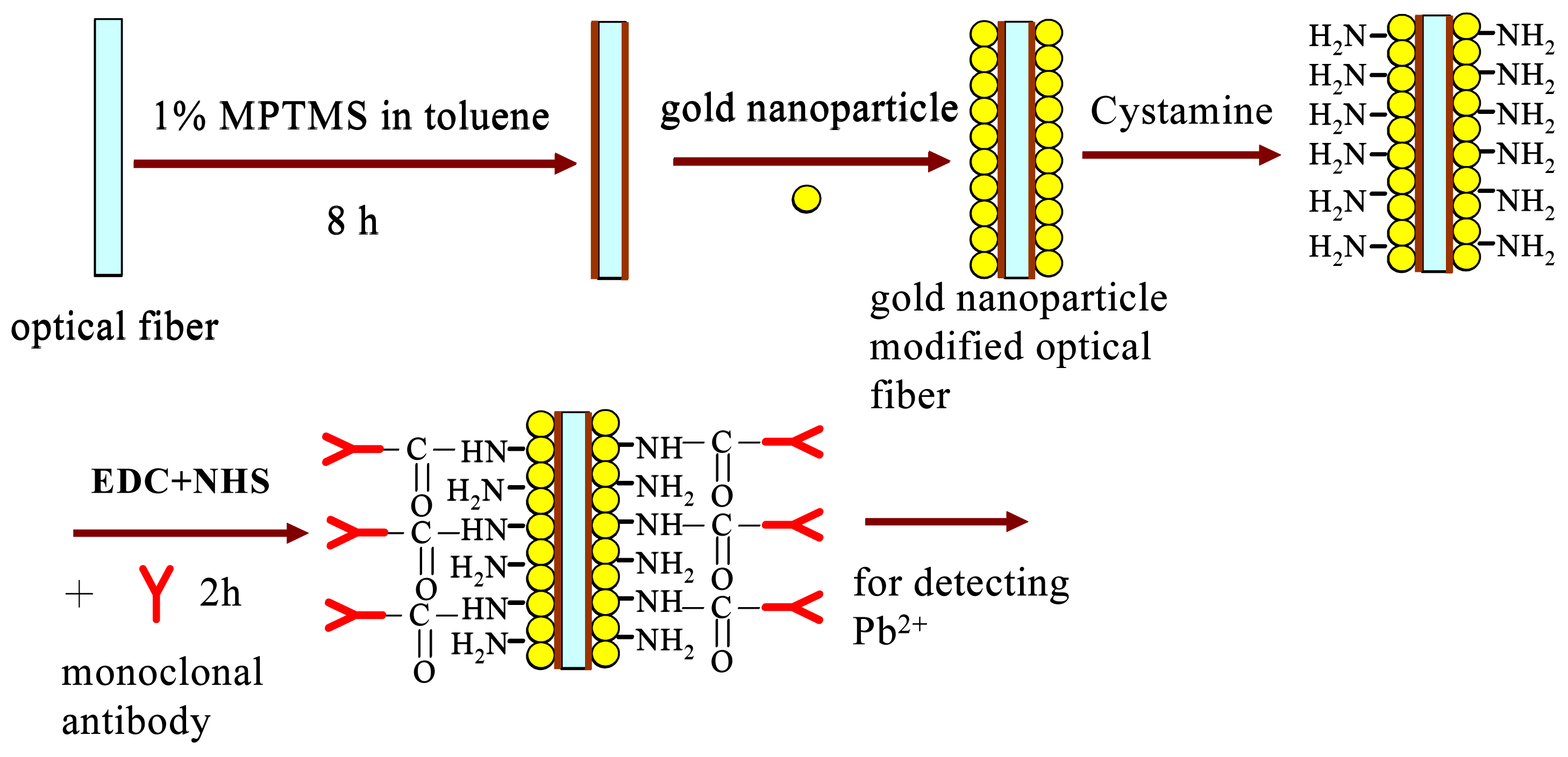
2.2 Preparation of gold nanoparticle-modified optical fiber

First, colloidal Au solution was prepared by mixing hydrogen tetracholoraurate (1.78 mL, 25.4 nM), 8.22 mL of chloroform, and 0.4 mL of a 0.02 M ethanol solution of CTAB for 10 min to form a 4.52×10⁻⁴ M hydrogen tetrachloroaurate solution. Freshly prepared NaBH4 ethanol solution (0.8 mL, 0.15 M) was added to the hydrogen tetrachloroaurate solution with vigorous stirring for 30 min. Then, the unclad portion (5 cm) of the optical fibers was cleaned for 30 min in a bath consisting of 3 volumes of 30% H2O2 and 7 volumes of concentrated H2SO4. The clean unclad portions of the optical fibers were then submerged into vials of 1% solution of MPTMS in toluene. After 8 h, the optical fibers were rinsed with methanol to remove unbound monomers from the surface. After thorough rinsing, the unclad portions of the optical fibers were immersed in Au solution for 5 h to form a self-assembled gold nanoparticle monolayer (NMAu) on the core surface. Subsequently, the modified optical fibers were rinsed sequentially with water, methanol, and chloroform. Figs. 1(a) and 1(b) were shown the 2-D and 3-D AFM images of surfaces on the prepared MPTMS and gold nanoparticle-modified optical fiber, respectively. From the images of Fig. 1(a), the surface modified by MPTMS became smoother with a 2.05 nm roughness. A Joel transmission electron microscopic (TEM) 1200EX was applied to observe the samples, which had been dispersed and allowed to dry on copper grids. Histograms derived from TEM image analysis showed that the mean diameter of Au nanoparticles was 9.6±2.3 nm, as shown in Fig. 1(b).

2.3 Immobilization of antibody onto NMAu

NMAu was initially modified to form a self-assembled monolayer (SAM) of cystamine by immersing NMAu in 0.02 M cystamine dihydrochloride (pH 7.4 in PBS) for 2 h to form an amine functional group. The cystamine-modified NMAu was further immersed in a PBS solution with containing 450 mM EDC, 90 mM NHS, and various concentrations of antibody for 2 h at room temperature, rinsed with PBS, and air-dried at room temperature. Then, the amine group of cystamine modified on the NMAu can couple with the activated succinimide esters reacting from antibody and NHS, and finally form an amino bond between cystamine and antibody, as shown in Fig. 2. The concentration of antibody, pH value of solution, and incubation time were varied to find the optimal immobilization conditions.

2.4 Pb(II) and other metals detection by antibody modified sensors

In LSPR sensors, the light attenuation will be affected as the analyte molecules are captured or reacted by a molecular recognition element immobilized on the sensor surface. When Pb(II) was given with an excess of EDTA, all Pb ions were bound in a 1:1 Pb(II)-EDTA complex. It is because of the high binding affinity of EDTA to insure that all the Pb(II) in the sample form a complex with EDTA. Besides, the antibody exhibited little or no ability to bind to the metal-free EDTA. In this study, the reaction was carried out in standard solution as mentioned in Eq. (1). When Pb(II)-EDTA complex binds with antibody, the decreasing light intensity is correlated to the concentration of Pb(II). The degree of absorbability can be calculated according to the following formula:
Absorbabilty ( % ) = I 0 I 1 I 0 × 100
where I0 and I1 are the averaged light intensity measured at the initial antibody activity without and with Pb(II)-EDTA complex, respectively. According to World Health Organization (WHO), the Pb(II) concentration in drinking-water should be less than 50 ppb. In this study, the detecting range of Pb(II) is chosen from 10 to 100 ppb in pH 7.4 PBS buffer solution. For comparison, other metals, including Cu(II), Ni(II), and Mg(II), had performed the same treatments and testing as Pb(II).

2.5 Stability of antibody modified sensors

For evaluate the stability, the immobilized LSPR sensors were immersed in a 5% D-(+)-Trehalose dehydrate solution and were stored in a 4°C refrigerator. The preserved sensors were taken out for the change of calibration curve on Pb(II)-EDTA complex detection after 1, 7, 14, 21, 28, and 35 days. The relative activity for each time of test can be calculated by the following formula:
relative activity ( % ) = A o A n A o × 100
Where Ao and An were the change absorbabilities of the sensor on the initial manufactured day and the preserved n days, respectively.

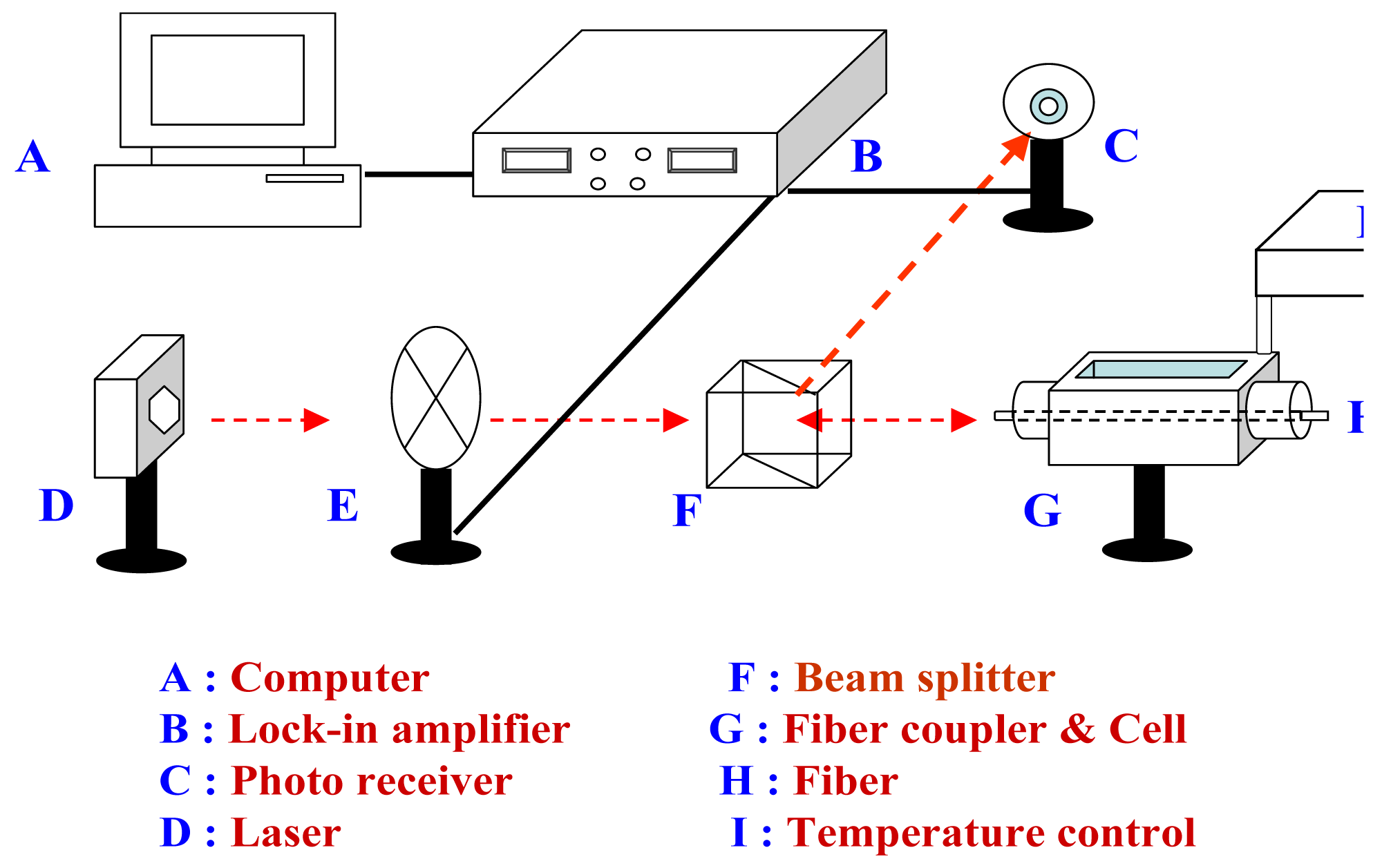
2.6 Instrumentation and measurements

The optical configuration for the gold nanoparticle-modified optical fiber biosensor is a reflection-based fiber-optic configuration, as shown in Fig. 3. The system consists of a laser (Hitachi HL6320G laser diode, 635 nm, 10 mW; Thorlabs LDC500 laser diode controller; Thorlabs TEC2000 temperature controller; Thorlabs TCLDM9 laser mount), a chopper (Stanford Research SR540), a fiber coupler, a beam splitter, a sensing fiber, a liquid cell (10 mL), a photo-receiver (Thorlabs PDA55), and a lock-in amplifier (Stanford Research SR830). For the detail description of the biosensor system, please refer to Chau et al. (2006) [19]. The temperature of the liquid testing cell was maintained by a circulatory waterbath (Wisdom, model LC-06) within ±0.05 K. The Pb(II)-EDTA complex solution was filled in the liquid testing cell, in which the LSPR sensor was installed horizontally. After reaching a balance, a new stable response can be attained in 5 min. However, to make sure reaction reaching to equilibrium state, the responding signals are taken over 10 minutes after addition of the Pb(II)-EDTA complex solution. In this work, at least three replicates were performed for each parameter.

3. Results and Discussion

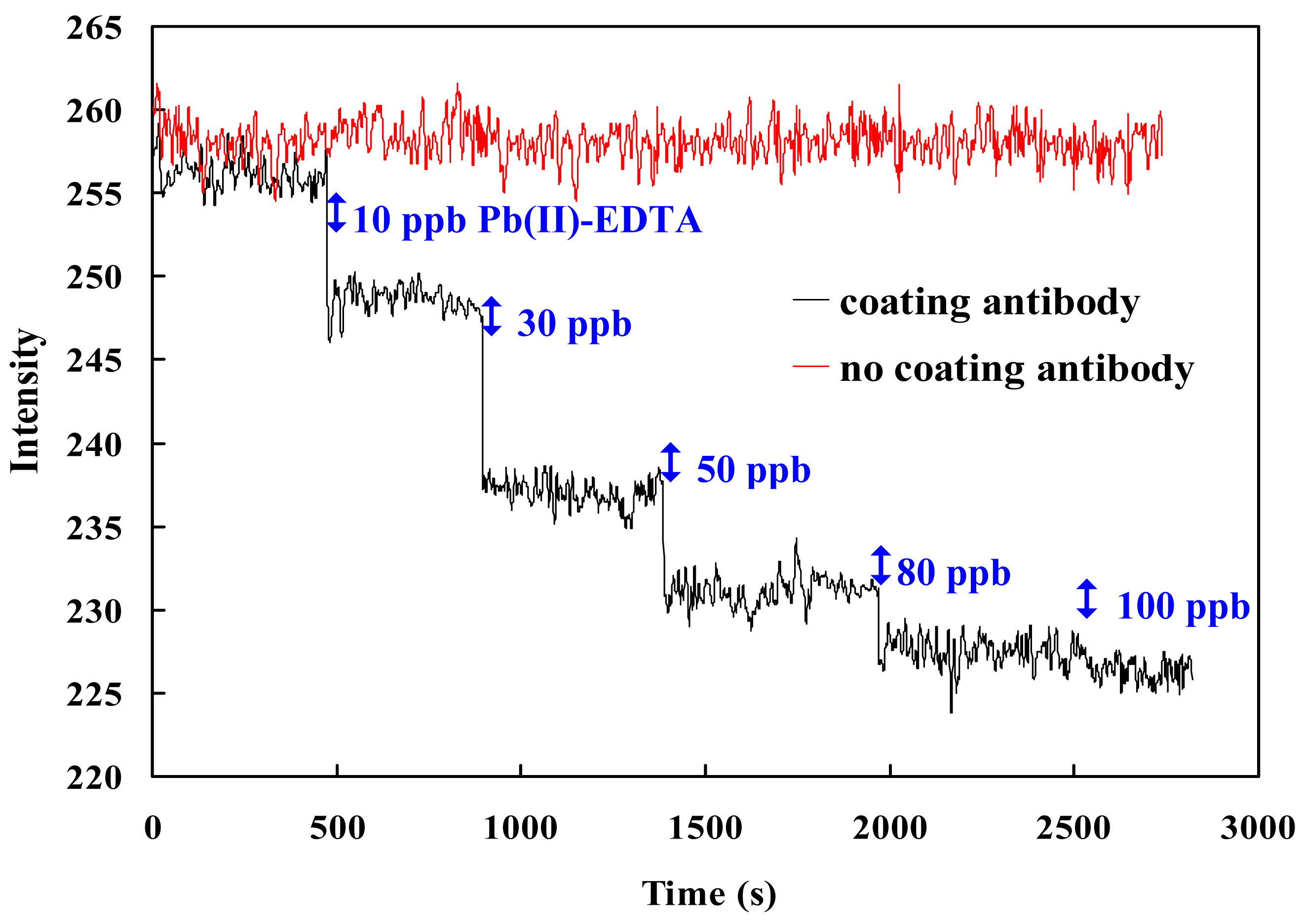
3.1 Comparing probe w/wt immobilizing monoclonal antibody on NMAu

While applying the LSPR sensor, the capture of Pb(II)-EDTA complex by the immobilized antibody causes a decrease of light intensity at the detector due to an increase in the local RI. Fig. 4 shows an example of serial responding signals from the antibody modified LSPR sensor immersing in a pH 7.4 PBS buffer solution after sequential addition of Pb(II)-EDTA complex to final concentrations of 100 ppb. The responding signals decrease with increasing Pb(II)-EDTA complex concentration at room temperature and with a good linearity (which will be shown later). Notably, under the same operating conditions, there is no significant responding change for the fiber without immobilizing antibody, suggesting that the antibody can substantially differentiate the change of Pb(II)-EDTA complex at low concentrations.

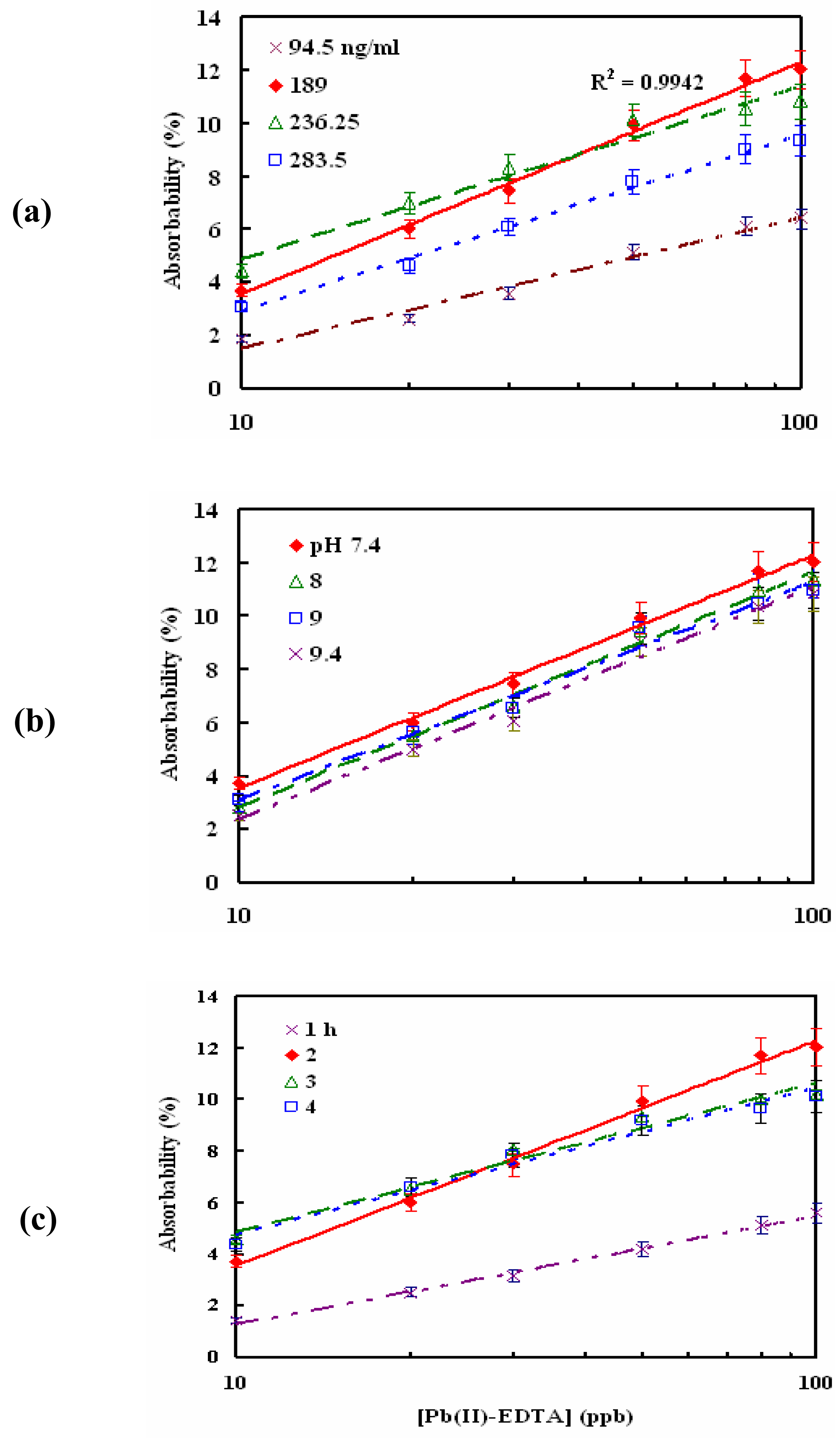
3.2 Optimal monoclonal antibody immobilizing conditions

The sensitivity of an optic-fiber sensor is related to the immobilizing conditions, such as the amount of immobilized ligand and pH value of the solution [23]. The amount of immobilized ligand is determined by the ligand concentration and incubation period. Fig. 5 shows the effects of the antibody concentration, and incubation time and pH on the response signals for Pb(II)-EDTA complex, respectively. In theory, higher incubating antibody concentration (or the longer the incubating time) revealed larger immobilization of monoclonal antibody onto the fiber, resulting in a stronger LSPR effect and a smaller I0. Besides, under a constant concentration of Pb(II)-EDTA complex the sensor immobilized with more antibody has the higher residua to react Pb(II)-EDTA complex with a smaller I1, results in a higher absorbability and resolution with a rather good linearity. From Fig. 5(a) and (b), it can be seen that as the incubating antibody concentration and time increases from 94.5 μg/mL and 1 h, respectively, to 189 μg/mL and 2 h, the absorbability reaches a maximal value of 12.2 % with a good linearity (R2=0.994) for Pb(II)-EDTA complex concentration ranging from 10 to 100 ppb. However, upon further increase in the incubating enzyme concentration or time, the amount of immobilized antibody onto the fiber becomes over saturated and causes steric hindrance from binding with Pb(II)-EDTA complex, leading to a less response in absorbability
Figure 5(c) shows the plots of the absorbability versus Pb(II)-EDTA complex concentration with different sensors incubated in various pH values of 189 μg/mL antibody solution for 2 h. In the lower pH, the coupling between succinimide esters formed by antibody and NHS and amine group on cystamine is less effective to form the amino covalent binding [24]. Hence, only the base conditions of incubation will be considered in the study. From Fig. 5(c), it shows that the maximal absorbability change is found at pH 7.4, after which the absorbability decreases again. This result can be attributed to two reasons: first, the protein denaturation of antibody in the higher pH condition may lead to its lower activity. Second, the charge of antibody becomes negative in the higher pH condition, which leads to over saturated immobilizing antibody onto the fiber. In summary, the optimal immobilization conditions for modifying the LSPR sensor are 189 μg/mL antibody and pH 7.4 for 2 h incubation, and will be applied for the rest of the studies. With the limit of detection (LOD) defined as the intensity at a Pb(II)-EDTA complex concentration that yields a signal-to-noise (S/N) ratio of 3, the LOD of the sensor with optimal immobilization is 0.27 ppb.

3.3 Temperature effect and comparison with other metal ions

Monoclonal antibody is a protein, of which activity depends on temperature. The antibody's binding and dissociation capabilities with metal-chelate complex are strongly affected by temperature. Fig. 6 shows the response of absorbability for detecting 10∼100 ppb Pb(II)-EDTA complex under different detecting temperatures. The optimal detecting temperature for antibody responding to Pb(II)-EDTA complex is at 25°C, after that the change of absorbability becomes less. An increasing temperature may enhance both the binding and dissociation rate. However, based on above results, it can be inferred that the temperature effects on the dissociation rate between antibody and Pb(II)-EDTA complex is larger than on the binding rate.
Another important issue on whether the monoclonal antibody-functionalized LSPR sensor is able to bind other metals needs to be addressed. Except Pb(II)-EDTA complex, the bindings of Mg(II)-, Cu(II)-, and Ni (II)-EDTA complexes to the sensor were also examined, as shown in Fig. 7. Basically, the sensor shows a rather good linearity for the above metal ions. However, for 10∼100 ppb detecting range the absorbability of these three metal-EDTA complexes was only 3% change, which was smaller than that of Pb(II)-EDTA complex (12.2%). That means the monoclonal antibody has a higher affinity to Pb(II)-EDTA complex.

3.4 Stability and dissociation constant

Note that protein can cause structural folding and becomes denatured if the surface water on it is not conserved after a long-time storage. D-(+)-Trehalose dehydrate, known as mycose, is an alpha-linked sugar that has the merits of rehydration and anti-oxidation to perverse the activity of protein [25]. To prove the stability of our sensor, we performed the measurement of Pb(II)-EDTA complex using the sensors stored in 5% D-(+)-Trehalose dehydrate solution at 4°C for up to 35 days after the initial measurement. The result shows that the relative activity of the sensor only decayed 7.2% after being stored for 35 days. Although there is some loss of the sensor activity after each cycle of use, the sensor can be recalibrated to determine Pb(II)-EDTA complex concentration. There was no significant change in the calibration curve after 35-day storage.

4. Conclusion

A monoclonal antibody-functionalized LSPR biosensor bas been successfully developed for determining the concentration of Pb(II). The optimal incubating conditions for the immobilization of monoclonal antibody on to the LSPR biosensor were found to be 189 μg/mL monoclonal antibody in pH 7.4 PBS for 2 h. With the optimal modification, the sensor can obtain a maximal 12.2 % change of the absorbability for detecting 10∼100 ppb Pb(II)-EDTA complex and a LOD of 0.27 ppb. The biosensor demonstrates a reproducible and stable response after over 35-day storage. Conclusively, the monoclonal antibody-modified LSPR biosensor can be used for the label-free detection of Pb(II)-EDTA with an excellent sensitivity.

Acknowledgments

The work was supported by the National Science Council grant NSC-96-2214-E-194-035 in Taiwan.

References and Notes

  1. Tong, S. Lead exposure and cognitive development: persistence and a dynamic pattern. J. Pediatr. Child Health 1998, 34, 114–118. [Google Scholar]
  2. Lilienthal, H.; Winneke, G.; Ewert, T. Effects of lead on neurophysiological and performance measures: animal and human data. Environ Health Perspect. 1990, 89, 21–25. [Google Scholar]
  3. CDC, Surveillance for elevated blood lead levels among children-United States, 1997-2001. Centers for Disease Control and Prevention MMWR Morb Mortal Wkly Rep. 2003, 53, 1–21.
  4. Rothenberg, S.J.; Poblano, A.; Schnaas, L. Brainstem auditory evoked response at five years and prenatal and postnatal blood lead. Neurotoxicol Teratol. 2000, 22, 503–510. [Google Scholar]
  5. Anderson, J.L.; Bowden, E.F.; Pickup, P.G. Dynamic electrochemistry: Methodology and application. Anal. Chem. 1996, 68, 379R–444R. [Google Scholar]
  6. Burlingame, A.L.; Boyd, R.K.; Gaskell, S.J. Mass spectrometry. Anal. Chem. 1996, 599R–651R. [Google Scholar]
  7. Jackson, K.W.; Chen, G. Atomic absorption, atomic emission, and flame emission spectrometry. Anal. Chem. 1996, 68, 231R–256R. [Google Scholar]
  8. Zhyliak, G.A.; Dzyadevici, S.V.; Korpan, Y.I.; Soldatkin, A.P.; Elskaya, A.V. Application of urease conductometric biosensor for heavy metal ion determination. Sens. Actuat. B 1995, 24-25, 145–148. [Google Scholar]
  9. Bontidean, I.; Ahlqvist, J.; Mulchandani, A.; Chen, W.; Bae, W.; Mehra, R.K.; Mortari, A.; Csoregi, E. Novel synthetic phytochelatin-based capacitive biosensor for heavy metal ion detection. Biosens. Bioelectron. 2003, 18, 547–553. [Google Scholar]
  10. Bontidean, I.; Berggren, C.; Johansson, G.; Csoregi, E.; Mattiasson, Bo; Lloyd, J.R.; Jakeman, K.J.; Brown, N.L. Detection of Heavy Metal Ions at Femtomolar Levels Using Protein-Based Biosensors. Anal. Chem. 1998, 70, 4162–4169. [Google Scholar]
  11. Khosraviani, M.; Blake, R.C., II; Pavlov, A.R.; Lorbach, S.C.; Yu, H.; Delehanty, J.B.; Brechbiel, M.W.; Blake, D.A. Binding properties of a monoclonal antibody directed toward lead-chelate complexes. Bioconjugate Chem. 2000, 11, 267–277. [Google Scholar]
  12. Blake, D.A.; Blake, R.C., II; Khosraviani, M.; Pavlov, A.R. Immunoassays for metal ions. Analytica Chimica Acta 1998, 376, 13–19. [Google Scholar]
  13. Delehanty, J.B.; Jones, R.M.; Bishop, T.C.; Blake, D.A. Identification of Important Residues in Metal-Chelate Recognition by Monoclonal Antibodies. Biochemistry 2003, 42, 14173–14183. [Google Scholar]
  14. Johnson, D.K.; Combes, S.H.; Parsen, J.; Jolley, A.A. Lead Analysis by Anti-Chelate Fluorescence Polarization Immunoassay. Environ. Sci. Technol. 2002, 36, 1042–1047. [Google Scholar]
  15. Chakrabarti, P.; Hatcher, F.M.; Blake, R.C., II; Ladd, P.A.; Blake, D.A. Enzyme immunoassay to determine heavy metals using antibodies to specific metal-EDTA complexes: optimization and validation of an immunoassay for soluble indium. Anal. Biochem. 1994, 21, 70–75. [Google Scholar]
  16. Reardan, D.T.; Meares, C.F.; Goodwin, D.A.; McTigue, M.; David, G.S.; Stone, M.R.; Leung, J.P.; Bartholomew, R.M.; Frincke, J.M. Antibodies against metal chelates. Nature 1985, 316, 265–268. [Google Scholar]
  17. Blake, R.C., II; Delehanty, J.B.; Khosraviani, M.; Yu, H.; Jones, R.M.; Blake, D.A. Allosteric Binding Properties of a Monoclonal Antibody and Its Fab Fragment. Biochemistry 2003, 497–508. [Google Scholar]
  18. Lin, T.-J.; Huang, K.-T.; Liu, C.-Y. Determination of Organophosphorous Pesticides by a Novel Biosensor Based on Localized Surface Plasmon Resonance. Biosens. Bioelectron. 2006, 22, 513–518. [Google Scholar]
  19. Chau, L.-K.; Lin, Y.-F.; Cheng, S.-F.; Lin, T.-J. Fiber-optic chemical and biochemical probes based on localized surface plasmon resonance. Sens. Actuat. B 2006, 113, 100–105. [Google Scholar]
  20. Jensen, T.R.; Duval, M.; Kelly, K.L.; Lazarides, A.A.; Schatz, G.C.; Richard, P.; Van, D. Nanosphere lithography: Effect of the external dielectric medium on the surface plasmon resonance spectrum of a periodic array of silver nanoparticles. J. Phys. Chem. B 1999, 103, 9846–9853. [Google Scholar]
  21. Nath, N.; Chilkoti, A. A colorimetric gold nanoparticle sensor to interrogate biomolecular interactions in real time on a surface. Anal Chem. 2002, 74, 504–509. [Google Scholar]
  22. Haes, A.J.; van Duyne, R.P. A nanoscale optical biosensor: sensitivity and electivity of an approach based on the localized surface plasmon resonance spectroscopy of triangular silver nanoparticles. J. Am. Chem. Soc. 2002, 124, 10596–10604. [Google Scholar]
  23. Jorgenson, R.C.; Yee, S.S. A fiber-optic chemical sensor based on surface plasmon resonance. Sens. Actuat. B 1993, 12, 213–218. [Google Scholar]
  24. Gestwicki, J.E.; Cairo, C.W.; Mann, D.A.; Owen, R.M.; Kiessling, L.L. Selective Immobilization of Multivalent Ligands for Surface Plasmon Resonance and Fluorescence Microscopy. Anal Chem. 2002, 305, 149–155. [Google Scholar]
  25. Higashiyama, T. Novel functions and applications of trehalose. Pure Appl. Chem. 2002, 74, 1263–1269. [Google Scholar]
Figure 1. (a) Images of 2-D and 3-D AFM surfaces on the prepared MPTMS; (b) Electron micrograph and particle size distribution of pure Au nanoparticles.
Sensors 08 00582f1
Figure 2. The chemical reactions scheme carried out for covalent binding of monoclonal antibody to the fiber-based LSPR sensor.
Sensors 08 00582f2
Figure 3. The schematic representation of the LSPR biosensor system.
Sensors 08 00582f3
Figure 4. An example of serial Pb(II)-EDTA complex response signal in the range of 10∼100 ppb by two sensors with/without monoclonal antibody coating.
Sensors 08 00582f4
Figure 5. Effects of (a) the incubating monoclonal antibody concentration, (b) incubating time, and (c) pH on the response for detecting 10∼100 ppb Pb(II)-EDTA complex in pH 7.4 PBS buffer solution at 25°C.
Sensors 08 00582f5
Figure 6. Effect of the detection temperature on the response for detecting 10∼100 ppb Pb(II)-EDTA complex in pH 7.4 PBS buffer solution at 25°C.
Sensors 08 00582f6
Figure 7. Comparison of the responses between different metal-chelate complexes (i.e., Mg(II)-, Cu(II)-, and Ni(II)-EDTA) through the monoclonal antibody-functionalized LSPR biosensor.
Sensors 08 00582f7

